http://vet.sagepub.com/
Veterinary Pathology Online
http://vet.sagepub.com/content/38/1/98 The online version of this article can be found at:
DOI: 10.1354/vp.38-1-98 2001 38: 98
Vet Pathol
P. Scocco, F. Mariotti, P. Ceccarelli, O. Fagioli, G. Renzoni and G. Vitellozzi
(Capra hircus):
A Glycohistochemical Approach
Origin of Enzootic Intranasal Tumor in the Goat
Published by:
http://www.sagepublications.com
On behalf of:
Pathologists.
American College of Veterinary Pathologists, European College of Veterinary Pathologists, & the Japanese College of Veterinary
can be found at:
Veterinary Pathology Online
Additional services and information for
http://vet.sagepub.com/cgi/alerts Email Alerts: http://vet.sagepub.com/subscriptions Subscriptions: http://www.sagepub.com/journalsReprints.nav Reprints: http://www.sagepub.com/journalsPermissions.nav Permissions: What is This? - Jan 1, 2001 Version of Record >>
98
Origin of Enzootic Intranasal Tumor in the Goat
(Capra hircus): A Glycohistochemical Approach
P. SCOCCO, F. MARIOTTI, P. CECCARELLI, O. FAGIOLI, G. RENZONI, ANDG. VITELLOZZI Dipartimento di Scienze Veterinarie, Universita` di Camerino, Italia (PS,1FM, OF, GR); and
Dipartimento di Scienze Biopatologiche Veterinarie, Universita` di Perugia, Italia (PC, GV)
Abstract. Enzootic intranasal tumor (EIT) appears glandular in type and has recently been classified as an adenocarcinoma of low malignancy. The aim of this study was to characterize the secretion of surface glycoconjugates (GCs) in EIT and in normal respiratory and olfactory mucosae of the goat by means of conventional and lectin histochemistry, in order to shed light on the histogenesis of EIT. Morphologic and ultrastructural investigations showed two growth types of EIT: i.e., tubular and papillary patterns. Conven-tional histochemistry revealed the presence of neutral and carboxylated GCs in the olfactory glands and in the tubular part of EIT, as well neutral and sulphated GCs in the respiratory glands and in the papillary part of EIT, suggesting that the papillary pattern tumor arises from the respiratory glands, whereas the tubular portion of EIT arises from the olfactory glands. Lectin histochemistry gave further information on the ex-pressed GCs.
Key words: Enzootic intranasal tumor; glycohistochemistry; goats; lectins; olfactory mucosa; respiratory mucosa.
Tumors are uncommon pathologies in sheep and goats; however, pulmonary adenomatosis, small intes-tinal carcinoma, lymphosarcoma, skin squamous cell carcinoma, and enzootic intranasal tumor (EIT) have been described, and EIT seems the most frequent neo-plasia. In goats, tumors seem to be less frequent, al-though some cases of EIT have been reported in recent years.4,5,10,14,17,23
The enzootic intranasal tumor is a chronic and afe-brile disease characterized by a profuse initial sero-mucous and then purulent nasal exudate associated with clinical signs of respiratory distress. In the late period of the disease, facial swelling, sometimes with seromucous exudation, is found because of na-sal bone erosion and skin ulceration.15EIT in sheep
is distributed worldwide, with a high prevalence in affected ovine flocks.2,8,12 Instead, EIT in goats has
been reported only in Italy, Spain, France, Greece, India, and Canada; in the affected flocks, frequence did not exceed 2–3% of the animals, but tumor oc-currence was quite constant.5,15Even though this
tu-mor has a low and only local malignancy, it is gen-erally lethal because of its particular position as it enlarges to completely invade the nasal and para-nasal cavities.
Histologically, the tumor appears glandular in type. 1Present address: Dipartimento di Scienze Veterinarie, Fa-colta` di Medicina Veterinaria, Universita` di Camerino, Via Circonvallazione, 93–95, 62024 MATELICA MC (Italy).
In the past it was classified as an adenopapilloma or adenoma, whereas it has recently been considered as an adenocarcinoma of low malignancy.5,6,11,23
After numerous etiologic hypotheses, from envi-ronmental to genetic factors, a viral cause was dem-onstrated. Viral-like particles, compatible with a type D retrovirus, were isolated from nasal fluid, and neo-plastic tissue and successful experimental transmis-sion of EIT from goat to goat confirmed this hypoth-esis.1,3,20
In spontaneous disease, the infection occurs through the respiratory route, and the incubation time is so long that the clinical disease is found in animals that are approximately 1 year old.23
The complete histogenesis of EIT still remains un-known. Its probable histologic origin could be in the olfactory glands, in the supporting cells of the olfac-tory epithelium, or in the brush cells of the respiraolfac-tory epithelium.7,19This paper reports on the histochemical
characterization of the glandular secretions from EIT and from the normal respiratory and olfactory mucosa of goats.
Materials and Methods
Five Alpine goats with clinical signs of EIT were obtained from flocks in Central Italy. At necropsy, after dissection of the cranium through sagittal medial cutting, samples of neo-plastic tissue were removed and fixed in Carnoy’s fluid for 24 hours, postfixed in 2% calcium acetate and 4%
parafor-Vet Pathol 38:1, 2001 Glycohistochemistry in Goat EIT 99
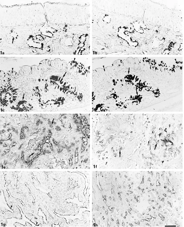
Fig. 1. LID and HID staining. Olfactory glands (arrow) react moderately to LID (a). HID reactivity ranges from moderate to slight (b). In the respiratory mucosa, LID (c) and HID (d) react intensely at both goblet cell (arrowhead) and gland level (arrows). In the tubular part of EIT, LID (e) moderately stains some cells (arrow), whereas HID (f) reacts slightly (arrows). In the papillary part of EIT, only the apical cytoplasm of cells appears LID-positive (g) (arrowheads) and, in minor measure, HID-positive (h). Bar5 40 mm.
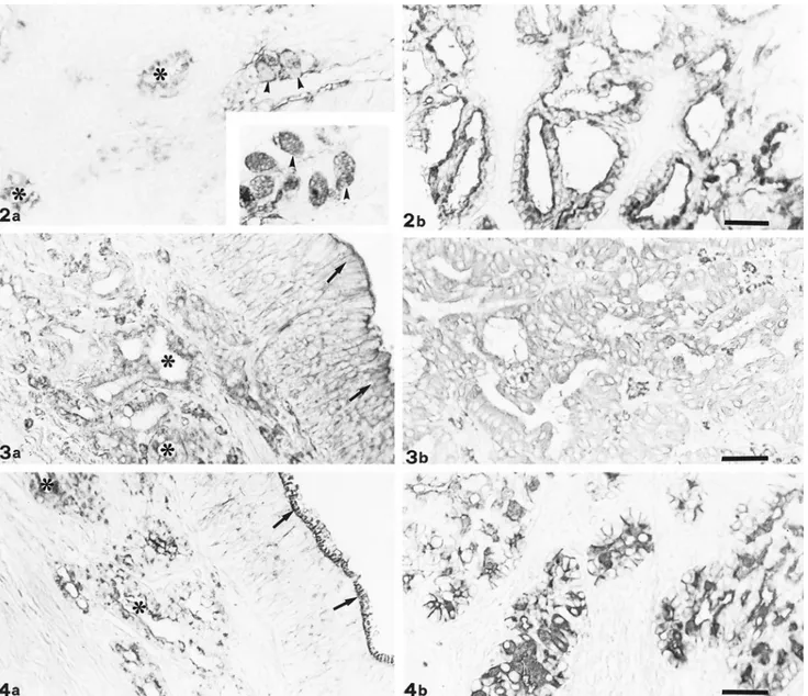
Fig. 2. DBA staining. In the respiratory mucosa, goblet cells (arrowheads) and respiratory glands (*) show reactivity ranging from weak to moderate (a); sialidase digestion enhances goblet cell (arrowheads) positivity (inset). Papillary portion of EIT appears positive with sialidase and DBA treatment (b). Bar5 33 mm.
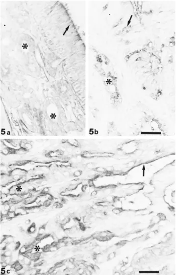
Fig. 3. Con A staining. Both olfactory epithelium (arrows) and glands (*) react strongly to Con A (a). Also tubular portion of EIT shows strong positivity (b). Bar5 33 mm.
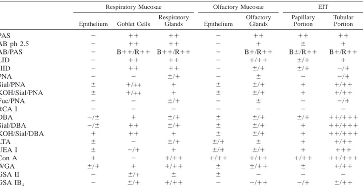
Fig. 4. WGA staining. Olfactory glands (*) react strongly to WGA; also, the cell coat of olfactory epithelium (arrow) appears positive (a). EIT tubular portion shows very strong staining (b). Bar5 33 mm.
maldehyde (1 : 1) for 3 hours, and then dehydrated and em-bedded in paraffin wax.
Serial sections (5mm thick) were treated for carbohydrate conventional histochemistry with periodic acid–Schiff (PAS; to detect vicinal hydroxyls), Alcian blue (AB) at pH 2.5 (to reveal acidic groups), Alcian blue–periodic acid–Schiff (AB/ PAS; to demonstrate acidic groups and vicinal hydroxyls), low-iron diamine (LID; to detect sulfate and carboxyl radi-cals) and high-iron diamine (HID; to discriminate sulfate groups).16,22
Lectin staining was performed as previously described.9,13 The following lectins were used: Peanut agglutinin from
Ar-achis hypogaea [terminalb-D-galactose (1–3)-N-acetylgalac-tosamine], Dolichos biflorus agglutinin from Dolichos
biflo-rus (terminal a-N-acetylgalactosamine), Ricinus communis
agglutinin I from Ricinus communis [terminal b-D
-galac-tose(1–4)-N-acetylglucosamine], Griffonia simplicifolia ag-glutinin IB4 from Griffonia simplicifolia (terminal a-D -ga-lactose), WGA from Triticum vulgaris (terminal and internal b-N-acetylglucosamine [sialic acid]), GSA II from Griffonia
simplicifolia (terminal N-acetylglucosamine), Con A from Canavalia ensiformis (terminal and internal a-D
–man-nose.a-D-glucose), Ulex europaeus agglutinin I from Ulex
a-Vet Pathol 38:1, 2001 Glycohistochemistry in Goat EIT 101
Fig. 5. UEA I staining. Both epithelium (arrow) and ol-factory glands (*) react moderately to UEA I (a); respiratory mucosa shows positive sites at goblet cell (arrow) and gland (*) level (b). Bar 5 33 mm. Cells of tubular portion (*) of EIT appear strongly reactive to UEA I, whereas in papillary portions, the reactivity is restricted to the luminal border (arrow; c). Bar5 25 mm.
L-fucose). Before lectin histochemical staining, some sec-tions were incubated at 37 C for 16 hours in 0.86 U/mg protein of sialidase (Type V, from Clostridium perfringens) with and without pretreatment with KOH 0.5% in 70% eth-anol in order to remove acyl groups.18,21Other sections were incubated at 37 C for 14 hours witha-fucosidase from bo-vine epididymis (2.3 U/mg prot.).21 Lectins and enzymes were purchased from Sigma Chemical Co. (St. Louis, MO). The respective peroxidase-conjugated lectins were omit-ted, or their hapten sugars (0.2–0.4 M) were added to control sections. As controls for enzyme digestion, sections were incubated with enzyme-free buffers under the same experi-mental conditions or with the lectins specific for sugar de-tached by enzyme pretreatment. Sections from normal olfac-tory and respiraolfac-tory mucosae of four goats killed in an
au-thorized abattoir were fixed, processed, and stained with the same methods.
Results
In normal goats, the respiratory mucosa is lined by a pseudostratified columnar epithelium with inter-mingled goblet cells. The propria-submucosa consists of a loose connective tissue in which tubuloacinar-mixed glands are located. The olfactory mucosa is grossly distinguishable from the respiratory one be-cause of its yellowish pigmentation. Microscopically, it is usually thinner than the respiratory mucosa, and the epithelium consists of three different cell types: sensory, substentacular, and basal cells. Many tubulo-acinar olfactory glands producing a serus secretion with occasional mucus-producing cells are located in the propria-submucosa.
The neoplasms were similar in all the cases exam-ined and were classified as low-grade adenocarcinoma. They showed two types of growth patterns: tubular, generally found in the deeper areas; and papillary, pre-sent mostly on the surface of the tumor. In the tubular part, there were regularly and irregularly shaped acinar and tubular or cystic structures separated by thin vas-cularized stroma; the neoplastic cells were pyramidal or cuboidal in shape. The papillary pattern consisted of undulating or branching villous projections with delicate connective tissue stalks, covered by one or several layers of cuboidal to columnar epithelial cells with abundant eosinophilic cytoplasm and round nu-clei located in a basal position.
The epithelial lining of both respiratory and olfac-tory mucosae appeared unstained after conventional histochemical procedures in normal subjects.
Olfactory glands showed strong reactivity to PAS, moderate staining intensity after AB pH 2.5 and LID (Fig. 1a) treatments, and slight reactivity to HID (Fig. 1b). Glands and goblet cells of the normal respiratory mucosa intensely reacted to PAS and AB pH 2.5 stain-ing; LID (Fig. 1c) and HID (Fig. 1d) treatments also elicited strong reactivity.
In the neoplastic tissue, the reactivity of the tubular and papillary parts were different, but no differences were found among the five cases examined. In the tu-bular part of EIT, all of the neoplastic cells showed diffuse cytoplasmic reactivity; PAS staining was in-tense, whereas AB pH 2.5 showed moderate staining. Response to LID was moderate (Fig. 1e), whereas re-activity to HID ranged from negative to moderate (Fig. 1f).
In the papillary part of EIT, only the apical cells of the papillary projections showed histochemical reac-tivity in the supranuclear region. Cells showed strong positivity to PAS, whereas AB pH 2.5, LID (Fig. 1g),
Table 1. Reactivity of samples to histochemical treatments.* Respiratory Mucosae
Epithelium Goblet Cells
Respiratory Glands Olfactory Mucosae Epithelium Olfactory Glands EIT Papillary Portion Tubular Portion PAS AB ph 2.5 AB/PAS LID HID PNA Sial/PNA KOH/Sial/PNA Fuc/PNA RCA I DBA Sial/DBA KOH/Sial/DBA LTA UEA I Con A WGA GSA II GSA IB4 2 2 2 2 2 2 6 6 2 2 2/6 2/6 1 6 6 1 6/1 2 2 11 11 B11/R11 11 11 2 1/++ 1/++ 2 2 1 11 11 2 2/1 2 1 6/+ 6/1 11 11 B11/R11 11 11 6/1 1 1 6/1 2 6/1 6/1 1 6/1 1 1/11 1/11 6 1/11 2 2 2 2 2 2 6 6 2 2 6 6 6 6/1 6/1 1/11 6 6 2 11 1 B1/R11 1/11 6/1 6 6/1 6/1 6 2 6/1 6/1 6/1 6 6/1 1/11 6/11 2 2/11 11 6 B6/R11 6/1 6/1 2 1 1 2 2 6/1 1 1 1 1 1/11 6 2 2/1 11 1 B1/R11 1 2/1 2/1 1/11 1/11 2/1 2 11/111 11/111 11/111 1/11 111 11/111 1/11 2 6/11
* Results are given in arbitrary units as follows:2 5 negative; 6 5 weak; 1 5 moderate; 11 5 strong; 111 5 very strong staining. R5 Red, B 5 blue, Sial 5 sialidase, Fuc 5 Fucosidase, PAS 5 periodic acid–Schiff, AB 5 Alcian blue, LID 5 low-iron diamine, HID 5 high-iron diamine.
and HID (Fig. 1h) gave a reactivity ranging from weak to moderate.
Lectin histochemistry showed weak PNA reactivity on the luminal border of both the olfactory and re-spiratory glands. Only the tubular portion of EIT showed moderate positivity. Sialidase treatment in-duced weak reactivity on the olfactory epithelium cell coat and in the apical zone of papillary EIT and en-hanced the positivity of respiratory glands. DBA showed binding patterns in both respiratory (Fig. 2a) and olfactory mucosae, as well as in the two growth patterns of EIT. Sialidase digestion affected only the positivity of the respiratory epithelium goblet cells (Fig. 2a) and the papillary portion of EIT (Fig. 2b). In any case, KOH treatment did not modify the ac-tivity of sialidase.
Con A showed moderate positivity in both normal mucosae and in the two parts of EIT (Fig. 3). WGA showed strong reactivity, especially in the respiratory and olfactory glands (Fig. 4a) and in the tubular por-tion of EIT (Fig. 4b). GSA II and WGA reactivities were overlapping only in the goblet cells of the respi-ratory epithelium. Binding sites for GSA IB4were pre-sent in the respiratory and olfactory glands and in the two parts of EIT.
LTA and UEA I showed overlapped binding sites, with the exception of goblet cells of the respiratory epithelium (Fig. 5). Fucosidase digestion did not affect
any lectin reactivity. Reactivities to the histochemical treatments are summarized in Table 1.
Discussion
The aim of this work was to determine the histo-chemical characteristics of secretion products in the EIT and in normal respiratory and olfactory mucosae of goats in order to clarify the histogenesis of EIT. We expected to find some histochemical differences be-tween normal and tumoral tissues as a consequence of both qualitative and quantitative modifications of cel-lular secretions caused by the neoplastic transforma-tion.
The results of our conventional histochemical pro-cedures led us to hypothesize a strong presence of neu-tral and prevalently sulphated acid and glycoconju-gates (GCs) in the glands of the respiratory mucosa. We also hypothesized that the olfactory mucosa seems to produce neutral GCs and a very small amount of carboxylated GCs.
Cells of the papillary portion of EIT prevalently ex-press neutral GCs and a smaller amount of sulphated GCs; the tubular portion of EIT synthesizes a great amount of neutral GCs and a small quantity of prev-alently carboxylated acid GCs.
The two recognizable growth patterns in EIT are not only different morphological expressions but also
re-Vet Pathol 38:1, 2001 Glycohistochemistry in Goat EIT 103
flect different histochemical behavior that could testify to a twofold histologic origin of the tumor.
In all the goats examined, the tubular portion of EIT produced neutral and prevalently carboxylated acid GCs like the glands of the normal olfactory mucosa. Staining intensity was almost the same in both tissues; the only difference concerned the distribution of stained products. Indeed, in the cells of the tubular portion, staining was widespread in the cytoplasm, whereas in the olfactory mucosa, only the apical cy-toplasm of the glandular cells reacted. The papillary portion of EIT showed the presence of neutral and prevalently sulphated acid GCs, as occurred in the glands of normal respiratory mucosa, although to a lesser extent.
On the basis of the above considerations, we may speculate on the hypothesis of a nonunivocal histolog-ic origin of EIT, as proposed by several authors.4,23The
neoplastic process could start from the olfactory glands for the tubular portion of EIT and from the respiratory mucosa glands for the papillary one, sug-gesting the possibility of a double glandular origin for the two growth patterns of goat EIT. In addition, the constant histochemical negativity of the epithelial lin-ing in the normal nasal mucosa seems to exclude its involvement in EIT histogenesis.
Lectin histochemistry confirms the strong presence of neutral glucidic residues in all the samples exam-ined. In particular, there is a strict correspondence be-tween the presence of a-N-acetylgalactosamine, a-L
-fucose, mannose, or glucose residues in respiratory glands and the papillary portion of EIT. In addition, in the papillary portion of EIT, the presence of subter-minal b-galactose was evidenced by sialidase and PNA sequence; b-galactose residues could be the ac-ceptor sites for sulphated groups revealed by conven-tional techniques.
In the olfactory glands and the tubular portion of EIT, there is a correspondence between the presence of a-N-acetylglucosamine and a-galactose residues and the sialic acid–b-D-galactose
(1-3)-N-acetylgalac-tosamine sequence. Carboxylated groups revealed by conventional histochemistry in both olfactory glands and the tubular portion of EIT could be ascribed to the sialic acid residues.
Lectin histochemistry partially confirms the hypoth-esis that EIT originates from both respiratory and ol-factory glands. However, because further evidence to strengthen this hypothesis on the histogenesis of EIT has emerged, the problem requires additional ap-proaches.
Acknowledgements
We wish to thank Ms. Natalina Cammertoni for technical assistance. This study was supported by a University Grant for Research.
References
1 Cousens C, Minguijon E, Garcia M, Ferrer LM, Dalziel RG, Palmarini M, Sharp JM, De Las Heras M: PCR-based detection and partial characterization of a retro-virus associated with contagious intranasal tumors of sheep and goat. J Virol 70:7580–7583, 1996
2 De Las Heras M, Garcia de Jalon JA, Badiola JJ: Tumor intranasal en una oveja. Med Vet 3:459–462, 1986 3 De Las Heras M, Garcia de Jalon JA, Minguijon E, Gray
EW, Dewar P, Sharp JM: Experimental transmission of enzootic intranasal tumors of goats. Vet Pathol 32:19– 23, 1995
4 De Las Heras M, Garcia de Jalon JA, Sharp JM: Pa-thology of enzootic intranasal tumor in thirty-eight goats. Vet Pathol 28: 474–481, 1991
5 De Las Heras M, Sharp JM, Garcia de Jalon JA, Dewar P: Enzootic nasal tumour of goats: demonstration of a type D-related retrovirus in nasal fluids and tumors. J Gen Virol 72:2533–2535, 1991
6 Fontaine JJ, Crespeau F, Guereaud JM, Parodi AL: Ob-servation d’une enzootie d’adenome pituitaire de la che`vre. Rev Med Vet Ec Alfort 159:383–388, 1983 7 Frisch D: Ultrastructure of mouse olfactory mucosa. Am
J Anat 121:87–120, 1967
8 Giauffret A, Russo P, Laserre M: Tumeurs transmisibles de la muqueuse nasale des petits ruminants. Bull Lab Vet 6:35–38, 1982
9 Graham RC Jr, Karnovski MJ: The early stages of ab-sorption of injected horseradish peroxidase in the prox-imal tubules of mouse kidney: ultrastructural cytochem-istry by a new technique. J Histochem Cytochem 14: 291–302, 1966
10 Gue´raud JM: A propos de cas de tumeur nasale de la che`vre. Bull Lab Vet 6:31–34, 1982
11 Lombard CH, Cabanie P, Crespin J: Ade´nopapillome de la muqueuse pituitaire chez la che`vre. Bull Acad Vet Fr 39:199–202, 1966
12 Mc Connel EE, Van Rensburg IBJ, Van Wik JA: A case of adenocarcinoma of the olfactory mucosa in a sheep of possible infectious origin. J S Afr Vet Med Assoc 41: 9–12, 1970
13 Menghi G: Reactivity of peroxidase-labeled lectins in rabbit submandibular and sublingual glands. Acta His-tochem 75:27–35, 1984
14 Moulton JE: Tumours of Domestic Animals, 3rd ed. Uni-versity of California Press, London, England, 1990 15 Palmarini M, Mughetti L, Tollis M, Alunni L, Vitellozzi
G: Il tumore intranasale enzootico della capra. Summa 3:49–53, 1994
16 Pearse AGE: Histochemistry Theoretical and Applied, 3rd ed., vol. 1, pp. 659–660. Churchill, London, Eng-land, 1968
17 Rajan A, Sulochana S, Sreekuraman T, Reddi MV, Nair MK: Tumours of the ethmoid mucosa in goats (Capra
hircus). Indian J Cancer 17:196–199, 1980
18 Reid PF, Culling CF, Dunn WL: A histochemical method for the identification of 9-O-acyl sialic acid. An inves-tigation of bovine submaxillary gland and intestinal mu-cins. J Histochem Cytochem 26:187–192, 1978
19 Rhodin JAG: Histology. A Text and Atlas, pp. 607–645. Oxford University Press, New York, NY, 1974
20 Rutili D, Vitellozzi G, Maresca C, Manocchio I: Exper-imental transmission of enzootic intranasal tumour (EIT) in goats. 13th Int Symp Proc WAVMI, 46, 1994 21 Scocco P, Accili D, Menghi G, Ceccarelli P: Unusual
glycoconjugates in the oesophagus of a tilapine polyhy-brid. J Fish Biol 53:39–48, 1998
22 Spicer SS, Horn RG, Leppi TJ: Histochemistry of con-nective tissue mucopolysaccharides. In: The Concon-nective Tissue, eds. Wagner BM, Smith DE, pp. 251–303. Wil-liam and Wilkins, Baltimore, MD, 1967
23 Vitellozzi G, Mughetti L, Palmarini M, Mandara MT, Mechelli L, Sharp JM, Manocchio I: Enzootic intrana-sal tumour of goats in Italy. J Vet Med B 40:459–468, 1993
Request reprints from Paola Scocco, Dipartimento di Scienze Veterinarie, Facolta` di Medicina Veterinaria, Universita` di Camerino, Via Circonvallazione, 93–95, 62024 MATELICA MC (Italy).