Exploiting MS-based techniques to unveil elusive
reaction intermediates of bioinorganic relevance
Thesis
in Partial Fulfillment of the Requirements for the Degree of
Doctor of Philosophy in Pharmaceutical Sciences
XXXI cicle
Author
Davide Corinti
Supervisor
Alle donne della mia vita che mi hanno insegnato la cultura, la dedizione e l’amore
Non quia difficilia sunt non audemus, sed quia non audemus difficilia sunt.
I
ABSTRACT V
INTRODUCTION 1
1.1 Life and its connection with metal complexes, the role of bioinorganic chemistry 1
1.2 Cisplatin and other metal-containing drugs 4
1.3 Analytical tools for bioinorganic chemists 7
1.3.1 Mass spectrometry and MS-based techniques 8
1.3.2 Theoretical methods 10
1.4 ESI-MS to understand mechanisms of liquid-phase reactions: gas- and liquid-phase dichotomy 12
References 13
METHODS 16
2.1 Mass Spectrometry 16
2.1.1 ESI source 17
2.1.2 Time-of-flight mass spectrometers 19
2.1.3 Quadrupole analyzers 21
2.1.4 FT-ICR analyzers 23
2.2 Tandem mass spectrometry 26
2.2.1 Collision induced dissociation 26
2.2.2 IR multiple photon dissociation 27
2.3 Ion mobility mass spectrometry 30
2.4 Computational methods 32
2.4.1 Basic principles of quantum chemistry applied to calculations 32 2.4.2 The Hartree-Fock and post-Hartree-Fock methods 33
2.4.3 Density functional theory 35
2.4.4 Basis functions and relativistic effective core potentials 38
References 41
GAS-PHASE KINETICS AND IRMPD SPECTROSCOPY TO UNVEIL ELEMENTARY STEPS IN THE REACTION OF CISPLATIN WITH N- O- AND S- CONTAINING NUCLEOPHILES 44
3.1 Introduction 44
3.2. Experimental details 47
3.2.1 Sample preparation 47
3.2.2 IRMPD spectroscopy 47
3.2.3 Ion-molecule reactions in FT-ICR mass spectrometry 48
II 3.3.2 Reactivity of [PtX(NH3)2(H2O)]+ (X = Cl, OH) complexes in ligand substitution reactions 56
S3.3 Supporting material 63
3.4 Probing the missing step in the understanding of metal complexes substitution reaction: the encounter complex of cisplatin with model ligands isolated and characterized by MS and IRMPD
spectroscopy 66
3.4.1 Cisplatin complexes with model ligands: formation of cis-[PtCl(NH3)2(L)]+ in solution 67 3.4.2 Vibrational and structural features of cis-[PtCl(NH3)2(L)]+ complexes 68 3.4.3 Structural features of cis-[PtCl(NH3)2(H2O)(L)]+ complexes 75 3.4.4 Ligand substitution in the encounter complex of L and cis-[PtCl(NH3)2(H2O)]+ 89
S3.4 Supporting Material 95
3.5 Summary 114
References 117
ELUSIVE PRIMARY COMPLEXES OF CISPLATIN WITH AMINOACIDS: EXPLOITING IRMPD
KINETICS TO ASSAY ISOMER COMPOSITION 123
4.1 Introduction 123
4.2 Experimental details 125
4.2.1 Sample preparation 125
4.2.2 Mass spectrometric experiments 125
4.2.3 IRMPD spectroscopy and ion mobility experiments 125
4.2.4 Computational details 127
4.3 Interaction of cisplatin with histidine: kinetically trapped isomers quantified by
photofragmentation kinetics 130
4.3.1 Mass spectrometric characterization of cis-[PtCl(NH3)2(His)]+ 131 4.3.2 Vibrational features and structural characterization of cis-[PtCl(NH3)2(His)]+ 134 4.3.3 Photofragmentation kinetics for isomer and conformer quantification 140
S4.3 Supporting material 145
4.4 Binding differences of cisplatin and transplatin with methionine as assayed by IRMPD
spectroscopy and kinetics 159
4.4.1 ESI-MS/MS and IRMPD spectroscopy of methionine platinum(II) complexes 160 4.4.2 Computational survey of cis-[PtCl(NH3)2Met]+ and trans-[PtCl(NH3)2Met]+ complexes 165 4.4.3 Gas-phase structural and vibrational features of cis-[PtCl(NH3)2Met]+ 167 4.4.4 Gas phase structural and vibrational features of trans-[PtCl(NH3)2Met]+ 169 4.4.5 IRMPD kinetics to probe N- or S-platination in [PtCl(NH3)2Met]+ complexes 171
S4.4 Supporting material 173
4.5 Summary 181
References 184
MOVING TO HIGHER OXIDATION NUMBERS: MS-BASED TECNIQUES FOR THE STUDY OF PROPERTIES OF PTIV-CONTAINING ANTINEOPLASTIC ACTIVE COMPLEXES 190
III
5.2.1 Sample preparation 193
5.2.2 Mass analysis 193
5.2.3 IRMPD experiments 195
5.2.4 Computational details 195
5.3 Investigation of the vibrational and structural features of protonated PtIV complexes 197
5.3.1 Collision-induced dissociation experiments 198
5.3.2. Vibrational and structural features of [EP32+H]+ 200
5.3.3. IRMPD spectroscopy of [EP417+H]+ 202
5.3.4 IRMPD spectroscopy of [EP440+H]+ 204
5.3.5 Discussion 207
5.4 Gas-phase dissociation patterns of deprotonated platinum(IV) complexes 210
5.4.1 Mass spectrometric analysis of pro-drug PtIV complexes 211 5.4.2 Breakdown behavior of pro-drug PtIV complexes 212 5.4.3 Computed paths for the gas-phase breakdown pattern 219 5.4.4 A structural assay of the sampled deprotonated complexes by IRMPD spectroscopy 222
S5.4 Supporting material 225
5.5 Summary 237
References 240
APPENDIX A: FURTHER JOINT PUBLICATIONS 244
A.1 Photoionization mass spectrometry of ω -phenylalkylamines: Role of radical cation- π
interaction 245
A.1.1 Introduction 245
A.1.2 Methods 248
A.1.3 Results and discussion 250
A.1.4 Conclusions 263
SA.1 Supporting material 264
References 268
A.2 Complexation of halide ions to tyrosine: role of non-covalent interactions evidenced by IRMPD
spectroscopy 270
A.2.1 Introduction 270
A.2.2 Methods 272
A.2.3 Results and discussion 275
A.2.4 Conclusions 296
SA.2 Supporting material 298
References 320
APPENDIX B: LIST OF PUBLICATIONS 322
APPENDIX C: CONTRIBUTION TO CONFERENCES, SCHOOLS AND AWARDS 324
V The periodic table for a medicinal chemist or a biochemist is usually restricted to very few elements. More than 95% of the mass of living systems is indeed composed by carbon, hydrogen, nitrogen and oxygen. Besides, elements present only in trace amount can have irreplaceable roles in the chemistry of life. Even more surprisingly, transition metals completely unrelated to living systems have found their way in therapy and, nowadays, antineoplastic drugs containing for example platinum are widespread. However, many techniques routinely exploited for analyzing chemical reactions in solution fail to characterize the properties of metal complexes, in particular in their interaction with biological molecules. The aim of this thesis is to show how electrospray ionization (ESI) mass spectrometry (MS) may excel in capturing elusive species from solution. One can thus shed light on reaction mechanisms of biological relevance and gain insight about coordination sites of biomolecules in binding metals. This work is focusessed on platinum complexes moving from the PtII-containing anticancer drug cisplatin to novel PtIV complexes,
which are promising candidates to be at the forefront of future platinum-based therapies. To obtain structural insights about the species of interest, several MS-based techniques have been exploited. IR multiple photon dissociation (IRMPD) spectroscopy was used to obtain the vibrational features of mass selected species. IRMPD spectroscopy combined with calculations at the DFT level, to interpret the experimental vibrational features, allowed us to tackle a variety of issues. Among them, we could unveil the nature of the encounter complex lying on the reaction coordinate of ligand exchange of cisplatin with model biological targets. IRMPD
VI by cisplatin reacting with histidine and methionine, major platination targets in proteins. Eventually, IRMPD kinetics on isomer- and conformer-specific vibrational modes were also used to obtain semi-quantitative information about the conformational landscape of cisplatin derived complexes. Collision induced dissociation (CID) was instead the MS/MS technique of choice to gain information about the gas-phase reactivity of platinum(IV) complexes. Using high-resolution mass spectrometry a complete fragmentation pattern was achieved by assigning an unambiguous molecular formula and so characterizing exotic species generated by reduction of PtIV.
1
Introduction
1.1 Life and its connection with metal complexes, the role of bioinorganic chemistry
Thinking about the chemistry of life usually makes our mind wandering among hydrogen andcarbon containing molecules more or less derivatized by oxygen or nitrogen containing groups, with the participation of a few sulfur atoms. However, the so called “inorganic” elements, which comprehend among others alkaline and transition metals as well as halogens, are essential to many biological functions. Therefore, the understanding of their properties is needed to obtain a thorough description of living system. Indeed, stating the importance of metals like calcium, sodium and potassium, which are present in considerable quantities in the human body and are involved in neuron signaling and cell homeostasis, is trivial. The same, however, cannot be said about transition metals, which have found their placement in directing many biological functions, from participating to the structural folding of proteins to acting as cofactors in enzymatic processes.1 The main transition metals
involved in biological functions are, in order of decreasing abundance in the body: iron, zinc (which is not strictly a transition metal, but shares many biochemical properties with them), copper, molybdenum, cobalt, chromium, vanadium and nickel. Their importance is confirmed by the fact that their concentration in the human body greatly exceeds that in seawater, as reported in table 1.1.2 Under
these premises, it is not surprising that in the last 50 years the study of metals in life, discipline identified as “Bioinorganic chemistry”, has risen as testified by the appearance in 1972 of the Journal of Inorganic Biochemistry and later in 1996 by the Journal of Biological Inorganic Chemistry, without mentioning the several books and manuals on the subject.
2
Table 1.1 Concentration of transition metals and zinc in sea water and human
plasma. Data are collected from Ref. 2 and references therein.
Element Sea water (M) * 108 Human plasma (M) * 108
Fe 0.005-0.002 2230 Zn 8.0 1720 Cu 1.0 1650 Mo 10.0 1000 Co 0.7 0.0025 Cr 0.4 5.5 V 4.0 17.7 Mn 0.7 10.9 Ni 0.5 4.4
Metals can be stored in several ways in the organism, for example iron is stored using ferritin, a highly conserved complicated protein composed by a protein coat that is important for its recognition by other cellular elements and helps to solubilize the Fe-containing inorganic core,3,4 or employing proteins like
metallothioneins that present clusters of cysteine residues whose SH groups can coordinate metals, in particular zinc and copper.5,6 Indeed, the most common
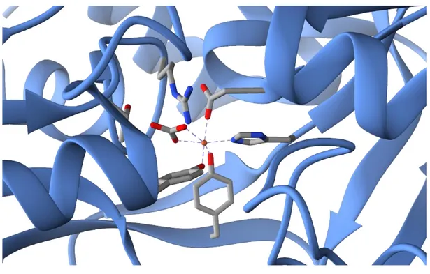
chemical tool used by living systems in order to make the stored metals available is complexation. Usually, metals are coordinated by the side chains of aminoacid residues like histidine, cysteine or tyrosine, however also small inorganic molecules may be involved as well as protein cofactors, e.g. porphyrins. Several examples can be gathered by the biochemistry of iron compounds which has been extensively studied being iron the metal with the highest abundance in all the living systems. In particular, the proteins of the transferrin family, which are implicated in the transfer of the iron ions, are a perfect example of how transition metals can be coordinated by the residues of a polypeptide. The iron ion in fact is coordinated in human transferrin by two phenoxide groups of tyrosine side chains, the imidazole nitrogen of a histidine residue and the carboxylate moiety of an aspartate. The octahedral coordination sphere is completed by the presence of a bidentate interaction with a bicarbonate ion. In figure 1.1 is reported a magnification of the
3 crystal structure of the N-lobe of a mutant human transferrin, which shows the wide range of interactions involved in the formation of the Fe-containing complex.7
Figure 1.1 Crystal structure of a human transferrin N-lobe coordinating an iron
4
1.2 Cisplatin and other metal-containing drugs
Inorganic elements are not only essential for life, their use has crossed the frontier of medicine leading to the introduction in clinical therapy of several metal-based drugs for a broad range of applications, as summarized in Scheme 1.1.8
Scheme 1.1 Some of the key areas involving medicinal inorganic chemistry.
Besides the multitude of therapeutic and diagnostic areas where metals and inorganic elements in general are involved, there is still the need of a better comprehension of the molecular basis behind their activity. An interesting example regards aluminum, a metal used as adjuvant in human and veterinary vaccines due to its enhancing activity of the pharmacological effect. Despite its widespread implementation, there are no clear evidences on the chemical basis of this.9
Moreover, as shown in section 1.1 metals in the biological media are mostly associated with ligands forming adducts that can show huge differences in their activity compared to the one of the element by itself. The dependence on both composition and environment on the behavior of metal complexes adds another complexity layer to the overall figure, thus explaining why it is so challenging to predict and control the behavior of inorganic pharmaceuticals.8
5 It is not surprising, though, that medicinal inorganic chemistry is a relatively young research area, stimulated by the progresses in analytical and spectroscopic techniques.1,8 Also, the growing of this discipline is connected with the success of
cisplatin, a Pt-based antineoplastic drug which is still widely used nowadays even if its discovery is dated back to the late ‘60s.10,11 The activity of cisplatin
(cis-[PtCl2(NH3)2]) derives from its interaction with the nucleobases of DNA but a
preliminary aquation step, which consists in the substitution of a chloride with a water molecule, is considered to be required for its activation.12,13 The necessity of
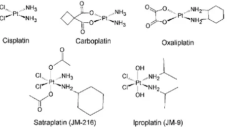
an in vivo transformation before reaching the target site is common among inorganic drugs. The possible transformations include a variety of reactions, from redox to substitution ones. Many efforts have been devoted to design more efficient and safer platinum drugs employing higher oxidation states of the metal, e.g. PtIV, unfortunately with scarce results. Nowadays, the FDA has approved only
three Pt-containing drugs: cisplatin, carboplatin and oxaliplatin, reported in figure 1.2. These three pharmaceuticals contain a PtII atom and share a square planar
geometry. However, several PtIV complexes have also been synthesized and are in
clinical trial. A few examples are shown in figure 1.2. However, no PtIV containing
complex has been currently approved for clinical use.
Figure 1.2. Cisplatin, carboplatin and oxaliplatin are square planar PtII complexes
and have been approved by the FDA for clinical use. They are widely employed worldwide. Satraplatin and iproplatin are PtIV octahedral complexes still in
6 The development of novel platinum drugs has been stimulated by the fact that cisplatin produces severe side effects and its use is strongly limited by drug resistance.14 However, many efforts devoted to prepare and screen new potential
platinum antineoplastic drugs have met with little success till now.15 In this regard,
recent advances in spectrometric and spectroscopic techniques may be useful in obtaining a better understanding of platinum complexes interaction and reactivity with biological molecules and in general in the mainframe of the chemistry of living system, hoping to gain insight towards an appreciable improvement in efficacy and safety for platinum-based chemotherapy.
7
1.3 Analytical tools for bioinorganic chemists
Exploring metal involvement in the chemistry of life has not been an easy task for decades. Most of the analytical methods developed for biochemistry were not meant to investigate the presence and the role of inorganic elements in biomolecules and, in addition, advanced spectroscopic techniques have to be developed to a state which makes it feasible to work with little amount of sample and limited stability. From the 80s, however, things have drastically changed and nowadays the bioinorganic chemist can choose among a vast number of techniques, which include electron paramagnetic resonance (EPR) and NMR (nuclear magnetic resonance) spectroscopy, IR (infrared) and Raman spectroscopy, X-ray-based techniques and mass spectrometry in all its developments.
Starting from the low energy end of the spectroscopic techniques, NMR is now widely used in the field of protein characterization, in particular after improvement in multiple dimensional methods and the use of isotopes with nuclear spin different from that of the naturally occurring elements (2H, 13C, 15N, 17O and 57Fe). NMR
spectroscopy has also been involved in several studies regarding platinum complexes and their interaction with biological targets. In 1985 Saudek was already studying the complexes of cisplatin with nitrogen containing ligands using NMR. In fact, platinum complexation on a nucleophilic N atom gives rise to two satellites and produces a characteristic chemical shift on the 1H NMR signals of the organic
ligand.16 Moreover, in the ‘90s many progresses were made in understanding donor
atom preferences of platinum complexes using 195Pt NMR, which shows
characteristic coupling constants for nucleophiles containing atoms such as the 15N
isotope.17 Moving on to the millimeter range, EPR was a decisive tool to determine
the aspect and function of copper proteins and it played an important role in the discovery of Fe-S proteins and in the exploration of their electronic structure.1,18
However, the pioneering work of Beinert on Fe-S clusters in proteins gained a new dimension using ray absorption spectroscopy (XAS) and in particular extended X-ray absorption fine structure (EXAFS) which allowed to discover and determine the structure of 3Fe clusters,19 showing the potential of high energy photons
8 spectroscopy in the study of metal clusters. Furthermore the analysis of the X-ray absorption near edge structure (XANES) has also demonstrated its importance, focusing on the pre-edge features that, used to determine the degree of covalency of metal-to-organic ligand bonding, provided new insights about the electronic structure of Fe-S proteins.20,21 XAS regions are schematized in figure 1.3
Figure 1.3 Schematic representation of an exemplary X-ray absorption spectrum.
XANES and EXAFS region are pointed out.
Vibrational spectroscopy had also its important role in bioinorganic chemistry. Resonance Raman (RR) spectroscopy for example, which is based on the enhancement of ordinary Raman lines by the presence in the molecule of a transition metal, is largely employed to have information about the position of the ligands near the metal center. Not to mention infrared spectroscopy that showed its analytical power when it helped to discover the presence of CO and CN ligands bound to the 2Fe clusters in hydrogenases.22 More recently, action IR spectroscopy
performed on mass-isolated bare ions has started to yield a fine characterization of the vibrational features of transition metal complexes with organic ligands.23,24
1.3.1 Mass spectrometry and MS-based techniques
Mass spectrometry can probably be seen as one of the most versatile tool in the hand of scientists from any field. Based on the implementation of different ion sources and/or analyzers, its function has shifted from a tool to detect cathode rays in the late 19th century to one of the forefront techniques for proteomic.25 The use
of MS in inorganic chemistry dates back to the first decades of the 20th century
where physicists like Aston and Nier used MS to discover new isotopes and determine their abundances and accurate masses.26 However, mass spectrometry
9 was relegated to volatile compounds, therefore with little applications in the biological field. Things drastically changed with the development of new ionization methods, in particular fast atom bombardment (FAB) in 1981, matrix-assisted laser desorption/ionization (MALDI) in 1988 and electrospray ionization (ESI) in the second part of the eighties. The last two methods have been particularly relevant for the analysis of macro biomolecules because they allow to analyze species up to and also above 100 kilodaltons, mass range that comprehends many of the proteins and other biological macromolecules. This was essentially the beginning of contemporary proteomics, which extensively exploits mass spectrometry in order to obtain information about the aminoacid sequence in a protein or the position of post-translational modifications. Two main methodologies are used: the bottom up approach that is based on the analysis of a proteolytic digestion;27 or the top-down
approach in which an intact protein is brought to the gas-phase and then activated and dissociated using several possible methods (e.g. CID, IR multiple photon dissociation or UV photodissociation) in order to obtain sequence coverage.28 In
addition, mass spectrometrists are now dealing with the study in the gas-phase of native species. Using soft conditions in the ionization source, it is possible to retain a protein conformation that can be attributed to the native form of the macromolecule.29 In this field, the developing of ion-mobility devices, which can be
used to separate isobaric ions based on their morphology or collisional cross-section (CCS), provides the perfect allied for seeking native molecular characterization.30
Now, what about bioinorganic chemistry? Mass spectrometry has been used in particular with inductively coupled plasma (ICP) sources to quantify and identify the presence of metals in biological samples due to its high sensitivity.31 However,
ICP-MS completely destroys the organic structure preventing to obtain structural information. Therefore soft sources like the above mentioned MALDI and ESI have been extensively exploited to understand the interaction of metals with biological molecules. The bottom-up approach on a protein such as calmoduline incubated with cisplatin allowed to obtain insights about the coordination sites of platinum within the protein and to have experimental evidences of its cross-linker character.32 In recent years, mass spectrometry has been coupled with IR
10 spectroscopy in order to obtain the vibrational features of mass isolated ions, removing thus the effect of solvent and other contaminant species on the absorptions and allowing to have a direct correlation of the obtained frequencies with calculated ones. This technique is based on the coupling of a tunable IR laser with mass spectrometers such as Paul ion traps and the ones based on Fourier-transform ion cyclotron resonance (FT-ICR). The number density of ions in the cell of this trapping instrument is too low to record the IR absorption. Therefore, the abundance of photofragments compared to the product ion is monitored. Fragmentation happens only when photons are resonant with the vibrations of the mass isolated molecules thus allowing to obtain an IRMPD spectrum plotting the fragmentation yield versus the photon frequency.33,34 IRMPD spectroscopy was
employed to characterize [Hemin]+ adducts with CO, N
2, and O235 and complexes of
nucleobases with metal cations36 as well as to assess the coordination sites of
cisplatin on adenine and guanine37, to cite just few examples among many
successful applications. Thanks to the absence of any solvent effect and to the possibility to analyze a single species from a complex mixture, IRMPD spectroscopy constitutes one of the most powerful tools in the hand on a bioinorganic chemist. 1.3.2 Theoretical methods
Theoretical simulations give an invaluable help in interpreting spectroscopic and kinetic data, in particular when dealing with a complex system involving metals interacting with organic molecules. At the same time, in the presence of a transition metal calculations have to be carefully planned and considered since usually a number of simplifications have to be made in order to obtain a fast and still reliable result. Therefore, despite the advances in computing power and in data storage have allowed to exploit ab initio or density functional theory (DFT) methods for simulating structures and properties of increasingly bigger molecules, we are still far from reaching a simple, homogeneous and automated protocol for calculations in the bioinorganic field. However, a careful choice of the calculation method, moving from DFT hybrid functionals to dispersion corrected ones, or employing post Hartree-Fock methods, permits to obtain fair results.38 In addition, when dealing
11 effects needs to be considered.39 Usually, an effective core potential (ECP) is used
that replaces the explicit quantum-mechanical treatment of the metal core electrons with an effective potential. The parameters of the ECP can be designed to mimic the relativistic effects therefore avoiding artifacts generated by the non-relativistic treatment of the Hamiltonian energy, but keeping low the computational cost of the calculations. Therefore, the selection of the right ECP is a critical factor in the simulation.40
Using these precautions and minding the limitations of each method, excellent results have been obtained for the simulation of structures, vibrational frequencies and reaction coordinates in the field of metal complexes.
12
1.4 ESI-MS to understand mechanisms of liquid-phase reactions: gas- and liquid-phase dichotomy
As anticipated in section 1.3 electro-spray ionization has opened mass spectrometry to biochemists and biologists. Moreover, ESI-MS has the unique possibility to analyze compounds that have been extracted with soft conditions from the solution to the gas-phase. An exemplary application is the exploration of native protein conformations, but the method can be also used to elucidate the mechanism of organic and inorganic reactions.41 Indeed, mass spectrometry has been extensively
exploited to study reactions happening in the gas-phase, allowing the ionic species of interest to interact with other molecules directly in the mass spectrometer. The novelty of the approach permitted by ESI is that a reaction can be studied as it occurs in solution, for example extracting an elusive ionic intermediate from the solution and assaying it in the gas-phase.42 One of the main concerns in this matter
is the possibility that the structure of the observed ion may be different from the one in solution, although there are studies showing superposition between the species investigated in gas-phase and the one studied by NMR in solution regarding the same reactions.43 What makes MS attractive to study reaction intermediates is
that they can be extremely stable in high-vacuum conditions even if they are short-lived in solution, thus allowing to assay their structure with spectroscopic techniques or CID and to probe their reactivity using ion/molecule reactions. In addition, ESI-MS permits to detect ions at very low concentration so it is ideal for the rapid screening of micro-scale reactions.
Finally, ESI-MS has allowed to address the reactivity of microsolvated cluster ions with the aim to understand solution chemistry from gas-phase behavior.44 From a
bioinorganic chemist point of view, this is one of the most appealing features of ESI-MS that can be exploited to reveal with the definition and precision of gas-phase measurement, reaction mechanisms as they happen in the liquid environment of the biological media.
13
References
[1] Beinert, H. Bioinorganic chemistry: A new field or discipline? Words, meanings, and
reality J. Biol. Chem. 277 (2002) 37967-37972.
[2] Bertini, I., Gray, H.B., Lippard, S.J., Valentine, J.S. Bioinorganic Chemistry University Science Books 20 Edgehill Rd., MIll Valley, CA, 1994.
[3] Theil, E.C. Ferritin: structure, function, and regulation Adv. Inorg. Biochem. 5 (1983) 1-38.
[4] Crichton, R.R., Charloteaux‐Wauters, M. Iron transport and storage Eur. J. Biochem. 164 (1987) 485-506.
[5] Palmiter, R.D. The elusive function of metallothioneins Proc. Nat. Acad. Sci. U.S.A. 95 (1998) 8428-8430.
[6] Coyle, P., Philcox, J.C., Carey, L.C., Rofe, A.M. Metallothionein: The multipurpose
protein Cell. Mol. Life Sci. 59 (2002) 627-647.
[7] Yang, A.H.-W., Macgillivray, R.T.A., Chen, J., Luo, Y., Wang, Y., Brayer, G.D., Mason, A.B., Woodworth, R.C., Murphy, M.E.P. Crystal structures of two mutants (K206Q,
H207E) of the N-lobe of human transferrin with increased affinity for iron Protein Sci.
9 (2000) 49-52.
[8] Alessio, E. Bioinorganic Medicinal Chemistry, Wiley-VCH Verlag GmbH, Weinheim (2011).
[9] Gupta, R. K. Aluminum compounds as vaccine adjuvants Adv. Drug. Deliv. Rev. 32 (1998) 155-172.
[10] Rosenberg, B., Van Camp, L., Krigas, T. Inhibition of cell division in Escherichia coli by
electrolysis products from a platinum electrode Nature 205 (1965) 698–699
[11] Rosenberg, B., Van Camp, L., Trosko, J.E., Mansour, V.H. Platinum compounds: A new
class of potent antitumour agents Nature 222 (1969) 385–386.
[12] Berners-Price, S. J.; Appleton, T. G. The Chemistry of Cisplatin in Aqueous Solution. In
Platinum-Based Drugs in Cancer Therapy in Platinum-Based Drugs in Cancer Therapy,
Kelland, L. R., Farrell, N., Eds.; Humana Press: Totowa, NJ, (2000) 3-35.
[13] Davies, M. S.; Berners-Price, S. J.; Hambley, T. W. Slowing of Cisplatin Aquation in the
Presence of DNA but not in the Presence of Phosphate: Improved Understanding of Sequence Selectivity and the Roles of Monoaquated and Diaquated Species in the Binding of Cisplatin to DNA. Inorg. Chem. 39 (2000) 5603−5613.
[14] Rabik, C.A., Dolan, M.E., Molecular mechanisms of resistance and toxicity associated
with platinating agents Cancer Treat. Rev 33 (2007) 9-23.
[15] Xin Zhang, C., Lippard, S.J. New metal complexes as potential therapeutics Curr. Opin.
14
[16] Saudek, V., Pivcova, H., Noskova, D., Drobnik, J. The reaction of Pt-antitumor Drugs
with selected nucleophiles. II. Preparation and Characterization of Coordination Compounds of Pt(II) and L-Histidine J. Inorg. Biochem. 23 (1985) 55-72.
[17] Appleton T.G. Donor atom preferences in complexes of platinum and palladium with
amino acids and related molecules Coord. Chem. Rev. 166 (1997) 313-359.
[18] Beinert, H. Spectroscopy of succinate dehydrogenases, a historical perspective
Biochim. Biophys. Acta, Bioenerg. 1553 (2002) 7-22
[19] Beinert, H., Emptage, M.H., Dreyer, J.L., Scott, R.A., Hahn, J.E., Hodgson, K.O., Thomson, A.J. Iron-sulfur stoichiometry and structure of iron-sulfur clusters in
three-iron proteins: evidence for [3Fe-4S] clusters Proc. Nat. Acad. Sci. U.S.A. 80 (1983)
393-396.
[20] Anxolabéhère-Mallart, E., Glaser, T., Frank, P., Aliverti, A., Zanetti, G., Hedman, B., Hodgson, K.O., Solomon, E.I. Sulfur K-edge X-ray absorption spectroscopy of 2Fe-2S
ferredoxin: Covalency of the oxidized and reduced 2Fe forms and comparison to model complexes J. Am. Chem. Soc. 123 (2001) 5444-5452.
[21] Glaser, T., Rose, K., Shadle, S.E., Hedman, B., Hodgson, K.O., Solomon, E.I. S K-edge
X-ray absorption studies of tetranuclear iron-sulfur clusters: μ-sulfide bonding and its contribution to electron delocalization J. Am. Chem. Soc. 123 (2001) 442-454.
[22] Pierik, A.J., Roseboom, W., Happe, R.P., Bagley, K.A., Albracht, S.P.J., Lacey, D. Carbon
Monoxide and Cyanide as Intrinsic Ligands to Iron in the Active Site of [ NiFe ] -Hydrogenases J. Biol. Chem.274 (1999) 3331-3337.
[23] MacAleese, L., Maitre, P. Infrared spectroscopy of organometallic ions in the gas
phase: from model to real world complexes Mass Spectrom. Rev.26 (2007) 583-605.
[24] Duncan, M. A. Structures, energetics and spectroscopy of gas phase transition metal
ion-benzene complexes Int. J. Mass Spectrom. 272 (2008) 99-118.
[25] Dass, C. Fundamentals of Contemporary Mass Spectrometry eds. John Wiley & Sons, Inc (2007).
[26] Vestal, M.L., Methods of ion generation Chem. Rev. 101 (2001) 361-375.
[27] Aebersold, R., Mann, M. Mass spectrometry-based proteomics Nature 422 (2003) 198-207.
[28] Sze, S.K., Ge, Y., Oh, H., McLafferty, F.W. Top-down mass spectrometry of a 29-kDa
protein for characterization of any posttranslational modification to within one residue Proc. Natl. Acad. Sci. U.S.A.99 (2002) 1774-1779.
[29] Heck, A.J.R. Native mass spectrometry: A bridge between interactomics and
structural biology Nat. Methods 5 (2008) 927-933.
[30] Lanucara, F., Holman, S.W., Gray, C.J., Eyers, C.E. The power of ion mobility-mass
spectrometry for structural characterization and the study of conformational dynamics Nat. Chem. 6 (2014) 281-294.
15
[31] Łobiński, R., Schaumlöffel, D., Szpunar, J. Mass spectrometry in bioinorganic analytical
chemistry Mass Spectrom. Rev. 25 (2006) 255-289.
[32] Li, H., Zhao, Y., Phillips, H.I., Qi, Y., Lin, T.Y., Sadler, P.J., O’Connor, P.B. Mass
spectrometry evidence for cisplatin as a protein cross-linking reagent Anal. Chem. 83
(2011) 5369-5376.
[33] Polfer, N.C., Oomens, J. Vibrational spectroscopy of bare and solvated ionic
complexes of biological relevance Mass Spectrom. Rev. 28 (2009) 468-494.
[34] Eyler, J.R. Infrared multiple photon dissociation spectroscopy of ions in Penning traps
Mass Spectrom. Rev. 28 (2009) 448-467.
[35] Dillinger, S., Lang, J., Niedner-Schatteburg, G. Cryo IR spectroscopy of [Hemin](+)
complexes in isolation J. Phys. Chem. A 28 (2017) 7191-7196.
[36] Salpin, J.-Y., Macaleese, L., Chirot, F., Dugourd, P. Structure of the Pb2+-deprotonated
dGMP complex in the gas phase: A combined MS-MS/IRMPD spectroscopy/ion mobility study Phys. Chem. Chem. Phys. 16 (2014) 14127-14138.
[37] Chiavarino, B., Crestoni, M.E., Fornarini, S., Scuderi, D., Salpin, J.Y. Interaction of
cisplatin with adenine and guanine: A combined IRMPD, MS/MS, and theoretical study J. Am. Chem. Soc. 135 (2013) 1445-1455.
[38] Ghosh, A. Ab initio wavefunctions in bioinorganic chemistry: More than a succès
d’estime? J. Biol. Inorg. Chem. 16 (2011) 819-820.
[39] Schwarz, H. Relativistic effects in gas-phase ion chemistry: An experimentalist’s view
Angew. Chemie – Int. Ed. 42 (2003) 4442-4454.
[40] Schwerdtfeger, P. The pseudopotential approximation in electronic structure theory
ChemPhysChem 12 (2011) 3143-3155.
[41] Leonardo S. Santos, Reactive intermediates: MS Investigations in Solution. Ed. Leonardo S. Santos (2010) WILEY-VCH Verlag GmbH & Co. KGaA, Weinheim.
[42] Aliprantis, A.O., Canary, J.W. Observation of Catalytic Intermediates in the Suzuki
Reaction by Electrospray Mass Spectrometry J. Am. Chem. Soc. 116 (1994) 6985-6986.
[43] Colton, R., D’Agostino, A., Traeger, J.C. Electrospray mass spectrometry applied to
inorganic and organometallic chemistry Mass Spectrom. Rev. 14 (1995) 79-106.
[44] Takashima, K., Riveros, J.M. Gas-phase solvated negative ions Mass Spectrom Rev. 17 (1998) 409-430.
16
Methods
2.1 Mass Spectrometry
Mass spectrometry (MS) is an analytical technique that permits to measure the mass of molecules after they are ionized and volatilized. More precisely, it records the m/z ratio of an ion where m is the mass in Da and z is the charge of the ion. MS presents unique features that have favored its use in several fields of scientific research1:
It has unmatched molecular specificity due to its ability to measure accurate molecular masses. Also using tandem MS or other hyphenated techniques, MS can provide useful structural information.
The detection sensitivity of mass spectrometry is extremely high allowing to identify molecules in attomole and zeptomole amounts.
Using different kinds of ionization sources, MS is applicable virtually to all kind of samples.
In addition mass spectrometry permits to study gas-phase reactivity of ions and to obtain data on physical properties such as ionization energy, enthalpy of reaction, proton affinities and so on.
The only major limit of mass spectrometry is that it can only measure the m/z ratio of charged species. Therefore, the experimental apparatus can be subdivided in different sections which are schematized in scheme 2.1.
17 Several ion sources and analyzers have been developed along the years allowing the application of mass spectrometry in different fields of science, from the analysis of petroleum products to proteomics. For the aim of this thesis, we have employed electrospray ionization (ESI) as ion source and three different analyzers: triple quadrupole, a Paul ion trap and a Fourier-transform ion cyclotron resonance (FT-ICR) cell.
2.1.1 ESI source
Electrospray ionization has been a groundbreaking development for mass spectrometry and in general in the field of analytical chemistry, as testified by the 2002 Nobel Prize in chemistry awarded to the developer of ESI, John Fenn.2,3 ESI is
an atmospheric pressure ionization (API) technique and it is applicable to a conspicuous range of liquid-phase samples. It has the characteristic to be a soft ionization source, thus leading to poor in source fragmentation and consistency between the gas-phase nature of the ion with the solution one. This is particularly true when the observed species is already ionized in the solvent, but this is not always the case. In fact, the ions can also be generated at the surface of the droplets that are implied in the spray process. The ionization process will therefore follow a gas-phase like regime and be regulated by gas-phase basicity.4 ESI has
allowed to successfully couple liquid chromatography with mass spectrometry.5
One of its main features is the possibility to form multiply charged ions, which can be useful for observing large molecules beyond the m/z limit of the chosen analyzer, thus enabling the study of proteins and other biological macromolecules with MS.6
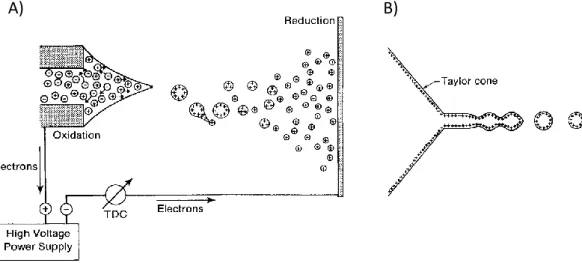
The overall electrospray ionization process can be rationalized in two independent phases: the electrostatic dispersion of the sample liquid into droplets containing both ions and neutral species; and the separation of the charged molecules from the neutrals and the solvent leading to bare ions.7 The formation of the droplets is
usually obtained applying an electrostatic potential between a narrow-bore injection capillary and an opposing counter-electrode, as depicted in figure 2.1 A. Often the evaporation of the charged droplets is assisted by a flow of hot nitrogen.
18 Applying a high electric field at the capillary tip generates a partial charge separation, thus in positive ion mode, cations are enriched at the surface of the liquid at the capillary tip. The combination of electrostatic repulsion of the ions and electric field pull eventually overcomes the surface tension of the solvent shaping the liquid at the tip as a cone, called Taylor cone. Finally, the tip of the cone elongates into a filament that afterward breaks forming charged droplets (figure 2.1 B).
Figure 2.1 Representation of ESI process (A) and of the liquid cone at the tip of
the capillary (B). The figure is adapted from ref. 7.
The electrospray formation process has been extensively studied and is now well understood.8 The onset of the spray requires an electric field given by the equation
2.1:
𝐸
0= (
2𝛾cos49°𝜀0𝑟𝑐
)
1/2
(2.1)
where γ is the surface tension of the liquid, cos49° is the half angle of the Taylor cone, ε0 is the permittivity of vacuum and rc is the capillary radius. From the
equation it is clear that solvents with high surface tension, such as water, will require higher electric field for the electrospray formation. Thus, special arrangements could be needed when water is the sole solvent.
The evolution of the charged droplets to form the naked gas-phase ions is instead still a discussed theme. The process starts with progressive Coulomb fissions of the droplets. After formation, the heat supplied from the air, or the hot nitrogen,
19 produces solvent evaporation decreasing the droplet radius until it reaches the Rayleigh limit (equation 2.2) in which the surface tension is not enough to counterbalance the Coulombic repulsion of the charges inside the droplet:
𝑞 = 8𝜋(𝜀
0𝛾𝑟
3)
1/2(2.2)
However, Coulombic fission has been observed only for big charged droplets, above 100 nm. Thus, two mechanisms have been proposed to explain the steps finally forming the bare ions: the ion evaporation model (IEM)9,10 and the charge residue
model (CRM)11. The CRM proposes that the droplets continue to shrink due to
Coulombic fission until no further evaporation of the solvent is possible, while the IEM suggests a direct emission of ions M+ from those droplets which present a size
reduced to a radius comprised from 10 to 20 nm after solvent evaporation and Coulombic fissions. At present, there is no final word on which theory provides a better model for the ESI process despite the many efforts devoted to unambiguously prove the mechanism of the ions formation. In fact, it is probably a combination of the CRM and the IEM that is responsible for the high sensitivity and versatility of the ESI source. In particular, the ion evaporation process is considered to be involved in the formation of small ions, while CRM is invoked for clusters and big ions.8 The overall ESI process clearly shows that the ions are formed in a gentle
fashion due to the cooling provided by solvent evaporation, making possible to obtain molecular and pseudo-molecular ions, both protonated and metallated, which may retain isomeric and/or conformational fetures from the solution.
2.1.2 Time-of-flight mass spectrometers
Time-of-flight (TOF) mass analyzers are among the simplest devices to run mass analysis and are currently mainstream due to the potentially unlimited mass range, making TOF mass spectrometers preponderant in the analysis of biomolecules.12
The developing of reflectron TOF instruments which have minimized the effect on mass resolution of initial spatial and energy spread,13 helped to overcome the major
limit of this technique, namely the low mass resolution at high masses, contributing to the popularity of the instrument.
20 Stephens presented the basics of TOF analysis in 1946.14 Ions with defined kinetic
energy present different velocities based on the inverse function of the square root of their m/z values. Therefore, when a packet of ions travels in a flight tube of length L the arrival time of the different species is given by:
𝑡 =
𝐿𝑉
= 𝐿 √
𝑚
2𝑧𝑉 (2.3)
Measuring the arrival time provides a time-domain spectrum, which can be converted with an appropriate calibrating function to a mass spectrum. As previously mentioned, mass resolution in TOF instruments is highly dependent on having pulsed ions with defined and equal kinetic energy and without spatial spread, thus in standard instruments mass resolution was usually low and inadequate to analyze proteins and other biomolecules. Reflectron instruments allowed to overcome this issue, in particular the device permits to reduce the effect of spatial and energy spread using an electrostatic mirror at the end of the first field-free region (FFR or flight tube) of the instrument, as represented in figure 2.2.
Figure 2.2 Reflectron time-of-flight mass spectrometer. Adapted from Ref. 1.
In principle an ion with excess energy +U0 will arrive earlier at the reflectron but it
will penetrate deeper in the field (d), thus slowing down and compensating for the excess of starting energy. Reflectron TOF opened the technique to proteomic and contributed to its success. To the scope of this thesis, a reflectron-TOF instrument coupled with an ion-mobility sector was employed for the mobility measurements.
21 2.1.3 Quadrupole analyzers
Quadrupole-based instruments are among the most common types of mass spectrometers mainly due to the low cost, mechanical simplicity and compactness of the device. The basic principles regarding the quadrupole mass filter can be dated back to the pioneering work of Paul and Steinwedel in the fifties.15 Four
metallic rods of ideally hyperbolic geometry are arranged symmetrically in a square array as shown in figure 2.3 and the mass separation is generated by the motion of ions into a high frequency oscillating electric field that is maintained by electrically connecting opposite pairs of rods.16
Figure 2.3 A) schematic and B) picture representation of a linear quadrupole
mass filter. Figure adapted from Ref. 17.
One of the pair receives a superimposed dc potential U and a time-dependent rf potential V cos ωt, where ω is the angular frequency (in rad sec-1), V is the
amplitude and t the time, while the other pair presents a dc –U and an rf potential equal in magnitude, but out of phase by 180°. The resulting oscillating field is given by:
Ф
(𝑥,𝑦)= (𝑈 + 𝑉 cos 𝜔𝑡)
𝑥2− 𝑦2
𝑟0 (2.4)
where Ф is the applied potential, r0 the inscribed radius so one-half the distance
between the opposite electrodes), and x and y the distances from the center of the field. The ions are injected in the z direction and their motion is influenced by the applied potential based on their m/z ratio. Therefore, for given values of U, V and ω
22 only the ions presenting a certain m/z are able to sweep through the quadrupole and reach the detector or the other sectors of the mass spectrometer, while the others, presenting wrong trajectories, are discharged on the rods. To obtain the mass spectrum the quadrupole field is varied in order to force ions of consecutively
m/z windows to break through the quadrupole. The advantages of a quadrupole
analyzer comprise high scan speed, high transmission, good sensitivity and linear mass range, however quadrupoles present a practical upper mass limit of 4000 m/z, which can be limiting for applications on biomolecules, and the impossibility of obtaining perfect hyperbolic rods set a practical resolution limit to the unit.
Quadrupole ion-trap MS
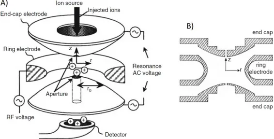
Quadrupole ion-trap (QIT), also called Paul trap from the name of its inventor who introduced it in 1958,18 and was awarded for this work of the Nobel Prize for Physics
in 1989, are popular mass analyzers that work as three-dimensional analogs of quadrupole mass filters. QIT functions both as an ion store and as a mass analyzer. The trapping of the ions is generated by a potential well applied to the electrode of the ion trap, which can be visualized like a bowl of parabolic cross-section in which the ions are confined in horizontal layers based on their m/z ratio. The electrodes are arranged as shown in figure 2.4.
Figure 2.4 A) Schematic representation of a QIT. B) Section in the rz-plane of a
23 Two of the three electrodes, called end-caps, have a hyperboloidal geometry and are identical but for the number of holes in each of them. One end-cap electrode has a single small central aperture that permits the inlet of the ions, while the other can have more than one small aperture allowing the passage of the ions to the detector. The third electrode has a two sheets hyperboloidal geometry and is called the ring electrode. The potential well is created from the field generated when an rf potential is applied to the ring electrode, while the two end-cap electrodes are grounded. The overall geometry of the trap in this conditions produces an ideal quadrupole field which in turn generates a parabolic potential well for the confinement of the ions.19 The ion trap acts as a mass spectrometer when the field
is changed making the trajectories of ions of consecutive m/z ratio sequentially unstable, following a method developed by Stafford et al.20 Therefore, the ions
leave the trap based on their m/z ratio to a detector and, knowing the initial amplitude and ramping rate, it is possible to correlate a specific electronic signal to its m/z ratio. A prerequisite for the mass-selective axial ejection method is that the ions have to be collision constrained at the center of the ion trap, thus requiring a certain neutral gas background pressure, usually ca. 1 mTorr of Helium. The motion of the ions and therefore their stability regions is governed by the Mathieu’s equation21 and can be expressed in term of parametrized coordinates:
𝑞
𝑧=
4𝑧𝑉 𝑚𝜔2𝑟 02𝑎
𝑧= −
8𝑧𝑈 𝑚𝜔2𝑟 02 (2.5) where V is the rf peak voltage, U is the voltage applied to the ring electrode, r0 itsradius, ω is the angular frequency of the rf voltage and m and z are respectively the mass and charge of the ion.
2.1.4 FT-ICR analyzers
Fourier transform ion-cyclotron resonance mass spectrometry (FT-ICR MS) is one of the oldest ion trap techniques. The basic principle behind its functioning was firstly described in 1930 by Lawrence and Edlesen22 and developed to mass spectrometry
by Sommers et al.23 in 1949, but its modern form is due to the work of Comisarow
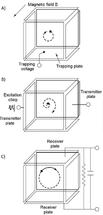
24 strong static magnetic field and axially by a static electric field. There are lots of designs for Penning traps and the functioning of a cubic cell is schematized in figure 2.5 for its simplicity.However, nowadays the most widely used cells are cylindrically shaped.
Figure 2.5 Schematic representation of FT-ICR MS operation. Mass analysis
involves three main steps: A) ion formation and storage; B) excitation of the ions by a broad-frequency range pulse; C) detection by measuring their image current. Figure adapted from Ref. 1.
25 Two basic concepts are at the basis of mass analysis in ICR, in particular (i) an ion traveling in a magnetic field will precess at a frequency given depending on its m/z; (ii) energy can be absorbed by a precessing ion from an rf source only when the rf frequency coincides with the ion cyclotron one. The cyclotron frequency of the ion
ωc is defined as the number of revolution per second according to the equation:
𝜔
𝑐=
𝑞𝐵2𝜋𝑚 (2.6)
where q and m are respectively the charge and mass of the ion and B the strength of the magnetic field. The operation in ICR experiments is along four time-spread events: quenching, ion formation/injection, excitation and detection. These events happen inside the cell which is composed by three pairs of opposing plates devoted to a specific function: trapping, excitation or detection,25 as shown in figure 2.5. To
begin, a quench pulse is applied to empty the cell of any remaining ions, subsequently the ions to be analyzed are either formed in the cell or, which is more common nowadays, pulsed in the cell from an external source. At this point, a few volts are applied to the trapping electrodes (figure 2.5 A), in order to store the ions, and gas may be introduced in the cell to cool them. The next step is the application of an excitation pulse through the excitation electrodes (figure 2.5 B). This way, the ions whose precessional frequency is resonant with that of the excitation pulse absorb energy and get promoted to larger orbits. Finally, the ions, as they orbit the ICR cell, are detected by measuring the image current generated in the receiving plates (figure 2.5 C). The excitation pulse in the FT-ICR instruments is dispensed in the form of a chirp, a fast sweep of frequencies over a broadband. The ions, whose cyclotron frequencies fall in the range of frequencies applied, are promoted to larger orbital radii in phase coherent packets generating an image current containing the frequency components of all the excited ions. The time-domain transient signal obtained is finally converted to a frequency-domain signal that can be translated to a mass spectrum using a convenient calibration file.
26
2.2 Tandem mass spectrometry
Mass spectrometry when coupled with soft ionization sources such as ESI, while gaining the possibility to observe the molecular or pseudomolecular mass of the assayed ions, tends to lose structural information that was gathered from the dissociation paths generated with harsher ionization methods, such as electron ionization. Tandem MS or MS/MS is a tool that permits to recover the information about structures retaining the advantages of soft ionization sources. It is based on the coupling of two or more mass analysis steps either in time or space. Tandem MS was firstly used in the late sixties and nowadays there are several devices and instruments with different characteristics and fragmentation/assaying techniques that permit to gather structural information. We will focus on collision-induced dissociation (CID) and infrared multiple photon dissociation (IRMPD)
2.2.1 Collision induced dissociation
CID is the most common activation and dissociation tool and it is usually available in every commercial mass-spectrometer. This technique, which was firstly introduced in 1968,26 is a two-step process: a preliminary collision activation followed by
unimolecular dissociation. In the first step, the mass-selected ions are accelerated and excited to higher-energy states after collision with an inert gas (e.g. Ar, He or N2). The kinetic energy is therefore transformed to internal energy based on the
mass of the precursor ion and that of the neutral gas, following the equation:
𝐸
𝑐𝑜𝑚=
𝑁𝑚𝑝+𝑁
𝐸
𝑙𝑎𝑏 (2.7)where Ecom is the center-of-mass kinetic energy of the ion, mp and N are respectively
the masses of the precursor ion and the neutral and Elab is the ion’s energy in the
laboratory frame.1 The unimolecular dissociation step has been fully theorized by
the Rice–Ramsperger–Kassel–Marcus (RRKM) theory and quasiequilibrium theory (QET). A thorough introduction to the unimolecular dissociation theory can be found in Ref. 27.
27 Depending on the instrument, CID can be performed in either high- or low- energy regime. We will focus on the low-energy regime which is primarly used in triple quadrupole- and ion trap- (QIT, FT-ICR-MS) based tandem instruments. Low-energy CID,28 has somewhat long activation time (milliseconds to seconds) and involves
high vibrational states. Usually high pressure is present in the collisional cell to allow more ions to participate to the CID process, which can lead to a stepwise activation of the precursor.
2.2.2 IR multiple photon dissociation
IRMPD is a slow heating process usually performed in ion traps (FT-ICR or QIT). The trapped ions are bombarded with IR photons that, if resonant with the vibrational modes of the ions, are absorbed in a stepwise fashion until the dissociation threshold is reached and the ion eventually fragments. Usually, the lowest-energy decomposition pathways are sampled. The IRMPD technique is not dependent on the mass of the ion, thus it has acquired popularity in the –omics, in particular for top-down approaches.29 The usual setup for this application requires a fixed
frequency continuous-wave CO2 laser coupled with FT-ICR-MS or Paul ion traps.
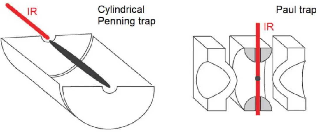
Attention has to be devoted to obtaining a proper overlapping of the laser beam with the ion-cloud. Taking into account the differences regarding the ion clouds shapes and dimensions between the Penning trap and the quadrupole ion trap, two different coupling geometries are conveniently used, as showed in figure 2.6.
Figure 2.6 Section of Penning and Paul traps. The ion clouds are represented in
black together with the optical access for the IR laser. Figure adapted from Ref. 30.
28 IRMPD spectroscopy
Ions in mass spectrometers are in extremely low density (<108 cm-3) thus measuring
the direct absorption of IR light is extremely challenging, de facto impeding to perform IR spectroscopy on mass selected ions in the gas-phase. The possibility to obtain dissociation upon IR absorption with IRMPD, however, showed a way to circumvent the need to measure light absorption in order to record the spectrum. In practice, a mass-selected ion is irradiated with a tunable laser beam at a particular frequency. If the laser frequency is resonant with one of the vibrational modes of the ion, the absorption may results in a dissociation, which can be recorded by the mass spectrometer. Finally, performing a scan with the tunable laser along the IR region of the electromagnetic spectrum, while monitoring the dissociation yield, permits to record an IRMPD spectrum related to the absorption one of the ion.30 IRMPD spectroscopy is therefore an “action” spectroscopy that
records a modification that happens when the absorption occurs, in this particular case the unimolecular dissociation of the starting ion. The measurement of the absorption consequence is more sensitive than directly monitoring the absorption, thus the method is well suited to study low-density samples such as the ions in a mass spectrometer. However, the advantages of this process are accompanied by an increased complexity of its mechanics, in particular due to the non-linear nature of IRMPD. In fact, the absorption of multiple photons is a non-coherent process where energy is quickly dissipated into the bath of vibrational degrees of freedom through intra-molecular vibrational redistribustion (IVR),31,32 as illustrated in figure
2.7.
29 The absorption of multiple photons leads to a progressive increase of the internal energy of the assayed ion until the dissociation threshold is reached and fragments can be observed. IVR is therefore avoiding the anharmonicity bottleneck that could happen for a “ladder-climbing” process, however since the normal modes of ions at higher internal energy are not orthogonal to the ground state ones, this process can shift the absorption frequency out of resonance with the laser. This effect is tempered by an increase of the density of states, which are functions of the internal energy, leading to a broadening of the absorption lines. Another factor to account when dealing with IRMPD is the presence of processes leading to loss of internal energy, in particular, stimulated emission, spontaneous emission and collision with background gas. Spontaneous emission among these can play an important role, while the others can be usually neglected in ultra-high vacuum condition. A collisionless environment has been estimated to exist at <10-7 mbar,33 which is a
pressure well above the one in the Penning trap, but can be reached in Paul trap, thus adding another layer of complexity when coupling IRMPD with this kind of instruments.
30
2.3 Ion mobility mass spectrometry
Ion mobility (IM) is a technique that separates ions based on their mobility, or the ability to move through a medium, usually an inert gas, under the influence of a driving force. The basic principle of this technique can be traced back at the beginnings of the 20th century, however the first drift tube with characteristics
similar to that of present IM instruments was developed by McDaniel in the fifties.34
A gas-filled drift tube in which the ions move under the influence of a static electric field represents an ion mobility spectrometer in its basic form. In this kind of instruments the velocity of an ion (v) can be defined as the product of the electric field (E) and the mobility of the ion (K), and can be measured as the time required (td) to move through a drift cell of dimension d.35
𝑣 = 𝐾𝐸 =
𝑑𝑡𝑑 (2.8)
The mobility costant normalized for pressure and temperature (T) can also be described based on the characteristics of the ion and the parameters of the drift cell:
𝐾
0=
3𝑧𝑒 16𝑁𝜔 1 𝛺(
2𝜋 𝜇𝑘𝐵𝑇)
1 2⁄ (2.9)where ze is the ion charge, Ω is the average collision cross section (CCS), N the number of density of the drift gas and μ is the reduced mass of the ion and buffer gas. Therefore, the velocity of an ion in a defined drift cell will depend on the mass, charge and collision cross section of the ion. If the IM sector (IMS) is coupled to a mass spectrometer, we can simultaneously obtain the m/z ratio, allowing the separation of isobaric ions through their CCS.
The first commercial IM-MS instrument was introduced by Waters in 2006 and consists in a reflectron-TOF analyzer preceded by a travelling wave (TW) ion-mobility device and a quadrupole sector which permits to mass-select ions (figure 2.8 A). 36 In the TWIMS (traveling wave ion-mobility sector), alternating phases of
RF voltage are applied to a ring ion guide on which a traveling potential wave is superimposed, as shown in figure 2.8 B. A reverse gas flow is present in the IMS
31 that, antagonizing the movement of low mobility ions, eventually makes them to roll over the crest of the wave and exit the cell later. It has to be noted that the mobility of ions in TWIMS is not directly related to their CCS as in drift tube (DT) based IM-MS. Despite this, the use of carefully selected standards, whose CCS has been previously recorded using DT IM-MS, allowed to correlate the arrival time of an ion through a TWIMS with the corresponding collision cross section.37
Figure 2.8 Schematic representations of B) the functioning of TWIMS and A) a
32
2.4 Computational methods
Computational chemistry is nowadays intertwined with all branches of experimental chemistry and accurate simulations of molecular features are required to obtain a proper description of observed phenomena. An impressive number of methods have been developed in order to employ calculations at all levels, starting from chemical physical problems on 2-atom systems to the screening of potential candidates to block the activity of an enzyme. This chapter will focus on specific problems related to the simulation of bioinorganic complexes by means of density-functional theory (DFT) calculations and post-Hartree Fock methods based on perturbation theory (MP2), in particular the implementation of dispersion energy in DFT and the use of relativistic pseudopotential to calculate the properties of species containing heavy atoms.
2.4.1 Basic principles of quantum chemistry applied to calculations
Quantum mechanics is based on postulates that have been experimentally tested in order to prove their validity. In particular, the first postulate states that to describe the state of a system exists a function Ψ of the particles coordinates called state function or wave function, which was firstly described by Schrödinger for a one-electron/one-dimension problem. The approach used to simulate a molecular system in ab initio computational chemistry is to resolve the wave function of that system. This is indeed not an easy task, in particular for polyatomic molecules, and appropriate mathematical tools as well as approximations have to be used.38
The wave function problem is nevertheless an eigenvalue problem, where to describe the energy of the system is necessary to find the eigenvalues Ei of the
Hamiltonian operator Ĥ with respect to the eigenfunctions ψ that represent in our case the time-independent wave functions of our system:
Ĥ𝜓
𝑖= 𝐸
𝑖𝜓
𝑖 (2.10)For a polyatomic molecule, the Hamiltonian operator of equation 2.10 is of formidable appearance containing terms regarding the kinetic energies for the nuclei and the electrons and the potential energies for the repulsion and attraction
![Figure 3.4 shows an example of kinetic plot for the reaction of cis-[PtCl(NH 3 ) 2 (H 2 O)] +](https://thumb-eu.123doks.com/thumbv2/123dokorg/2895840.11645/67.892.236.674.469.753/figure-shows-example-kinetic-plot-reaction-cis-ptcl.webp)
![Figure 3.5 Computed profile for the reaction of cis- and trans-[PtCl(NH 3 ) 2 (H 2 O)] +](https://thumb-eu.123doks.com/thumbv2/123dokorg/2895840.11645/70.892.154.770.317.758/figure-computed-profile-reaction-cis-trans-ptcl-nh.webp)
![figure 3.18 together with the calculated IR spectra of the lowest lying species, conforming to either [PtCl(NH 3 ) 2 L] + · H 2 O (such as DMA-H2O_1) or](https://thumb-eu.123doks.com/thumbv2/123dokorg/2895840.11645/96.892.162.760.381.676/figure-calculated-spectra-lowest-lying-species-conforming-ptcl.webp)
