DIPARTIMENTO DI NEUROSCIENZE UMANE
Dottorato di Ricerca in Neuroscienze Clinico- Sperimentali e Psichiatria
Curriculum Disturbi Neurologici, Neuroevolutivi e Psichiatrici in età evolutiva
Ciclo XXXII
“Identification and functional characterization of new disease
genes and variants implicated in pediatric encephalopathies:
from exome sequencing to in vivo investigation”
Tutor: Candidata:
Prof. Vincenzo Leuzzi Martina Venditti
Co-Tutor:
“Don't let anyone rob you of your imagination,
your creativity or your curiosity.”
1
INDEX
INDEX
1
ABSTRACT
4
1.
INTRODUCTION
6
1.1 Undiagnosed Diseases Program 6
1.2 Infantile encephalopathies with neurodevelopmental defects 7
1.3 Next generation sequencing 9
1.3.1 “Wet lab” workflow: library preparation, exome capture and sequencing technology 12
1.3.2 “Dry lab” workflow: sequence alignment, variant calling and gene annotation 14
1.4 The model organism Zebrafish (Danio Rerio) 19
1.4.1 Why does zebrafish make such good animal model? 24
1.4.2 Use of zebrafish to investigate pediatric rare disease affecting the neurodevelopment 25
1.5 Early neurogenesis in zebrafish 28
1.6 Morphogenesis of axes formation in zebrafish 32
1.7 AP patterning and segmentation of the vertebrate head and trunk: role of krox20
and myoD. 34
2. AIMS OF MY PHD THESIS
40
3. MATERIALS AND METHODS
43
3.1 Patients selection 43
3.2 Whole exome sequencing (WES) 43
3.2.1 Sample preparation 45
3.2.2 Hybridization and Capture 49
2
3.2.4 Clusters generation and sequencing 59
3.3 Exome sequencing data analysis 60
3.4 Zebrafish husbandry 62
3.5 Zebrafish breeding, embryos collection and staging 62
3.6 Whole-mount in Situ Hybridization of Zebrafish Embryos 64
3.6.1 Sample preparation: early zebrafish embryos 64
3.6.2 RNA extraction and cDNA synthesis 65
3.6.3 PCRs of zebrafish fragments of krox 20 and myoD 66
3.6.4 Cloning of zebrafish krox20 and myoD PCR products into PGEM T-easy vector 67
3.6.5. Synthesis of zebrafish krox20 and myoD digoxygenin-labeled RNA probes 70
3.6.6 Whole-mount in situ hybridization (protocol) 71
3.7 Statistical analysis 75
4. RESULTS
76
4.1 Whole exome sequencing (WES) projects and results 76
4.2 Clathrin Heavy Chain (CLTC) 78
4.2.1 Case study 78
4.2.2 WES data 79
4.2.3 Genotype-phenotype correlation 80
4.3 Potassium Channel, Subfamily K, Member 4 (KCNK4) 82
4.3.1 Case study 82
4.3.2 WES data 82
4.3.3 Genotype-phenotype correlation 83
4.4 ADP Ribosylation Factor 3 (ARF3) 84
4.4.1 Case study 84
4.4.2 WES data 85
3
4.5 Zebrafish arf3 characterization 89
4.5.1 Protein sequence conservation 89
4.5.2 Phenotype characterization in the zebrafish ARF3 mutants 90
4.5.3 In-depth body axis and brain phenotype analysis 94
4.5.4 Defects in Convergence and Extension movements analyzed by in situ hybridization of krox20
and myoD 96
5. DISCUSSION
105
5.1 New perspectives in healthcare: from whole exome sequencing to in vivo validation 105
5.1.1 KCNK4, CLTC: two cases of previously unknown disease-genes 106
5.1.2 ARF3: in-depth understanding of the impact of specific mutations of a previously unknown
disease gene on organism physiology 108
5.2 Towards detailed investigation of body axis perturbation and microcephaly caused
by ARF3 mutations in vertebrates 109
6. CONCLUSIONS
111
APPENDIX
112
4
Abstract
Rare diseases are a heterogeneous group of clinical conditions that affect pediatric patients in about two-thirds of the cases and, most of them, have a genetic cause. These are chronic conditions, often complex to treat. The diagnosis is very challenging for many of these patients and, in about half of the pediatric cases presenting syndromes associated with cognitive impairment, identifying the cause of the disease remains complex. The Ospedale Pediatrico Bambino Gesù (OPBG) and above all the Unit of “Molecular genetics and functional genomics”, directed by Dr. Marco Tartaglia, carries out projects dedicated to find the genes implicated in rare diseases in orphan patients and explore the underlying pathogenetic mechanisms. In my PhD thesis, I have contributed to discover the molecular cause of forms of complex encephalopathies participating to whole-exome sequencing (WES) efforts that allowed to identify new disease genes of previously unknown pathologies. For one case, I investigated the pathogenesis using zebrafish as an in vivo model under expert supervision on animal experimentation. Specifically, the goal of my thesis was: 1. Initial identification, through the use of WES, of new genes responsible, when mutated, for rare neurodevelopment disorders (different forms of pediatric, isolated and syndromic encephalopathies); 2. The functional validation of a panel of mutations affecting one of these genes, through the use of small freshwater fish,
Danio rerio (zebrafish), as an ideal model system for studying pathophysiology in
embryonic development programs.
During the first part of my PhD activity, I became confident with the WES technology starting from the DNA preparation library (from patients’ peripheral blood DNA samples) to sequencing reactions and row data analysis (derived from bioinformatics core of our research group). In this context, I focused my PhD work on the study of news forms of encephalopathies caused by mutations in genes encoding channels or subunits of ion channel (KCNK4; Bauer., et al., 2018) and encephalopathies caused by mutations in genes encoding proteins involved in the control of cytoskeletal dynamics and intracellular trafficking (CLTC; Manti., et al., 2018; and ARF3; manuscript in preparation). The identification of new disease-causing genes or genetic variants allow me to begin with the in vivo functional validation.
5 Indeed, I focused the last period of my PhD work on the functional study of the variants identified in the ARF3 gene modeled in zebrafish. First, I performed studies of arf3 sequence to define the conservation among orthologs genes in human and in zebrafish. Second, I actively participated in the analysis of the phenotype of ARF3 transiently expressed mutants generated by my colleagues. Given the clinical features of patients (short stature with skeletal abnormalities and microcephaly), I investigated morphological characteristics of embryonic development leading to body axis formation and the possible impact on cephalic development. I deepened my analyses by looking at markers of early axis establishment (convergence and extension movements during early embryogenesis) via whole-mount in situ hybridization of specific mRNAs (krox20 and myoD) used as markers to reveal abnormalities in those processes. An enlargement of the medio-lateral (ML) axis as compared to a shortening of the anterior-posterior (AP) axis, which was evident in early mutant embryos after gastrulation as early as 15h of development. Fish had a shortened body axis and microcephaly.
In summary, in my PhD I have contributed to find genetic diagnosis in some previously unsolved cases via next-generation sequencing and have actively participated in the in vivo validation of a new disease-gene causing a severe form of encephalopathy.
6
1. Introduction
1.1 Undiagnosed Diseases Program
Rare diseases are heterogeneous clinical conditions, which, in the European definition, affect 1 person per 2.000 (Rommel K, et al., 2010). They are pediatric conditions in about two thirds of the cases and have a genetic origin approximately in 90% of those. Currently, over 8.000 nosological entities are known. Their overall number is a social problem of the national healthcare (approximately at least 750.000 people are affected in Italy excluding rare cancer forms). About 5-10% of rare patients are orphan of diagnosis and cure, but the percentage of patients with disabilities and without diagnosis even reaches 40%. Furthermore, a conspicuous number of them, about 5.000 are ultra-rare (<1/100.000). In Italy, there are ~1.000.000 individuals affected by a form of ultra-rare disease, 60% of which in pediatric age (Eurordis, 2007; Boycott et al., 2017).
Generally, more than 50% of children who approach a pediatric hospital is affected by a medical condition of genetic cause or by a multifactorial disorder with a strong incidence of genetic component (Chong et al., 2015). These forms include diseases caused by chromosomal (i.e. number and structural anomalies) and genetic alterations (i.e. specific mutations in a single/multiple gene/s). Despite a significant investment of time and money, many of these rare pathologies are orphans of diagnosis for the lack of disease-causing gene and knowledge on the pathogenetic mechanisms involved.
The Bambino Gesù Children’s Hospital (OPBG) is the largest pediatric institution in
Italy that welcomes~ 13.000 patients suffering from these chronic diseases, mostly of high clinical complexity and difficult to cure (Italian register of rare diseases in 2017). Diagnosis remains challenging for most of these patients (~ 50% of the cases). Understanding the molecular causes and the pathogenetic mechanisms of a disease is an essential requirement for diagnosis, correct stratification of patients, definition of natural history and its clinical variability for the optimization of the successful treatment protocols. Indeed, the identification of new disease-causing genes and their pathogenetic mechanism can: 1) promote the development of new diagnostic tests, 2) characterize
7 new nosological entities, 3) differentiate similar diseases, outlining new correlations between genetic alterations and clinical features, 4) contribute to implement early diagnosis offering targeted genetic counseling to the family, 5) offer regular prevention programs as well as 6) monitor any future high-risk pregnancy.
Since 2015, the “Molecular genetics and functional genomics” research unit in particular has focused part of the research activities on an ambitious program dedicated to undiagnosed patients. This program recruited 560 patients, selected in the context of 48 multidisciplinary multi-center teleconsultation sessions. The causes of disease in 32% of the recruited patients were investigated at least at two levels. The first one comprised the analysis of the coding regions of genes known to be associated with human disease (from now on called clinical exome), based on a specific diagnosis hypothesis. This investigation step allowed to reach a diagnosis in 66% of cases. On the other hand, for the rest of patients (~ 42%), with unclear clinical features and whose clinical exome investigation did not highlight any known genetic aberration in disease genes, a deeper level of investigation towards previously undescribed variants likely causative of the unknown disease, object of the study. This approach is referred to as “research exome analysis”. This second level of analysis aimed to understand the molecular mechanisms of disease caused by known genes already associated to a disease in that 42% of patients and identify potential new disease-causing genes in the remaining 13% of patients. Overall, combining different approaches, this program has led to the identification of several new disease-causing genes and new clinical-genetic classification of previously uncharacterized pediatric conditions (Kortüm et al., 2015; Chong et al., 2016; Flex et al., 2016; Muto et al., 2018; Niceta et al.,2015). Alongside the development and application of these genomic technologies in the diagnostic and research fields, there is a growing need to develop complementary experimental in vitro and in vivo models aimed at the functional validation of new disease-causing genes /gene variants identified by genomic sequencing approach.
1.2 Infantile encephalopathies with neurodevelopmental
defects
The developmental and epileptic encephalopathies (DEEs) are a heterogeneous group of rare and ultra-rare syndromes characterized by seizures, behavioral disturbances, or
8 abnormalities of spontaneous electrical neuro-activity. These syndromes involve the impaired brain development (i.e. developmental encephalopathies) or developmental regression and progressive cerebral dysfunction (i.e. epileptic encephalopathies) (Nashabat, et al., 2019); the cause of DEEs remains unknown in the majority of cases. Epilepsy is often associated with major comorbidities, most frequently intellectual disability (ID), which affects 25% of cases (Berg et al., 2008; Tuchman and Cuccaro, 2011). Conversely, the frequency of lifetime history of epilepsy ranges from 7%–15% for individuals with mild to moderate ID to 45%–82% for those with severe ID. The co-occurrence of epilepsy and ID can involve at least two non-exclusive mechanisms. In some cases, uncontrolled seizures can be detrimental to developing cortical networks and can lead to regression and poor cognitive outcomes in children (Ben-Ariand Holmes, 2006). The term epileptic encephalopathy (EE) has been used to designate disorders where the epileptic activity itself contributes to cognitive slowing or regression, and EE can occur in a child with or without preexisting developmental delay (Schefferet al., 2017). In other cases, a single genetic or environmental process is sufficient to induce both seizures and cognitive impairment (Brooks-Kayal, 2011). For instance, mutations that induce specific synaptic defects might result in aberrant connectivity and seizures, as well as alter synaptic plasticity and cause learning disabilities. The term developmental encephalopathy (DE) has been proposed to designate disorders where developmental delay emerges before the presence of epileptic activity or in the presence of infrequent seizures. Because it is not always easy to dissect the contribution of each of these mechanisms and because some genetic disorders can involve both mechanisms in the same or in different individuals, the term developmental and epileptic encephalopathy (DEE) has been coined to refer to conditions characterized by ID and epilepsy where both mechanisms might play a role (Hamdan et al., 2017). Epileptic encephalopathies are complex and heterogeneous disorders that make difficult not only the diagnosis but also the treatment decisions. Recent exome sequencing data suggest that mutations causing epileptic encephalopathies are often sporadic, typically resulting from de novo dominant mutations in a single autosomal gene, although inherited autosomal recessive and X-linked forms also exist (Nieh and Sherr, 2014).
If, on the one hand, the use of modern genomic sequencing technologies generate valid informations and allow the identification of new disease-causing genes/genetic variants underlying different forms of pediatric encephalopathies, on the other it is important to
9 approach their functional validation. For most of these diseases, in vitro analyses have already provided the first level of information on pathogenetic mechanisms. However, studies based on the in vivo model are necessary for 1) the understanding of the impact that these genetic variants have on morphogenetic and developmental programs and physiology cellular, focusing on the cellular processes that coordinate the development of the central nervous system (CNS) and the neuronal functions, and 2) identifying possible pharmacological approaches able to contrast, slow or stop the onset or progression of the pathological phenotype. Although in vitro approaches are used successfully for this purpose, these are not highly informative in understanding patho-physiology and the pleiotropic effects on development programs, especially in the case of genes whose coded protein has not been fully characterized and the relationship between it and the biological processes behind the disease are not clear. For all these reasons, alongside the use of exome sequencing, the use of an in vivo model system becomes necessary. For this purpose, the small teleost fish of freshwater, zebrafish, represents an ideal animal model to study different forms of pediatric encephalopathies.
1.3 Next generation sequencing
Rare diseases, or orphan diseases, caused by altered functions of single genes can be chronically debilitating and life limiting. Some rare diseases are compatible with a good quality of life if they are diagnosed early and optimally managed. Although the individual diseases are rare, they collectively affect millions of individuals worldwide. Currently, >9000 diseases are estimated to exist, 75% of which affect children. Unfortunately, effective therapies for these diseases are themselves comparatively rare. Thus, in addition to the effects on patients and their families, these diseases have a tremendous cost for health care systems and societies (Boycott et al, 2017; Shen., 2014). It is very difficult to define the precisely number of rare genetic diseases. An interrogation of Online Mendelian Inheritance in Man (OMIM), a catalogue of human genes and associated genetic diseases, and Orphanet, a comprehensive reference portal for rare diseases, results in a best estimate of between 6,000 and 7,000 rare genetic diseases. The advent of next-generation sequencing (NGS) has changed the landscape of rare-genetic-disease research, with causative genes being identified at an accelerating rate.Next generation sequencing (NGS) represents an entirely new principle of
10
sequencing technology following Sanger (first generation) sequencing, which was first described in 1977 (Sanger et al., 1977). NGS became available to the community in 2009 when the first NGS machines entered the market (Lohmann and Klein, 2014). Technical improvements of this sequencing technology enabled automation of this approach, thereby increasing the sequencing capacity from a few hundred base pairs to several thousands of them within a single analysis (Metzker et al., 2005). NGS, or massively parallel sequencing, represents a method of simultaneously sequencing millions of fragments of DNA. It has become crucial in several fields, including human genetics, virology, system biology, and forensic biology, among others and it has been rapidly adopted in the clinical laboratory because of its ability to simultaneously analyze several genes or gene regions with a single test and in a short time compared to traditional methods. The Sanger sequence reaction produces DNA chains arbitrarily terminated at each of the different positions by introducing a dideoxy-nucleotide and, subsequently, separating the pool of these chains according to size by electrophoresis; conversely, NGS is based on the principle of “sequencing-by-synthesis”. This concept means that the complementary integration of a single nucleotide during chain prolongation (i.e. the sequencing reaction) is directly monitored by the sequencing machine (Lin et al., 2008). In medical genetics, DNA sequencing has been proven as a powerful tool in finding the human disease-causing genes (Rabbani et al., 2014). Understanding the pathogenetic mechanism underlying genetic diseases mostly depends on finding the causative genes/genetic variants associated with a specific phenotype. The identification of new genes causing human rare monogenic disorders is crucial to understand the biological pathways underlying diseases and, consequently, improve the genetic counseling in respect to diagnosis, prognosis, and risk assessment as well as treatment and therapeutic management (Rabbani et al., 2014). Using NGS approach, it is possible to study human DNA through: 1) Whole Genome Sequencing (WGS), which is able to sequence the whole genome of an individual in a single experiment; by this sequencing strategy about 4 million variants per individual can be detected (Lam et
al., 2012), and 2) Whole Exome Sequencing (WES), which captures and sequence the
exome of the human genome (~ 1% of the genome). By this sequencing strategy it is possible to detect about 20.000 variants per individual. Both sequencing processes represent powerful and unbiased methods for detecting genetic variations (Boycott et
al., 2013). WGS/WES of a patient eliminates the sequencing of a targeted and limited
11 leading from a two-step to a one-step approach (Gilissenet al., 2012).Currently, WES is the most used approach for the discovery of rare disease-causing genes because it has been estimated that 85% of the disease-causing genes/genetic variants are located in the coding portion of the human genome because of its feasibility in terms of computational infrastructures, costs and interpretation effort (Boycott et al., 2013; Rabbani et al., 2014). WES presents fundamental limitations compared to WGS such as the inability to assess the impact of non-coding alleles. Despite this, WES is a well-justified strategy for discovering rare alleles underlying Mendelian phenotypes and complex traits (Bamshad et al., 2011) as well as predisposing genetic variants in common diseases and cancers (Boycott et al., 2013). The WES analysis in a research or diagnostic setting
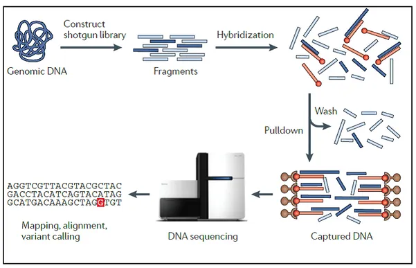
provides for a) a wet laboratory workflow, including library preparation and the actual sequencing of the library (refers to par. 1.3.1), and b) a dry laboratory part involving informatics and bioinformatics analyses (sequence alignments and single nucleotide variant calling), and variant filtering and interpretation (annotation, gene variant mapping against genetic variation databases) (refers to par. 1.3.2). The key steps of exome sequencing are shown in figure 1:
Figure 1: Workflow for the exome sequencing. First step is DNA fragmentation and the construction of a library. Fragments corresponding to exons (in dark blue) are hybridized to DNA or RNA probes (in orange) and then they are recovered through a streptavidin-based pulldown assay. Following steps are represented by amplification and massive parallel sequencing of the enriched and amplified library and the mapping and calling of candidate causal variants (Bamshad et al., 2011).
12
1.3.1 “Wet lab” workflow: library preparation, exome capture and
sequencing technology
The library preparation is the first step and refers to the process of DNA fragment preparation (by randomized fragmentation) and collection followed by a ligation of specialized adaptors (called also linkers) to both fragment ends. Few micrograms of genomic DNA are sufficient to construct an in vitro shotgun library. Next, as the second step, the DNA fragment library is enriched for sequences corresponding to exons through aqueous-phase hybridization capture and the exon fragments are hybridized to biotinylated DNA or RNA single-stranded oligonucleotides (also called probes or
baits). Recovery of targeted fragment-probe hetero duplexes (through a streptavidin
beads pulldown assay) is followed by the amplification reaction.
The exome capture sequesters protein coding regions by hybridization of genomic DNA library to biotinylated oligonucleotide baits that are DNA or RNA complementary to targeted exons. Magnetic streptavidin beads are used to selectively pull-down and enrich baits to which the target sequences are linked. The sample preparation methods are highly similar across the different technologies but there are considerable differences among those technologies in terms of target regions, and in the total number of bases targeted (Chilamakuri et al., 2014). Currently, there are four major solution-based human exome capture systems available: Nimble Gen’s SeqCap EZ Exome
Library, Agilent’s SureSelect Human All Exon, Illumina’s TruSeq Exome Enrichment,
and Illumina’s Nextera Exome Enrichment. The major differences between the technologies correspond to the choice of their respective target regions, bait lengths, bait density, molecules used for capture, and genome fragmentation method (Table 1).
13 Table 1: Performance and design comparison of four exome capture systems for deep sequencing (Chilamakuri et al., 2014).
The sequencing of the enriched and amplified library (range from 25-500 base pair, bp) can be performed through different technologies based on fluorescence or
chemiluminescence methods (Bamshad et al., 2011).
Among the most used sequencing platforms two NGS systems are normally used, which differ in the underlying chemistry technology and offer different advantages and disadvantages.
- Illumina platform (including the HiSeq, MiSeq, and NexSeq benchtop sequencers): it uses a flow cell to immobilize each DNA fragment and clonally amplify it for generating a large enough signal for the detection. In detail, the flow cell contains sequences that hybridize to a part of the adaptor on the DNA fragments. The clonal amplification step creates a cluster with approximately 1000 identical copies of a unique parent DNA molecule that are physically isolated from other molecules. Illumina platform, taking advantage of the principle of the “sequencing by synthesis” with fluorescent detection, it produces sequence reads of several hundreds of bp length (up to 300 bp) from tens of millions of simultaneously amplified DNA fragments.
- Ion Torrent technology (including the IonPGM, IonProton, and IonS5 systems): it uses a bead emulsion for the immobilization and the clonal amplification. Conversely, to Illumina platform, Ion Torrent sequencing adds a single base during the chain prolongation in each round. When a base is incorporated, a hydrogen ion is released,
14 accompanied by a pH change that is detected for each bead within a well; if a base is not incorporated there is no voltage generated (Yohe and Thyagarajan, 2017).
1.3.2 “Dry lab” workflow: sequence alignment, variant calling and
gene annotation
The exome sequencing experiments provide a large amount of reads, 140 million on average, depending on capture and sequencing technologies used (Chilamakuri et al., 2014). The several parameters to evaluate NGS experiments include sequencing depth and coverage. Depth represents the average number of reads that align to a specific position in the reference genome, while coverage refers to the degree of enrichment of target sequences (exons in case of WES). In the first step of WES analysis, the raw data of reads (fastq files) are analyzed to remove sequence adaptors and low quality reads (DePristo et al., 2011). The raw data of reads deriving from the sequencers undergo a series of bioinformatics processes (also referred as a bioinformatic pipeline) to ultimately deliver a variant call file (VCF), which contains the genomic variants and allows to describe in tabular format the most common genomic variants of a genome, together with the possibility of inserting annotations and metadata (Danecek et al, 2011).The bioinformatic pipeline include three key phases (summarized in Fig.2; Meena, et al., 2017).
15 Figure 2: The pipeline involving three important phases, viz. preprocessing, variant discovery and prioritization of variants. (Meena, et al., 2017).
1) NGS data pre-processing, demultiplexing, defined as a process for the separation of an individual sample’s reads from the pooled reads of multiple samples by unique identifier codes that were attached before pooling), quality analysis, mapping of the reads to a reference genome (also called re-sequencing), in the first step of WES analysis, raw reads data (FASTQ files, files with consensus assessment of sequence and variation) are analyzed to remove sequence adaptors and low quality reads (DePristo et
al., 2011). The next step is represented by the alignment to the reference genome, a
process that determines the exact position of each read on the human genome (Ruffalo
et al., 2011). There are different tools that can achieve this task, and that take into
account several factors, e.g. genetic variation in the population, sequencing error, short read length and the huge volume of short reads to be mapped (Ruffalo et al., 2011). 2) Variant discovery, the output is a BAM file (Binary version of sequence alignment/map), of about 6 GB, containing all retained reads (Sophia Yohe; Bharat Thyagarajan., 2017).
16 3) Variant prioritization, the next step is the variant calling that consists in the identification of DNA sequence variation relative to the reference genome. Variations that can be recognized by WES are single-nucleotide variants and small insertion-deletions (indels); the output file of this analysis is a Variant Calling File (VCF), a text format file containing variants calling (Van der Auwera et al., 2013).
The final step is characterized by the annotation of variants and of genes in which the variants localize, in order to characterize them and to investigate their possible involvement in a specific pathology or biological process. Variant interpretation is complex when applied to whole genes (as opposed to well-defined hotspots) and a large number of genes. There are several kinds of functional annotations that can be retrieved for DNA variants such as position in the genome (e.g. genes, intron/exon, etc.), effect on amino acid sequence (silent, non-synonymous, missense, etc.) and conservation (Torkamani et al., 2011). Variants can also be annotated using information present in population databases as dbSNP (Database of Single Nucleotide Polymorphisms -
http://www.ncbi.nlm.nih.gov/SNP/), 1000 Genomes Project
(http://www.1000genomes.org/) and ExAC (Exome Aggregation Consortium -
http://exac.broadinstitute.org/). At this point, the interpretation of the variants, which concerns with evaluating whether they could be potentially pathogenic and therefore causative of the disease studied) is a critical step. To this aim, the genetic information is collected and evaluated for categorization of the effect on an encoded protein, for in silico prediction of the consequences on protein function, and for previously reported knowledge on the gene and the specific mutation in question from databases. For this step can be used a functional annotation to predict the potentially damaging effect of identified variants on protein function (CADD score - Combined Annotation Dependent Depletion - http://cadd.gs.washington.edu/), the involvement in pathogenic mechanisms (OMIM - Online Mendelian Inheritance in Man - http://www.ncbi.nlm.nih.gov/omim),
animal disease models (MGD - Mouse Genome Database -
http://www.informatics.jax.org/ and ZFIN database-https://zfin.org/), and gene
expression (GXD Gene Expression Database -
http://www.informatics.jax.org/mgihome/GXD/aboutGXD.shtml) (Torkamani et al., 2011). These approaches explore the predicted proteins changes caused by specific amino acid substitutions and enrich for functional sites at which observed variants are more likely to affect phenotype (Bamshad et al., 2011). This allows to quantitatively
17 prioritize functional, deleterious, and disease causal variants across a wide range of variants.
Finally, when one or a few candidate nucleotides changes are selected as potential pathogenic variants, a comprehensive functional validation is needed to confirm the pathogenicity and the causative link with the disease object of the study. This functional investigations include: 1. re-sequencing the human samples to exclude false positive; 2. Setup an experimental workflow to investigate the impact of the new variants found in the disease causing mechanism using ideally both in vitro and in vivo models.
18
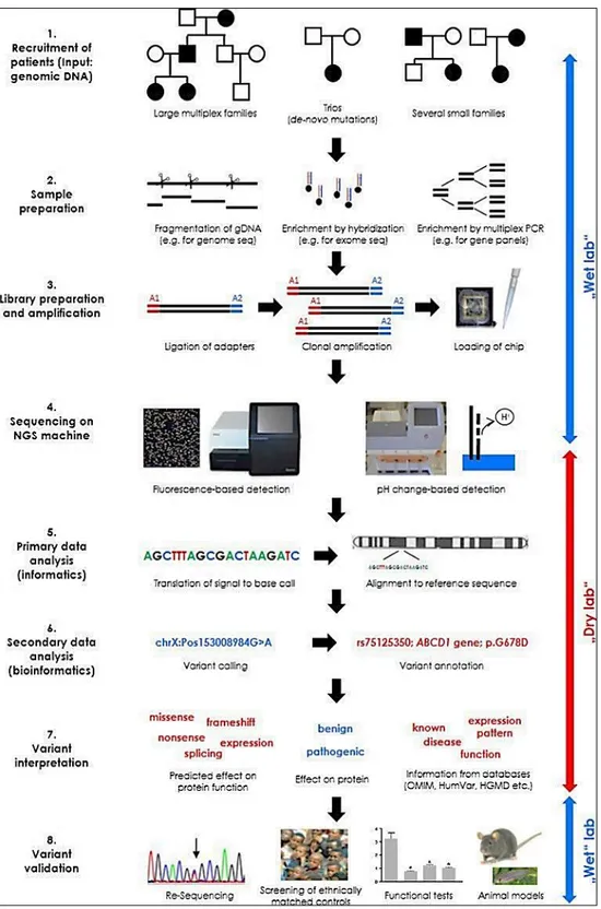
Figure 3: Workflow of NGS analysis. The figure provides a simplified overview of the main phases in
NGS and demonstrates some of the different options at each step (Figure modified by Katy Lohmand
19
1.4 The model organism Zebrafish (Danio Rerio)
The zebrafish, Danio Rerio, is a small tropical freshwater fish belonging to Cyprinidae family. The zebrafish is native to the streams of the south-eastern Himalayan region and it is found in parts of India, Pakistan, Bangladesh, Nepal, and Burma. This specie arose in the Ganges region in eastern India and commonly inhabits streams, canals, ditches, ponds, and slow-moving or stagnant water bodies, including rice fields.
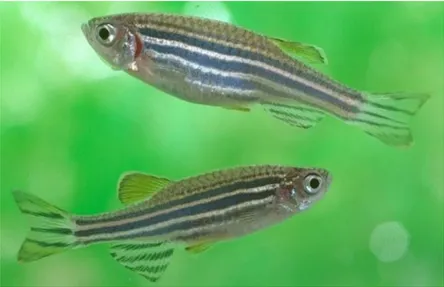
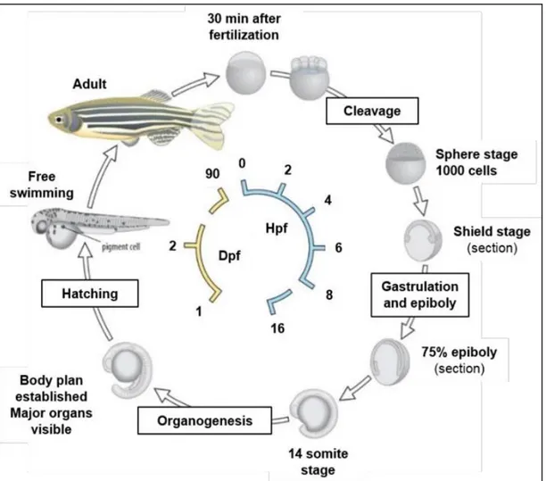
The zebrafish is so called because of five uniform, pigmented, horizontal, blue or purple stripes on the side of the body, which are reminiscent of a zebra's stripes and which extend to the end of the caudal fin. Its shape is fusiform and laterally compressed, with its mouth directed upwards. The male is torpedo-shaped, with gold stripes between the blue stripes; the female has a larger, whitish belly and silver stripes instead of gold (Figure 4). The zebrafish can grow to 2.5 cm in length, although it seldom grows larger than 4 cm in captivity. Its lifespan in captivity is around two to three years, although in ideal conditions, this may be extended to five years (Spence et al., 2008). Controlled cross-breeding has given rise to different varieties: wild-type (with the classic stripes, from which it derives its name), leopard, longfin (characterized by long fins), and the cross-breeding of longfin type with spotted skin leopard variety.
Females can spawn every 2-3 days and a single clutch may contain several hundred eggs. The generation time is short, typically 3-4 months, making it suitable for selection experiments. Zebrafish eggs are large relative to other fish, (0.7 mm in diameter at fertilization) and optically transparent, a characteristic that makes the zebrafish a convenient research model in genetics. Furthermore, taking advantage of the external fertilization, live embryos are accessible to manipulation and can be monitored through all developmental stages under a dissecting microscope (Kimmel et
al., 1995). The organogenesis and development is rapid (within 72 hours post
fertilization, hpf) and larvae display food seeking and active avoidance behaviors within five days post fertilization (dpf), 2-3 days after hatching (Kimmel et al., 1995). The sex of juveniles cannot be distinguished and sex determinants are not clearly understood. The zebrafish are omnivorous, primarily eating zooplankton, insects, larvae and small crustaceans (i.e. Artemia salina).
20
Figure 4: Male and female zebrafish
Zebrafish development is divided into seven broad periods of embryogenesis: the zygote, cleavage, blastula, gastrula, segmentation, pharyngula, and hatching. These divisions highlight the changing spectrum of major developmental processes that occur during the first 3dpf (Kimmel et al., 1995):
1) ZYGOTE PERIOD (0-3/4 h): the newly fertilized egg is in the zygote period until the first cleavage occurs about 40 minutes after fertilization. The zygote is about 0.7 mm in diameter at the time of fertilization. In this period many changes are occurring. Fertilization also activates cytoplasmic movements. Non-yolky cytoplasm begins to stream toward the animal pole, segregating the blastodisc from the clearer yolk granule rich vegetal cytoplasm;
2) CLEAVAGE PERIOD (3/4-21/4 h): after the first cleavage the cells, or blastomeres, divide at about 15-minute intervals. The cytoplasmic divisions are meroblastic; they only incompletely undercut the blastodisc (Kimmel and Law, 1985). The six cleavages that comprise this period frequently occur at regular orientations and are characterized by different numbers of cells (2-cells stage,4-cells stage, 8-cells stage, 16-cells stage, 32-cells stage and 64-cells stage);
21
3) BLASTULA PERIOD (2 1/4-5 1/4h): a period in which the blastodisc begins to look ball-like, at the 128-cell stage, and until the time of onset of gastrulation. Important processes occur during this blastula period; the embryo enters mid-blastula transition (MBT), the yolk syncytial layer (YSL) forms, and epiboly begins. This stage ends at 30 % epiboly;
4) GASTRULA PERIOD (5 1/4-10 h): the epiboly goes on with the following period of
gastrula stage, ranging from 5 to 10 hpf. During this developmental stage, the morphogenetic cell movements of involution, convergence and extension take place, leading to the formation of mesoderm, ectoderm and endoderm. The beginning of involution defines the onset of gastrulation. It is also possible to recognize the anterior (that will generate the head) from the posterior (that will generate the tail) part of the main body axis, the ventral from the dorsal and medial tissue from the lateral ones. (Figure5)
Figure 5: Gastrulation movements. (a) Dome stage. Cells intercalate radially, contributing to
epiboly. (b) Shield stage. Cells at the margin internalize and migrate toward the animal pole. Cells converge dorsally, with lateral mesodermal cells starting convergence at later stages than cells closer to the shield (282). (c) 90% epiboly stage. Epiboly, internalization, convergence and extension continue. (Schier and Talbolt, 2005).
5) SEGMENTATION PERIOD (10-24 h): the segmentation period is so called because of segmentation of the nervous system (Figure6). It’s characterized by a variety of morphogenetic movements, the formation of somites, which are the structures giving rise to the trunk muscles, the rudiments of the primary organs become visible, the tail bud becomes more prominent and the embryo elongates. The anterior-posterior and dorso-ventral axes are unambiguous. The first cells
22 differentiate morphologically, and the first body movements appear;
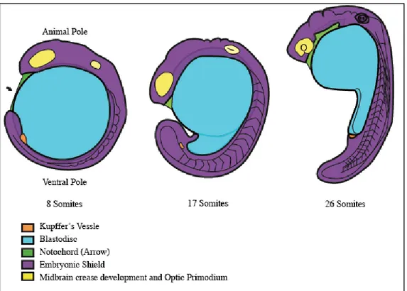
Figure 6: https://embryology.med.unsw.edu.au/embryology/index.php/Zebrafish_Segmentation_Period. The image shows the development of the Kupffer Vessel and the theformation of the subdivisions of the brain located at the animal pole. The straightening out of the posterior trunk also occurs. The lumps along the dorsal neural tube show the formation of the hindbrain rhombomeres; divided segments of the neural tube within the hindbrain
6) PHARYNGULA PERIOD (24-48 h): the term “pharyngula” refers to the embryo that has developed to the phyolotypic stage, when it possesses the classic vertebrate
bauplan. The embryo is most evidently now a bilaterally organized creature, entering
the pharyngula period with a well-developed notochord, and a newly completed set of somites that extend to the end of a long post-anal tail. The nervous system is hollow and expanded anteriorly. The embryo is fully transparent up to 22 hpf, by which stage the melanocytes start to differentiate and the embryo begins to be pigmented. The movements are irregular and non-controlled. Towards the end of this period all organs
23 were formed but the intestine;
7) HATCHING PERIOD (48-72 h): this is the last embryonic phase, in which the embryo comes out of the chorion (membrane surrounding the zygote) and the embryonic development is completed. During the hatching period the embryo continues to grow at about the same rate as earlier. The morphogenesis of many organ rudiments is now rather complete. After hatching, the embryo becomes a larva able to swim freely and all anatomical structures are fully established (Kimmel et al., 1995).
24
1.4.1 Why does zebrafish make such good animal model?
In recent years, zebrafish has become widely used for human diseases. This organism has convincingly demonstrated the usefulness of fish for improving the understanding of the molecular and cellular mechanisms leading to pathological conditions, and for the development of new diagnostic and therapeutic tools (Schartl, 2014). Zebrafish are widely used in research due to their numerous beneficial properties:
- Fully sequenced genome: the zebrafish share~ 70% of their genes with humans, and 84% of disease-causing genes have a zebrafish counterpart. The zebrafish genome has been fully sequenced and over 140,000 genes have been mutated to study their function in development and disease (Hannah Simmons, M.Sc.). In teleosts, as in zebrafish, the specific genome duplications led to the generation of paralogous gene copies, whose mechanism of divergence resulted in small (“subfunctionalization”) or large (“neofunctionalization”) changes in paralogues; this feature represent a good advantage for studying the contribute of each paralogue. (MátéVarga, et al 2018):
- Easy manipulation of its genome: transparent zebrafish embryos allows study of the different developmental stages starting from the early embryogenesis. Until now, it has been possible to generate more than 10.000 mutants in protein-coding genes and several transgenic lines of zebrafish to study human diseases (Howe et al., 2013). The easy availability of multiple strains (wild type, transgenic and mutant) of zebrafish is another important advantage of this species, which allows studying (considering the transparency of embryos) several biological process in vivo (Teame, et al., 2019).
- High fecundity and external fertilization: breeding and getting eggs from the zebrafish is relatively easy. A single reproductive event can ensure ~ 150 - 200 eggs, externally fertilized and produced regularly in large numbers (multiple hundreds of eggs/week). The external fertilization also helps the study of developmental processes avoiding the use of invasive techniques (Clark and Ekker. 2015) and for pharmacological high-throughput screenings. (MacRae and Peterson 2015)
- Short generation time and rapid and transparent embryonic development: zebrafish embryos form complete organ systems, including heart, intestine and blood vessels
25 within 48-72 hpf. Their organs share the same main features as humans and, for this reason, it is powerful to study human developmental processes (Clark and Ekker, 2015). Importantly, all the main phases of embryogenesis (from blastula formation, gastrulation movements to segmentation) are readily visible in the externally developing zebrafish embryos and occur in relatively short period of time. This makes the model ideal for addressing specific alterations of embryogenesis processes (including gastrulation defects and body axis formation, cell migration and early differentiation signaling for organogenesis) (Clark and Ekker, 2015). For instance, zebrafish is even used for studying hematopoiesis because these fish have the same sequential multi lineage hematopoiesis process as humans, investigating cardiovascular diseases because the gene determinants involved in cardiac development and differentiation are evolutionary conserved (Sakai and Hoffman., 2018). In addition, zebrafish is an excellent model also for modelling neurological disorders given the high conservation of the main neuronal cell populations and circuit architecture with other vertebrates and the underlying genetics (Veldmanand Lin, 2008). Zebrafish individuals grow rapidly and reach the sexual maturity about at third month of the development. This shortens the overall experimental processes and is particularly useful in mutant generation (Sakai and Hoffman., 2018).
- Easy maintenance: it is very affordable to maintain zebrafish colonies in a relatively small amount of laboratory space. Although zebrafish lines require relatively easy management, it is extremely important to optimize a healthy diet and adequate water quality to ensure the fish health and growth (Spence, et al., 2008).
1.4.2 Use of zebrafish to investigate pediatric rare disease affecting the
neurodevelopment
The advent of the NGS technology allows to sequence large regions of DNA rapidly and economically, which has been particularly advantageous for the diagnosis of rare genetic diseases. This possibility triggered rapid progress in the identification of their causative mutations in recent years (Koboldt et al., 2013) and led to the establishment of several large-scale collaborative initiatives, including national and international networks of clinicians and researchers, with the aim of accelerating the discovery of the
26 rare disease-causing genes. In many cases, these new sequencing technologies allowed a fast and accurate identification of new disease-causing genes and/or new variants in genes already associated with diseases (Máté Varga et al., 2018). Together with the integration of these technologies in the clinical practice as diagnostic tool, development of complementary experimental approaches is representing an important step to shed light on the pathological mechanisms underlying the newly discovered genes and variants. This is crucial for future test and development of pharmacological molecules; given that for the majority of these pediatric rare disease a cure is missing. For new genes and variants found, the functional approaches, based on in vitro and in vivo model systems, are therefore necessary to 1. Confirm the causative role of genetic variants in the rare diseases orphan of diagnosis as well as 2. For the understanding of the pathogenetic mechanisms involved and find effective cures. Over the past four decades zebrafish has become one of the most in-demand genetic organisms (Kinth et al., 2013; Grunwald et al., 2002; Varga et al., 2018; Lieschke et al., 2007). Zebrafish became so important in the genetics research because it has several characteristic of an ideal vertebrate genetic model organism (for details see paragraph 1.4.1). Especially, the high genetic conservation make it an ideal genetic model organism to study pediatric disease. The sequencing of the zebrafish genome revealed that 71% of all human proteins and 82% of those that cause disease have a zebrafish ortholog (Howe, K. et al., 2013). About 75% (13.217 / 17.727) of the genes included in the PedAM database, published on the record of pediatric diseases, have a zebrafish ortholog (Jiaet al., 2018) (Figure 8). Taking advantage of its unique features zebrafish is an ideal in vivo model for the study of genetic disorders (for details see par. 1.4.1), especially with respect to genetic neurodevelopmental disorders (Sakai and Hoffman., 2018).
27 Figure 8: A high level of genetic conservation makes zebrafish an ideal genetic model organism to study pediatric disease. (OMIM–Online Mendelian Inheritance in Man database) (MátéVarga, et al 2018).
Indeed, several successful zebrafish models exist for neurodevelopmental disorders depending on genetic causes: 1) Autism Spectrum Disorders (ASD), a devastating group of disorders affecting the neurodevelopment characterized by marked impairments in social behavior and communication, and by the presence of restricted, repetitive behaviors; 2) epilepsy accompanied by intellectual disability, a common neurological condition characterized by recurrent seizures and 3) Attention-Deficit/Hyperactivity Disorder (ADHD), marked by an ongoing pattern of inattention and/or hyperactivity-impulsivity that interferes with functioning or development, and 4) schizophrenia, a psychotic disorder characterized by hallucinations, disorganized thought processes or behavior which severely impacts overall functioning. Most studies of the zebrafish models affecting the neurodevelopment have focused on early morphological and behavioral phenotypes to identify the molecular and cellular mechanisms underlying these disorders. Recent advances in neuro-functional imaging allow also to follow the altered biological processes ‘in real time’ leading to the neuronal circuit-level characterization of phenotype resulting from risk gene disruption (Simvoulidis et al., 2017, Sakai and Hoffman., 2018).
28
1.5 Early neurogenesis in zebrafish
To understand how to model a pediatric neurological disorder from the molecular to the morphological level in zebrafish, it is essential to understand how the fish brain develops as compared to other vertebrates and humans. “Neurogenesis” describes the process by which undifferentiated neural progenitor cells generate mature and functional neurons, starting at the beginning of gastrulation, around 6 hpf. The induction of neural progenitors and the cell division, that enlarges the pool of progenitors, are the early phases of neurogenesis. Subsequently, a sequence of progenitor specification and commitment to differentiation of post-mitotic neurons occurs. Each of these phases is spatially and temporally orchestrated to generate the multiple neuronal and glial cell types that will populate the mature CNS.
1) The first step in the development of the vertebrate nervous system is the neural induction by extrinsic factors, or specification of the neuroectoderm, during the early embryonic development (Schmidt et al., 2013). At the onset of gastrulation, the forming mesodermal layer involutes and meets the overlying ectoderm (Doniach T and Musci TJ, 1995; Lumsden A and Krumlauf R, 1996; Spemann H and Mangold H, 2001). This presumptive mesodermal layer secretes important factors locally to induce or inhibit neural induction in the ectodermal layer. The extrinsic signaling factors, which are involved in the neural induction, are members of the bone morphogenetic protein (BMP), wingless-integrated (Wnt) and fibroblast growth factor (Fgf) families (Streit A,
et al., 2000; Wilson SI, et al., 2001). Ventral secretion of BMPs, especially BMP2, 4
and 7, blocks neural induction by inducing an epidermal fate (Sasai and De Robertis, 1997; Wilson, et al., 1997). BMP antagonists such as Noggin and Chordin are produced early in the dorsal pre-organizer region, which later forms the Spemann organizer, corresponding to the shield organizer in fish. These secreted proteins act permissively for the establishment of the neural fate in the dorsal ectoderm and allow the formation of the neural plate (Wessely O, et al., 2001).
2) The neural fate commitment is determined by intrinsic factors involving especially members of the SRY-box containing genes B1 (SoxB1) family (Avilion AA, et al., 2003; Streit A, et al., 1997). Together with the extrinsic factor participation, intrinsic networks, programmed by both transcriptional and epigenetic factors, participate in the regulation of the neural fate commitment as well. Recent findings focus on the initiation of the nervous system, elaborately regulated by the intrinsic programs, which are
29 mediated by transcriptional factors such as Sox2, Zfp521, Sip1 and Pou3f1, as well as epigenetic modifications, including histone methylation/demethylation, histone acetylation/deacetylation, and DNA methylation/demethylation (Tang, et al., 2015). 3) Once specified, the neural ectoderm forms the neural plate, a pseudostratified epithelial
structure in zebrafish. During the process of neurulation, the zebrafish differs from other vertebrates: instead of folding the neuronal plate immediately in a tube with a lumen, it is formed first a solid neural keel. However, the topological arrangement of cells in zebrafish during neuronal formation of the keel from the neural plate is similar to that of other vertebrates. In the so-called "secondary neurulation", the neural rod of the fish swells and forms a typical vertebrate tube. Thus, although there are differences, neurulation in fish and mammals it leads to the formation of a highly similar structure in each case, the neural tube (Papan C and Campos-Ortega JA, 1999). After the formation of the lumen, there is an increasing number of cells dividing asymmetrically into the neural tube; indeed the most apically derived daughter cell becomes the neuron, while a more basal daughter fills the apex pool of progenitors. Important for this asymmetric division is the Notch signaling: basal self-renewal daughter cells show high Notch activity while apical daughter cell differentiation shows low Notch activity (Alexandre,
et al., 2010; Dong, et al., 2012).
4) Following neural plate formation, the early anterior-posterior patterning of the neural plate is established. The early segmentation in the brain emerge morphologically during early gastrulation as a series of bulges along the A-P axis of the neural tube, including 7-8 hindbrain rhombomeres. Each rhombomere contains the same basic cell types (e.g., commissural interneurons, branchiomotor neurons), but also have distinct identities and contribute to different cranial nerves (Lumsden 2004). In the embryo, each rhombomere also forms a compartment, within which cell lineages are confined and separated by distinct boundary regions (Kimelman, and Martin, 2012). As development proceeds, the major zebrafish brain structures develop and, from 10 hpf, CNS structures can already be identified. By 24 hpf, three primary vesicles develop in the rostral portion of the neural tube: the prosencephalon (forebrain), mesencephalon (midbrain) and rhombencephalon (hindbrain). The forebrain, the anterior-most part of the embryonic brain, is perhaps one of the most complex domains of the embryonic brain, as it develops into the telencephalon, the diencephalon, the hypothalamus, and the retina. These structures represent an important functional part of the brain, responsible for receiving and processing sensory information and directing behavior. The zebrafish
30 telencephalon composed by the pallium, the subpallium, and the olfactory bulb. The thalamus, pineal body, and habenula are part of the zebrafish diencephalon. The olfactory bulb is responsible for perceiving odor information, and it contains neurons that transmit that information to other regions of the brain, such as the telencephalon, thalamus, and habenula (Friedrich and Korsching, 1997; Miyasaka, et al., 2009). The telencephalon is associated with regulating the social behavior, memory, and emotion, while the hypothalamus regulates fundamental aspects of physiological homeostasis and behavior. The midbrain is a relatively small structure in the adult zebrafish brain, but important for vision and hearing. The major structures derived from the midbrain are the tectum and the tegmentum. The tectum, or optic tectum, is the visual processing and response center, with retinal ganglion cells connecting this structure to the retina. The tectum and its connecting neurons are especially important for survival, since they make up the startle and reflex response center (Karlstrom, 1997; Portugues and Engert, 2009; Kita, 2015). The hindbrain is one of the most studied parts of the zebrafish brain and can easily be identified during embryonic development since it is physically separated anteriorly from the midbrain by the midbrain-hindbrain boundary (MHB). The hindbrain is composed of the posteriorly located medulla oblongata, the ventro anterior pons, and the dorso anterior cerebellum. The medulla and pons are often referred to as the “brainstem”, containing a conglomerate of cell groups that form a complex network termed the reticular formation, which is involved in higher order behaviors such as respiration, circulation, and wakefulness. Indeed, the primary role of the cerebellum is to coordinate movement, and it thus receives information from many sources while transmitting information primarily to motor cortical areas (Moens and Prince 2002). The cerebellum is easily identified by the physical specification of eight compartments along the neural tube called rhombomeres (r1–r7), as early as 10 hpf (Kimmel, C.B. et
al., 1995; Moens and Prince 2002). Each rhombomere gives rise to cells that
differentiate into specific neurons, which innervate the head but also project through the spinal cord to other parts of the body (Oxtoby and Jowett, 1993) (Figure9).
31 Figure 9. Development of the zebrafish brain. (A) Schematic representation of the embryonic brain (30 hpf), showing the forebrain (in yellow), midbrain (in blue), MHB (in green), and hindbrain (in orange). Coronal and sagittal section schemes show brain structures primordia. Forebrain is composed by the telencephalon (in darker gray) and the diencephalon (containing the hypothalamus, lighter grey). (B) Simplified representation of the adult brain and main domains.C: cerebellum; D: diencephalon; M: midbrain; MHB: midbrain-hindbrain boundary; H: hindbrain; Ha: habenula; Hyp: hypothalamus; OB: olfactory bulb; ON: optic nerve; ORR: optic recess region; OV: otic vesicle; Pal: pallium; PB: pineal body; R: retina; r1–r7: rhombomeres 1 to 7; Sub: sub-pallium; T: telencephalon; and Teg: tegmentum. (Raquel Vaz et al., 2019).
32
1.6 Morphogenesis of axes formation in zebrafish
The basic vertebrate body plan of the zebrafish embryo is established in the first 10 hours of development. Especially during the gastrulation periods, the movements of epiboly, internalization, convergence, and extension transform the radially symmetric blastula into the gastrula embryo with clear dorso-ventral and anterior-posterior axes (Adams RJ and Kimmel CB. 2004). The morphogenetic movements of gastrulation are a series of co-ordinated movements of cell groups that give rise to the three-germ layersectoderm, mesoderm and endodermand overtlyshape the embryonic axis, mostly orchestrated by the non-canonical Wnt (Schier and Tabolt, 2005). During the past 10 years, a combination of genetic, embryological, and molecular analyses has provided detailed insights into the mechanisms underlying this process. Maternal determinants control the expression of transcription factors and the location of signaling centers, responsible for patterning of the blastula and gastrula. Instead, Bmp, Nodal, FGF, canonical Wnt, and retinoic acid signals generate positional information that leads to the restricted expression of transcription factors that control cell type specification. One such movement is convergent extension, when cells of the mesoderm and ectoderm accumulate on the dorsal side of the gastrula by means of highly directed and integrated movements. This results in both medio–lateral (ML) narrowing (convergence) and anterior–posterior (AP) elongation (extension) of tissues to create the embryonic axis. This mechanism is regulated by a process that is called “medio-lateral intercalation behavior (MIB)”, which underlies the rearrangements (Figure10). In according to MIB hypothesis, a single force-generating cellular machine, distributed across a field of cells, produces both convergence and extension, both narrowing and elongation of the field. By the MIB hypothesis, as applied particularly to the domain of notochord-forming cells within dorsal mesoderm, motile and adhesive cells become polarized along one particular axis, the medio-lateral (ML) axis. The polarity may depend on, and be coordinated within the field, by a non-canonical Wnt signaling planar polarity pathway (Choi and Han, 2002; Heisenberg et al., 2000). The cells take on a bipolar shape, elongating along the ML axis. This process requires that the individual cells all correctly orient actin-based cytoskeletal machinery that mediates motility, and perhaps also orient associated adhesion complexes on their plasma membranes (Zalik et al., 1999).
33 Figure 10: The mediolateral intercalation behavior (MIB) hypothesis. (A) Early protrusive activity is random. (B) The cells take on a bipolar shape as their protrusions are restricted to the ML axis. (C) The field narrows and elongates (converges and extends) as the cells exert traction upon one another and pulltogether (Nathalia S. Glickman et al., 2002).
With this phenomenon, cells intercalate medio laterally and, to accomplish this, they all protrude filopodial processes both medially and laterally that extend between immediate cellular neighbors, and new cell-cell contacts are made, perhaps involving adhesion molecules of the cadherin/protocadherin superfamily localized to their filopodial tips. The newly contacting cells then contract their processes. They exert traction upon one another and pull together. Such events, which occur across the entire field of cells, narrow the field (convergence). The resulting intercalations push previously neighboring cells apart along the AP axis, lengthening the field along this axis (extension). The cells disperse along the AP axis by intercalating with neighbors to form a discontinuous AP string, as expected if MIB mediates extension (Kimmel and Warga, 1986; Kimmel et al., 1994). It is clear that convergent extension of the mesoderm is the major driving force of vertebrate gastrulation. During this process, mesodermal cells move toward the future dorsal side of the embryo, then radically change behavior as they initiate extension of the body axis.
34 AP patterning in the vertebrate embryo can be roughly divided into two major phases: 1) An initiation phase, in which the embryo is generally divided into the head and the
body. In zebrafish, the initiation phase occurs prior to the start of gastrulation, such that by the start of gastrulation, the different territories of the final body plan can be roughly mapped onto the embryo. The mesoderm of the head, which comprises part of a very important signaling center called “the Organizer”. It is first specified near the equator on what is defined as the dorsal side of the embryo (Saude et al., 2000). These cells migrate toward the animal pole during gastrulation, where the brain forms. In contrast, the major mesodermal derivative of the body (the fast muscle) and the spinal cord, are at the gastrula stage oriented with their AP axes along what is termed the dorsal-ventral axis (Kimelman and Martin, 2012);
2) An elaboration phase, in which the body progressively forms toward the posterior end, forming the trunk and tail (this process involves the formation of blocks of muscle tissue called somites and it is defined the somitogenesis stages).
1.7 AP patterning and segmentation of the vertebrate head
and trunk: role of krox20 and myoD.
The neuroepithelium of the developing hindbrain undergoes a transient AP segmentation into seven segmental progenitor cell compartments, derived largely, or entirely, from ectoderm, named rhombomeres (r1–r7). (Lumsden and Krumlauf, 1996). This conserved organization underlies the segmental organization of cranial nerves in the developing head of all vertebrate embryos. The vertebrate hindbrain segmentation is an evolutionarily conserved process that involves a complex interplay of transcription factors and signaling pathways that show spatially restricted patterns of expression along the AP axis, with limits corresponding to prospective or established boundaries between adjacent rhombomeres (Lumsden and Krumlauf, 1996; Schilling, 2008). They constitute compartments and developmental units for neuronal differentiation, branchiomotor nerve organization and neural crest specification (Lumsden and Keynes, 1989). The rhombomeres are units of cell lineage restriction, they are serially reiterated and each of them containing the same basic cell types (e.g. commissural inter-neurons, branchiomotor neurons), but also have distinct identities and contribute to different cranial nerves (Lumsden 2004). In the embryo, each rhombomere also forms a
35 compartment, within which cell lineages are confined and separated by distinct boundary regions (Fraser et al. 1990). Before boundary formation, cells can intermingle into adjacent territories, but once sharp borders with distinct cellular properties are formed between adjacent rhombomeres, neuroepithelial cells and their progeny can no longer cross into adjacent segments. This cellular segregation process is paralleled by progressive establishment and sharpening of rhombomere-specific gene expression domains, providing distinct segmental identities to each compartment (Lumsden and Krumlauf, 1996). The rhombomere identities are determined, at least in part, by a combinatorial code of transcription factors such as the Hox genes (Lumsden 2004). Stripes of rhombomere-specific Hox gene expression domains appear in the neural ectoderm by the end of gastrulation and these are positioned by early signals along the AP axis. In detail, three signals form an AP patterning network: 1) Wnt signals initially repress expression of anterior genes, 2) fibroblast growth factor (FGF) and 3) retinoic acid (RA) activate posterior genes in a concentration-dependent manner (Cho and De Robertis 1990; Sive, et al. 1990; Holowacz and Sokol 1999; Domingos, et al. 2001; Kudoh, et al. 2002). FGF and RA, in particular, are good candidates for the molecular link between AP patterning and segmentation, potentially acting as graded ‘‘morphogens’’ because they establish the boundaries of segments with distinct concentration thresholds. FGF signalling plays a major role because its signaling is necessary to promote the expression of the transcription factor krox20 (also known as
Egr2) which is required for the formation and specification of two segmental units:
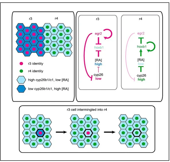
rhombomeres 3(r3) and 5(r5) (Aragon and Pujades, 2009; Marin and Charnay, 2000; Charlotte Labalette, et al., 2011). Krox20encodesa protein with three C2H2-type zinc finger. This protein was shown to bind to a specific DNA sequence and to act as a transcriptionfactor. It belongs to a small subfamily of proteins, with similar zinc fingers, which recognize identical or very closely related GC-rich sequences (Oxtoby and Jowett, 1993). This process is regulated by a specific signaling pathway: during segmentation, krox20 expression in r3 and r5 represses the expression of the RA-degrading enzyme genes, cyp26b1 and cyp26c1, thus maintaining high RA levels.
Krox20 auto regulates itself and represses the r4 marker hoxb1. In r4, high cyp26b1 and cyp25c1 expression levels contribute to maintain a low RA environment, with is
permissive for hoxb1 expression. Hoxb1 also auto regulates itself and represseskrok20 expression. When an isolated r3-derived cell intermingles into r4, it becomes exposed to environmental low RA signaling from surrounding cells, and the RA level is reduced