Characterization of human memory NK cells ex vivo and
study of therapeutic monoclonal antibody capability to
promote their expansion in vitro
PhD School in Immunological, Haematological and Rheumatological Sciences Curriculum: Immunology and Immunopathology, XXXI cycle
Lavinia Franchitti
Identification number 1338404
Supervisor Co-supervisor
Prof. Gabriella Palmieri Dr. Simone Battella
TABLE OF CONTENTS
INTRODUCTION ... 1
1. HUMAN NATURAL KILLER CELLS ... 1
1.1 Main biological and functional characteristics ... 1
1.2 NK receptors ... 9
CD16 ... 10
Natural cytotoxicity receptors (NCR) ... 12
Killer Immunoglobulin-like Receptor (KIR) family ... 14
LIR ... 15
C-type lectin receptors ... 16
DNAM-1 family receptors ... 17
PD-1 ... 18
Pattern-recognition receptors (PRR) ... 19
1.3 NK development and differentiation ... 19
2. NK CELLS AND TUMORS ... 23
2.1 NK cells in anti-tumor immune response ... 23
2.2 NK cell-based cancer therapies ... 25
Cytokine-based therapies ... 26
Antibody-based therapies ... 27
NK cell adoptive transfer therapies ... 30
3. MEMORY NK CELLS ... 32
3.1 HCMV-induced memory NK cells ... 32
Intracellular signalling molecules and functional signature of memory NK cells ... 36
3.2 Cytokine-induced human memory NK cells ... 39
3.3 Memory NK cells in mice and non-human primates ... 39
AIM OF THE WORK ... 42
MATERIALS AND METHODS ... 43
Anti-HCMV IgG detection ... 43
Cell lines ... 43
NK cell expansion ... 43
Immunostaining, flow cytometric analysis and gating strategy ... 44
Characterization of CD16A polymorphism ... 46
Functional assays ... 47
Statistical analysis ... 48
RESULTS ... 49
1. ex vivo analysis of memory NK cells ... 49
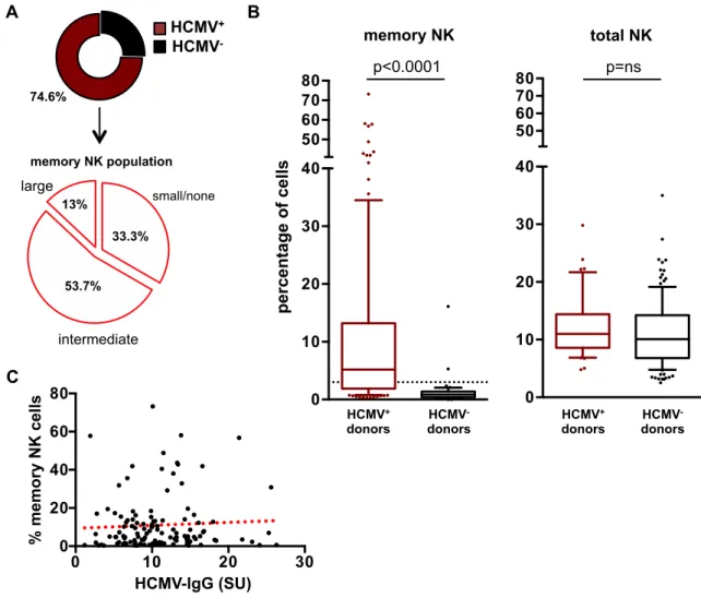
1.1 Impact of HCMV seropositivity ... 49
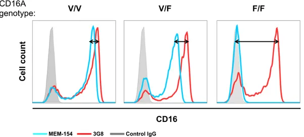
1.2. Impact of CD16 polymorphisms ... 51
1.3. Analysis of CD16 expression ... 52
1.4. Memory NK cell distinctive surface markers and intracellular mediators . 53 2. in vitro expansion of memory NK cells ... 55
2.1 Characterization of the ability of tumor-targeting mAb-opsonized lymphoblastoid cells to promote memory NK cell proliferation ... 55
2.3. Functional profile of fresh and in vitro cultured memory NK cells ... 60
DISCUSSION ... 62
INTRODUCTION
1. HUMAN NATURAL KILLER CELLS
Natural Killer (NK) cells are lymphocytes belonging to the innate branch of the immune system. They are included in the innate lymphoid cell (ILC) hemopoietic lineage, a recently identified heterogeneous group of lymphoid effector cells, largely tissue-resident and important orchestrators of early immune responses (Mjösberg J and Spits H, 2016; Vivier E et al., 2008).
NK cells have been initially identified in the peripheral blood (PB) by the unique ability to kill tumor cells without major histocompatibility complex (MHC) restriction and any prior activation, thus defined “natural” (Jondal M and Pross H, 1975; Kiessling R et al., 1975; Pross HF and Jondal M, 1975). In the last 40 years, our perception of NK cells is dramatically changing as, beside their ability to kill transformed cells, they have been recognized to kill virus-infected, damaged and allogenic cells, and also to contribute to immunoregulation and participate in tissue remodelling (Cooper MA et al., 2001a; Vivier E, 2006). Currently, over 30,000 NK cells subsets displaying peculiar features can be identified, reflecting the huge plasticity and diversification of this population (Horowitz A et al., 2013). Moreover, in the last decade, the characteristic of immunological memory has been recognized to the NK cell population, challenging the rigid classification that relegates them in the innate immune system (Rölle A and Brodin P, 2016).
1.1 Main biological and functional characteristics
NK cells represent 5-15% of circulating lymphocytes in humans, although significant populations permanently reside in tissues such as bone marrow, liver, spleen, gut, skin, lungs, and uterus during pregnancy. Moreover, NK cells were also found in other tissues: kidney, joints, and breast, under pathological conditions (Law BMP et al., 2017; Mamessier E et al., 2011; Teo T et al., 2015). Their different localization reflects their phenotypic and functional heterogeneity (Björkström NK et al., 2016; Peng H and Tian Z, 2017; Vivier E, 2006).
In the lungs, for example, NK cell ability to respond rapidly to infection suggests an important role for these cells in airway protection. However, it is emerging that they
are also important in the regulation of chronic infections, as tuberculosis, and chronic inflammatory disorders, as asthma, and lung fibrosis (Culley FJ, 2009; Marquardt N et al., 2017). The liver is a tolerogenic environment, where NK cells are important with their anti-viral properties, for example in HBV and in HCV infection (Aw Yeang HX et al., 2017; Maini MK and Peppa D, 2013; Peng H and Tian Z, 2017).
During pregnancy, uterine NK cells importantly contribute to spiral artery remodelling and trophoblast invasion, by producing cytokines, and angiogenic and growth factors (Faas MM and de Vos P, 2017; Gamliel M et al., 2018; Moffett-King A, 2002).
NK cells are an important component of tumor immunosurveillance; they can be recruited in the tumor microenvironment where they contribute to malignant cell killing and also exert immunoregulatory function (Larsen SK et al., 2014; Malmberg KJ et al., 2017; Stojanovic A and Cerwenka A, 2011).
In animal models, they have been recognized as important in controlling tumor growth and metastasis spreading (Guillerey C and Smyth MJ, 2015; Hayakawa Y and Smyth MJ, 2006; Kärre K et al., 1986; Ljunggren HG and Kärre K, 1985). Indeed, deficiencies in NK number (Kim S et al., 2000) and function (Talmadge JE et al., 1980), or NK cell depletion (Seaman WE et al., 1987), increase the rate of tumor and metastasis.
Retrospectives studies correlate a lower NK cell activity with tumor incidence (Imai K et al., 2000). While, NK cell infiltrate in gastrointestinal sarcoma (Delahaye NF et al., 2011) and in lung metastasis correlates with a better prognosis (Remark R et al., 2013). Moreover, the defective expression of NK activating receptors has been correlated with the incidence of multiple myeloma (Fauriat C et al., 2006), hepatocellular carcinoma (Jinushi M et al., 2005), metastatic melanoma (Konjević G et al., 2007) and other tumors. All these reports suggest a direct correlation between NK cells presence/functionality with tumor incidence and outcome (Guillerey C and Smyth MJ, 2015).
NK cell most recognized functional abilities are target cell killing and the secretion of a vast array of cytokines and chemokines. NK cytotoxic activity can be exerted through three main lytic mechanisms, which are shared with cytotoxic T lymphocytes (CTL):
1) The exocytosis, into a tight cytotoxic synapse, of granules containing perforin membrane-disrupting molecule and granzyme serine-proteases, resulting in the apoptosis of the target cell, by caspase cascade activation. Moreover, granzyme-induced target cell death can be also induced by a caspase-independent pathway that leads to mitochondria swelling and disruption (Arias M et al., 2017; Chowdhury D and Lieberman J, 2008; Estaquier J et al., 2012).
2) Receptor-dependent target cell apoptosis, mediated by TNF-related apoptosis-inducing ligand (TRAIL) and Fas ligand (FasL or CD178), which are induced on the surface of activated NK cells. These death ligands bind to their respective death receptors, Fas (CD95) for FasL and TRAIL-R1/2 for TRAIL, on target cell surface, promote receptor oligomerization and the death domain (DD)-mediated recruitment of Fas-Associated Death Domain (FADD) adaptor protein, which in turn activates the caspase apoptotic pathway (Martínez-Lostao L et al., 2015; Peter ME and Krammer PH, 2003).
3) Tumor necrosis factor (TNF)-α is secreted in large amount by NK cells. It is produced as a membrane protein that is later released in a soluble form by the action of TACE metalloprotease. TNF-α is recognized by two receptors: TNFR1, possessing conserved death domains (DD) and expressed at low level in all cell types, and TNFR2, regulated and expressed only in specific cells, as neurons, endothelial and immune cells. Upon ligand binding by TNFR1, the activation of adapter proteins through intracellular DD converges on FADD activation, as for TRAIL and FasL, and consequently in the formation of IIa and IIb complexes that induce caspase activation and subsequent apoptosis (Aggarwal BB et al., 2012; Kalliolias GD and Ivashkiv LB, 2016). Moreover, TNFR1 downstream signalling pathways can also promote the formation of IIc complex, which leads to necroptosis (Silke J et al., 2015).
Upon activation, NK cells are also able to secrete a vast array of cytokines and chemokines which exert a strong immunomodulatory activity, and orchestrate traffic and functional responses of other immune components in the inflammatory
sites (Figure 1): interferon γ (IFN-γ), TNF-α, granulocyte-macrophage colony-stimulating factor (GM-CSF), and the chemokines macrophage inflammatory protein (MIP)-1α, MIP-1β, the factor regulated on activation, normal T cell expressed and secreted (RANTES/CCL5), among others (Fauriat C et al., 2010a). IFN-γ, or type II interferon, plays critical roles in both host defence and immune regulation, by exerting multiple and pleiotropic effects (Castro F et al., 2018; Schoenborn JR and Wilson CB, 2007; Schroder K et al., 2004).
IFN-γ receptor is expressed in nearly all cell types and composed by two subunits, IFNGR1 and IFNGR2 that, upon ligand binding, rapidly activate JAK1 and JAK2 Janus tyrosine kinases, responsible for the activation of STAT-1, a member of the STAT (signal transducer and activator of transcription) protein family; STAT-1 then translocates into the nucleus, and promotes the transcription of IFN-γ-inducible genes. Most of them are transcription factors themselves, such as interferon regulatory factor (IRF)-1 -2 and -9, which promote, in turn, the transcription of downstream genes. In parallel, IFN-γ induces a JAK/STAT-independent pathway, involving MAP kinases. As a result, global changes are induced in the cell, influencing cell cycle, adhesion and functions (Castro F et al., 2018; Schroder K et al., 2004). This cytokine is also endowed with anti-tumor activity, by inhibiting cell proliferation, via upregulation of p21 and p27, or through apoptosis induction (Mojic M et al., 2017). For instance, IFN-γ induces IRF1, a tumor suppressor gene, which in turn reduces B-cell lymphoma-2 (Bcl-2) and increases Bcl-2 homologous antagonist/killer (Bak). As a result of cytochrome C release from mitochondria and reactive oxygen (ROS) and nitrogen species production, apoptosis is induced (Aqbi HF et al., 2018).
Moreover, IFN-γ influences antigen processing and presentation by both MHC class I and class II molecules, by inducing the replacement of some of the proteasome subunits; by promoting the synthesis of transporter associated with antigen processing (TAP) 1 and 2, which deliver cytosolic peptides in the endoplasmic reticulum for their processing; by up-regulating MHC subunits (Castro F et al., 2018; Schroder K et al., 2004).
IFN-γ acts on many immune cell populations, mainly by: a) resulting in an increased microbe ingestion and destruction by phagocytes, either by directly targeting macrophages, and indirectly by up-regulating CD40 ligand on TH1 cells;
b) polarizing the immune response toward a TH1 response, by promoting CD4+ T
cells differentiation in TH1 subset, and, in parallel, by blocking TH2 and TH17
subset differentiation; c) promoting the switch to opsonizing IgG subclasses by activated B cells (Schroder K et al., 2004); d) enhancing NK and CTL cytotoxicity. However, several studies have outlined the role of chronic IFN-γ production in tumorigenesis and in tumor immune evasion. Under homeostatic conditions, IFN-γ up-regulates genes designed to limit tissue damage and facilitate tissue repair upon resolution of inflammation. Chronic exposure to IFN-γ induces an epigenetic signature leading to the expression, in the tumor microenvironment, of immune suppressive molecules, such as programmed death-1 (PD-1) and its ligands PD-L1 and PD-L2, cytotoxic T-Lymphocyte antigen-4 (CTLA-4), that induce a hypofunctional/exhausted phenotype in T lymphocytes and NK cells (Benci JL et al., 2016). IFN-γ also up-regulates the expression of HLA-E non canonical HLA molecule that, by interacting with inhibitory receptors, promotes tumor escape from CTL and NK cells (Derré L et al., 2006).
Besides the previously described direct cytotoxic effects on target cells through the IIa, IIa and IIc complexes downstream of TNFR1 receptor, TNF-α also exerts immunoregulatory activity. Through TNFR1-induced complex I assembly, comprising adapter molecules TRADD and TNFR-associated factor 2 (TRAF2), signals converge on mitogen-activated kinase (MAPK) cascades, and Activator protein 1 (AP1) and nuclear factor kappa-light-chain-enhancer of activated B cells (NF-κB) activation, important promoters of inflammation, host defence, cell proliferation and survival (Aggarwal BB et al., 2012; Brenner D et al., 2015). TNFR2, instead, is believed to bind preferentially to the transmembrane form of TNF-α. This receptor lacks DD and it is not able to induce cell death, instead it promotes local homeostatic effects, such as cell survival and tissue regeneration, by recruiting TRAF2 and the formation of complex I (Kalliolias GD and Ivashkiv LB, 2016).
GM-CSF is an important hematopoietic growth factor and modulator, mostly involved in inducible hemopoiesis. It mainly acts on multipotent progenitor cells to drive the production of granulocytes and monocytes. It also induces the recruitment of monocytes, induces their differentiation to macrophages and dendritic cells (DCs), depending on other cytokines present in the
microenvironment (Becher B et al., 2016).
NK cells can be triggered to perform their effector functions either upon cell-cell contact, mediated by a vast array of surface receptors (vide infra), and by the action of several cytokines, which tune their activation status together with other aspects of their biology, as development, homeostasis and proliferation. The main cytokines whose receptors are expressed on human NK cells are: IL-2 family (IL-2, IL-15, IL-21), IL-12, IL-18, IL-27, TNF-α, IFN-α/β, IFN-γ and transforming growth factor (TGF)-β (Cooper MA et al., 2009; Parrish-Novak J et al., 2002; Wu Y et al., 2017; Zwirner NW and Domaica CI, 2010; Zwirner NW and Ziblat A, 2017).
IL-2, mainly produced by activated T cells, and IL-15, produced by macrophages, DC and bone marrow stroma, have several positive effects on NK cell functions, as they enhance proliferation and survival, co-stimulate cytokine production and amplify cytotoxic activity. Upon receptor engagement, their downstream signals induce the activation of JAK1/3 and STAT3/5, PI3K pathway, MAPK pathway and NF-κB (Waldmann TA, 2015). IL-2 and IL-15 share the IL-2/15Rβ (CD122) and γc (CD132) receptor subunits, which allow ligand binding with intermediate
affinity, at nanomolar concentration. The inclusion of CD25/IL-2Rα subunit forms the high affinity heterotrimeric IL-2Rαβγ receptor, which responds to picomolar concentration of IL-2. The high affinity receptor is constitutively expressed on a subset on NK cells named CD56bright (vide infra), but it can be induced also on CD56dim NK cells, upon stimulation with IL-2, IL-15 (Pillet AH et al., 2009), or combinations of IL-15, IL-12 and IL-18 (Chase JM et al., 2012; Leong JW et al., 2014). IL-15Rα is primarily expressed on activated dendritic DC and macrophages, it has high affinity for IL-15 as a single subunit, and trans-presents bound IL-15 to the IL-2/15Rβγc complex on NK cells. It has been also described,
after exposure to IFN-β, a cis-presentation of 15 by 15Rα to the dimeric IL-2/15Rβγc complex expressed on the same NK cell (Stonier SW and Schluns KS,
2010; Waldmann TA, 2015; Zanoni I et al., 2013). IL-21 can be expressed by a large number of cell types, such TH17, TH2, follicular TH cells; its receptor is
composed by IL-21R (CD360) and the common γc chain to form the functional
heterodimeric receptor that, upon ligand binding, induces JAK1/3 activation. On human NK cells, the stimulation with IL-21 increases cytolytic activity (Parrish-Novak J et al., 2000, 2002; Venkatasubramanian S et al., 2017) in vitro and in HIV
patients (Iannello A et al., 2010); in mice, IL-21 stimulation promotes the acquisition of functional markers and decreases their proliferation (Brady J et al., 2004).
IL-12 is produced by DC and macrophages in response to pathogens; during infection with intracellular parasites, IL-12 promotes IFN-γ production and target recognition in cooperation with other stimuli (Hashimoto W et al., 1999; Thierfelder WE et al., 1996; Vignali DAA and Kuchroo VK, 2012; Zwirner NW and Ziblat A, 2017). IL-12 receptor is composed by two subunits, IL-12R β1 and IL-12R β2; upon ligand binding it activates JAK1 and TYK2, leading to T-bet activation, crucial promoting factor for IFN-γ transcription (Trinchieri G, 2003; Zwirner NW and Ziblat A, 2017).
Another DC- and macrophage-produced cytokine important for NK activity is IL-27, which displays proinflammatory effects. IL-27 affects NK cell activation state, up-regulates IL-2Rα and NKp46 expression, enhances NKp46-driven cytotoxicity, and rituximab- and other therapeutic monoclonal antibody (mAb)-dependent cytotoxicity and cytokine production (Ziblat A et al., 2015; Zwirner NW and Ziblat A, 2017). IL-27 also primes NK cells for responsiveness to IL-18-induced IFN-γ production, by up-regulating T-bet (Matsuda JL et al., 2007).
IL-18 belongs to the IL-1 family, and promotes NK cell activation and effector functions, in cooperation with IL-12 (Chaix J et al., 2008). IL-18R signal transduction acts through the Toll/IL-1 receptor (TIR) domains, and triggers the binding of MyD88, phosphorylation of the Interleukin-1 receptor-associated kinases (IRAKs), and leads to the activation of NF-κB (Dinarello CA et al., 2013). Type I IFN (IFN α and β) are innate cytokines abundantly produced during viral infections, especially by specialized plasmacytoid pDC. Their shared receptor (IFNAR) is broadly expressed, and upon ligand binding, it initiates the JAK/STAT pathway (Ivashkiv LB and Donlin LT, 2014). Type I IFNs potentiate NK cell functional activity, increase TRAIL expression and also IL-15 production by NK cells (Nguyen KB et al., 2002; Paolini R et al., 2015). Moreover, DC are themselves stimulated by type I IFN to produce IL-15, and to indirectly activate NK cells (Ivashkiv LB and Donlin LT, 2014; Lucas M et al., 2007).
TGF-β is a powerful immunosuppressive cytokine, secreted by a variety of cell types, including macrophages and regulatory T cells (Treg). It is produced in an
inactive form that needs processing to become active. The receptor is composed by two TGF-βRI chains and two TGF-βRII chains, and it is broadly expressed; intracellular signal propagation occurs thanks to the phosphorylation of small mother against decapentaplegic (Smad)-2 and-3 that translocate into the nucleus, activate Smad-4 transcription factor, leading to downstream gene expression (Massagué J, 2012). Moreover, in NK cells there is a Smad-independent signal transduction that involves other pathways, such as the phosphoinositide 3-kinase (PI3K)/Akt axis, leading to mammalian target of rapamycin (mTOR) inhibition. As a result, NK cells reduce their metabolism and proliferation, and decrease their functional activity (Viel S et al., 2016). Moreover, TGF-β suppresses the expression of T-bet and Gata3, crucial transcription factors in NK cell maturation (Marcoe JP et al., 2012). IFN-γ NK cell Target cell Macrophage DC T cell Antigen presentation co-stimulatory signals IFN-γ TNF-α IL-12 IL-15 IL-18 IFN I IFN-γ TNF-α
Figure 1. NK cell biological functions and cellular crosstalk
NK cells can be activated by the recognition of target cells. This leads to target cell lysis, and to the production of various cytokines and chemokines. NK cells participate in shaping the subsequent adaptive response, engaging a crosstalk with DC, macrophages and T cells. Adapted from Vivier E and Ugolini S 2010- Nature Immunology Poster https://www.nature.com/nri/posters/nkcells/ nri1012_nkcells_poster.pdf
1.2 NK receptors
NK cells interact with other immune or non-immune cells through the engagement of a variety of receptors, resulting in inhibitory or activating signals, whose overall balance determines the activation state and the strength of NK cell responses, which result in target cell killing and/or immune regulation (Figure 2). Differently from TCR and BCR, NK cells receptors are germ-line encoded; moreover, the expression of members of the killer-cell immunoglobulin-like receptor (KIR) family is stochastic, resulting in different receptor combinations in each cell, which provide individualized target recognition capability (Guia S et al., 2018).
NK cells display several recognition strategies that allow the sensing of:
a) the levels of self-molecules expressed on normal cells, but down-modulated in infected and malignant cells. HLA molecules are indeed recognized as hallmark of “self” cells by inhibitory receptors on NK cells, blocking self-reactivity (Shifrin N et al., 2014; Yokoyama WM and Kim S, 2006);
b) the levels of self molecules that undergo overexpression in malignant and infected cells, named stress-induced ligands, that are recognized by activating receptors (Kruse PH et al., 2014; Lanier LL, 2015);
c) the presence of pathogen-derived molecules, as a result of the infection process of either bacteria and viruses (Kruse PH et al., 2014; Lanier LL, 2015). Many NK receptors are grouped in families, based on sequence homology. In several cases, activating and inhibitory receptors belong to the same family, and often recognize shared ligands.
NK cell activation is the result of the synergistic engagement of activating receptors, in most cases. Indeed, except for CD16, none of the other activating receptors is sufficient alone to trigger cytotoxicity and cytokine production (Bryceson YT et al., 2006, 2009; Long EO et al., 2013). On the other hand, NK cell activation threshold and responsiveness to the engagement of individual activating receptors may be deeply affected by the exposure to some cytokines, such as IL-2, IL-1IL-2, IL-15, IL-18, and IL-21 (Berrien-Elliott MM et al., 2015; Bryceson YT et al., 2006). A recent paper, based on the impedance variation study, has also confirmed these results, by comparing the response triggered by selected receptors (Fasbender F and Watzl C, 2018).
crosstalk with other immune system populations, for example with DC and macrophages. Here, NK cells recognize and kill the immature forms of these antigen-presenting cells (APC), selecting those cells able to cooperate in an efficient immune response (Malhotra A and Shanker A, 2011).
CD16
CD16, or FcγRIIIa, is the low affinity receptor for the Fc fragment of IgG. It is a multi-chain receptor, possessing one α-chain, responsible for ligand binding, coupled to Immunoreceptor Tyrosine-based Activation Motif (ITAM)-containing CD3ζ and FcεRIγ disulfide-linked homo- and hetero-dimers. CD3ζ has three ITAM domains, while FcεRIγ has only one, possibly resulting in a different signal transduction capability of the different CD16 complexes (Lanier LL et al., 1991). Oligomerization of CD16 complex subunits occurs thanks to the presence of charged residues with opposite sign, in the transmembrane regions of the different components (Blázquez-Moreno A et al., 2017).
Healthy Missing self Stress-induced self Activating receptor Inhibitory receptor Inhibitory ligand Activating ligand TARGET CELL NK CELL
Figure 2. NK cell recognition of target cells
The integration of inhibitory (blue) and activating (red) pathways, resulting from the interaction with target cell, determines the dynamic equilibrium of NK cell activation. Adapted from Vivier E and Ugolini S 2010- Nature Immunology Poster https://www.nature.com/nri/posters/nkcells/ nri1012_nkcells_poster.pdf
ITAM motifs are found in the components of different immunoreceptor complexes, such as TCR, BCR, FcγRs, FcεRI, among others. Motif consensus sequence: D/E xxYxxL/I x-6/8 Yxx L/I is characterized by the presence of two phosphorylable tyrosines, both necessary and sufficient for the induction of downstream intracellular signals (Reth M, 1989; Romeo C et al., 1992). CD16 aggregation triggers ITAM phosphorylation by Lck and Fyn Src-family kinases. This event leads to the recruitment of SH2 domain-containing ζ-associated protein (ZAP70) and Syk tyrosine kinases. Bound ZAP70 and Syk becomes themselves substrate for Src kinases, and contribute to signal propagation by phosphorylating the transmembrane adapter molecules LAT, resulting in the recruitment and activation of different signaling molecules (Humphrey MB et al., 2005; Samelson LE et al., 1999). Among them, Grb2 leads to Ras and Erk kinase activation, which promote AP-1 transcription complex activation. In parallel, phosphorylated LAT also scaffolds and activates phosphatidyl-inositol-3-OH kinase (PI3K), phospholipase Cγ (PLCγ) and Vav-SLP-76 complex. Guanine nucleotide exchange factor Vav acts on Rac, and initiates a distinct MAP kinase cascade, while JNK activation converges on the activation of AP-1 components. Both PI3K and PLCγ use the membrane phospholipid phosphatidylinositol-4,5- bisphosphate (PIP2) as common
substrate. PI3K activity converges on Vav family proteins and Akt kinase, while PLCγ pathway contributes to ERK and NF-κB activation. All these signaling pathways lead to cytoskeletal re-organization, cytotoxic granule polarization and secretion, and gene activation (Samelson LE, 2002). In contrast to other activating receptors, CD16 aggregation by IgG-coated target cells is by itself sufficient to activate antibody-dependent cell-mediated cytotoxicity (ADCC) and cytokine production in resting human NK cells (Bryceson YT et al., 2006; Fauriat C et al., 2010a).
A genetic factor that affects CD16 responsivity is represented by an allelic dimorphism in the CD16A gene, which results in a functional aminoacidic difference (valine or phenylalanine) at position 158. In fact, the presence of valine (V) confers to CD16 receptor a higher affinity for Fc portion of IgG with respect to the phenylalanine (F)-carrying one (Koene HR et al., 1997). In that respect, individuals can be FF or VV homozygous, or VF heterozygous.
proliferative and apoptotic stimuli in specific contexts. CD16 triggering with IgG or agonist antibodies, induces NK cell proliferation (Lee HR et al., 2017). Conversely, CD16 aggregation inhibits the proliferation induced by IL-2, partially through the induction of the apoptosis. It has been hypothesized that this mechanism helps to control and limit NK cell functionality in the late stages of immune response when the adaptive branch of immune system is finally working (Azzoni L et al., 1995; Eischen CM et al., 1996; Ortaldo JR et al., 1995; Warren HS and Kinnear BF, 1999).
CD16 expression is regulated during NK cell differentiation (see below), and is susceptible to modulation: some studies have proposed a post-transcriptional regulation of CD16 expression, through miR-218 microRNA, which negatively affects CD16 mRNA transcription (Victor AR et al., 2018). Moreover, CD16 is sensitive to disintegrin and metalloproteinase (ADAM)-10 and -17 cleavage (Pham DH et al., 2017); these proteases are induced after stimulation with IL-2 and IL15, and with tumor cells (Feldinger K et al., 2014; Romee R et al., 2013).
Natural cytotoxicity receptors (NCR)
Natural cytotoxicity receptors (NCR) NKp30, NKp44 and NKp46 are a specialized group of activating receptors that mediate a key role in target cell recognition, identified in the 90s (Pende D et al., 1999; Pessino A et al., 1998; Vitale M et al.,, 1998). NCR are transmembrane proteins that belong to the immunoglobulin superfamily, and are associated with ITAM-containing accessory chains that allow surface expression and signalling capability; NKp44 pairs with DNAX-activating protein of 12kD (DAP12), while NKp30 and NKp46 interact with CD3ζ and FcεRIγ (Hudspeth K et al., 2013; Kruse PH et al., 2014; Moretta A et al., 2001).
NKp46 and NKp30 are constitutively expressed by NK cells, and in some restricted groups of T cells and ILC (Kruse PH et al., 2014). NKp44 is expressed on activated NK cells, on specialized tissue NK cell subsets, such as in the decidua, gut lamina propria, and tonsils (Cella M et al., 2009; Horton NC and Mathew PA, 2015; Takayama T et al., 2010), and also in a subset of plasmacytoid DC in the tonsils (Fuchs A et al., 2005).
NCR are crucial receptors in recognition and killing of several tumors including melanoma, carcinoma and multiple myeloma (Sun C et al., 2015), but their ligands
have not been conclusively identified yet. It has been reported that the three NCR, with different individual specificities, can recognize three different heparan sulphate glycosaminoglycans typical of tumor cells (Hecht ML et al., 2009; Kruse PH et al., 2014). Other self-ligands are HLA-B associated transcript 3 (BAT3) and Vimentin, deriving from the intracellular milieu of malignant cells, and recognized by NKp30 and NKp46, respectively (Garg A et al., 2006; Pogge von Strandmann E et al., 2007). B7-H6 (NCR3LG1), also expressed on the surface of tumor cell lines and primary tumors, is the main ligand for NKp46 (Gutierrez-Franco J et al., 2018; Horton NC and Mathew PA, 2015; Matta J et al., 2013). Recently, it has been reported by Colonna’s group that platelet-derived growth factor (PDGF)-DD is a relevant ligand for NKp44. This protein is produced by many tumors and promotes the malignant transition and cell proliferation. Engagement of NKp44 by PDGF-DD stimulated the secretion of IFN-γ and TNF-α, and other proinflammatory cytokines, which in turn induced the downregulation of tumor cell-cycle genes and tumor growth arrest (Barrow AD et al., 2018). Moreover, proliferating cell nuclear antigen (PCNA), which is overexpressed by tumor cells and interacts with HLA I, can be recognized by NKp44. Interestingly, this binding is generating an inhibitory signal for the effector NK cell; indeed, NKp44 also has an active Immunoreceptor Tyrosine-based Inhibition Motif (ITIM) motif (vide infra), able to inhibit cytotoxicity, IFN-γ release, and representing a tumor immune escape strategy (Horton NC and Mathew PA, 2015).
Several studies have demonstrated the involvement of NCR in NK cell-mediated antiviral responses (Brusilovsky M et al., 2012). NKp46 and NKp44 can directly recognize cell surface expressed pathogen-derived molecules as hemagglutinins (Orthomyxovirus) and hemagglutinin-neuraminidases (Paramyxovirus) (Arnon TI et al., 2001; Mandelboim O et al., 2001). Human cytomegalovirus (HCMV) tegument protein pp65 is recognized by NKp30, whose binding disrupts the association with CD3ζ adapter, blocking the transduction signal (Arnon TI et al., 2005).
Different NCR can cooperate, by recognizing multiple ligands on the same target cell, for optimal recognition and functional activation of effector NK cells (Long EO et al., 2013).
Killer Immunoglobulin-like Receptor (KIR) family
The KIR polygenic locus includes 15 genes and 2 pseudogenes, on chromosome 19 in the position 19q13.4. All genes code for transmembrane glycoproteins expressed on NK cells and a subset of T cells, and present either 2 or 3 Ig-like domains in the extracellular region, which allows a classification in 2D or 3D receptors, respectively. Their major ligands are HLA class I molecules. KIR ligands include C1 and C2 groups of HLA-C alleles (for 2D receptors), some HLA-A and HLA-B alleles (for 3D receptors), and HLA-G (for KIR2DL4) (Campbell KS and Purdy AK, 2011). KIR family includes highly homologous activating and inhibitory members, that differ for the sequence and the length of the cytoplasmic domains (Thielens A et al., 2012).
Inhibitory KIR (iKIR) display a longer cytoplasmic tail (identified as L), which includes an ITIM motif; this is characterized by V/I/LxYxxL/V sequence whose tyrosines, upon ligand recognition, undergo phosphorylation by SRC family kinases, and promote recruitment of SHP-1 and SHP-2 phosphatases that block the propagation of activating signals (Campbell KS and Purdy AK, 2011; Long EO, 2008; Long EO et al., 2013).
Activating KIR (aKIR) have a short truncated cytoplasmic tail (identified as S), and associate with DAP-12 adapter, except for KIR2DL4 that couples with FcεRIγ chain (Kikuchi-Maki A et al., 2005). Despite the high homology of their extracellular domains with iKIR, the identification of aKIR ligands remains in most cases elusive; only for some of them, the recognition of HLA-C2, -C1 and A alleles has been demonstrated (Della Chiesa M et al., 2015; Varbanova V et al., 2016).
The genetic asset of the KIR locus is highly complex. The repertoire of KIR genes inherited by an individual represents a haplotype. Two types of haplotypes can be distinguished: type A, with a fixed number of inhibitory receptors and few activating receptors, and type B, with a variable number of inhibitory receptors and several activating members; KIR3DL3, KIR2DL4 and KIR3DL2 genes are common to all haplotypes. Moreover, KIR genes show an extensive polymorphism (Parham P, 2005; Uhrberg M et al., 1997; Vilches C and Parham P, 2002). Expression of inherited KIR genes occurs stochastically during the differentiation process of individual NK cell precursors (see below), generating a high number of different mature effectors, expressing different KIR combinations (more than 104) (Horowitz
A et al., 2013; Parham P, 2005; Vierra-Green C et al., 2012).
The physiological role of iKIR interaction with self-HLA class I molecules is to provide a mechanism that protects healthy self cells from potential NK cell self-reactivity. In selected pathological conditions, i.e. infection by several viruses, or tumor progression, HLA class I down-modulation or loss may occur, as an immunoevasion strategy to avoid recognition by CTL. Diminution of HLA class I expression, named “missing self”, lowers NK cell activation threshold, as the missing engagement of iKIR conveys weakened inhibitory signals (Yokoyama WM and Kim S, 2006; Shifrin N et al., 2014).
Noteworthy, some studies described a lower affinity of aKIR with respect to iKIR for HLA class I molecules (Ivarsson MA et al., 2014; Moesta AK and Parham P, 2012), and it has been proposed that aKIR recognition of HLA molecules may be highly dependent on viral peptides, or altered self proteins, allowing them to actively participate to target cell recognition (Ivarsson MA et al., 2014).
Several reports have shown a significant association between certain KIR haplotypes and the susceptibility to some viral infections, some tumors and autoimmune diseases, suggesting a role for KIR in their onset/progression (Boyton RJ and Altmann DM, 2007; Falco M et al., 2013).
LIR
LIR family members are transmembrane proteins, widely expressed in hematopoietic-lineage cells and interact with HLA molecules. They are encoded by 13 genes; among them, members classified as LIRB has inhibitory effects, and just two of them are expressed on NK cells, LIRB1 and LIRB5.
LIRB display 2 or 4 extracellular Ig-like domains, and a long ITIM-containing cytoplasmic tail. LIRB1 is implicated in the recognition of HLA-I and of the human HCMV HLA class I homolog UL18 protein (Cosman D et al., 1997). HCMV-driven expression of UL18 by the infected cells is probably a viral immune escape strategy, to avoid NK cell-mediated recognition and killing of the infected cell (Prod’homme V et al., 2007). Moreover, some LIR gene polymorphisms have been associated with autoimmune disease predisposition (Zhang J et al., 2017).
C-type lectin receptors
The genes coding for several NK receptors that belong to the C-lectin superfamily are clustered in the Killer Lectin-like Receptor (KLR), on human chromosome 12. The C-lectin superfamily cluster members have different roles and ligand specificities (Bartel Y et al., 2013; Seliger B et al., 2016).
NKG2A and NKG2C heterodimerize with CD94, to form an inhibitory and an activating receptor, respectively, specific for HLA-E, a non-canonical HLA class I molecule. CD94/NKG2A provides the inhibitory signal through its intracellular tail that contains two ITIM domains, while CD94/NKG2C couples to DAP12 to provide activating signals. Their expression is regulated during NK cell differentiation, as CD94/NKG2A is expressed earlier during the maturation pathway, while it is less expressed on the terminal stage, in parallel with the progressive acquisition of NKG2C (Stabile H et al., 2018).
HLA-E molecules are constitutively expressed on the cell surface at low level, but may undergo up-regulation in stressed and malignant cells. HLA-E stabilization on the cell membrane requires loading with self and non self peptides, such as heat shock protein 60 (hsp60)-derived peptide (Michaëlsson J et al., 2002), the leader peptides derived from classical HLA class I A, B, and C, hence representing the overall expression of HLA molecules on the cell surface (Borrego F et al., 2005), and peptides from the HCMV encoded protein UL40 (Ulbrecht M et al., 2000). Although it has not been definitively understood how HLA-E-associated peptides affect recognition of CD94/NKG2A and CD94/NKG2C, it has been shown in different contexts that HCMV infection accelerates NK cell terminal differentiation, and promotes the expansion of NKG2Cbright NK cells (Gumá M et al., 2004;
Hammer Q and Romagnani C, 2017).
NKG2D was initially identified in 1991 by Houchins and colleagues (Houchins JP et al., 1991), but its role has been ignored until 1999, when MIC (MHC class I chain related) A, one of its ligands, was identified (Bauer S et al., 1999). NKG2D homodimers are expressed on NK cells, but also on γδTCR+ and on CD8+ T cells, and on some CD4+ T cells (Lanier LL, 2015).
NKG2D recognizes multiple ligands that are structurally related to HLA I molecules and are expressed by stressed, infected, and transformed cells. These are MICA, MICB and UL16-binding proteins (ULBP)-1-6 (Dhar P and Wu JD, 2018).
After ligand recognition, NKG2D signalling ability depends on the association with DAP10 adapter protein. It carries a YINM motif, leading to an ITAM-independent pathway that, upon ligand-triggered tyrosine phosphorylation, allows the binding of PI3K, PLCγ and Grb2. Grb2 mediates the recruitment of Vav proteins (Lanier LL, 2015). NKG2D engagement alone can induce chemokine production and release, while cytotoxic activity, instead, needs a synergy with other receptors (Bryceson YT et al., 2006; Fauriat C et al., 2010a; Lanier LL, 2015). NKG2D receptor thus represents an important tool for NK cell-mediated protection against pathogens and tumors. Indeed, NKG2D ligand shedding from tumor cell surface, either by protease cleavage, or by exosome release, is considered a mechanism of tumor evasion (Lanier LL, 2015). Moreover, HCMV displays several strategies to interfere with NKG2D ligand expression on infected cells (Dunn C et al., 2003; Lanier LL, 2015; Slavuljica I et al., 2011).
DNAM-1 family receptors
The DNAX accessory molecule-1 (DNAM-1) family comprises four recognized members: DNAM-1 (CD226), T cell Ig and ITIM domain (TIGIT, Vstm3) T cell activation, increased late expression (TACTILE, CD96), and CRTAM (CD355), all belonging to the Ig superfamily.
DNAM-1 is expressed on almost the totality of NK cells, on a fraction of CD8+ and CD4+ T cells, and on some myeloid cells (De Andrade LF et al., 2014). It has 3 putative phosphorylation sites in the intracellular domain that are phosphorylated upon ligand binding. The association with Lymphocyte function-associated antigen 1 (LFA-1) is necessary for the signal transduction, indeed, it recruits Fyn SRC kinase to phosphorylate the other tyrosines in DNAM-1 and to initiate DNAM-1 downstream signalling. The cooperation with other activating signals is essential to promote NK activation (De Andrade LF et al., 2014).
TIGIT is an inhibitory receptor expressed on NK cells, on memory T cells and Treg
and on CD4+ T cells upon activation. The percentage of TIGIT+ NK cells can vary among individuals, and also among cells of the same individual, where its intensity inversely correlates with NK cell functional ability (Wang F et al., 2015). It has one ITIM domain and an immunoglobulin tail tyrosine (ITT)-like motif in the intracellular portion, both actively working in decreasing cell activating status and functional
ability (Liu S et al., 2013; Stanietsky N et al., 2013).
DNAM-1 and TIGIT have two shared ligands, poliovirus receptor (PVR) and Nectin-2, widely expressed on epithelial cells and in immune system populations under physiological conditions. These ligands are stress-induced, hence increased, during cell transformation and viral infections, and also upon activation of hemopoietic cells (Cerboni C et al., 2014). TIGIT binds PVR with high affinity, and competes with DNAM-1, contributing to the inhibition of NK cell functionality (Levin SD et al., 2011; Stanietsky N et al., 2013). It is also important in the crosstalk between immune cell populations, as it has been recently demonstrated that NK cells can be suppressed in their degranulation potential by PVR-expressing myeloid-derived suppressor cells (MDSC), through TIGIT engagement (Sarhan D et al., 2016).
Others not extensively studied members of the family are TACTILE and CRTAM. TACTILE is expressed on all NK cells, while CRTAM is confined on activated NK cells. Both can be also expressed on CD4+ and on CD8+ T cells. Their ligands are stress-induced ligands, respectively PVR and Necl-2. In human NK, their activating role in NK function, has been described (Chan CJ et al., 2014; Dessarthe B et al., 2013; Fuchs A and Colonna M, 2006; De Andrade LF et al., 2014)
PD-1
Several MHC class I-specific and non-specific inhibitory receptors are expressed on human NK cells and restrain their functional response. Among them, and in addition to inhibitory KIR, inhibitory member of DNAM-1 family, TIM-3 and LAG-3 (Anderson AC et al., 2016), there is Programmed Death-1 (PD-1). PD-1 is a receptor crucial in the suppression of the immune response, also expressed on activated T cells, and is especially involved in the tumor immune escape triggered by the engagement of its ligands, PD-L1 and PD-L2, which are expressed by malignant cells and activated immune cells. PD-1 possesses an ITIM and an immunoreceptor tyrosine-based switch motif (ITSM) domain. In T cells, both can associate with SHP phosphatases and block activation through the suppression of PI3K/AKT signaling; in NK evidence of a similar signaling are reported (Bardhan K et al., 2016; Liu LL et al., 2017; Sharpe AH and Pauken KE, 2017).
PD-1 has been recently has been identified on CD56dim subset of NK cells, in around 1/4 of healthy subjects, and its presence is restricted to HCMV+ donors
(Della Chiesa M et al., 2016; Pesce S et al., 2017). PD1+ NK cells have been
found enriched in the tumor microenvironment. From the functional point of view, PD-1+ NK cells are hypofunctional and hyporesponsive in terms of tumor cell
killing, and this effect can be reverted by using anti-PD-1 blocking antibodies (Della Chiesa M et al., 2016; Liu LL et al., 2017).
Pattern-recognition receptors (PRR)
Innate immune cells express Pattern-recognition receptors (PRR) specifically able to recognize recurrent motifs carried by viruses, bacteria and fungi, named pathogens-associated molecular patterns (PAMP), and also involved in the recognition of damage-associated Molecular Patterns (DAMP) (Mogensen TH, 2009; Tang D et al., 2012). NK cells express only some Toll-like Receptors (TLR), such as TLR2, TLR3 and TLR5. Moreover, as mentioned before, the capability of some NCR to directly recognize microbial ligands has been reported. TLR engagement leads to IRFs and NF-κB activation, resulting in type I IFN and other proinflammatory cytokine production. Moreover, they can co-operate with receptors belonging to other classes to induce a more efficient NK cell activation (Adib-Conquy M et al., 2014; Sivori S et al., 2014)
1.3 NK development and differentiation
NK cells development occurs in the bone marrow from a CD34+ hematopoietic
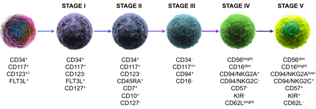
stem cells (HSCs), in a common pathway that originates all Innate Lymphoid Cell (ILC) subsets; however, several studies report that NK cells can further differentiate in tissues, especially in secondary lymphoid organs, liver, uterus and thymus (Fauriat C et al., 2010b). Human NK cells differentiation is a not completely understood multistep process, where some markers are acquired and in parallel some others are lost (Figure 3). This process results in the progressive restriction toward the NK cell lineage and acquisition of recognition repertoire and functional competence (Freud AG and Caligiuri MA, 2006).
the common lymphoid progenitor (CLP), which has the potential to give rise to B, T, NK, ILCs and DCs. Stage 1 progenitors have been found in cord and adult blood, in bone marrow, in several adult tissues and in fetal liver. Interestingly, from this progenitor all the ILC types can originate, through a recently identified intermediate stage that is Lin-CD7+CD127+CD117+. The differentiation program is
influenced by the microenvironment as required, under the stimulation of different cytokines, at every age and in every tissue, to replenish tissue resident ILC pools, including NK cells (Lim AI et al., 2017).
Stage 2, or NK progenitors (NKP) are CD34+CD117+CD123-FLT3L+CD127+ and are lineage-restricted. A key step is the acquisition of the IL-2/IL-15Rβ chain (CD122), whose expression is induced by stem cell factor (SCF) and FLT-3, confers responsivity to IL-15 stimulation and allows further differentiation of NK cells, which is dependent on the action of Eomes and Tbx21 transcription factors (Scoville SD et al., 2017).
Stage 3, or immature NK cells are: CD34-CD117+CD123 -CD45RA+CD7+CD10+CD127- (Stabile H et al., 2018). Despite they are committed to be NK cells, they are not yet endowed with the ability to perform the two hallmark functions of NK cells: degranulation and IFN-γ production (Freud AG and Caligiuri MA, 2006).
Stage 4 cells are identified as CD34-CD117+/-CD94+CD16-. They also express
CD56 at high levels, hence defined CD56bright NK cells (Stabile H et al., 2018).
CD56, or Neural Cell Adhesion Molecule (N-CAM), is a member of the Ig superfamily and represents an important marker for NK cells. Its functional role on NK cells has not been definitively assessed yet, although it has been reported to mediate homotypic adhesion, to bind fibroblast growth factor, and to mediate direct recognition of some microorganisms, as the Aspergillus fumigatus, resulting in the activation of the NK cell (Van Acker HH et al., 2017; Ziegler S et al., 2017).
CD56bright NK cells mainly localize in secondary lymphoid organs, while they
represent only 10% of NK cells in the peripheral blood and in the liver. CD56bright are preferentially CD94/NKG2A+CD94/NKG2C-CD57-KIR-CD16dim, they proliferate in response to cytokines, and produce large amounts of IFN-γ and TNF-α, but they have a low cytotoxic potential and low ability to perform ADCC. The CD56bright subset is believed to precede the final stage of NK differentiation (Freud AG et al.,
2006; Luetke-Eversloh M et al., 2013).
Indeed, stage 5, or CD56dim, represent the majority of peripheral blood NK cells,
express CD16 receptor at high density, and are thought to represent the final stage of NK cell differentiation. They also typically display CD16 at high levels (CD16bright), are CD94/NKG2AlowCD94/NKG2C+CD57+KIR+, and show low
proliferative capability. Historically, the functional activity of these cells was thought to be confined to their high cytotoxic potential, while more recently it has been shown that they can also produce a large amount of cytokines (Cooper MA et al., 2001b; Luetke-Eversloh M et al., 2013; Montaldo E et al., 2013).
The unique milieu available in different peripheral tissues might influence in situ differentiation of resident NK cells, and could contribute to explain the heterogeneity of NK cell subsets (Freud AG and Caligiuri MA, 2006). Even in the same tissue, heterogeneity of NK cells is observed, reflecting different stages of differentiation. The tissue distribution and homing ability of NK cells depend on their expression of chemokine receptors (Freud AG and Caligiuri MA, 2006). CD56dim cells express chemerin-R, CXCR1 and CX3CR1, which determine their ability to migrate in inflamed tissues. Differently, CD56bright express CCR7 and CD62L, and are attracted to secondary lymphoid organs (Bernardini G et al., 2013; Del Zotto G et al., 2017; Griffith JW et al., 2014; Soriani A et al., 2018).
NK cell functional competence is acquired during differentiation, in a still partially undefined process, named education (Boudreau JE and Hsu KC, 2018a). It has been established that the acquisition of full competence requires the contact of stochastically expressed KIR inhibitory receptors with self-HLA molecules, during bone marrow differentiation. In parallel, this mechanism provides a protection of self cells from NK cell attack. NK cells without any inhibitory receptors are hypofunctional, thus preventing self-reactivity (Boudreau JE and Hsu KC, 2018b; Fauriat C et al., 2010b; Goodridge JP et al., 2015).
Figure 3. Stages of human NK cell differentiation
The multiple-stage model of NK cell development is based on the sequential acquisition of activating/inhibitory receptors, adhesion molecules, and by the progressive functional maturation. Five discrete stages have been described, on the basis of characteristic arrangement of cell surface receptors. Modified from Stabile H et al., 2018.
CD34+ CD117+ CD123+/- FLT3L+ CD34+ CD117+ CD123- FLT3L+ CD127+ CD34+ CD117+ CD123- CD45RA+ CD7+ CD10+ CD127- CD34- CD117+/- CD94+ CD16- CD56bright CD16dim CD94/NKG2A+ CD94/NKG2C- CD57- KIR- CD62Lbright
STAGE I STAGE II STAGE III STAGE IV STAGE V
CD56dim CD16bright CD94/NKG2Alow/- CD94/NKG2C+ CD57+ KIR+ CD62L-
23
2. NK CELLS AND TUMORS
NK cells are relevant players in the anti-tumor response for their ability to interact with the tumor directly, by recognizing and lysing tumor cells, and indirectly, by orchestrating the immune response in the tumor microenvironment. For their important role, NK cell have been exploited in anti-tumor therapies in multiple strategies and successful approaches (Fang F et al., 2017).
2.1 NK cells in anti-tumor immune response
NK cells provide a first line of defence against tumor development and metastasis spreading, thanks to their ability to recognize and become activated upon recognition of transformed cells (Gajewski TF et al., 2013). Historically, NK cells have been recognized as outstanding contributors in the control of haematological tumors (Baier C et al., 2013; Farnault L et al., 2012); lately, their potential in solid tumor infiltration and killing has also been considered (Stojanovic A and Cerwenka A, 2011). Interestingly, increasing evidences also report the NK cell contribution to the killing of tumor stem cells, and hence in preventing tumor relapse (Luna JI et al., 2017; Tallerico R et al., 2013, 2016).
In parallel with the multistep acquisition of mutations, epigenetic alterations, and metabolic imbalance, stress-induced ligands are also up-regulated on tumor cells, which are recognized by several NK activating receptors. For example, MICA, MICB and ULBP 1-6 trigger NKG2D activation, B7-H6 and BAT3 are recognized by NKp30, PVR and Nectin-2 interact with DNAM-1 activating receptor (Marcus A et al., 2014; Waldhauer I and Steinle A, 2008). Moreover, tumor variants expressing lower or absent levels of HLA class I molecules, which emerge as a result of immune pressure exerted by CTL, are more sensitive to NK cell recognition, thanks to loss of the inhibitory brake provided by HLA I-specific inhibitory receptors (Garrido F et al., 1997; Kageshita T et al., 1999). NK cells not only directly contribute to the recognition and killing of tumor cells, but also enhance the effector function of other immune subsets, by exerting immunomodulatory activities. This is mainly accomplished by the capability of NK cells to secrete cytokines and chemokines. The development of M1 macrophages requires IFN-γ, produced in large amounts by activated NK cells. M1
macrophage presence at tumor site promotes tumor regression via the activation of TH1 response and by the secretion of nitric oxide (Aqbi HF et al., 2018). They
also produce IL-12 that stimulates NK cells to produce IFN-γ and to up-regulate CD25/IL-2Rα expression (Duggan MC et al., 2018).
Another innate immune subset recruited and activated by NK cells is the DC. They promote antigen-specific T cell activation, and contribute to the orchestration of anti-tumor adaptive response. Their presence in the tumor microenvironment is a positive prognostic factor, and is also associated with an increased number of infiltrating T cells (Chen DS and Mellman I, 2013; Lee SC et al., 2014; Spranger S et al., 2017). NK cell-secreted chemoattractants, including CCL5 and CXCL1/2, are necessary for DC recruitment (Böttcher JP et al., 2018). NK-produced IFN-γ induces DC maturation and cross-presentation, and T cell priming (Martín-Fontecha A et al., 2004; Walzer T et al., 2005; Wittrup KD, 2017). In parallel, IL-12 and type I IFN produced by DC contribute to NK activation and boost their IFN-γ production and cytotoxicity (Walzer T et al., 2005). NK-DC cell-cell contacts are also necessary, such as IL-15 trans-presentation, and the interactions between other receptor/ligand couples. NKp30 plays a central role in the NK/DC crosstalk. The maturation of monocyte-derived DC is induced by NKp30 triggering, and to the subsequent release of TNF-α and IFN-γ by NK cells. Moreover, NK cells kill immature, but not mature, DC in a NKp30-dependent way (Moretta A, 2005; Walzer T et al., 2005; Wehner R et al., 2011).
Nevertheless, many tumors evolve under the pressure of the immune response, selecting clones that can resist/evade NK cell recognition and effector capabilities (Hanahan D and Weinberg RA, 2011; Vinay DS et al., 2015). For example, it has been demonstrated in animal models, and confirmed in patients, that some tumor aggressive variants lose the expression or shed the ligands for NKG2D activating receptor. This last strategy, moreover, impairs the immune system also in other locations, far from the tumor site (Dhar P and Wu JD, 2018; Marcus A et al., 2014; Zhang J et al., 2015). To hamper NK cell activation, the tumor secretes immunosuppressive soluble factors, such as TGF-β (Massagué J, 2012), that decreases NK cell IFN-γ production, proliferation and cytotoxicity, through mTOR repression (Viel S et al., 2016), and ADCC, through SMAD3
activation and T-bet repression (Trotta R et al., 2008).
Beside TGF-β, tumor cells also secrete other immunomodulatory factors, including IL-6, IL-10, VEGF and indoleamine 2,3-dioxygenase (IDO). These factors contribute to the generation and maintenance of tolerogenic immune subsets (Vinay DS et al., 2015). A second strategy is the recruitment of Treg or myeloid
derived suppressor cells (MDSC). Treg, upon stimulation through their TCR,
produce immunosuppressive cytokines such as TGF-β and IL-10. Further, via their high expression of CD25 they sequester IL-2, blocking NK cell and CTL activation and survival. Moreover they express inhibitory molecules such as CTLA-4 or PD-1, able to inhibit other immune populations (Beatty GL and Gladney WL, 2015; Chaudhary B and Elkord E, 2016). MDSCs produce nitric oxide and ROS, increasing inflammation, tissue damage and apoptosis of T cells. They produce anti-inflammatory cytokines, which promote Treg formation, they block T cell
recruitment by decreasing L- and E- selectin expression, and inhibit T cell activation by expressing PD-L1 (Parker KH et al., 2015). MDSC interfere with NK activation, induce the down-modulation of NK activating receptors and the decrease of perforin production (Elkabets M et al., 2010; Liu C et al., 2007; Mao Y et al., 2014).
Tumor-derived prostaglandin E2 (PGE2) interferes with NK cell and DC contribution to anti-tumor immunity by impairing NK cell viability and chemokine production, as well as by causing downregulation of CCL5 and CXCL1/2 receptors on DC (Böttcher JP et al., 2018). In addition, PGE2 and IDO also down-modulate tumor-expressed ligands of NK cell activating receptors, contributing to immune escape (Pietra G et al., 2012). NK cells also interact with tumor-associated neutrophils, which can be polarized toward the alternative N2 phenotype, thus promoting tumor progression (Fridlender ZG et al., 2009; Shaul ME et al., 2016). Finally, as IFN-γ induces vascular endothelial growth factor (VEGF) production, it can positively contribute to tumor growth and neoangiogenesis (Ogura K et al., 2018).
2.2 NK cell-based cancer therapies
number of studies have focused on NK exploitation in cancer therapy. These strategies include the in vivo administration of NK-activating cytokines, or of monoclonal antibodies to manipulate the balance between activating and inhibitory receptor signalling, and the adoptive transfer of selected and/or in vitro-manipulated NK cell populations.
Cytokine-based therapies
For their well-known effect on NK proliferation and activation, IL-2 and IL-15 have been used in cancer therapy. IL-2 was one of the first approved cytokines for the treatment of metastatic renal cell carcinoma and metastatic melanoma. This treatment gave long lasting remission only in a fraction of patients. Moreover IL-2 high dose administration has many side effects, as capillary leakage and organ injuries (García-Martínez E et al., 2018; Sim GC and Radvanyi L, 2014). IL-2 infusion has not only effects on NK cells, but also on T cells, and on Treg, which
display high levels of IL-2Rα/CD25 (García-Martínez E et al., 2018). IL-2 mutants have also been created to increase the benefits of IL-2 administration. One of them, for example, is the superkine that displays higher agonist functions and less side effects by increasing its affinity for the IL-2Rβγ (Jiang T et al., 2016).
In contrast to IL-2, IL-15 does not promote Treg activation. The intravenous
administration of IL-15 induces the amplification of the NK subset, without all the side effects induced by 2. Several clinical studies are verifying the effect of IL-15 as a monotherapy and in combination with other chemotherapeutics and NK cell adoptive transfer (Evans R et al., 1997; Robinson TO and Schluns KS, 2017). Since IL-15 also enhances the ADCC of NK cells, its efficacy has also been experimented in association with therapeutic tumor-targeting monoclonal antibodies, such as cetuximab (anti-EGFR) and rituximab (anti-CD20) (Roberti MP et al., 2012; Robinson TO and Schluns KS, 2017; Yu P et al., 2010).
Because of the short half-life of 15, super-agonist compounds composed by IL-15 bound to its receptor have been produced, to increase its half-life. Improved molecules include two IL-15/IL-15Rα complexes, fused to IgG Fc portion, to increase functionality and stability (Robinson TO and Schluns KS, 2017).
IL-12, important to increase NK cell functional activities, particularly IFN-γ production and cytotoxicity, was tested for cancer therapy but suspended because
of toxicity due to high dose administration (Gokhale MS et al., 2014; Gollob JA et al., 2000, 2003).
Antibody-based therapies
Considering the power of ADCC, the idea of exploiting it to implement new effective strategies was developed, such as the driving of NK cell activity against cancer cells through tumor-targeting therapeutic mAb. For instance: anti-CD20 mAb, used in therapy of B cell malignancies and of some autoimmune diseases; anti-CD38 mAb, for the treatment of multiple myeloma and chronic lymphocytic leukaemia; anti-Her2 mAb, for invasive breast cancer; anti-CD133 mAb, for acute myeloid leukaemia (Albanell J et al., 2003; Scott AM et al., 2012a; Smith MR, 2003; Weiner GJ, 2010).
The ability of NK cells to recognize mAb-bound targets is crucial for ADCC triggering. Variations in the affinity of CD16 for antibody Fc region can make the difference. As previously mentioned, two main allelic forms of CD16 are present in the population, characterized by different binding affinity (Koene HR et al., 1997); the correlation between CD16 functional polymorphisms and clinical outcome of mAb-based therapy has been reported, even if with conflicting results (Bibeau F et al., 2009; Cartron G et al., 2002; Dall’Ozzo S et al., 2004; Farag SS et al., 2003; Weng WK and Levy R, 2003).
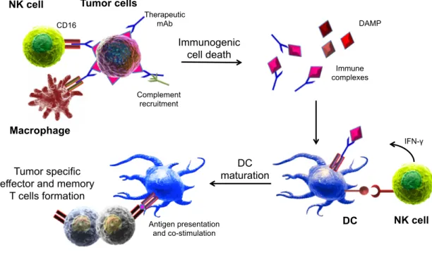
Besides ADCC, tumor-targeting mAb can induce other responses that promote tumor cell killing, such as complement-mediated target lysis and phagocytosis (Gajewski TF et al., 2013; Pincetic A et al., 2014; Scott AM et al., 2012b). Tumor-specific antibodies, moreover, can induce long-lasting protective effects in the treated patient by enhancing the development of adaptive immune response; this phenomenon has been called vaccinal effect, and consists in the fact that tumor-targeting mAb favour the uptake of tumor antigens by DC, and promote the generation of DAMP and a proinflammatory environment, that stimulate DC maturation and activation (Abes R et al., 2010; Pincetic A et al., 2014; Wittrup KD, 2017). NK cells are crucial promoting components of this process, as they induce DC maturation through the secretion of IFN-γ and TNF-α and by cell-cell contacts (Walzer T et al., 2005). Activated and mature DC then allow the formation of efficient and long-lasting T cell-mediated anti-tumor responses (Martín-Fontecha A et al., 2004) (Figure 4).
In particular, therapeutic mAb directed against CD20 antigen were the first to demonstrate clinical efficacy. CD20 is an antigen widely expressed on the surface of B lymphocytes in all stages of development, from pre-B precursor up to mature B cell, and also in several B cell malignancies. CD20-targeting mAb are widely studied and developed (Salles G et al., 2017; Weiner GJ, 2010). In 1997 the Food and Drug Administration (FDA) approved rituximab (RTX) anti-CD20 chimeric mAb for the treatment of refractory and relapsing non-Hodgkin lymphoma. In the last 20 years, RTX was used effectively also in many other B cell malignancies and in some autoimmune diseases, including rheumatoid arthritis, multiple sclerosis, haemolytic anaemia (Gürcan HM et al., 2009; Salles G et al., 2017). In combination with chemotherapeutic drugs, it gives effective results on
progression-Figure 4. Model for the development of anti-tumor vaccinal effect
Therapeutic mAb induce tumor cell death by ADCC, phagocytosis and complement-induced cytotoxicity. Resulting immune complexes (antigen-containing debris+ mAb) and damage- associated molecular patterns (DAMP) can be captured and recognized by immature DC leading to their maturation and consequent expansion of tumor-specific T cells. DC activation can be strengthened by NK cell-derived IFN-γ and by cell-cell interactions. In parallel, DC-produced IL-12 results in an increased function of NK cells. The formation of a tumor-specific memory T cell pool guarantees the long term protection against tumor recurrence.
Tumor cells NK cell Immunogenic cell death CD16 Immune complexes DC Tumor specific effector and memory
T cells formation Macrophage Therapeutic mAb DAMP Complement recruitment IFN-γ DC maturation NK cell Antigen presentation and co-stimulation
free and overall survival of patients with diffuse large B-cell lymphoma (DLBCL) or follicular lymphoma (Harrison AM et al., 2014; Tilly H and Zelenetz A, 2008). Upon RTX binding, low-level direct death of the target cell is induced, while ADCC, phagocytosis and complement-mediated death are triggered (Mossner E et al., 2010).
Several mechanisms of resistance to RTX have been recognized that limit its therapeutic efficacy (Freeman CL and Sehn LH, 2018); this knowledge led to the development of second-generation anti-CD20 mAb. Obinutuzumab (GA101) is a glycoengineered humanized anti-CD20 mAb that more powerfully induces direct cell death, independently from caspase activation. Ex vivo studies, however, suggest that this is not the principal mechanism through which the antibody is working in vivo (Bologna L et al., 2011; Tobinai K et al., 2017). In fact, thanks to its defucosylated Fc domain, GA101, although inducing minimal complement-mediated death, is recognized with higher affinity by CD16, improving both ADCC and phagocytosis (Bologna L et al., 2011; Mossner E et al., 2010). Its improved efficacy has been demonstrated in untreated follicular lymphoma and CLL, in combination with chemotherapy. Tumor histological subtype, host immune integrity, as well as CD16 polymorphisms may represent important factors for the preference of one mAb over the other (Freeman CL and Sehn LH, 2018).
mAb may also act with other therapeutic mechanisms, for example by targeting NK receptors that regulate their functional activities. Some examples are: anti-NKG2D, anti-CD137, anti-NKG2A, anti-PD-1 (Carotta S, 2016; Chester C et al., 2015; Kohrt HE et al., 2011, 2014; Vadstrup K and Bendtsen F, 2017; Battella S et al., 2016). A mAb that blocks iKIR was tested until phase I trial, however with disappointing results. Indeed, mAb-iKIR interaction blocked the NK education process, thus hampering their functional maturation (Benson DMJ et al., 2012; Vey N et al., 2012).
The scientific community is now investigating the efficacy of bi- and tri-specific antibodies, made to increase the effectiveness of mAb-based treatments. For instance, the CD16xCD33 bi-specific (BiKe) construct engages CD16 and CD33, resulting in an increased NK cell function against CD33+ tumor and MDSC targets (Gleason MK et al., 2014). New generation TriKe is based on the addition of IL-15 to the CD16xCD33 BiKe, to increase the stimulation and the effector functions of