© 2019 Journal of Cardiovascular Echography | Published by Wolters Kluwer - Medknow
62
Abstract
Case Report
I
ntroductIonIatrogenic aortic dissection (AD) during percutaneous coronary intervention (PCI) is a complication that occurs in 3.4%–2.0% of diagnostic angiography and 14.3% of ad hoc PCI.[1] We
present a case of iatrogenic right coronary artery (RCA) dissection complicated by a retrograde subtle AD, promptly treated with coronary stents implant from the medium tract of right coronary to the ostium, to arrest the progression of dissection and rupture of the coronary wall.
c
aser
eportA 65-year-old female presented at the emergency department of our hospital with acute anterior ST-elevation myocardial infarction (STEMI) associated with moderate dyspnea. In her history, there was a family history of cardiovascular disease, hypertension, dyslipidemia, smoke, and polycythemia vera.
The patient was directly referred to the Cath lab for primary percutaneous transluminal coronary angioplasty (PTCA). Coronary angiography revealed a severe proximal subocclusive stenosis (99%) of the left anterior descending, successfully treated with stent implantation. After 5 h, there was a worsening of patient’s condition, with recurrence of chest pain and electrocardiogram (ECG) ST-elevation in the inferior leads (inferior STEMI); therefore, the patient was submitted to a new coronary angiography. A diagnosis of spiral dissection, extending from the ostium to the medium tract of the RCA with a contrast media extravasation, due to coronary ostium fissure, was made, and coronary stents were implanted from
We report a rare case of iatrogenic right coronary artery (RCA) dissection complicated by a retrograde subtle aortic dissection, which occurred during a primary percutaneous transluminal coronary angioplasty (PTCA). A 65-year-old female, with acute anterior ST-elevation myocardial infarction (STEMI), promptly underwent primary PTCA in the left anterior descending artery. After 5 h, the patient’s condition becomes worse with recurrence of chest pain and new electrocardiogram modifications suggestive of inferior STEMI. A second coronary angiography revealed a spiral dissection extending from the ostium to the medium tract of the RCA. At the same time, a contrast media extravasation due to coronary ostium fissure occurred. Coronary stents were implanted from the medium tract of the right coronary to the ostium, to promptly arrest the active bleeding and to treat the dissection. After cardiosurgical advice, the patient was referred to the radiology department, where she underwent computed tomography angiography (CTA), which showed a small hematoma in the anterior wall of the ascending aorta. The stable clinical conditions of the patient suggested a conservative therapeutic approach. During the following 6 weeks CTA and transesophageal echocardiography were performed to rule out any other complication, and the patient was fortunately discharged with almost complete resolution of the hematoma.
Keywords: Aortic intramural hematoma, computed tomography angiography, coronary artery dissection, transesophageal
echocardiography
Address for correspondence: Dr. Ugo Barbaro, Consolare Pompea Street, 13, Messina 98168, Italy. E‑mail: [email protected]
Access this article online Quick Response Code:
Website:
www.jcecho.org
DOI:
10.4103/jcecho.jcecho_13_19
This is an open access journal, and articles are distributed under the terms of the Creative Commons Attribution-NonCommercial-ShareAlike 4.0 License, which allows others to remix, tweak, and build upon the work non-commercially, as long as appropriate credit is given and the new creations are licensed under the identical terms.
For reprints contact: [email protected]
How to cite this article: Salamone I, Carerj ML, Barbaro U, Virga V, Zito C, Bracco A, et al. The usefulness of a multimodality approach in a case of subtle iatrogenic aortic dissection: Sometimes is better to look and wait. J Cardiovasc Echography 2019;29:62-4.
The Usefulness of a Multimodality Approach in a Case of
Subtle Iatrogenic Aortic Dissection: Sometimes is Better to
Look and Wait
Ignazio Salamone, Maria Ludovica Carerj, Ugo Barbaro, Vittorio Virga1, Concetta Zito1, Antonio Bracco1, Alfredo Blandino, Sergio Racchiusa
Departments of Biomedical Sciences and Morphological and Functional Imaging and 1Clinical and Experimental Medicine, University of Messina,
Policlinico “G. Martino,” Messina, Italy
Salamone, et al.: Iatrogenic aortic dissection
Journal of Cardiovascular Echography ¦ Volume 29 ¦ Issue 2 ¦ April - June 2019 63 the medium tract to the ostium to promptly arrest the active
bleeding and to treat the dissection.
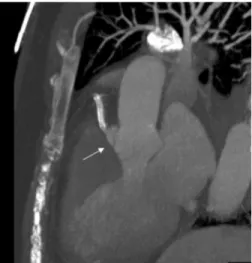
Although the patient was hemodynamically stable, once that consent informed was obtained, a computed tomography (CT) scan was promptly performed with a 64-detectors scanner (SOMATOM Definition Siemens), to rule out an AD. Unenhanced scan showed extravasation of contrast media, administered during the second PTCA, in the upper pericardial recesses [Figure 1]. After intravenous administration of contrast media, the examination showed the absence of pericardial active bleeding and the presence of stents in the RCA ostium [Figure 2]; however, CT showed also a small hematoma of the right coronary sinus, as a result of the dissection, treated with stents of the right coronary [Figures 2 and 3]. The stable clinical conditions of the patient suggested a conservative therapeutic approach.
The follow-up of this finding was made with serial transesophageal echocardiography (TEE) [Figures 4a and b], which revealed a progressive and complete hematoma resolution [Figures 5a and b].
d
IscussIonCoronary artery dissection could be spontaneous or traumatic. The real incidence of spontaneous coronary artery dissection (SCAD) is not well known widely, since, in many cases, it is rarely diagnosed. This pathology was considered very rare, nevertheless contemporary angiographic series report SCAD diagnosis rates of 0.07%–0.2% of all angiograms, and 2%–4% of angiograms performed for acute coronary syndromes.[2]
Figure 3: An axial view shows the presence of a subtle hematoma of the
right coronary sinus (black arrow)
Figure 1: Unenhanced computed tomography scan shows extravasation
of contrast medium (asterisk) in the anterior recess due to right coronary artery dissection
Figure 2: Sagittal reconstruction shows the absence, after contrast
medium intravenous injection, of pericardial active bleeding, the presence of a stent in right coronary, and the subtle hematoma of right coronary sinus (white arrow)
Figure 4: (a) (Top) transesophageal echocardiography at 44° and (b)
(bottom) at 110° multiplane angle display a complete resolution of the aortic hematoma
b a
Salamone, et al.: Iatrogenic aortic dissection
Journal of Cardiovascular Echography ¦ Volume 29 ¦ Issue 2 ¦ April - June 2019
64
Nowadays, the pathophysiology of SCAD is still unknown. The combination of predisposing factors such as female sex and pregnancy, fibromuscular dysplasia, inflammatory conditions, atherosclerotic risk factors, mechanical stressors, emotional stressors, inherited connective tissue disorders, and genetics could increase the susceptibility of minor trigger events.[2]
Nevertheless, a great amount of coronary artery dissections is iatrogenic, since these latter ones have been caused by the tip of guiding catheter applied during PCI. Iatrogenic coronary artery dissection extending from the coronary sinuses during PCI is present in 3.4%:2.0% of diagnostic angiography and 14.3% of ad hoc PCI.[1] In the majority of cases, a proximal
RCA dissection with retrograde extension toward the aorta is the first complication,[3] as in our case. The origin of most
dissections of the ascending aorta is located at the ostium or in the proximal segment of the coronary artery. This complication occurs following a trauma caused by the tip of the catheter or balloon dilation in case of PTCA; furthermore, a vigorous manual injection of contrast material may play a role in extending the dissection to the aortic root.[3] Clinical
risk factors for the extension of the coronary dissection into the aortic sinuses include older age, hypertension, both present in our patient, and calcification of the aortic root.[4,5] The prompt
treatment of the coronary dissection in the area of the tear with the implant of covered stents blocks the bleeding and extension of the dissection, with a subsequent disappearance of the supply of the false lumen, as in our case. In some cases, despite a stent implantation covering the coronary dissection’s
tear, the retrograde extension of dissection has made necessary a coronary artery bypass grafting and ascending aorta replacement.[6] To detect an acute AD,[7] a transthoracic
echocardiogram is inadequate, while TEE, due to the relative proximity of the esophagus to the thoracic aorta and high resolution of the images, plays an important role. ECG-gated CT and magnetic resonance imaging are the leading techniques for diagnosis and classification of ADs. The first imaging modality plays a pivotal role in the diagnosis, risk stratification, and management of aortic diseases. Its advantages over other imaging modalities include the short time required for image acquisition and processing, the ability to obtain a complete 3D dataset of the entire aorta, and its widespread availability.[7]
A combined diagnostic approach, with multimodality imaging, plays a fundamental role in patients with aortic complications during PTCA. In the present case, this combined approach has been helpful for clinical decision-making and ultimately to reach an agreement for avoiding cardiac surgery.
Declaration of patient consent
The authors certify that they have obtained all appropriate patient consent forms. In the form the patient(s) has/have given his/her/their consent for his/her/their images and other clinical information to be reported in the journal. The patients understand that their names and initials will not be published and due efforts will be made to conceal their identity, but anonymity cannot be guaranteed.
Financial support and sponsorship Nil.
Conflicts of interest
There are no conflicts of interest.
r
eferences1. Prakash R, Starovoytov A, Heydari M, Mancini GB, Saw J. Catheter-induced iatrogenic coronary artery dissection in patients with spontaneous coronary artery dissection. JACC Cardiovasc Interv 2016;9:1851-3.
2. Adlam D, Alfonso F, Maas A, Vrints C; Writing Committee. European Society of Cardiology, acute cardiovascular care association, SCAD study group: A position paper on spontaneous coronary artery dissection. Eur Heart J 2018;39:3353-68.
3. Welch TD, Foley T, Barsness GW, Spittell PC, Tilbury RT, Enriquez-Sarano M, et al. Iatrogenic aortic dissection…Or intramural hematoma? Circulation 2012;125:e415-8.
4. Tanasie C, Chandonnet M, Chin A, Kokis A, Ly H, Perrault LP, et al. Catheter-induced aortic dissection after invasive coronary angiography: Evaluation with MDCT. AJR Am J Roentgenol 2011;197:1335-40. 5. Pérez-Castellano N, García-Fernández MA, García EJ, Delcán JL.
Dissection of the aortic sinus of valsalva complicating coronary catheterization: Cause, mechanism, evolution, and management. Cathet Cardiovasc Diagn 1998;43:273-9.
6. Li JC, Guan XL, Gong M, Zhang HJ. Iatrogenic aortic dissection during percutaneous coronary intervention: A case report and review of the literature. J Int Med Res 2018;46:526-32.
7. Erbel R, Aboyans V, Boileau C, Bossone E, Bartolomeo RD, Eggebrecht H, et al. 2014 ESC guidelines on the diagnosis and treatment of aortic diseases: Document covering acute and chronic aortic diseases of the thoracic and abdominal aorta of the adult. The Task Force for the Diagnosis and Treatment of Aortic Diseases of the European Society of Cardiology (ESC). Eur Heart J 2014;35:2873-926.
Figure 5: (a) (Top) transesophageal echocardiography at 44° and (b)
(bottom) at 110° multiplane angle display a complete resolution of the aortic hematoma
b a