International PhD Program in Neuropharmacology
XXVI Cycle
Implications of dopamine D3 receptor for glaucoma: in-silico
and in-vivo studies
PhD thesis
Chiara Bianca Maria Platania
Coordinator: Prof. Salvatore Salomone
Tutor: Prof. Claudio Bucolo
Department of Clinical and Molecular Biomedicine Section of Pharmacology and Biochemistry.
University of Catania - Medical School
2
Copyright ©: Chiara B. M. Platania - December 2013
3
TABLE OF CONTENTS
ACKNOWLEDGEMENTS... 4
LIST OF ABBREVIATIONS ... 5
ABSTRACT ... 7
GLAUCOMA ... 9
Aqueous humor dynamics ... 10
Pharmacological treatments of glaucoma... 12
Pharmacological perspectives in treatment of glaucoma ... 14
Animal models of glaucoma ... 18
G PROTEIN COUPLED RECEPTORS ... 21
GPCRs functions and structure ... 22
Molecular modeling of GPCRs ... 25
D2-like receptors ... 31
CHAPTER I ... 37
CHAPTER II ... 75
GENERAL DISCUSSION AND CONCLUSIONS ... 102
REFERENCES... 105
4
A
CKNOWLEDGEMENTSI would like to thank prof. Claudio Bucolo, who has fully supported me during the last two years of my PhD studies. With his great experience and knowledge, he taught me how to approach “Research”.
I wish to thank prof. Filippo Drago, he helped me not to give up with the PhD program. Prof. Drago, along with prof. Bucolo and prof. Salomone, has welcomed me in his research group, that is the best I’ve ever worked for.
I am grateful to prof. Salomone, for each constructive scientific advice he gave me.
I want to thank also all the former and actual lab. mates, researchers and friends of the Department of Pharmacology of University of Catania.
The last but not the least, my thanks go to my beloved parents, for their efforts in supporting me during university studies, till the highest degree. I’m also grateful to Riccardo, who has continuously supported me either in the worst and best moments.
5
L
IST OF ABBREVIATIONSIOP Intraocular Pressure TM Trabecular Meshwork
GPCRs G protein Coupled Receptors hDx human Dopamine Dx receptor
D2L Dopamine D2 receptor long isoform
D2S Dopamine D2 receptor short isoform
wt wild-type
5HTxy Serotonin 5HTxy receptor
D3-/- D3 receptor gene knockout
AH Aqueous Humor
POAG Primary Open Angle Glaucoma RGCs Retinal Gaglion Cells
PACG Primary Angle Closure Glaucoma AQPs Aquaporins
CA Carbonic Anhydrase
ATP Adenosin Triphosphate ADP Adenosin Diphosphate ECM Extracellular Matrix
NICE National Institute for Health and Clinical Excellence cAMP cyclic Adenosin MonoPhospate
CAI Carbonic Anhydrase Inhibitor BBB Blood Brain Barrier
PG Prostaglandin
EP Prostaglandin E receptor Mx Muscarinic Mx receptor
siRNA Small Interference RNA
11- HSD1 11- hydroxysteroid dehydrogenase-1 NPE Non-Pigmented Epithelial cells Ax Adenosin Ax receptor
MMP Matrix Metallic Proteinase
AEA Anandamide
6 CBx Cannabinoid CBx receptor
PEA Palmitoyl ethanolamide
PPARP- Peroxisome Proliferator Activated Receptor-alpha Gs G stimulatory protein
Gi G inhibitory protein
DEX Dexamethasone
PBS Phosphate Buffered Saline
IUPHAR International Union of Basic and Clinical Pharmacology FDA Food and Drug Administration
NCE New Chemical Entities FP Prostaglandin F receptor MW Molecular Weight
3D Three Dimentional
Hx Histamine Hx receptor
x-OR x-Opiod Receptor NTSR Neurotensin Receptor PAR Protease Activated Receptor ECL Extracellular Loop
ICL Intracellular Loop
S1P1 Sphingosine 1-phospate receptor
R&D Research and Development
FG-MD Fragmente Guided Molecular Dynamics MD Molecular Dynamics
IR Infrared
NMR Nuclear Magnetic Resonance
CHARMM Chemistry at Harvard Macromolecular Mechanics VdW Van der Waals
oop out of plane movement
GB Generalized Born
CPU Computing Processing Unit
CUDA Computed Unified Device Architecture GPU Graphic Processing Unit
MM/GBSA Molecular Mechanics/Generalized Born Solvent Accessible surface area MM/PBSA Molecular Mechanics/Poisson Boltzmann Solvent Accessible surface area
7
A
BSTRACTGlaucoma is a progressive optic neuropathy and it is considered by the World Health Organization to be the cause of 12% of visual impairment and 2% of blindness. Glaucoma is characterized by alterations of optic disc and visual field. High intraocular pressure (IOP) is the main risk factor of glaucoma. The pathogenesis of glaucoma is still evanescent, and unfortunately the disease becomes symptomatic when irreversible and severe damage has occurred at the optic nerve head. IOP reduction represents the first step in the management of glaucoma which is eventually followed by laser surgery of the trabecular meshwork (TM) and glaucoma-filtering surgery.
Currently, there are five main classes of approved ocular hypotensive drugs: beta-blockers, carbonic anhydrase inhibitors, prostaglandin analogs, sympathomimetics and miotics. However, there is still the need to have more potent medications available for this disease, and pharmacological management of IOP is one of the most interesting and challenging endeavors facing the ocular pharmacology scientists.
In the panorama of pharmacological targets for regulation of IOP, there are some interesting G protein coupled receptors (GPCRs) such as dopaminergic receptors. The work of the present thesis has been focused on GPCRs and in particular on dopamine D3
receptor as pharmacological target for ocular hypotensive drugs. Cabergoline, bromocriptine, cianergoline and legotrile, classical D2 receptor agonists, have been shown
to decrease intraocular pressure. D3 receptor belongs to the D2 class of dopaminergic
receptors, along with D2 and D4 receptor. It shares high sequence homology and identity
with D2 receptor and several efforts have been carried out in order to design selective
ligands for either D3 or D2 subtype. Drug design and discovery, based on structure based
approach, need the knowledge of the tertiary structure of the target protein. In 2010 the x-ray structure of human D3 receptor (mutated hD3-lysozime chimera) was solved, then this
structure was used to carry out the homology modeling of wild-type (wt) hD3 and hD2L
receptors. The homology models of these receptors were not able to discriminate selective ligands by a molecular docking study, thus these structures have been subjected to optimization by means of molecular dynamics in a water-membrane environment. After optimization the structures differentiated in the binding pockets and have been validated, strengthen the validity and reliability of the in silico approach. A similar computational approach was carried out in order to study the structure differences between the D3
8 receptors and 5HT1A, 5HT2A-C receptors, known to be involved in regulation of intraocular
pressure.
The role of D3 receptor activation by cabergoline in lowering IOP was confirmed in
C57BL/6J wt and D3-/- mice, using a pharmacological approach along with D3 gene
deletion. Animals were pre-treated with U99194A, D3 selective antagonist, that
antagonized the effects of cabergoline. Ocular hypertension was induced in mice, implanting subcutaneously an osmotic micropump delivering dexamethasone. Cabergoline was not effective in ocular hypertensive D3-/- mice, whereas exerted a greater and longer
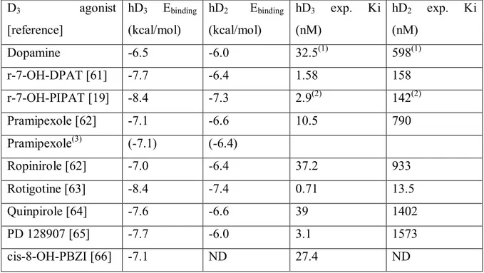
hypotensive effect in ocular hypertensive wt mice, in comparison to normotensive animals. The in silico approach, validated for D3 and D2L receptors, has been used to model and
optimize the structures of 5HT1A, 5HT2A-B-C receptors which are other putative ocular
targets of cabergoline. In silico results showed that cabergoline binds in a similar way into pockets of D3 and 5HT2A-C and it has higher affinity for D3 receptor in comparison to
serotonergic receptors, according to experimental affinity data. Moreover docking revealed that binding of cabergoline into D3 and 5HT1A receptors is associated with a better
desolvation energy in comparison to 5HT2A–C binding.
The structure-based computational approach hereby adopted was able to build, refine, and validate structure models of homologous dopaminergic and serotonergic receptors that may be of interest for structure-based drug discovery of ligands, with dopaminergic selectivity or with multi-pharmacological profile, potentially useful to treat optic neuropathies such as glaucoma.
Finally, the present work represents an excellent example of successful integration of two different approaches to biomedical research, in silico and in vivo, which are not in contrast but complementary.
9
GENERAL INTRODUCTION
GLAUCOMA
Glaucoma refers to a series of progressive optic neuropathies that involve optic disc degeneration and visual field aberration, with or without ocular hypertensions. Ocular hypertension in glaucomatous patients is related to an imbalance between aqueous humor (AH) production and drainage [1]. The most common form of glaucoma is the primary open angle glaucoma (POAG), that is an optic neuropathy characterized by optic disc damage and partial loss of visual acuity, associated to retinal ganglion cells (RGCs) death and atrophy of optic nerve [2]. The progression of POAG leads to irreversible blindness. Gene association studies have been carried out and more than 20 genetic loci have been reported for POAG; up to now only three have been found to have a causal relationship with the disease (myocillin, optineurin and WDR36) [3]. POAG is associated to increased IOP. However there are about 20-52% of glaucomatous patients with normal IOP and those patients develop damage at the optic nerve head similar to POAG patients. Ocular hypertension ( > 21 mmHg), can arise asymptomatically and patients develop symptomatic visual field loss when damage at optic nerve head is irreversible. Progression of optic disc damage leads to irreversible blindness. Up to know the only therapeutic approach in glaucoma therapy is aimed at decreasing the IOP, also in normotensive glaucomatous patients [4].
Glaucoma could be classified in primary and secondary glaucoma. The former is related to glaucoma forms not correlated to previous and concomitant ocular disease. Primary glaucoma has most probably genetic etiology and it is often bilateral. On the contrary secondary glaucoma is associated to previous ocular or systemic diseases, trauma or iatrogenic causes.
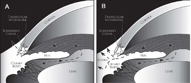
Glaucoma can be classified into major categories according to the appearance and obstruction of the drainage pathway at the iridocorneal angle. In POAG, despite normal clinical appearance the outflow of AH is restricted, possibly due to pathologic changes at the TM (Fig. 1A). In the primary angle-closure glaucoma (PACG) the block of drainage of AH is related to the contact of the iris at the TM, causing its permanent obstruction (Fig. 1B). PACG is often characterized by a rapid increasing of IOP, along with eye pain. Two potential pathogenetic mechanisms of glaucoma have been postulated. Mechanic hypothesis: high levels of IOP could lead to mechanic compression of fibers at the optic
10 nerve head and lamina cribosa where they pass. The axons of RGCs are grouped at the optic nerve head, and death of RGCs leads to visual field loss.
Vascular hypothesis: a presumed perfusion deficit at optic disc could lead to papillar atrophy leading to optic nerve head damage. The assumption of vascular deficit has not been demonstrated by validated scientific results; however it is an hypothesis able to explain normotensive glaucoma, although a perfusion deficit could be explained by an imbalance between AH production/outflow.
Aqueous Humor Dynamics
In healthy eye the flow of AH against resistance generates an average intraocular pressure of approximately 15 mmHg. IOP is necessary to inflate the eye and maintain the proper shape and then optical properties of the globe [5]. High intraocular pressure values are the main risk factor of glaucoma. Epidemiological studies have shown that per each 1 mmHg increase of IOP, the risk of incidence of glaucoma increases of 12% [6]. Up to now long term lowering of IOP is the only strategy to counteract RGCs death, than optic nerve head damage and visual field loss.
Increasing of IOP is due to imbalance between AH production and AH drainage. The ocular sites involved in the production of AH are the processes of ciliary bodies, that have a glomerular structure, due to basal and inner interdigitations. The epithelium of the ciliary processes has two layers: an inner non-pigmented layer in contact with AH in the posterior chamber, and the external pigmented epithelium in contact with the ciliary process stroma; those two epithelial layers have the apical surfaces opposite to each other. Non-pigmented epithelium is the continuation of retina, whereas the pigmented epithelium is a continuation of retinal pigmented epithelium. Both sympathetic and parasympathetic nerves supply the ciliary body. Production of AH is regulated by vascular contraction-dilatation and by neurovegetative inputs from the sympathetic and parasympathetic systems. Three mechanisms are involved in AH production: diffusion, ultrafiltration and active secretion. Diffusion occurs when solutes, especially lipid soluble molecules, are transported through the lipid portion of tissues between capillaries and posterior chamber. Ultrafiltration is related to the flow of water and water soluble molecules through fenestrated ciliary capillary endothelia into the ciliary stroma. Diffusion and ultrafiltration are responsible of accumulation of plasma ultrafiltrate in the stroma behind tight junction of non-pigmented ciliary epithelium.
11 The active secretion of AH from ciliary bodies is related to active transport of Na+, Cl- and
HCO3-, than water passively passes the blood-water barrier of ciliary bodies. Proteins
responsible of rapid bulk water flux are Aquaporins (AQPs) and in particular AQP1 and AQP4 [7]. The carbonic anhydrase (CA) is the enzyme responsible for secretion of hydrogen carbonate, and Na+/K+ pump leads to secretion of Na+. Na+-K+ ATPase is devoted to hydrolysis of ATP (adenosine triphospate) to ADP (adenosine disphosphate) providing energy for active transport of solutes. Na+-K+ ATPase can be inhibited by different molecules [8, 9], and this enzyme has been of particular interest for pharmacological studies on aqueous humor production. Furthermore the chloride ion is secreted through chloride channels.
AH is secreted in the posterior chamber, then it passes in the anterior chamber where there is the TM (Fig. 2A), the prominent outflow facility for AH. The TM is a structure that overpasses the sclera sulcus and converts it into the Schlemm’s canal. The TM is a spongy tissue, that consists of connective tissue surrounded by epithelium. TM can be divided in three components: uveal meshwork, corneoscleral meshwork and juxtacanalicular meshwork, ordered from innermost to outermost part. Three systems innervate TM: the sympathetic from superior sympathetic ganglion, the parasynmpathetic innervations from ciliary ganglion and sensory nerves that originate from trigeminal ganglion. The AH passes all TM parts and is collected in the Schlemm’s canal, which is connected to episcleral and conjunctival veins through external collector canals. Fluid movements take place under a pressure gradient from the TM to Schlemm’s canal and through its inner wall. This flux appears to be related to a passive pressure-dependent transcellular mechanism, associated with paracellular routes such as giant vacuoles and pores [10]. After exiting from Schlemm’s Canal, AH enters in aqueous veins, mixes with blood in episcleral veins where pressure is 8-10 mmHg; considering that normal pressure of AH conventional drainage system is 3-4 mmHg, average normal IOP is 15 mmHg. In humans 75% of the AH outflow resistance is localized at TM and in particular at juxtacanalicular portion [11]. The role of the extracellular matrix (ECM) on AH drainage has been investigated. ECM is constituted by glycosaminoglycans that hydrate TM leading to obstruction; catabolic enzymes depolymerize glycosaminoglycans preventing the excessive TM hydratation. Such effects of lysosomal enzymes is inhibited by corticosteroids, explicating a possible mechanism of steroid-induced glaucoma [12, 13]. AH outflow through the conventional route is influenced by two contractile structures, the iris and ciliary muscle innervated by cholinergic nerves. Contraction of such structures results in spreading of TM and dilation
12 of Schlemm’s canal resulting in increased outflow, on the contrary relaxation of such structures leads to decreased outflow [14].
The TM is assisted in AH outflow by an unconventional route (Fig. 2B), the uveo-scleral pathway through the uveal meshwork and ciliary muscle. That unconventional pathway accounts to a variable percentage of AH excretion, likely age related [15, 16]. Ciliary muscle contraction influences the uveo-scleral outflow, which is increased by prostaglandin F2 by decreasing the flow resistance of the interstitial spaces in the ciliary muscle [17].
Pharmacological treatments of glaucoma
The described AH inflow and outflow facilities are the pharmacological targets of ocular hypotensive drugs. Up to know there are five classes of IOP lowering approved drugs; beta-blockers, carbonic anhydrase inhibitors, prostaglandin analogs, sympathomimetics and miotics [18]. The first two drug classes can be named as “inflow” and the letter three can be named “outflow” drugs. The UK National Institute for Health and Clinical Excellence (NICE) has published a guideline for glaucoma treatment (guideline CG85), in which algorithms for treatment of glaucoma are stated; beta-blockers and prostaglandin analogs are advised for first-line and second line-treatment of glaucoma, whereas the remaining drug classes are mostly used as second-line drugs.
Beta-blockers are used in clinics since 30 years and timolol is the most used drug belonging to this class. Timolol antagonizes β-adrenergic receptors of iris-ciliary body system inhibiting the synthesis of the second messanger cAMP due to adrenergic stimulation. The elevation of cAMP leads to AH production. Timolol is effective at the iris-cilary body, where there is an endogenous adrenergic stimulation; TM lacks of this adrenergic tone even if there are expressed β-adrenergic receptors. Timolol is not a selective beta-blocker, and the role of either 1 or 2 receptors is still evanescent; because
of a 2 agonist (solbutamol) increases AH production. Several beta-blockers have been
approved for treatment of glaucoma: timolol, betaxolol, levobunol and carteolol. Beta-blockers are associated to adverse effects, involving respiratory (increased severity of bronchial occlusive disease) and cardio-vascular systems (arrhythmia, hypotension); thus the anamnesis of patients is important before treatment with beta-blockers.
Carbonic anhydrase inhibitors (CAI) are effective at epithelial cells of ciliary processes, inhibiting the release of HCO3- in the posterior chamber and then production of AH. The
13 Acetazolamide as ocular hypotensive drug has been, in the fifties, an advance in treatment of glaucoma relied at that time mostly on pilocarpine and cholinesterase inhibitors. However carbonic anhydrases are ubiquitous enzymes and several adverse effects arose in glaucomatous patients treated with acetazolamide, such as metabolic and respiratory acidosis, paraesthesias, hypokalemia, hyperuricemia, liver failure and respiratory failure. Thus several efforts were carried out to design new CAI as eye drops. Effective topical CAI must be highly active at CA enzyme, should be amphifilic in order to pass the cornea and be formulated as eye drops [19]. Drug design of new CAI led to dorzolamide, a thienothiopyran-2-sulfonamide. Dorzolamide has shown a low Ki=0.3 nM, inhibiting the 98% of CA. Dorzolamide is effective in decresing IOP of about 26% with a prolonged action (12%). Secondary effects of dorzolamide, compared to acetazolamide, are negligible.
Sympathomimetics ocular hypotensive drugs are 2-adrenergic and non-selective
adrenergic agonists, activating presynaptic adrenergic receptors in the ciliary body and inhibiting the release of noradrenaline that stimulate AH secretion. Sympathomimetics have shown not only to block the inflow of AH, but also to increase the uveo-scleral outflow. Clonidine was the first sympathomimetic drug effective in decreasing the IOP, but due to its high lipophilicity it crosses the blood-brain barrier (BBB) acting at vasomotor center, then inducing systemic hypotension and bradycardia. Apraclonidine is less lipophilic, it does not pass the BBB and does not lead to systemic side effects. It is absorbed through the conjunctival-scleral pathway and it is effective in decreasing IOP. Brimonidine is another 2-adrenergic agonist approved as ocular hypotensive drug [20].
Dipivefrin is a non-selective adrenergic agonist used in treatment of glaucoma and it is the prodrug of epinephrine.
Prostaglandin (PG) analogs are first-line, along with beta-blockers, drugs for treatment of ocular hypertension. PG analogs increase primary the uveo-scleral outflow, even though effects on TM have been reported. The uveo-scleral outflow is increased due to less resistance by means of remodeling of the ECM within the ciliary muscle and sclera. This effect of PG is mediated by activation of the GPCRs EP2 and EP4 activating the synthesis
of cAMP by stimulation of phosphoinositide turnover. It was reported in isolated TM a relaxant activity of EP2 agonists due to coupled activation of Ca2+ -activated K+ channels.
The current PG analogs approved in USA and EU are latanoprost, bimatoprost, travoprost and tafuprost, which are PGF2 analogs [18]. Topical treatment with PG analogs is not
14 associated to systemic side effects. PG analogs side effects are mostly local and not severe: changes in eye color and eyelid skin, stinging, blurred vision, eye redness, itching, burning. Pilocarpine has been used since 100 year for the treatment of glaucoma; it is a miotic or parasympathomimetic drug. It is an alkaloid extracted from plants belonging to Pilocarpus specie. Pilocarpine mimics acetylcholine activating muscarinic receptors of ocular parasympathetic nerve. Pilocarpine leads to myosis by activation of muscarinic M1 and M3
receptors, it induces increasing of Ca2+ levels and contraction of ciliary muscle thus leading to opening of the TM and to increased AH outflow. Due to the miotic effect of pilocarpine, treated patients report blurring of the vision. Moreover continuous treatments can lead to systemic side effects such as nausea, bradycardia and hypotension.
It is worth to be mentioned an experimental pharmacological approach for the treatment of glaucoma, in particular of normotensive glaucoma, focused on neuroprotection. In fact normotensive glaucomatous patients have optic nerve damage and atrophy comparable to ocular hypertensive patients. Thus it has been proposed a neurodegenerative etiology of glaucoma, assuming that RGCs death could be related to glutamate excitotoxicity. Thus NMDA noncompetitive antagonists, memantine [21] and bis-7-tacrine[22], have been studied in order to assess their properties as neuroprotectors at RGCs. Although phase III clinical trial results assessed the inefficacy of memantine in glaucoma treatment, the therapeutic application of neuroprotectors compounds should not be abandoned [23]. Considering neuroprotection as a potential endpoint in glaucoma, several studies about the role of autophagy modulation on RGCs death have been carried out. It is still not clear if promotion of authophagy might prevent RGCs death, however this field is worthy to be explored [24].
Pharmacologic perspectives in treatment of glaucoma
Even thought there are five approved classes of drugs for treatment of glaucoma, the pharmacological management of the disease is still a challenge. New pharmacological perspectives are facing the possibility to becoming ocular hypotensive treatments.
Gene therapy, by means of small interference RNA (siRNA), have been studied to silence 2-receptors. Treatment with siRNA appears to be promising even if the drug delivery of
such small polynucleotides is challenging. SYL040012 is a small siRNA able to interfere with the transduction of 2-receptor gene, potentially reducing IOP by means of 2
15 Within the GPCRs pharmacological targets for treatment of glaucoma the melatonin receptor deserves to be mentioned. Melatonin receptors have been found to be expressed in the ciliary body, and several melatonin analogs have been studied and have showed ocular hypotensive effects [26, 27]. Furthermore the pharmacological effects of melatonin has been confirmed by treatment of melatonin receptor 1 knockout mice, insensitive to melatonin effects [28].
Another promising “inflow” drug target is the hydroxysteroid dehydrogenase-1 (11-HSD1), this enzyme is localized in the ciliary body, in particular in the non-pigmented epithelial cells (NPE). 11-HSD1 catalyzes the conversion of cortisone to cortisol, thus increasing the sodium transporting capacity of NPE. Carbenoxolone, inhibitor of 11-HSD1, has shown to be able to lower IOP in rabbits treated topically [29, 30].
Considering “outflow” drugs, all current approved ocular hypotensive drugs, do not modify directly the structure of TM. Resistance to AH outflow has been identified mainly in TM, which has a spongy structure with interwoven beams of ECM, TM cells and cell layer in the inner wall of Schlemm canal. In glaucoma patients TM is stiffer in comparison to healthy people [31] and the use of drugs altering cytoskeleton of TM has been proposed. In this direction latruculins and rho-associated protein kinase inhibitors have been studied and reduced IOP in animals and humans. Moreover rho-kinase inhibitors have showed improved blood-flow, might helping survival of RGCs [32].
Since TM cells have a role in regulating AH outflow by modulation of their own cell volume and contractility and those function are regulated by NO/CO system, there is an emerging role of these biatomic molecules in regulation of IOP [33, 34]. It has been reported that NO releasing molecules are able to regulate IOP [35], on the contrary also inhibitors of nitric oxide synthase have shown an ocular hypotensive effect in ocular hypertensive animals [36]; thus positive or negative control of IOP by NO is not clear [37]. The potential role of NO donor molecules in lowering IOP, has led to preclinical and clinical studies on prostaglandin analogs able to release NO such as BOL-303259-X [38]. Considering the role of CO, induction of heme-oxygenase has shown a IOP lowering effect [39] and a CO releasing molecule has shown an ocular hypotensive activity, thus CO might facilitate AH outflow or block its production.
Studies report the involvement of GPCRs in modulation of TM outflow facility. Adenosin receptors have been identified as pharmacological target for IOP modulation and a A2
receptor agonist OPA-6566 is currently in development. OPA-6566 lowers IOP by increasing outflow through TM [40]. Also a A1 receptor agonist has shown to decrease
16 IOP, through activation of matrix metalloproteinases (MMP) leading to a remodeling of TM [41, 42]. The role of A3 subtype appears different, since A3 knockout mice have lower
IOP than wild-type due to decreasing AH inflow [43].
The role of cannabinoids in decreasing IOP, is controversial. Cannabinoids are able to decrease IOP, however due to cannabinoids systemic effects efforts to alternative pharmacologic strategies have been carried out. Anandamide (AEA) an endocannabinoid, is transported to cells then is hydrolyzed by fatty acid amide hydrolase (FAAH), that is expressed in TM tissue [44, 45]. Thus FAAH inhibitors have been studied for their potential modulation of IOP, and they have showed ocular hypotensive effects most likely prolonging AEA activity [46, 47]. Several CB1 and CB2 agonists have been tested and
showed IOP decreasing effects in animals and humans. Also palmitoyl-ethanolamide (PEA) that competitively binds to FAAH showed an ocular hypotensive activity blocking the degradation of AEA [48]. Recently Kumar et al [49] demonstrated that PEA acts in a peculiar way, increasing AH outflow through GPR55, a cannabinoid related receptor, and through activation of peroxisome proliferator activated receptor-alpha (PPARP-).
Serotonergic and dopaminergic receptors as pharmacological targets in modulation of IOP are the objectives of the present work. Serotonergic receptors are a large family of GPCRs (5HT1A-F, 5HT2A-C, 5HT4, 5HT5A-B, 5HT6, 5HT7) and a ligand-gated ion channel, the 5HT3
receptor. Several serotonergic receptor subtypes are expressed in ocular tissue: iris-ciliary body, ciliary epithelium, ciliary muscle and TM [50]. Moreover serotonin has been found in AH of humans. The serotonin receptor subtypes that seem to be strictly involved in the regulation of IOP, 5HT1 and 5HT2 subfamilies, are characterizing their self as interesting
targets. In particular extensive pharmacological studies suggested that 5HT2A activation
has a crucial role in lowering IOP, by means of activation of phospholipase C and releasing of diacylglicerol and inositol-3-phosphate than increasing of free intracellular Ca2+ [51]. A series of 5HT2A agonists have shown IOP lowering effects [52]; even though ketanserine,
5HT2A receptor antagonist, decreases IOP in glaucomatous patients [53]. For this reason
further studies, about the role of 5HT2A in IOP modulation, are needed. 5HT2A agonists and
antagonists have been proposed for IOP reduction, but some obstacles in development arose, possibly pertaining efficacy, adverse effects or lack of selectivity of those compounds. Swedish Orphan Biovitrum completed a phase IIa clinical trial in 2008 on BVT.28949, a 5HT2A antagonist showing an ocular hypotensive activity (10%), however
17 Within the pharmacological targets hereby analyzed only three of them are GPCRs that bind small molecules. Adenosine receptors diverge significantly from serotonergic and dopaminergic receptors, which are both aminergic. Dopaminergic receptors are a family of GPCRs characterized by two subfamilies of receptors: D1-like (D1 and D5) coupled to Gs
(stimulatory) proteins and D2-like (D2, D3 and D4) coupled to Gi proteins (inhibitory). The
pioneering study, about D2-like receptors as pharmacological targets of glaucoma and in
particular D3 receptor, is the work of Chu and collaborators. Chu and co-workers [55] used
a pharmacological approach to identify D3 receptor and its role in lowering IOP. Topical
7-OH-DPAT, a selective D3 receptor agonist was able to decrease IOP in rabbits, and a D3
selective antagonist U99194A reverted this effect. The suggested location of D3 receptor in
the eye, are sympathetic fibers afferent to ciliary body, thus activation of D3 receptor might
block AH inflow. Moreover, ibopamine, D1 receptor agonist, increases AH inflow, and
activation of D3 receptor leads to decreased IOP. The role of D3 receptor as potential
pharmacological target of ocular hypotensive drugs has been confirmed by means of pharmacological approaches along with gene deletion study, using D3-/- mice. Bucolo et al.
[56] have treated wt and D3-/- mice, either normotensive and steroid-induced ocular
hypertensive, with 7-OH-DPAT that exerts its IOP lowering effects only in wt mice. The effects of 7-OH-DPAT have been counteracted by the pretreatment with U99194A. Since all dopaminergic receptors have been identified in ciliary body of mice, it could be stated that inhibition of D3 receptor blocks AH production. In the present work of thesis a study
with a similar experimental paradigm was carried out in order to explain cabergoline lowering IOP effect. The ocular hypotensive effect of cabergoline has been claimed to the activation of 5HT2A receptor [51]. However, cabergoline, along with other ergot
derivatives with IOP lowering activity, is a mixed serotonergic and dopaminergic agonist. Cabergoline has been used, in the present work, as a pharmacological tool in order to confirm the role of D3 receptor in modulation of intraocular pressure, at least in the
18
Animal models of glaucoma
Research animal models have helped in understanding causes of diseases and in particular the discovery and development of new drugs. Animal models, sometimes do not mimic all features of a disease, although even one mimicked feature is useful to find a drug target and to develop new drugs.
Glaucoma is a complex disease and as written before its etiology is still unknown. Several animal models of glaucoma have been reported [57], including a wide variety of species with spontaneous or induced features of glaucoma [58, 59]. These include large animals such as monkeys, dogs, cats and pigs, with disadvantages of breeding and management. Small animals such rodents are used as animal model of glaucoma [60] and relative advantages exceed disadvantages. Rodent models of POAG are reported below.
Steroid-induced glaucoma is reported in humans and it is associated to ocular hypertension [61, 62]. A rat model of glaucoma, induced by topical application of dexamethasone (DEX), was developed to study the expression of myocilin. After two weeks of DEX treatment IOP increased but not significant changes in mRNA levels of myocilin, one causative gene of glaucoma in human [63], have been found. Thus increasing of IOP in steroid-induced glaucoma could be not related to myocilin [64], at least in the described animal model. Rats, in contrast to other non-primate models, have anatomical features of anterior chamber similar to the human especially regarding the AH outflow facility [65-67]. Retinal and optic nerve damage in ocular hypertensive rats resembles damage in POAG patients. However it was found that some glaucoma medications do not have identical effects to those observed in humans [68]. Rats, as well mice, are easy to maintain in laboratory, can be manipulated genetically and be used in large numbers.
A mouse strain expressing the Tyr423His myocilin mutation, corresponding to the human Tyr437His point mutation, was developed to study POAG [69, 70]. At 18 months of age, the myocilin mice model demonstrated loss of 20% of RGCs in peripheral retina, axonal degeneration in the optic nerve and detachment of endothelial cells of the TM. Myocilin mutant mice show a moderate but persistent increasing of IOP, 1-2 mmHg higher than normal. Another transgenic mouse model of glaucoma has been reported: mice with a mutation in the 1 subunit of collagen type I develop POAG [71, 72]. In this model mice have progressive optic nerve axonal loss and gradual elevation of IOP, thus IOP modulation can be related to fibrillar collagen turnover.
19 The steroid-induced ocular hypertension in mice, used in the present work, was first reported by Whitlock et al. [73]. Osmotic micropumps filled with either DEX (water soluble cyclodextrin-DEX complex) or PBS as control, have been implanted in B6.129 hybrid mice. Administration of DEX for four weeks resulted in increased IOP in comparison to baseline and PBS treated mice. This animal model of glaucoma was also used to induce ocular hypertension in C57BL/6J wt and D3-/- mice by Bucolo et. al. [56].
Implantation of osmotic micropumps allows delivery of DEX at constant concentration, 0.09 mg per day. The continuation of administration of DEX is crucial to maintain stable ocular pressure. Main risks for implanted animals are immune-depression and infections. A maximum of 50% of IOP increase, in comparison to baseline and PBS treated animal, was reached. Ocular hypertension in that animal model is mainly related to impairment of AH outflow facility, even if the related molecular mechanism is still unknown.
20
Figure 1. Schematic rapresentetion of POAG (A) and PACG (B).
www.glaucoma.org
Fig. 2 Conventional (A) and unconventional (B) root of aqueous humor outflow.
21
GPROTEIN COUPLED RECEPTORS
G Protein Coupled receptors are integral membrane proteins, with seven trasmembrane helices bundle embedded in the lipid bilayer. In human genome more than 800 genes have been identified encoding GPCRs [74]. In accordance to International Union of Basic and Clinical Pharmacology (IUPHAR), GPCRs are classified in four main classes (excluding sensory GPCRs): (1) class A rhodopsin-like; (2) class B secretin-like, (3) class C metabotropic glutamate/pheromone, (4) and frizzled receptors. Up to now such GPCRs classes are accounted to be pharmacological targets of about 30-50% of approved drugs [75-78]. The potential of GPCRs as pharmacological targets is huge as reported by Garland in the 2013 [79]. Garland has arisen an issue: about 400 drugs exert their effect through no more than 100 receptors, accounting for less than 30% of those expressed in human genome; thus a lot of target/pharmacology combination are still hidden.
Analyzing the new Food and Drug Administration (FDA) approved drugs in the last three years 2011-2013, 19% of approved new chemical entities (NCE) target GPCRs. Within these 17 NCE, four account for new activity/target combination and the remaining for a drug action improvement on known mechanism. One of these 17 approved drugs is tafluprost, which is new ocular hypotensive drug targeting the prostaglandin F (FP) receptor. Looking at drugs targeting aminergic receptors, two of them have been approved: lorcaserin a 5HT2C agonist for treatment of obesity showing a 100-fold and 17-fold
selectivity over 5HT2B and 5HT2A receptor and mirabegron a selective 3 adrenergic
receptor agonist for treatment of overactive bladder. Analysis of last approved drugs, making attention to drug molecular weight (MW), shows that 80% of those drugs targeting GPCRs are small molecules. 20% of molecules with high MW account for peptides; opening new potential studies in drug discovery of such molecules targeting GPCRs, taking also in account the challenges regarding drug stability and delivery issues. The taking home message is that GPCRs, with known or unknown (orphan) functions, are an important source for drug discovery of NCE but also for the repositioning of shelved drugs.
22
GPCRs functions and structure
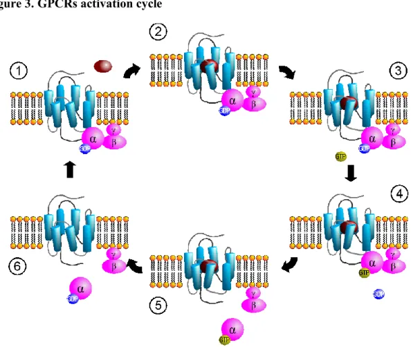
GPCRs are membrane proteins that share the seven transmembrane helices bundle as main structural feature. GPCRs respond to variety of extracellular signals: photons, ions, small organic molecules and peptides. After the action of those signals on GPCRs, the receptor undergoes to a conformational transition, causing the activation of cytosolic signaling networks. GPCRs are conventionally viewed to transmit signals trough activation of G heterotrimeric proteins (Fig. 3). There are encoded 21 , 6 , 12γ subunits, the assembly of which characterize four kinds of G proteins; (1) Gs proteins which upon activation
stimulate cAMP production, (2) Gi which inhibit cAMP production, (3) Gq/11 which
leads to intracellular calcium mobilization, (4) G12/13 which activates monomeric
GPTase RhoA. Some GPCRs are coupled with just one G protein, but other receptors couple to more than one G, depending i.e. from localization, leading to various physiological functions. Alternative signaling pathways of GPCRs have been identified to be exploited in the drug discovery process, such as the -arrestin signaling. Agonist binding to a receptor promotes both G protein activation but also sensitization and internalization. Receptor sensitization arises from phosphorilation of intracellular site of GPCRs, leading to increased affinity for -arrestins, than to block of later receptor activation and to subsequent promotion of receptor internalization. -arrestin signaling had been characterized as negative regulator of GPCRs, but now it has been studied for peculiar signal pathways such as MAP and Src kinases activation, transcriptional regulation and receptor transactivation [80-83]. Thus there is the emerging research field on discovery and development of biased agonists. Biased agonists differentiate themselves from other ligands, activating a subset of signal pathways of the receptor, that can be useful either for activation of clinically beneficial effects or inhibition of side effects. Recently some structural features have been identified to have a role in the biased signaling of GPCRs. A functional and structural approach was carried out to elucidate the behavior of ergotamine, which acts as biased agonist on 5HT2B receptor but not at 5HT1B receptor,
identifying the conformational changes at VII helix crucial for -arrestin signaling [84]. This result highlights the scientific contribution of structural studies on GPCRs. In the past of 12 years, more than 75 x-ray structures of 18 different classes of GPCRs have been solved in complex with several ligands [85]. The availability of such 3D structures, provides an unprecedented opportunity to study deeply the structural and functional
23 features of GPCRs, a protein family characterized by a great heterogeneity even if the overall structure is conserved.
Research now is helped by structural data in addressing questions about: molecular signature of GPCRs, fold and molecular changes that undergo upon receptor activation. Moreover the knowledge of receptor structure is fundamental in the drug discovery and development process that takes advantage of structure-based drug design approaches. So far, high-resolution structure of GPCRs have been solved for the following class A GPCRs: rhodopsin, 1/2 adrenoreceptors, muscarinic M2,3 receptors, H1 histaminergic
receptor, dopamine D3 receptor, 5HT1B and 5HT2B receptors, adenosine A2A, CXCR4
chemokine receptor, opiod receptors (nociceptin receptor, -OR, -OR and -OR), neurotensin receptor NTSR1, protease activated receptor PAR1, and the lipid activated GPCR sphingosine-1 phosphate S1P1 receptor. Within these structures an important
turning point is the crystal structure of 2-adrenoreceptor in complex with heterotrimeric G
protein [86].
The structure of a GPCR can be divided into three parts. The extracellular domain that consists of the N-terminus (often not solved due to its flexibility), and three extracellular loops (ECL). The transmembrane region that includes the orthosteric binding site. The third part is characterized by the intracellular part of a GPCR, consisting of intracellular loops (ICL); the ICL3 is generally longer than the other ICLs and binds to the effectors G proteins.
Sequence analysis shows that N-terminus and ECLs sequences are not highly conserved. At a structural analysis, the class A rhodopsin-like is characterized by two types of receptors: the one with occluded binding pocket that binds hydrophobic ligands, the other with open binding pocket typical of receptors activated by hydrophilic ligands. The ECL2 is involved in the receptor activation and can be folded as helix or sheets. Molecular dynamics simulations suggest that ECL2 is involved in the first stage of ligand recognition and selectivity [87], moreover pharmacological studies have identified the role of ECL2 in binding kinetics [88]. GPCRs have another conserved structural feature, disulfide bridges, that contribute to structural stability. The disulfide bridge involving cysteine residues of the III helix and of ECL2 is conserved, with exception of S1P1 receptor. Furthermore an
accessory disulfide bridge within the ECL3 has been found in the crystal structure of D3,
5HT1B and 5HT2B receptors. This accessory disulfide bridge seems to influence the overall
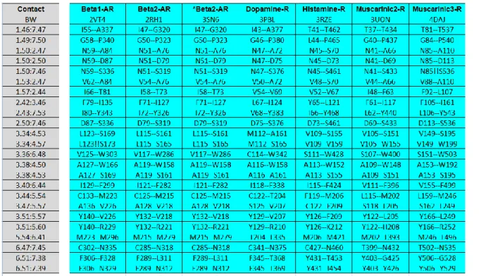
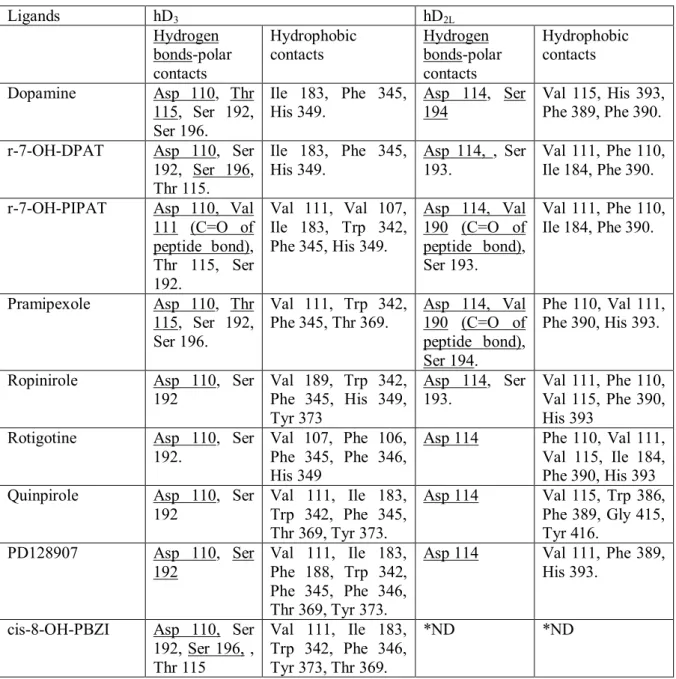
24 The seven transmembrane helices bundle serves as a link between the ligand binding pocket and the G protein. A comparison between the GPCRs structures solved so far using a network representation [85], has revealed that tertiary contacts in the trasmembrane region are conserved, independent of sequence diversity and receptor conformation (active or inactive). A consensus network of 24 contacts mediated by 36 residues have been identified (Fig. 4), and provide an evolutionarily conserved structural scaffold of non-covalent contacts for the GPCR fold. These tertiary contacts involve the central and cytoplasmatic site of the helices bundle, and are clustered at the interface of helices I-II, III-IV, III-V, and III-VI-VII. Another remarkable feature of GPCRs is to bind ligands with different shape and physical chemical properties. All ligands bind in a pocket, but some of these ligands bind deeply in that cavity. Despite the diversity of ligands that bind to GPCRs, residues within 4 Å of the ligands are similar. Topologically equivalent residues of III, VI and VII helices bind ligands, characterizing themselves as consensus contacts with ligands across the class A of GPCRs. In the present thesis (Chapter I and II) analyzing D3, D2L, 5HT2A-C receptors; the binding mode of agonists involves also hydrophilic
residues in the V helix, which seems to have a role in the affinity of compounds within these homologous receptors [90, 91]. In the consensus network of contacts two interaction patterns between residues 3.36-6.48 and 6.51-7.36 belong to the binding pocket (Fig 4). The described consensus network [85] can be useful for improvement of techniques for homology modeling of GPCRs.
Residues in the intracellular region and the cytoplasmatic part of helices bundle are involved in binding with signaling effectors. Moreover some conserved residues in the ICL2 have been identified to have a role in the activation of GPCRs, such as the conserved motif D(E)RY in several receptors [84, 92-94]. Other structural motifs have been identified to have a role in receptor activation, such as NPXXY at the VII helix, and P-I-F motif localized at the interface between the extracellular and intracellular part of helices bundle [84].
Significant differences, in those mentioned structural motifs, have been found between the x-ray structure of 5HT2B receptor in complex with ergotamine, and the simulated receptor
in the unbound state (Chapter II). The simulated 5HT2B receptor, reported in the present
work, have been found to have partially inactivated conformation [91].
The described molecular features, the growing literature and x-ray structures of GPCRs will help to fulfill the need for new targets and new drugs [79]. Tools such as structure-based drug research and discovery (R&D) might help to understand molecular features of
25 drug efficacy and selectivity and also to carry out high throughput screening of compounds in order to identify new hits and leads [95]. Moreover the knowledge of the structure of a GPCRs, either x-ray or modeled structure could help in the elucidation of the pharmacodynamic profile of multi-target ligands, i.e. trying to predict side effects [96].
Molecular modeling of GPCRs
The progress in crystallization techniques has been associated to the advances in computational (in-silico) techniques such as: structural modeling and molecular dynamics simulations. Computational approaches are important for GPCRs characterization. There is a small ratio between the number of x-ray structures and the genes that encodes for GPCRs, since there is still a lack of information. The comparative modeling of GPCRs structures has an important role in fulfillment of this gap.
Comparative modeling is defined as a computational approach that generates a predicted structure of a target molecule, such as a GPCR, with unknown structure. Comparative modeling is a knowledge-based method; in the present thesis two different comparative modeling approaches have been used, homology modeling and threading methods. Homology modeling is based on the assumption that the three dimensional structure of a protein depends on its sequence; moreover the overall structure of proteins is more conserved than their sequences [97]. This assumption is confirmed by experimental data; GPCRs are classified in different classes, families and sub-families of receptors, which diverge in sequences but not in the overall fold. With homology modeling it is possible to predict the structure of a “target protein”, building the 3D coordinates of its atoms using as reference the structure of a “template protein”, which is high homologous to the target. The reliability of an homology model depends on the homology and sequence identity shared by the target and template protein. The percentage identity cut-off, for large proteins with more than 150 residues, is about 40%. Inside a GPCR family such as dopaminergic GPCRs, the identity shared by D1 and D3 receptors is 30% and homology is 47%. Within
aminergic GPCRs these values are redundant. Although the percentage identity is below the suggested cut-off, the homology modeling of GPCRs is still an useful and validated approach. An example of software, that carry out homology modeling predictions, is MODELLER [98], moreover in the field of in-silico methods several web-servers have been developed and provide access to modeling software, such as the Swiss-Model web server [99]. Another strategy that can be aimed to obtain comparative modeling of protein is the treading method or fold-recognition approach. Threading is useful when non high
26 homologous templates are identified for a target. Threading methods employ multiple template proteins, and the main difference in comparison to homology modeling is that prediction is carried out by placing each amino acid of target primary structure to a position in the template structure, evaluating iteratively the fitness of target in the template. The fitness is based on a scoring function built on an established structure template database; the scoring function takes into account several knowledge-based structural restraints derived from experimental data. Threading methods are provided by software such as RAPTOR [100] but also web-servers such as HHpred [101] and I-Tasser [102]. Zhang and collaborators have provided an efficient threading pipeline for modeling of GPCRs, the G Protein Coupled Receptor Research Database (GPCRRD), which in addition to treading method performs conformational search of receptors and a Molecular Dynamics Fragment Guided simulation (FG-MD) [103]. Either homology modeling and threading provide useful predicted structural data of proteins with unknown structure. Several caveats affect those prediction approaches, such warnings are related to structure energy optimization. Comparative modeling approaches do not carry out extensive energy optimization of proteins, with some exceptions such as GPCRRD. However GPCRs are membrane proteins, thus the role of membrane-protein environment should be taken into account.
In the present thesis it is presented a computational approach that along with comparative modeling either homology modeling [90] and threading [91] was used to carry out the structural optimization of GPCRs, by means of molecular dynamics simulations in an explicit water-membrane environment. Molecular dynamics (MD) is a simulation technique that relies on physical chemistry laws. A molecule, atoms connected by bonds, is treated in MD as a set of beads connected by springs; during an MD the movement of such set of beads and springs is simulated by numerical integration of the Newton Law of motion (eq. 1). ) ,..., , ( 1 2 2 2 n i i i i i F V r r r t r m i=1,N (1)
In equation (1), m is the mass and r are the coordinates of the i atom. The force Fi experienced by the i particle is the derivative of potential energy. This potential energy is described in MD by a mathematical function called force field. The force field is characterized by a sum of contributes that accounts for bound, no-bound and cross-terms energies that govern the degrees of freedom of the simulated system.
crossterms bound no bound E E E E (2)
27 oop torsion E E stretching bending bond E E E (3) Jones -Lennard Coulomb bound -no E E E (4) ... stretching torsion stretching torsion stretching bending bending bending crossterms E E E E (5)
The physical reliability of MD is stated by the force field and its parameters, that are derived by ab inizio quantum chemical calculations but also by experimental data such as: Infrared (IR), Raman and Nuclear Magnetic Resonance (NMR) spectroscopy. In fact IR, Raman and NMR techniques measure energies respectively related to the stretching, rotational and torsional degrees of freedom of molecules. Several force field have been implemented in MD software, but CHARMM it is worth to be mentioned. CHARMM (Chemistry at Harvard Macromolecular Mechanics) force field and the homonymous implementing software have been developed by the “2013 Nobel Prize for chemistry” Martin Karplus and his collaborators [104]. The CHARMM forcefield is peculiar in comparison to other force fields, such as AMBER, since it describes the out-of-plane “oop” motions of bounds (eq. 6). Furthermore in CHARMM the Van Der Walls term includes the description of hydrogen bond (eq. 7), where sw is a function that describes the geometrical properties of hydroben bond.
2 0
)
(
K
E
oop pot (6))
,
,
(
2 2 2 6 12 ij on off ij ij ij ij Hbond VdWr
sw
r
r
r
b
r
A
E
(7)MD allows the inclusion of solvent in the simulation; since water is fundamental for folding and function of proteins several explicit water models are described by force fields parameters, such as TIP3P and SPC/E. Also an implicit water model is used in MD in order to speed simulations; the Generalized Born (GB) model where solvent is treated as continuum medium. GB is useful for simulation of small peptides but this approximate solution lacks of reliability when simulating proteins bigger than 30 amino acids. Moreover implicit water models do not account for solvent effects such as viscosity, hydrophobic effect and water-molecule hydrogen bonds. Since biomolecules are charged, ions can be added to the simulation system in order to reach neutralization. Moreover molecule structure is influenced by the ionic strength, then in the simulation of biological
28 molecules ions are added up to 150 mM concentration in order to resemble the physiologic condition.
Several kind of physical ensembles can be simulated:
- microcanonical ensemble NVE, where particle number N, volume V and energy E are constant;
- canonical ensemble NVT, where T stands for constant temperature;
- isothermal isobaric ensemble NPT, where P stands for constant pressure; a variation of this ensemble is NPγT or NPAT, where γ stands for costant surface tension of lipid bilayer and A for constant area of membrane.
The principal steps of MD simulation of a GPCR are:
- building of simulation box, i.e a GPCRsembedded in a lipid bilayer, often 100x100 Å or bigger lipid leaflet,
- primary equilibration steps such as lipid melting, water-ions equilibration, lipid-water-ions equilibration around the protein kept fixed with harmonic constraints, - equilibration of the whole system, with small integration time-step (1 fs),
- MD production run with 2 fs integration time-step.
MD is a statistical method; in statistical mechanics the average of observables are defined as average ensemble, thus the average of a huge number of simultaneous replicas of the analyzed system. Just one replica is simulated in MD and it is statistically reliable because of the ergodic principle: “the average system is equal to the time average of the system” (eq.8); it means that a system that is free to evolve in a time span, explores all possible allowed energetic states.
<A>ensemble= <A>time (eq. 8)
The time span or time-scale depends on which molecular event would be observed with MD. During a classical MD simulation, the most CPU (Computing Processing Unit) intensive task is the evaluation of non-bonded or non-covalent terms of force field. In Big O notation, common molecular dynamics simulations scale by O(n2) if all pair-wise
electrostatic and Van der Waals interactions are accounted explicitly. This computational cost can be reduced by employing electrostatics methods such as Particle Mesh Ewald ( O(nlog(n)) ), or good spherical cutoff techniques ( O(n) ). Another factor that impacts total CPU time required by a simulation is the size of the integration time step. This is the time length between two recurrent evaluations of the potential or integration of motion. The time step must be chosen small enough to avoid discretization errors (i.e. smaller than the fastest vibrational frequency in the system). Typical time steps for classical MD are in the
29 order of 1 fs (1E-15 s). This value may be extended by using algorithms such as SHAKE, which fixes the vibrations of the fastest atoms (e.g. hydrogen bound to carbon). Up to now MD simulations could be scaled-up (speeded up) not only by distribution of calculation on large number of CPU, up to 64 threads, but also by using MD software that support CUDA (Computed Unified Device Architecture) acceleration. With CUDA acceleration calculation of non-bond interactions could be speeded up by distributing the calculation also on GPU (Graphic Processing Units). Thus hardware development is going to pursue to MD the ability to carry out longer simulations in less time. Molecular dynamics time-scale depends from molecule dimensions and structure; moreover the simulation time extension depends on which molecular event is studied. In example simulation of protein folding may require from 30 ns to μs of simulation, protein aggregation requires μs [105]. Looking at GPCRs long time-scale all-atom MD, up to 15 μs for each trajectory, have been carried out to perform the analysis of 2-adrenergic receptor activation [106] and analyze the
allosteric binding at M2 muscarinic receptor [107]. Such long time-scale all-atom MD
simulation of GPCRs have been carried out on a special purpose computer, Anton, running the software Desmond, in order to perform acceleration of classical molecular dynamics [108].
In the present thesis, short time-scale (3 ns) simulation have been carried out in order to optimize the structural models of D3, D2L dopaminergic receptors [90] and 5HT1A, 5HT2A-C
serotonergic receptors [91]. Three ns of molecular dynamics simulation have been enough to reach a relative conformational minimum for both D3 and D2L receptors, which
differentiated in structure and have been validated for discrimination of selective agonists. Also the serotonergic receptors reached a relative minimum within 3 ns of simulation, and prediction of binding of cabergoline are comparable to the experimental binding of ergotamine at crystallized human 5HT2B receptor.
The binding of compounds have been studied by molecular docking. Docking is frequently used to predict the binding orientation of small molecules to potential protein targets in order to predict affinity and activity of compounds. Docking could play an important role in the rational design of drugs and considerable efforts have been directed towards improved docking methods. Molecular docking can be thought of as a problem of and-key; where the key in the ligand which is active toward the receptor (lock). The lock-and-key vision is quite rigid, because ligand and receptor are flexible. For this reason the ligand-receptor binding could be described as a hand that wears a glove (hand-in-glove). In fact during the binding process, ligand and protein adjust their conformations to achieve an
30 overall “best-fit” and these kind of conformational adjustments resulting in binding are referred as “induced-fit”. Molecular docking, simulating the molecular recognition process, aims to achieve an optimized conformation at least of the ligand. This conformation called “pose” is the relative orientation between protein and ligand, characterized by the best interaction energy. Two approaches are popular within molecular docking software. The matching technique that describe the protein and the ligand as complementary surfaces. In the second approach ligand-protein pairwise interaction energies are calculated iteratively; the most validated approach is the semi-flexible docking where ligand is flexible and receptor atoms are kept fixed. Input information of docking are protein structure and a set of potential ligands. The success of a docking program depends on two components: the search algorithm and the scoring function. The search space consists of all possible orientations and conformation of the protein paired with the ligand. However up to now it is hard to exhaustively explore the search space of the whole system, than the search space is in practice limited to the ligand. A variety of conformational search strategies have been applied to the ligand and to the receptor: systematic or stochastic torsional searches about rotable bonds, molecular dynamics simulations and genetic algorithms. The scoring function takes a pose as input and returns a number indexing the likelihood that the pose represents a favorable binding interaction. Existing scoring functions can be divided into three main classes: force field-based methods, empirical score functions and knowledge-based methods [109]. Docking software fail often to predict absolute values of binding energy, but could be successful in prediction of the trend of binding energy of congeneric class of compounds [90]. This capability is useful in order to identify hit or lead compounds. Sometimes there is the need of rescoring methods, in example by rescoring of poses with another scoring function. Rescoring could be atime expensive approach but can help to obtain reliable results. The evaluation of binding energy in terms of prediction of binding free energy has been shown to be successful by means of time expensive calculations such as MM/GB(PB)SA [Molecular Mechanics/Generalized Born (Poisson Boltzmann) Solvent Accessible surface area] [110, 111]. This kind of calculations are very predictive of drug affinity, however those are not yet extensively available for virtual screening studies in terms of codes and hardware. Application of MM-GBSA scoring or re-scoring to GPCRs is straightforward, because of receptor encounters to conformational changes upon binding and interaction with lipid bilayer should be taken into account. This consideration implies, i.e. in virtual screening, the use of multiple conformations of receptor obtained from MD, with more or less approximations [112].
31
D2-like receptors
The dopaminergic system is characterized by two subfamilies of receptors, D1-like (D1, D5)
and D2-like (D3, D2, D4); that respectively are coupled to Gs and Gi proteins. D2 and D3
receptors are highly homologous, whereas D4 receptor is phylogenetically distant from the other D2 like receptors (Fig.5). Several efforts have been carried out to model selective
ligands toward either D3 or D2 receptor [113-119]; recently also D4 receptor [120, 121] has
been investigated by means of ligand or structure-based in silico approaches.
Since the D3 receptor x-ray structure was solved [122] important information have been
revealed in comparison to previous structural knowledge. An interesting study was published after the releasing of dopamine D3 receptor structure, published by Carlsson et
al. in 2011 and entitle “Ligand discovery from a dopamine D3 receptor homology model
and crystal structure”. In that paper it is highlighted that the screening on the homology model of D3 receptor carried out before releasing of D3 receptor structure has led to
unbiased results since hit rates of virtual screening on homology models (23%) was comparable to the one (20%) carried on crystal structure of D3 receptor. The main bias of
these two virtual screening studies was found to be related to the binding at the allosteric binding pocket, which is more open in the homology modeling in comparison to the x-ray structure [95]. Differences in the allosteric binding pocket between D3 and D2 receptors
have been claimed by Chien et al. [122] to influence the selectivity either at D3 or D2
receptors, possibly due to different electrostatic properties of the two cavities. In chapter I of the present thesis it is showed that upon molecular dynamics simulation there is an evolution of binding pockets of D3 and D2 receptors, and the whole pocket of D2 receptor
became bigger than D3, possibly due to the lack of the accessory disulfide bridge within the
3ECL [90]. Recently, an interesting update about the role of allosteric pocket in D2-like
receptor and the 1ECL have been reported by Michino et al. 2013 [123]. In this publication it is reported that a single glycine residue, in the 1ECL of D3 receptor, influences 1ECL
flexibility and II helix movement upon ligand binding. Moreover it has been recognized a high energy hydration site in the allosteric binding pocket of D3 receptor, thus compounds
that are able to displace such high energy water molecules have high affinity for D3
receptor. Those calculations have been carried out with Water-Map [124].
Starting from latest literature about structural features of dopaminergic D2-like receptors,
medicinal chemists must move toward evaluation of NCE or drugs for repositioning that could interact with the allosteric binding pocket of D3 receptor.
32 In chapter II it is reported an analysis of structural features between D3 receptor and
several serotoninergic receptors: 5HT1A, 5HT2A-C [91]. Ergot derivatives ocular
hypotensive drugs such as lisuride, cabergoline, cianergoline and bromocriptine are reported to be serotonergic and dopaminergic agonists. Moreover multi-pharmacological profiles are also accounted to antipsychotics that bind with high affinity to 5HT2A and D2
receptors such as iloperidone and risperidone [125]. Up to date few modeling studies have compared the structural features of binding of mixed serotonergic and dopaminergic ligands at D2-like and serotonergic receptors [96, 126, 127]. The in silico study reported in
chapter II is the first that shows an extensive comparative modeling study on D3 receptors
and four homologous serotonergic receptors. In that study it is reported that cabergoline has a conservative binding mode at D3 and 5HT2A-C. Furthermore affinity of this compound
is higher for D3 and 5HT1A receptors in comparison to 5HT2A-C, due to the better
desolvation energy associated to the predicted complexes cabergoline/ D3 and 5HT1A.
Recently an interesting paper suggested new perspective studies for drug development of biased agonists active on D3 receptor. D3 receptor, but not D2 receptor, undergoes to fast
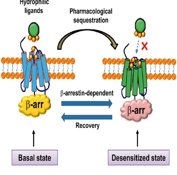
pharmacological sequestration and desensitization which is GRK-independent [128]. The molecular mechanism of pharmacological sequestration (Fig. 6), upon agonist action, seems to be related to a D3 receptor conformation that forbids the binding of hydrophilic
agonists, but allows the binding of hydrophobic compounds. -arrestin binding through Gγ interaction regulates such pharmacological sequestration. Pharmacological sequestration and desensitization of D3 receptors are related to each other;
7-OH-DPAT>quinpirole>dopamine pharmacological sequestration pattern resembles the sensitization induced by those compounds. However sensitization characterizes a long term modification of D3, since it is maintained after five washes; whereas pharmacological
sequestration is reverted in the middle of one wash. It could be stated that pharmacological sequestration is the event the precedes the sensitization. This highlight on the novel results about biased signal of D3 receptor, open research on D2-like receptors to R&D of biased
33
34
Figure 4. Consensus network of contacts in aminergic GPCRs
BW stands for Ballesteros Weinstein definition of residues.