FACOLTÁ DI SCIENZE MATEMATICHE FISICHE E NATURALI
DOTTORATO DI RICERCA IN BIOLOGIA AMBIENTALE ED
EVOLUZIONISTICA – BOTANICA
XXXII ciclo
PhD Thesis
Camilla Badiali
Response of root cultures and in vitro-grown plantlets systems
of Hypericum perforatum L. to biotic and abiotic stress
Tutor
Prof. Alessio Valletta
Contents
Abbreviations chapter 1 ... 1
1. Introduction ... 3
1.1 Hypericum perforatum ... 3
1.2 Traditional use, pharmacological activity and clinical properties of H. perforatum ... 5
1.3 Phytochemical content of aerial organs ... 5
1.4 Phytochemical content of roots ... 6
1.5 Structure and chemical properties of xanthones ... 7
1.5.1 Xanthone biosynthesis ... 8
1.5.2 Biological activities of xanthones in plants ... 9
1.5.3 Pharmacological activities of xanthones ... 11
1.6 Plant defense responses ... 11
1.7 Induction of responses in plant cultures: advantages of in vitro systems and elicitation techniques ... 16
1.7.1 Biotic stress ... 17
1.7.1.1 Chitin and chitosan ... 17
1.7.1.2 Chitosan oligosaccharides ... 19 1.7.1.3 Methyl jasmonate ... 19 1.7.1.4 Salicylic acid ... 21 1.7.1.5 Hydrogen peroxide ... 22 1.7.2 Abiotic stress ... 24 1.7.2.1 Toxic metals ... 24 1.8 Research objectives ... 26 References ... 26 Abbreviations chapter 2 ... 54
2. Chitosan oligosaccharides affect xanthone and VOC biosynthesis in Hypericum perforatum root cultures and enhance the antifungal activity of root extracts ... 55
References ... 79
Abbreviations chapter 3 ... 86
3. Response of Hypericum perforatum root cultures and in vitro-grown plantlets to chitosan oligosaccharides (COS), methyljasmonate (MeJA), salicylic acid (SA) and hydrogen peroxide (H2O2) ... 87
References ... 102
Abbreviations chapter 4 ... 107
4. Effect of cadmium and arsenic on xanthone production in Hypericum perforatum root cultures ... 108
References ... 113
5. Conclusions ... 116
1
Abbreviations chapter 1
13-HPOT 13-hydroperoxylinoleic acid AOC allene oxide cyclase
AOS allene oxide synthase APX ascorbate peroxidase As arsenic
BPS benzophenone synthase Cd cadmium
CHIT chitosan
CHS chalcone synthase
COS chitosan oligosaccharides ET ethylene
ETC electron transport chains ETI effector-triggered immunity ETS effector-triggered susceptibility GR glutathione reductase
HPLC high performance liquid chromatography HR hypersensitive response
IAA indole acetic acid IBA indole butyric acid ICS isochorismate synthase ISR induced systemic resistance JA jasmonate
JMT JA carboxyl methyltransferase LOX lipoxygenases
MAMP microbial-associated molecular pattern MAO monoamine oxidase
MAPK mitogen-activated protein kinases MeJA methyl jasmonate
MeSA methyl salicylate NO nitric oxide
2
OPR 12-oxo-phytodienoic acid reductase PAL phenylalanine ammonia lyase PAMP pathogen-associated molecular PCD programmed cell death
PIs proteinase inhibitors
PR pathogenesis-related proteins PRR pattern recognition receptors PTI PAMP-triggered immunity ROS reactive oxygen species SA salicylic acid
SAG SA O-β-glucoside SAGT SA glucosyltransferase SAMT SA methyl transferase SAPB2 MeSA methyl esterase SAR systemic acquired resistance SOD superoxide dismutases
3
1. Introduction
1.1 Hypericum perforatum
The genus Hypericum, the most numerous of the nine genera of Hypericaceae family (Stevens 2007; APG III 2009), includes almost 470 species of herbs, shrubs and small trees. It is distributed in Eurasia (>230 species), North and South America (c. 40 and >130 species respectively), Southeast Asia (c. 47) and Africa (c. 40 species). Typical habitats are rocky, sometimes calcareous, and dry to moist grasslands or acidic fens and shallow swamps. In the tropics it is generally confined to high elevation habitats (Robson 2003).
The most studied species of the genus is Hypericum perforatum, a medicinal plant used since antiquity with a broad range of pharmacological activities.
H. perforatum is commonly known as St. John’s wort because of its blooming period which
occurs at the end of June (June 24th is St. John’s day). Christians believed that H. perforatum
kept evil spirits away, for this reason on St. John’s day they used to burn plants to purify the air and ensure good crop harvest, indeed the origin of the genus name “Hypericum” comes from Greek words hyper (over) and eikon (image, in the sense of ghost) because people believed in its exorcistical properties. The specific name “perforatum” refers to leaves that seem to be perforated due to the presence of secretory structures.
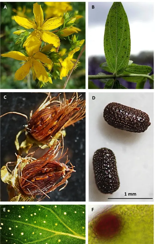
H. perforatum (Fig.1) is a completely glabrous herbaceous plant species, characterized by
an erect stem, 30 to 100 cm long and branched in the upper section; the leaves (Fig. 1B) are yellow-green in color, opposite, shortly petiolate; yellow flowers (Fig. 1A) are numerous, forming a broadly paniculate, almost corymbose, inflorescence with numerous stamens free or into three bundles at the base. The ovary is superior and ovoidal with three widely divergent styles, flowers have five sepals and five petals with dark glands along the margins; the fruit (Fig. 1C) is a small septate capsule; seeds (Fig. 1D) are 1 mm long cylindric, brown, with a pitted or finely patterned surface (Bombardelli and Morazzoni 1995).
An anatomical characteristic of H. perforatum is the presence of two types of secretory structures on stems, leaves and flowers: translucent glands (Fig. 1B, E), which are colorless and accumulate hyperforin and essential oils, are localized all over the leaf lamina;dark glands (Fig. 1B, F), which are dark red and produce hypericin and its derivatives, are distributed on stems, leaves, flowers and anthers.
4
Fig. 1 Hypericum perforatum. A) Flowers with numerous stamens; B) leaves showing dark and translucent glands; C) fruits, capsules; D) seeds; E) translucent glands; F) dark gland.
A B
C D
E F
5
1.2 Traditional use, pharmacological activity and clinical properties of H.
perforatum
The properties of H. perforatum have been known for centuries and its use in folk medicine has an ancient tradition. The Europeans have used it in order to treat a broad number of diseases, like anxiety, colds, depression, flue, hemorrhoids, womb muscle contractions during menstruation, skin irritations or infections, burns and wounds (Saddiqe et al. 2010 and literature cited therein). The oldest references about the use of H. perforatum come from the Greek botanist of the I century a.C., Dioscórides, the Roman student of the I century a.C., Plenius, and the Greek physician and father of medicine of the V century b.C., Hippocrates. In the XII century, Templars were the first to discover that H. perforatum was very useful for improving the mood of warriors forced to bed for months.
In last decades, H. perforatum has been studied primarily for the antidepressant activity of its extracts which have been widely sold in health food stores and pharmacies in Europe and the USA for the treatment of depression (Lecrubier et al. 2002; Kasper et al. 2006). Clinical studies found H. perforatum extracts to be efficacious in the treatment of mild to moderate depression (Kasper et al. 2010; Chen et al. 2011; Ng et al. 2017). The effectiveness of H.
perforatum extracts has been also demonstrated in the treatment of somatoform disorders
(Volz et al. 2002; Müller et al. 2004), obsessive-compulsive disorder (Kobac et al. 2005), anxiety (Singewald et al. 2004) and seasonal affective disorder (Wheatley 1999; Pjrek et al. 2005). Moreover, studies about other H. perforatum extracts properties have been conducted, including anti-Alzheimer (Hofrichter et al. 2016) wound-healing (Rao et al. 1991; Öztürk et al. 2007), anti-inflammatory (Kumar et al. 2001), antibacterial (Conforti et al. 2005), antifungal (Milosevic et al. 2007), and antiviral activities (Richer and Davies 1995).
1.3 Phytochemical content of aerial organs
The phytochemical content of leaves and flowers of H. perforatum has been deeply investigated because secondary metabolites with anti-depressant activity are produced and accumulate in the aerial organs of the plant (Mennini and Gobbi 2004). Hypericum preparations rich in bioactive secondary metabolites are obtained through hydroalcoholic extraction of the flowering tops and leaves; these are exploited above all for the treatment of depressive states, neurovegetative disorders and anxiety (Par 1.2).
6
Hypericins (naphthodianthrones) (Fig. 2A) and hyperforins (phloroglucinols) (Fig. 2B) are considered the main bioactive constituents of the aerial organs of H. perforatum, they are produced in black globules and translucent glands, respectively (Fig. 1E-F); hypericin was initially described as a monoamine oxidase (MAO) inhibitor (Suzuki et al. 1984), but later studies indicated that this effect was not clinically significant (Mennini and Gobbi 2004 and literature cited therein). In last decade the anti-depressant activity of hyperforin has been investigated, neglected at the beginning because of its instability in most organic solvents and fast air oxidation (Medina et al. 2002 and literature cited therein; Isacchi et al. 2007). However, in addition to naphtodianthrones and phloroglucinols, several molecules have been characterized such as essential oils and volatile compounds (hydrocarbons, monoterpenes, sesquiterpenes) (Nahrstedt and Butterweck 1997; Crockett et al. 2010), flavonoids (flavonols, flavones, glycosides, biflavonoids, catechins), tannins and other phenols (caffeic, chlorogenic, p-coumaric, ferulic, p-hydroxybenzoic and vanillic acids) (Barnes et al. 2010 and literature cited therein).
Fig. 2 Main bioactive secondary metabolites accumulated in H. perforatum shoots. Chemical structure of: A) hypericin and pseudohypericin; B) hyperforin and adhyperforin (Barnes et al. 2010).
1.4 Phytochemical content of roots
Although many therapeutic properties of aerial organ metabolites are known, little information is available on the chemical composition and biological activities of H.
perforatum root extracts (Tocci et al. 2013; Simonetti et al. 2016).
7
The phytochemical content of St. John’s wort root includes compounds also found in shoots such as phenolic acids (e.g. chlorogenic acid) and flavonoids (aglycon or glucoside flavonols like quercetin or hyperoside, respectively) (Cui et al. 2011) (Fig. 3A-C), but also xanthones, which are compounds specifically biosynthesized and accumulated in roots (Crockett et al. 2011).
Fig. 3 Main polyphenols accumulated in H. perforatum. Chemical structures of: A) chlorogenic acid; B) of quercetin and C) hyperoside.
1.5 Structure and chemical properties of xanthones
Xanthones, from Greek “ξανθός” (xanthόs) which means “yellow”, are phenolic compounds with a limited distribution in plant kingdom. Xanthone (9H-xanthen-9-one) (Fig. 4) is the central core to which substituents are added to form different xanthones usually found as aglycones, O-glycosides and C-glycosides. They are produced mainly by plants, but also by some lichens, fungi and bacteria (Masters e Brase, 2012). Xanthones are typical secondary metabolites of plants belonging to the Hypericaceae family in particular of Hypericum species, but they are also synthesized in species belonging to Gentianaceae, Moraceae and Polygalaceae families (Negi et al. 2013).
A B
8 Fig. 4 Chemical structure of xanthone.
1.5.1 Xanthone biosynthesis
Exodermis and endodermis are the sites of xanthone biosynthesis in H. perforatum roots (Tocci et al. 2018); they are synthesized through the phenylpropanoid pathway which has different branches leading to many different compounds including simple phenylpropanoids, flavonoids, lignin and others (Demirkiran 2007).
Key enzymes regulate this pathway moving the metabolism depending on plant’s needs. The phenylpropanoid pathway originates from phenylalanine, which is deaminated by phenylalanine ammonia lyase (PAL). This is one of the well-studied and characterized enzyme of plant secondary metabolism, it catalyzes the reaction which links primary metabolism (shikimate pathway) to secondary metabolism: phenylalanine deamination to
trans-cinnamic acid (Fig. 5) with release of nitrogen in the form of ammonia. The
biosynthetic pathway divides in two branches from trans-cinnamic acid to give xanthones or flavonoids. p-coumaroyl-CoA and malonyl-CoA are produced from trans-cinnamic acid and two enzymes act on these compounds respectively: chalcone synthase (CHS) and benzophenone synthase (BPS) (Liu et al. 2003; Vogt 2010).
CHS in presence of one molecule of p-coumaroyl-CoA and three of malonyl-CoA catalyzes consecutive decarboxylations and condensations leading to chalcone, the flavonoid precursor from which tannins, flavones, isoflavones, flavonols and anthocyanins are synthesized.
BPS, a transferase, in presence of one molecule of benzoyl-CoA and three of malonyl-CoA catalyses the reaction which leads to benzophenone, the precursor of xanthones.
9 Fig. 5 Phenylpropanoid pathway. Branches leading to xanthone and flavonoid synthesis. PAL: phenylalanine ammonia lyase; CHS: chalcone synthase; BPS: benzophenone synthase (Liu et al. 2003).
1.5.2 Biological activities of xanthones in plants
Phenolic compounds are known to be molecules involved in protecting plants against microbial attacks (Ahuja et al. 2015); in vitro studies have shown that xanthones, as observed for other polyphenols, are constitutively biosynthesized, although their content may significantly increase in response to pathogenic attack (phytoalexins). An increase in xanthone accumulation in cell cultures of many plant species subjected to elicitor treatments
10
has been observed: Centaurium erythraea elicited with yeast extract and methyl jasmonate (MeJa) (Beerhues and Berger 1995); H. perforatum elicited with Colletotrichum
gloeosporioides cell wall extracts, Agrobaterium tumefaciens, MeJa, salicylic acid (SA) and
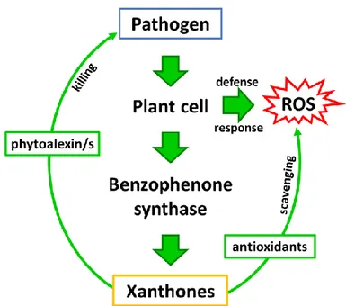
chitosan (CHIT) (Conceicao et al. 2006; Franklin et al. 2009; Tocci et al. 2010). Franklin and colleagues (2009) proposed a double role of xanthones in the defense response as antioxidant and antimicrobial compounds: in vivo xanthones on one hand constitute a powerful antioxidant system to protect the host cells from reactive oxygen species (ROS) and on the other hand have also the potential to act as phytoalexins against pathogenic microorganisms (Fig. 6).
In the last decades the inducible role of xanthones has been also investigated in organ cultures (Ishimaru et al. 1990; Vinterhalter et al. 2008; Tocci et al. 2011, 2012; Valletta et al. 2016). In vitro cultures of H. perforatum roots elicited with chitosan or chitosan oligosaccharides (which mimic a fungal attack) synthesized xanthones significantly increasing the concentration compared to non-treated samples (Tocci et al.2010, 2011; Brasili et al. 2014; Badiali et al. 2018).
Fig. 6 Hypothetical role of xanthones in the plant pathogen-interaction. In the model proposed by Franklin et al. (2009) xanthones play dual function in plant cells during biotic stress: (1) as antioxidants to protect the cells from oxidative damage and (2) as phytoalexins to impair the pathogen growth (modified from Franklin et al. 2009).
11
1.5.3 Pharmacological activities of xanthones
The interest in studying xanthone pharmacological activity had its beginning in ‘60s when diuretic and cardiotonic effect of mangiferin, a compound belonging to this class, was demonstrated (Finnegan et al. 1968 and literature cited therein). Following studies led to the discovery that xanthones can reverse various types of tumor (Abe et al. 2003; Chen et al. 2004; Su et al. 2011; Núñez et al. 2016) and that they have antimutagenic and antiangiogenic activities (Mackeen et al. 2000; Pinto et al. 2003; Almanza et al. 2011; Núñez et al. 2016). Xanthones have also anti-inflammatory properties (Chung et al. 2002; Park et al. 2006; Chen et al. 2008), antioxidant activity (Panda et al. 2013), they are effective in the treatment of cardiovascular diseases (Ishiguro et al. 2002) and have an enzyme inhibitory activity against mono amine oxidase (MAO) enzyme which plays an important role in the regulation of some neurologically active amines (Ohishi et al, 2000; Gnerre et al. 2001; Urbain et al. 2008); the inhibitors of MAO are useful in the therapy of several neurodegenerative conditions, including Parkinson's disease and Alzheimer's disease, psychosis, depression and schizophrenia (Laban and Saadabadi 2019 and literature cited therein; Wang et al. 2019). In the last decades antifungal, antibacterial and antiviral activities against human pathogens have attracted the attention of scientists because of the onset of resistance of pathogens to conventional antibiotics and antifungals (De Vita et al 2012). Xanthones, in particular prenylated and oxygenated xanthones, showed a significant antimicrobial activity against microorganisms pathogenic to humans (Fotie and Bohle, 2006; Tocci et al. 2011, 2012, 2013; Simonetti et al. 2016; Badiali et al. 2018) and plants (Cortez et al. 1998; Crockett et al. 2011).
1.6 Plant defense responses
Plants are sessile organisms in environments inhabited by living beings potentially dangerous to them (pathogens or phytophages) (biotic stress); moreover, they are subjected to stress caused by non-living factors depending on climate conditions (abiotic stress). For these reasons they had to evolve specific mechanisms to detect and consequently act against complex stress combinations, minimizing damage while conserving resources for growth and reproduction.
12
Plants have evolved different ways to perceive external attacks. As shown in Fig. 7, the recognition of microbial- or pathogen-associated molecular pattern (MAMP or PAMP) perceived by host encoded pattern recognition receptors (PRRs) leads to the PAMP-triggered immunity (PTI, called basal resistance). Successful pathogens secrete pathogen-encoded effector proteins which suppress PTI leading to the effector-triggered susceptibility (ETS), these factors are in turn recognized by plant resistance (R) proteins leading to the effector-triggered immunity (ETI). Natural selection drives pathogens to avoid ETI diversifying or acquiring new effectors.
Fig. 7 “Zig-zag” model for plant immune system (Jones and Dangl 2006).
The typical manifestation of ETI, mostly against biotrophic pathogens and viruses (Glazebrook et al. 2005) is hypersensitive response (HR) (Chisholm et al. 2006; Bari et al. 2009). The latter consist in the developing of necrotic lesions at the pathogen entry site to prevent the invasion into plant tissues and to deprive the pathogen of nutrients. ETI usually causes the accumulation of reactive oxygen species (ROS) and nitric oxide (NO), and the activation of defense-related genes including those encoding pathogenesis-related (PR) proteins (Dempsey et al. 1999). Ion fluxes (in: Ca2+ and H+; out: K+ and Cl-) occur in cells
next to the invasion site causing the production of toxic compounds formed by molecular oxygen reduction, such as superoxide anion (O2-), hydrogen peroxide (H2O2), and hydroxyl
radical (OH•); these reactive species start radical chain reactions involving a wide variety of organic substances leading to lipid peroxidation, enzymatic inactivation and nucleic acid
13
degradation (Lamb and Dixon 1997), contributing to apoptosis and acting directly against the pathogen. Moreover, ROS are involved in cell wall fortification causing proline-rich protein modification (Bradley et al. 1992), this adds to lignification and callose apposition to create a barrier against the invader. At the same time NO is produced. Both NO and ROS can act also as signaling molecules (although their long-distance action is unlikely). They appear to function in a positive feedback loop with SA and their production is necessary for HR activation (Fig. 8). NO induce SA accumulation and SA is required in NO defense signaling. H2O2 increases following a pathogen attack and activates SA synthesis; SA then
cooperates with ROS generated during the second phase of the oxidative burst potentiating cell death and genes involved in plant defense. SA also induces an increase in H2O2
production, which in turn activates the synthesis of more SA and programmed cell death (PCD) in a self-amplifying loop which regulates defense responses (Dempsey et al. 1999; Durner et al. 1997; Overmyer et al. 2003).
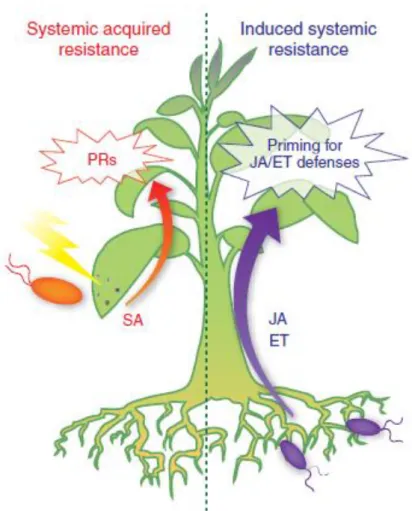
The phytohormone SA has a prominent role in HR (Par. 1.7.1.4) activating non-expressor of PR genes 1 (NPR1) which in turn activates PR proteins; moreover, SA is also necessary for systemic acquired resistance (SAR) induction, probably acting via its volatile methyl ester methyl salicylate (MeSA) (Park et al 2007). Among PR proteins hydrolytic enzymes are the principal group, they attack the pathogen cell wall and degrade it. At last, phytoalexins are produced, secondary metabolites with a high toxicity against pathogens. A few hours to several days after HR, also in distant portions of the plant PR gene expression levels increase leading to the development of SAR, a long-term resistance to infection by a broad diversity of pathogens.
14 Fig. 8 Plant response against pathogen infection. SA: salicylic acid; ROS: reactive oxygen species; NO: nitric oxide; MeSA: methyl salicylate; PCD: programmed cell death; PR proteins: pathogen related proteins; HR: hypersensitive response; SAR: systemic acquired resistance.
Response against herbivore insects and necrotrophic pathogens involves other signalling molecules. The principal signalling pathway involved in plant defense responses against pests is the octadecanoid pathway which produces the phytohormones jasmonic acid (JA), methyl jasmonate (MeJA) and derivatives (Par. 1.7.1.3). This pathway is activated by pest oral secretions (Halitschke et al. 2001) and mechanical wounding stress leading to the release of systemin, a signal peptide of 18 amino acids which is cleaved from the C-terminal region of a 200-amino acid precursor protein called prosystemin; it induces a signal cascade leading to the activation of octadecanoid pathway for JA production which in turn causes the expression and the accumulation of defensive proteinase inhibitors (PIs), which play a defensive role by inhibiting the activity of digestive enzymes in the guts of insects (Sun et al. 2011) and induces the production of phytoalexins both locally and systemically.
Jasmonates, synergistically with ethylene (ET), are also involved in induced systemic resistance (ISR) activated by non-pathogenic microorganisms; this response alerts the plant and causes the establishment of an advanced state of preparation against pathogenic attacks
15
(priming for enhanced defense). This type of systemic defense does not include the production of SA or PR proteins (Hase et al. 2008; De Vleesschauwer et al. 2008; Segarra et al. 2009) (Fig. 9). The defense regulatory protein NPR1 is an important regulator in SA-dependent SAR and acts also in JA/ET-SA-dependent ISR (Dong 2004; Pieterse and Van Loon 2004; Leon-Reyes et al. 2009) but its activation does not induce the expression of SA-responsive PR-genes. This suggests that NPR1 plays a key role in regulating and connecting different induced defense pathways (Dong 2004; Pieterse et al. 2009) via some factors (WRKY and MAPKs – mitogen activated protein kinase) involved in mediating the bidirectional antagonism between SA- and JA-mediated signaling (Li et al. 2004, 2006; Brodersen et al. 2006; Qiu et al. 2008) although synergism between these pathways has been observed (Mur et al. 2006).
16
1.7 Induction of responses in plant cultures: advantages of in vitro
systems and elicitation techniques
The major source of bioactive compounds for medicine, food additives, pigments, insecticides, cosmetics, and fine chemicals has historically been the plant kingdom. However, the extraction of bioactive compounds directly from plants is often not efficient, the extract may contain only very small quantities, or its composition may vary with the season or the environment. Often the biosynthesis of some compounds is activated in specific stages of development or in response to stress, specific environmental conditions and nutrients availability. Also, the characteristics of the producer organism may limit the availability of the molecules of interest: the plant could be rare, could have a slow or a difficult growth, or could be a protected species (Verpoorte et al. 2002). To obtain qualitatively and quantitatively standardized extracts, plant biotechnology can represent a valuable alternative. This technology is advantageous compared to the conventional agricultural production because it is independent of geographical and seasonal variations and various environmental factors; it offers a defined production system; it allows to obtain a rapid production and an efficient recovery; it allows to use plants as biotransformers for the production of novel compounds from cheap precursors.
For these reasons, in the last decades many studies have focused on plant cell cultures as a possible method to produce plant secondary metabolites of commercial interest (Buitelaar and Tramper 1992; Lipsky 1992; Verpoorte et al. 1993, 1998; Su 1995). Many authors demonstrated that in undifferentiated calli and suspended cells of H. perforatum xanthones are the main secondary products accumulated (Dias et al. 1999; Dias et al. 2001; Pasqua et al. 2005; Conceicao et al. 2006; Mulinacci et al. 2008). However, for many of the desired compounds, the production from cell cultures is too low, not exploitable for applicative purposes. This is usually because some compounds require tissue differentiation to be correctly synthesized.
To increase the production of secondary metabolites (including xanthones) elicitors can be used. The elicitation is a technique commonly used in plant biotechnology to enhance secondary metabolite production (Zhao et al. 2005; Namdeo 2007).
Elicitation techniques have been applied to suspended cell cultures of H. perforatum (literature cited in Par. 1.5.2) and the xanthone biosynthesis was significantly stimulated but it was insufficient for a large-scale production. The use of elicitors on organ cultures allowed to obtain the highest xanthone production, the adventitious root cultures of H. perforatum
17
demonstrated to be a promising reliable way for production of pharmaceutically and nutraceutically important metabolites (literature cited in Par. 1.5.2).
1.7.1 Biotic stress
Biotic stress is stress caused by a living organism to another; plants are continuously exposed to biotic stress factors such as the attack by fungi, bacteria, viruses, nematodes and herbivores. Nonetheless, disease takes place only if the pathogen overcomes the diverse defense strategies that plants put in place against the invader. After the attack, plants activate signaling pathways to organize a response against pest, the response culminates in the production of secondary metabolites (Par. 1.6). The involved signal molecules can be used in plant biotechnology as elicitors to mimic the attack and consequently induce the biosynthesis of secondary metabolites of interest or to investigate plant responses.
1.7.1.1 Chitin and chitosan
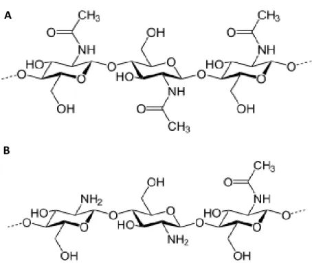
Chitin is a linear polysaccharide composed by N-acetyl-D-glucosamine repeat units linked by β-(1 → 4) glycosidic bonds (Fig. 10A). It is the principal component of the exoskeleton of arthropods and fungal cell wall, and the second-most abundant polysaccharide on earth following plant cellulose. Plants do not contain chitin but possess enzymes to degrade it (chitinases), probably because of the coevolution with their fungal pathogens (Passarinho and de Vries 2002). Chitinases act both directly on fungi degrading their cell wall, and indirectly generating chitin fragments that are recognized as stress signals by plant cells (Boller 1995). Unfortunately, chitin has limited utility for human applications due to its low solubility.
Chitosan is obtained from a partial deacetylation of chitin (Fig. 10B) so it is an heteropolymer composed by N-acetyl-D-glucosamine and D-glucosamine, whose relative amount may vary resulting in different degrees of deacetylation, molecular weights, viscosities etc. (Raafat and Sahl 2009 and literature cited therein). Chitosan is more soluble than chitin, is biodegradable, atoxic and non-allergenic (Raafat and Sahl 2009 and literature cited therein). Among chitosan biological activities there are antimicrobial (Rebea et al. 2003; Eaton et al. 2008), antioxidant (Yen et al. 2008), and hypocholesterolemic activity
18
(Xia et al. 2011 and literature cited therein). Moreover, chitosan promotes plant growth (Chibu et al. 2000) and it was reported to induce plant defense responses via raising of cytosolic Ca2+, activation of mitogen-activated protein kinases (MAPK), callose apposition, generation of ROS, hypersensitive response (HR), synthesis of abscisic acid (ABA), jasmonate, pathogenesis related proteins (PR) (Iriti and Faoro 2009 and literature cited therein) and phytoalexins (Fan et al. 2010; Sivanandhan et al. 2012; Sathiyabama et al. 2016) including xanthones in various species (Tocci et al. 2010; Krstić-Milošević et al. 2017). Despite many advantages, it has been reported that the use of chitosan at high concentrations (necessary for massive industrial production of secondary metabolites) has also inconveniences including the inhibitory effect on biomass growth in vitro and the irreversible morpho-anatomical alterations which make elicited biomass in vitro no longer usable for further production cycles (Brasili et al. 2014). Moreover, chitosan is insoluble in neutral water and other organic solvents, for this reason it is solubilized in water acidulated with acetic acid. This makes its use difficult in food and biomedical applications. Moreover, its use in basic research is also limited, at least in H. perforatum, as it has been recently demonstrated that acetic acid acts as an elicitor exerting itself a chitosan-like effect on xanthone biosynthesis (Valletta et al. 2016).
Fig. 10 Chemical structure of chitin (A) and chitosan (B).
A
19
1.7.1.2 Chitosan oligosaccharides
Chitosan oligosaccharides (COS) are obtained through chemical or enzymatic hydrolysis from chitosan and their use has recently increased. Unlike chitosan, COS are soluble in aqueous solutions in all proportions, due to the short chain length and to the free amino groups in D-glucosamine units (Jeon et al. 2000). Moreover, they are biodegradable, biocompatible, atoxic and have a low viscosity. These characteristics attracted researchers’ attention for being promising oligosaccharides that have potentials in agriculture and in cosmetic, pharmaceutical and food industry. Several biological activities of COS have been demonstrated in recent years, including antimicrobial (Jeon and Kim 2000 and literature cited therein; Jeon et al. 2001; Choi et al. 2001), antitumoral (Nam et al. 1999; Jeon and Kim 2002; Nam et al. 2007; Shen et al. 2009), antioxidant (Xing et al. 2005), hypocholesterolemic (Kim et al. 2005), hypoglycemic (Miura et al. 1995), anti-Alzheimer's (Yoon et al. 2009) and accelerating calcium absorption (Jung et al. 2006). Moreover, COS have proven to promote plant growth, improving the capacity of plants against salt and drought stress (Dzung et al. 2011; Chatelain et al. 2014; Zou et al. 2015) and to be effective elicitors of innate immunity against diseases of plants such as tobacco, rice, grape and other (Agrawal et al. 2002; Eikemo et al. 2003; Cabrera et al. 2006; Chen et al. 2009). Studies on their use as biopesticides have been conducted, indeed they have a powerful protective effect on various species of plants of economic interest (Yin et al. 2010; Zhao et al 2007). They are an effective post-harvest treatment for inhibiting diseases, which affect many fruits such as citrus fruits, tomato, pear, apple and peach (Chien et al. 2007; Badawy and Rebea 2009; Meng et al 2010; Yang et al 2010, 2012; Yan et al. 2011). It was also demonstrated that a pre-harvest administration of COS determines a higher post-harvest resistance to pathogens (Yan et al. 2012; Ma et al. 2013). Their biological activity strictly depends on its chemical and physical properties such as viscosity, polymerization degree and deacetylation degree (Cabrera et al. 2006 and literature cited therein; Zou et al. 2015).
1.7.1.3 Methyl jasmonate
Methyl jasmonate (MeJA) is a volatile compound which plays a long-distance signaling role in many cellular responses such as plant-environment, plant-herbivore and plant-plant interactions. In addition, MeJA and its precursor jasmonic acid (JA) are involved in many
20
developmental processes such as seed germination, root growth, stomatal closure, fruit maturation, leaf movement, leaf senescence, tuber formation and trichome formation (Creelman and Mulpuri 2002; Wasternack and Hause 2002; Wasternack 2014) (Fig. 11). Jasmonates (JA, MeJA and derivatives) are also involved in defense responses to biotic stress such as injury, wound caused by pests (Browse and Howe 2008), pathogen attack (Glazebrook 2005) and abiotic stress such as high salinity (Qiu et al. 2014), drought (Savchenko et al. 2014), cold (Du et al. 2013; Hu et al. 2013) and heat (Clarke et al. 2009). In stressed plants MeJA enhances protease inhibitors production against herbivores; these inhibitors cause the sensing of bad taste in pests and in some cases even cannibalistic tendencies. MeJA is also responsible for the production of phytoalexins, which act as antimicrobial agents. Moreover, a MeJA release to the atmosphere through stomata is recognized by nearby plants which activate defense responses (Farmer et al. 1990; Karban et al. 2000; Baldwin et al. 2006).
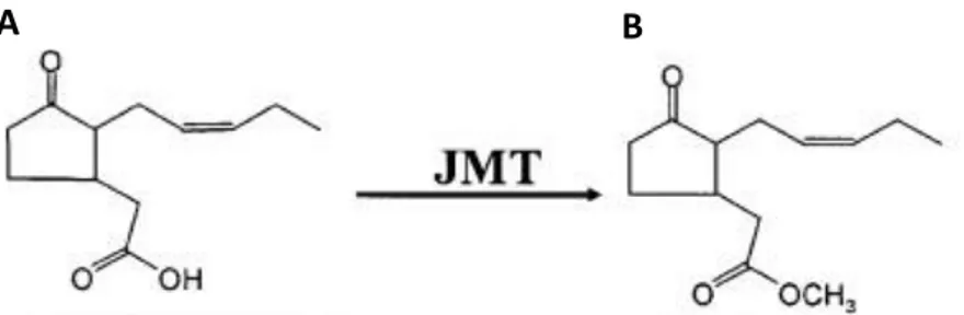
Fig. 10 Chemical structure of A) jasmonic acid and B) methyl jasmonate. JMT: JA carboxyl methyltransferase (Yang et al. 2006).
In plants MeJA is synthesized through the octadecanoid pathway (Fig. 11): external stimuli via the release of systemin cleaved from the C-terminal region of prosystemin, activate a phospholipase which releases linoleic acid from lipids of the chloroplast membrane. α-linoleic acid is oxygenated by specific lipoxygenases (LOX) to 13-hydroperoxyα-linoleic acid (13-HPOT), which is converted in 12,13-epoxyoctadecatrienoic acid by allene oxide synthase (AOS). On the latter compound acts allene oxide cyclase (AOC) to form 12-oxo-phytodienoic acid. This intermediate is then processed in peroxisomes through one reduction made by 12-oxo-phytodienoic acid reductase (OPR) and three β-oxidation cycles which form JA. The conjugation with isoleucine is required for its activation (JA-Ile). Free-acid JA might not be able to move across the cellular membrane because of its acidic characteristics so JA is catabolized by JA carboxyl methyltransferase (JMT) to MeJA (Yang et al. 2006; Browse et al. 2009 and literature cited therein) (Fig. 10).
21
Because of its role as signal molecule involved in defense responses, MeJA has been studied as elicitor in in vitro cultures of various plant species such as Vitis vinifera (Repka et al. 2004), Rubus sp. (Wang et al. 2008), Mentha piperita (Krzyzanowska et al. 2012) and H.
perforatum (Wang et al. 2015).
Fig. 11 Octadecanoid pathway for jasmonic acid (JA) biosynthesis (Zhai et al. 2017).
1.7.1.4 Salicylic acid
For two centuries, salicylic acid (SA) has been studied for its medicinal use in humans. Contrarily, its regulatory functions in plants as phytohormone have only been investigated in the last 30 years.
SA influences various processes in plant development and growth such as seed germination, cell growth, respiration, stomatal closure, senescence-associated gene expression, responses to abiotic stresses, and basal thermotolerance (Rate et al. 1999; Morris et al. 2000; Metwally et al. 2003; Clarke et al. 2004; Norman et al. 2004; Rajou et al. 2006; Clarke et al. 2009; and
22
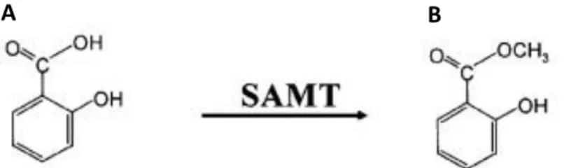
literature cited therein). SA in plants is produced via two distinct pathways that require chorismate. This primary metabolite can be converted into SA via L-phenylalanine, involving a series of enzymatic reactions initially catalyzed by phenylalanine ammonia lyase (PAL) (Verberne et al. 1999). Chorismate can also be converted into SA via isochorismate in a two-step process involving isochorismate synthase (ICS) and isochorismate pyruvate lyase (IPL), well known in bacteria, but whose existence in plants is supported by various papers (Verberne et al. 2000; Wildermuth et al. 2001; Strawn et al. 2007). Genes regulated by SA can be divided into two classes: early-responsive genes, induced within 30 minutes of SA treatment, and genes induced later, including NPR1, a master regulator of the SA-mediated induction of PR genes. Most of the SA produced in planta is converted into SA O-β-glucoside (SAG) by a pathogen-inducible SA glucosyltransferase (SAGT). MeSA and/or its glucosylated derivative MeSAG also accumulates to relatively high levels in vivo. MeSA, obtained from SA by salicylic acid methyl transferase (SAMT) (Fig. 12), is probably involved in signal translocation to distal portions of the plant and then it is hydrolyzed by MeSA methyl esterase (SAPB2) to SA, activating the defense genes (Park et al 2007). Treatments with exogenous SA have been investigated in in vitro cultures of diverse plant species enhancing plant resistance against both biotic and abiotic stress (Németh et al. 2002; Ali et al. 2006; Hussain et al. 2008; Popova et al. 2009; Sivanandhan et al. 2012; Gadzovska et al. 2013) making SA a cheap elicitor of plant defense responses.
Fig. 12 Chemical structure of A) salicylic acid and B) methyl salicylate. SAMT: salicylic acid methyl transferase (Yang et al. 2006).
1.7.1.5 Hydrogen peroxide
Hydrogen peroxide (H2O2) is a ROS whose production can be either enzymatic or
non-enzymatic. There are numerous ways of H2O2 production in plant cells, such as
photorespiration, electron transport chains (ETC), and redox reactions (Mittler 2002); in case
23
of high rates of production it is normally balanced by very efficient antioxidant systems which consist of both non-enzymatic and enzymatic. H2O2 scavengers include the enzymes
catalase (CAT) (Willekens et al. 1997), peroxidase (POX) (Fan and Huang 2012), ascorbate peroxidase (APX) and glutathione reductase (GR) (Jahan and Anis 2014) (Fig. 13).
H2O2 has been considered mainly as a toxic cellular metabolite for decades; however, it is
now clear that it acts also as a signaling molecule which may move between cells through aquaporin channels. H2O2 plays important roles in plant developmental and physiological
processes including seed germination (Barba-Espín et al. 2011), PCD (Cheng et al. 2015; Vavilala et al. 2015), senescence (Liao et al. 2012), flowering (Liu et al. 2013), root system development (Liao et al. 2009; Ma et al. 2014; Hernández-Barrera et al. 2015), stomatal aperture regulation (Ge et al. 2015). This suggests that H2O2 is involved in cellular signaling
transduction pathways and gene expression modulations in plants.
H2O2 is produced also through several enzymes including cell wall peroxidases (Francoz et
al. 2015), oxalate oxidase (Hu et al. 2003), amine oxidases and flavin-containing enzymes (Cona et al. 2006). Moreover, nicotinamide adenine dinucleotide phosphate (NADPH) oxidases on the plasma membrane also increase H2O2 level through generating O2-, which
in turn is converted to H2O2 and OH• by superoxide dismutases (SOD) (Grivennikova and
Vinogradov 2013; Brewer et al. 2015).
24
An oxidative burst with rapid H2O2 synthesis and its release into the apoplast, is induced by
biotic stress such as pathogens, elicitors and wounding or abiotic stress including drought (Ashraf et al. 2015), low and high temperatures (Orabi et al. 2015; Wu et al. 2015), salinity (Mohamed et al. 2015), ultra-violet light (He et al. 2013), ozone (Oksanen et al. 2004) and heavy metals (Wen et al. 2013), causing rapid responses in plant cells.
1.7.2 Abiotic stress
Abiotic stress is stress caused by non-living factors such as drought (Chaves and Oliveira 2004), salinity (Sahi et al. 2006; Munns and Tester 2008), heat (Scharf et al. 2012), cold (Chinnusamy et al. 2007), freezing (Sakai and Larcher 2012), nutrient (Hirel et al. 2007), high light intensity (Rossel et al. 2002), ozone (O3) (Welfare et al. 2002) and anaerobic
stresses (Agarwal and Grover 2006) (Wang et al. 2003; Nakashima et al. 2014). Abiotic stresses represent the primary cause of crop loss worldwide with reductions in production of more than 50% (Alcázar et al. 2006; Hussain et al. 2011) and probably these stresses will become more intense because of the expected climate change. Abiotic stresses can be used as plant defense stimulators to produce molecules of interest or to investigate plant responses pathways.
1.7.2.1 Toxic metals
Pollution with toxic metals is a phenomenon that is constantly increasing since it is closely connected to the anthropization process and to the exponential increase of human population. Alarming data show that in China 2.9% of the soils for agricultural use (corresponding to about 4 million hectares) is heavily contaminated by the presence of heavy metals (Su 2014), while in Europe 58% are characterized by quantities of toxic metals above the threshold values recommended and applied by UNEP (Tòth et al. 2016). The presence of toxic metals in the soil and their absorption by plants, can give rise to phenomena of food chain contamination, with repercussions either on animal and human health.
Heavy metal phytotoxicity may alter numerous physiological processes such as enzyme activity, metabolism of essential elements (Dong et al. 2006), and membrane integrity
25
(Gadallah 1999). Moreover, heavy metals enhance the production of reactive oxygen species (ROS) leading to oxidative stress.
Plants activate several defense mechanisms which control uptake, accumulation and translocation of heavy metals and detoxify them; furthermore, antioxidant systems which counteracts oxidative stress are activated (Srivastava et al. 2004). Although these plant mechanisms, heavy metals are often found in shoots, leaves, flowers, or, worse, seeds and fruits (Muchuweti et al. 2006; Unterbrunner et al. 2007; Shaheen et al. 2016). One common strategy is preventing the entrance of heavy metals into root cells by trapping them in the apoplast by detoxifying them via chelate complex formation (Watanabe and Osaki 2002) or to anionic groups of cell walls (Dalla Vecchia et al. 2005; Rascio et al. 2008). Most of the heavy metal amount that enters the plant is then kept in root cells, where it is detoxified by complexation with amino acids, organic acids or metal-binding peptides (e.g. phytochelatins) and/or sequestered into vacuoles (Salt and Rauser 1995; Piechalak et al. 2002). These trapping strategies protect the leaf tissues from damage.
The effects of high concentrations of heavy metals on plants are various and different depending on the pollutant, they include the reduction in photosynthesis, water and nutrient uptake, chlorosis, growth inhibition, browning of root tips, and death (Yadav 2010 and literature cited therein). Roots, being in direct contact with soils, are the most and first affected organ which show alterations both in their normal hormonal metabolism and in the development and morpho-anatomical differentiation, with damage that affects the growth of the entire plant.
In Arabidopsis thaliana and Oryza sativa it has been shown that both cadmium (Cd) and arsenic (As), respectively metal and half-metal toxic elements, frequently present in polluted soils, express their toxicity by altering both biosynthesis and transport of auxins, fundamental phytohormones for plant organogenesis (Ronzan et al. 2018; Fattorini et al. 2017). The correct distribution, carried out both through transport and conversion of the specific indole-3-butyric acid precursor (IBA) into its chemically active form indol-3-acetic acid (IAA), is required in various processes such as the genesis, development and maintenance over time of a functional root system (Strader et al. 2010). Moreover, effects of Cd on secondary metabolism were demonstrated in several species such as Catharanthus
roseus (Zheng and Wu 2004), Phyllanthus amarus (Rai et al. 2005), Brassica juncea
(Ahmad et al. 2016); otherwise, little is known about As effect on secondary metabolism. Many species that survive in soils characterized by high heavy metal concentrations behave as “excluders”, they retain and detoxify most of the heavy metals in the root tissues, with a minimized translocation to the leaves (Hall 2002). Otherwise, the term “hyperaccumulator”
26
is used for plants which actively accumulate large amounts of one or more heavy metals from the soil and which translocate and accumulate them in aerial organs at concentrations hundreds-fold higher than non-hyperaccumulating species. These plants show no symptoms of phytotoxicity (Reeves 2006 and literature cited therein), and due to this, they could be more dangerous for human health especially in case of crops and medicinal plants. Among hyperaccumulating plants, there are species of numerous families such as Brassicaceae, Poaceae, Asteraceae, Fabaceae (Reeves et al 2006 and literature cited therein) and Hypericaceae including H. perforatum (Pavlova et al. 2015).
1.8 Research objectives
This work aims to elucidate the effect of biotic and abiotic stress on H. perforatum roots, administering chitosan oligosaccharides (COS), methyl jasmonate (MeJA), salicylic acid (SA), hydrogen peroxide (H2O2), cadmium (Cd) and arsenic (As). This project has both
applicative and basic purposes: biotic elicitors may be used in order to stimulate secondary bioactive metabolite biosynthesis for drug production and to elucidate the influence of shoot/root interaction on elicitor perception in H. perforatum. Moreover, the treatment with toxic metals could help in understanding the processes that occurs when H. perforatum grows on polluted soils.
References
Abe F, Nagafuji S, Okabe H, Higo H, Akahane H (2003). Trypanocidal constituents in plants 2. Xanthones from the stem bark of Garcinia subelliptica. Biol Pharm Bull. 26(12):1730-1733
Agarwal S, Grover A (2006). Molecular biology, biotechnology and genomics of flooding-associated low O2 stress response in plants. Crit Rev Plant Sci. 25(1):1–21
Agrawal GK, Rakwal R, Tamogami S, Yonekura M, Kubo A, Saji H (2002). Chitosan activates defense/stress response(s) in the leaves of Oryza sativa seedlings. Plant Physiol
27
Ahmad P, Allah EA, Hashem A, Sarwat M, Gucel S (2016). Exogenous application of selenium mitigates cadmium toxicity in Brassica juncea L. (Czern & Cross) by up-regulating antioxidative system and secondary metabolites. J Plant Growth Regul. 35(4):936-950
Ahuja I, Kissen, R, Bones AM (2012). Phytoalexins in defense against pathogens. Trends
Plant Sci. 17(2):73-90
Alcázar R, Marco F, Cuevas JC, Patron M, Ferrando A, Carrasco P, Tiburcio AF, Altabella T (2006). Involvement of polyamines in plant response to abiotic stress. Biotechnol Lett. 28(23):1867-1876
Ali MB, Yu KW, Hahn EJ, Paek KY (2006). Methyl jasmonate and salicylic acid elicitation induces ginsenosides accumulation, enzymatic and non-enzymatic antioxidant in suspension culture Panax ginseng roots in bioreactors. Plant Cell Rep. 25(6):613-620
Almanza GR, Quispe R, Mollinedo P, Rodrigo G, Fukushima O, Villagomez R, Akesson B, Sterner O (2011). Antioxidant and antimutagenic polyisoprenylated benzophenones and xanthones from Rheedia acuminata. Nat Prod Commun. 6(9):1934578X1100600916
Angiosperm Phylogeny Group (2009). An update of the Angiosperm Phylogeny Group classification for the orders and families of flowering plants: APG III. Bot J Linn Soc. 161(2): 105-121
Ashraf MA, Rasheed R, Hussain I, Iqbal M, Haider MZ, Parveen S, Sajid MA (2015). Hydrogen peroxide modulates antioxidant system and nutrient relation in maize (Zea mays L.) under water-deficit conditions. Arch Agron Soil Sci. 61(4):507-523
Badawy MEI, Rabea EI (2009). Potential of the biopolymer chitosan with different molecular weights to control postharvest gray mold of tomato fruit. Postharvest Biol
Technol. 51(1):110–117
Badiali C, De Angelis G, Simonetti G, Brasili E, de Castro Tobaruela E, Purgatto E, Yin H, Valletta A, Pasqua G (2018). Chitosan oligosaccharides affect xanthone and VOC
28
biosynthesis in Hypericum perforatum root cultures and enhance the antifungal activity of root extracts. Plant Cell Rep. 37(11):1471-1484
Baldwin IT, Halitschke R, Paschold A, von Dahl CC, Preston CA (2006). Volatile signaling in plant-plant interactions: “talking trees” in the genomics era. Science. 311(5762):812–815
Barber MS, Bertram RE, Ride JP (1989). Chitin oligosaccharides elicit lignification in wounded wheat leaves. Physiol Mol Plant Pathol. 34(1):3–12
Barba-Espín G, Diaz-Vivancos P, Job D, Belghazi M, Job C, Hernández JA (2011). Understanding the role of H2O2 during pea seed germination: a combined proteomic and
hormone profiling approach. Plant Cell Environ. 34(11):1907-1919
Bari R, Jones JD (2009). Role of plant hormones in plant defence responses. Plant Mol Biol. 69(4):473-488
Barnes J, Anderson LA, Phillipson JD (2001). St John's wort (Hypericum perforatum L.): a review of its chemistry, pharmacology and clinical properties. J Pharm Pharmacol. 53(5):583-600
Beerhues L, Berger U (1995). Differential accumulation of xanthones in methyl-jasmonate and yeast-extract treated cell cultures of Centaurium erythraea and Centaurium littorale.
Planta. 197(4):608–612
Boller T (1995). Chemoperception of microbial signals in plant cells. Annu Rev Plant
Physiol Plant Mol Biol. 46(1):189–214
Bombardelli E and Morazzoni P (1995). Hypericum perforatum. Fitoterapia 66: 43-68
Bradley DJ, Kjellbom P, Lamb CJ (1992). Elicitor-and wound-induced oxidative cross-linking of a proline-rich plant cell wall protein: a novel, rapid defense response. Cell. 70(1):21-30
Brasili E, Pratico G, Marini F, Valletta A, Capuani G, Sciubba F, Miccheli A, Pasqua G (2014). A non-targeted metabolomics approach to evaluate the effects of biomass growth
29
and chitosan elicitation on primary and secondary metabolism of Hypericum perforatum in
vitro roots. Metabolomics. 10(6):1186-1196
Brewer TF, Garcia FJ, Onak CS, Carroll KS, Chang CJ (2015). Chemical approaches to discovery and study of sources and targets of hydrogen peroxide redox signaling through NAPDH oxidase proteins. Annu Rev Biochem. 84:765-790
Brodersen P, Petersen M et al (2006) Arabidopsis MAP kinase 4 regulates salicylic acid- and jasmonic acid/ethylene-dependent responses via EDS1 and PAD4. Plant J. 47(4):532-546
Browse J (2009). Jasmonate passes muster: a receptor and targets for the defense hormone.
Annu Rev Plant Biol. 60:183-205
Browse J, Howe GA (2008). New weapons and a rapid response against insect attack. Plant
Physiol. 146(3):832-838
Buitelaar RM, Tramper J (1992). Strategies to improve the production of secondary metabolites with plant cell cultures: a literature review. J Biotech. 23(2):111-141
Cabrera JC, Messiaen J, Cambier P, Van Cutsem P (2006). Size, acetylation and concentration of chitooligosaccharide elicitors determine the switch from defence involving PAL activation to cell death and water peroxide production in Arabidopsis cell suspensions.
Physiol Plant. 127(1):44-56
Chatelain PG, Pintado ME, Vasconcelos MW (2014). Evaluation of chitooligosaccharide application on mineral accumulation and plant growth in Phaseolus vulgaris. Plant Sci. 215:134-140
Chaves MM, Oliveira MM (2004). Mechanisms underlying plant resilience to water deficits: prospects for water-saving agriculture. J Exp Bot. 55(407):2365-2384
Chen JJ, Chen IS, Duh CY (2004). Cytotoxic xanthones and biphenyls from the root of
30
Chen LG, Yang LL, Wang CC (2008). Anti-inflammatory activity of mangostins from
Garcinia mangostana. Food Chem Toxicol. 46(2):688-693
Chen XW, Serag ES, Sneed KB, Liang J, Chew H, Pan SY, Zhou SF (2011). Clinical herbal interactions with conventional drugs: from molecules to maladies. Curr Med Chem. 18(31):4836-4850
Chen Y, Zhan Y, Zhao X, Guo P, An H, Du Y, Han Y, Liu H, Zhang Y (2009). Functions of oligochitosan induced protein kinase in tobacco mosaic virus resistancenand pathogenesis related proteins in tobacco. Plant Physiol Biochem. 47(8): 724–731
Cheng XX, Yu M, Zhang N, Zhou ZQ, Xu QT, Mei FZ, Qu LH (2015). Reactive oxygen species regulate programmed cell death progress of endosperm in winter wheat (Triticum
aestivum L.) under waterlogging. Protoplasma. 253(2):311-327
Chibu H, Shibayama H (2000). Effects of chitosan application on the growth of several crops. Chitin chitosan in life science, Kodansha Scientific LTD, Tokyo. 5(3):182
Chien PJ, Sheu F, Lin HR (2007). Coating citrus (Murcott tangor) fruit with low molecular weight chitosan increases postharvest quality and shelf life. Food Chem. 100(3):1160–1164
Chinnusamy V, Zhu J, Zhu JK (2007). Cold stress regulation of gene expression in plants.
Trends Plant Sci. 12(10):444-451
Chisholm ST, Coaker G, Day B, Staskawicz BJ (2006). Host-microbe interactions: shaping the evolution of the plant immune response. Cell. 124(4):803-814
Choi BK, Kim KY, Yoo YJ, Oh SJ, Choi JH, Kim CY (2001). In vitro antimicrobial activity of a chitooligosaccharide mixture against Actinobacillus actinomycetemcomitans and
Streptococcus mutans. Int J Antimicrob Agent. 18(6):553–557
Chung MI, Weng JR, Wang JP, Teng CM, Lin CN (2002). Antiplatelet and anti-inflammatory constituents and new oxygenated xanthones from Hypericum geminiflorum.
31
Clarke SM, Cristescu SM, Miersch O, Harren FJ, Wasternack C, Mur LA (2009). Jasmonates act with salicylic acid to confer basal thermotolerance in Arabidopsis thaliana. New Phytol. 182(1)175–187
Clarke SM, Mur LAJ, Wood JE, Scott IM (2004). Salicylic acid dependent signaling promotes basal thermotolerance but is not essential for acquired thermotolerance in
Arabidopsis thaliana. Plant J. 38(3):432–47
Cona A, Rea G, Botta M, Corelli F, Federico R, Angelini R (2006). Flavin-containing polyamine oxidase is a hydrogen peroxide source in the oxidative response to the protein phosphatase inhibitor cantharidin in Zea mays L. J Exp Bot. 57(10):2277-2289
Conceição LFR, Ferreres F, Tavares RM, Dias ACP (2006). Induction of phenolic compounds in Hypericum perforatum L. cells by Colletotrichum gloeosporioides elicitation.
Phytochem. 67(2):149–155
Conforti F, Statti GA, Tundis R, Bianchi A, Agrimonti C, Sacchetti G, Andreotti E, Menichini F, Poli F (2005). Comparative chemical composition and variability of biological activity of methanolic extracts from Hypericum perforatum L. Nat Prod Res. 19(3):295-303
Cortez DG, Young MCM, Marston A, Wolfender JL, Hostettmann K (1998). Xanthones, triterpenes and a biphenyl from Kielmeyera coriacea. Phytochem. 47(7):1367-1374
Creelman RA, Mulpuri R (2002). The oxylipin pathway in Arabidopsis. The Arabidopsis
Book/American Society of Plant Biologists. 1
Crockett SL (2010). Essential oil and volatile components of the genus Hypericum (Hypericaceae). Nat Prod Commun. 5(9):1493
Crockett SL, Poller B, Tabanca N, Pferschy‐Wenzig EM, Kunert O, Wedge DE, Bucar F (2011). Bioactive xanthones from the roots of Hypericum perforatum (common St John's wort). J Sci Food Agric. 91(3):428-434
32
Cui XH, Murthy HN, Jin YX, Yim YH, Kim JY Paek KY (2011). Production of adventitious root biomass and secondary metabolites of Hypericum perforatum L. in a balloon type airlift reactor. Bioresour Technol. 102(21):10072-10079
Dalla Vecchia F, La Rocca N, Moro I, De Faveri S, Andreoli C, Rascio N (2005). Morphogenetic, ultrastructural and physiological damages suffered by submerged leaves of
Elodea canadensis exposed to cadmium. Plant Sci. 168(2):329-338
Demirkiran O (2007). Xanthones in Hypericum: synthesis and biological activities. Springer, Berlin, Heidelberg. In Bioactive Heterocycles III pp.139-178
De Vita D, Scipione L, Tortorella S, Mellini P, Di Rienzo B, Simonetti G, D’Auria FD, Panella S, Cirilli R, Di Santo R, Palamara AT (2012). Synthesis and antifungal activity of a new series of 2-(1H-imidazol-1-yl)-1-phenylethanol derivatives. Eur J Med Chem. 49: 334– 342
De Vleesschauwer D, Djavaheri M, Bakker PAHM, Höfte M (2008). Pseudomonas
fluorescens WCS374r- induced systemic resistance in rice against Magnaporthe oryzae is
based on pseudobactin-mediated priming for a salicylic acid-repressible multifaceted defense response. Plant Physiol. 148(4):1996-2012
Dempsey DA, Shah J, Klessig DF (1999). Salicylic acid and disease resistance in plants.
Crit Rev Plant Sci. 18(4):547–75
Dias ACP, Seabra RM, Andrade PB, Fernandes-Ferreira M (1999). The development and evaluation of an HPLC-DAD method for the analysis of the phenolic fractions from in vivo and in vitro biomass of Hypericum species. J Liq Chromatogr Relat Technol. 22(2):215-227
Dias ACP, Seabra RM, Andrade PB, Ferreres F, Ferreira MF (2001). Xanthone production in calli and suspended cells of Hypericum perforatum. J Plant Physiol. 158(7):821-827
Dong HP, Peng J, Bao Z, Meng X, Bonasera JM, Chen G, Beer SV, Dong H (2004). Downstream divergence of the ethylene signaling pathway for harpin-stimulated Arabidopsis growth and insect defense. Plant Physiol. 136(3):3628-3638
33
Dong J, Wu F, Zhang G (2006). Influence of cadmium on antioxidant capacity and four microelement concentrations in tomato seedlings (Lycopersicon esculentum). Chemosphere. 64(10):1659-1666
Du H, Liu H, Xiong L (2013). Endogenous auxin and jasmonic acid levels are differentially modulated by abiotic stresses in rice. Front Plant Sci. 4:397
Durner J, Shah J, Klessig DF (1997). Salicylic acid and disease resistance in plants. Trends
Plant Sci. 2(7):266-74
Dzung NA, Khanh VTP, Dzung TT (2011). Research on impact of chitosan oligomers on biophysical characteristics, growth, development and drought resistance of coffee. Carbohyd
Polym. 84(2):751-755
Eaton P, Fernandes JC, Pereira E, Pintado ME, Malcata FX (2008). Atomic force microscopy study of the antibacterial effects of chitosans on Escherichia coli and
Staphylococcus aureus. Ultramicroscopy. 108(10):1128-1134
Eikemo H, Stensvand A, Tronsmo A M (2003). Induced resistance as a possible means to control diseases of strawberry caused by Phytophthora spp. Plant Dis. 87(4):345-350
Fan G, Li X, Wang X, Zhai Q, Zhan Y (2010). Chitosan activates defense responses and triterpenoid production in cell suspension cultures of Betula platyphylla Suk. Afr J
Biotechnol. 9(19):2816-2820
Fan Y and Huang Y (2012). The effective peroxidase-like activity of chitosan-functionalized CoFe2O4 nanoparticles for chemiluminescence sensing of hydrogen peroxide and glucose.
Analyst. 137(5):1225-1231
Farmer EE, Ryan CA (1990). Interplant communication: airborne methyl jasmonate induces synthesis of proteinase inhibitors in plant leaves. Proc Natl Acad Sci. 87(19):7713-7716
Fattorini L, Ronzan M, Piacentini D, Della Rovere F, De Virgilio C, Sofo A, Altamura MM, Falasca G (2017). Cadmium and arsenic affect quiescent centre formation and maintenance
34
in Arabidopsis thaliana post-embryonic roots disrupting auxin biosynthesis and transport.
Environ Exp Bot. 144:37-48
Felix G, Baureithel K, Boller T (1998). Desensitization of the perception system for chitin fragments in tomato cells. Plant Physiol. 117(2):643-650
Felix G, Regenass M, Boller T (1993). Specific perception of subnanomolar concentrations of chitin fragments by tomato cells: induction of extracellular alkalinization, changes in protein phosphorylation, and establishment of a refractory state. Plant J. 4(2):307-316
Finnegan RA, Stephani RA, Ganguli G, Ganguly SN, Bhattacharya AK, (1968). Occurrence of mangiferin in Hiptage madablota Geartn. J Pharm Sci. 57(6): 1039 1040
Fotie J, Bohle DS (2006). Pharmacological and biological activities of xanthones.
Anti-Infective Agents Med Chem. 5(1):15-31
Francoz E, Ranocha P, Nguyen-Kim H, Jamet E, Burlat V, Dunand C (2015). Roles of cell wall peroxidases in plant development. Phytochemistry. 112:15-21
Franklin G, Conceição LF, Kombrink E, Dias AC (2009). Xanthone biosynthesis in
Hypericum perforatum cells provides antioxidant and antimicrobial protection upon biotic
stress. Phytochemistry. 70(1):60-68
Gadallah MAA (1999) Effects of proline and glycine betaine on Vicia faba responses to salt stress. Biol Plant. 42(2):249-257
Gadzovska S, Maury S, Delaunay A, Spasenoski M, Hagège D, Courtois D, Joseph C (2013). The influence of salicylic acid elicitation of shoots, callus, and cell suspension cultures on production of naphtodianthrones and phenylpropanoids in Hypericum perforatum L. Plant
Cell Tiss Org. 113(1):25-39
Ge XM, Cai HL, Lei X, Zou X, Yue M, He JM (2015). Heterotrimeric G protein mediates ethylene-induced stomatal closure via hydrogen peroxide synthesis in Arabidopsis. Plant J. 82(1):138-150
35
Glazebrook J (2005). Contrasting mechanisms of defense against biotrophic and necrotrophic pathogens. Annu Rev Phytopathol. 43:205-27
Gnerre C, von Poser GL, Ferraz A, Viana A, Testa B, Rates SM (2001). Monoamine oxidase inhibitory activity of some Hypericum species native to South Brazil. J Pharm Pharmacol. 53(9):1273-1279
Grivennikova VG, Vinogradov AD (2013). Partitioning of superoxide and hydrogen peroxide production by mitochondrial respiratory complex I. Biochim Biophys Acta
Bioenerg. 1827(3):446-454
Halitschke R, Schittko U, Hermsmeier D, Baldwin IT (2001). Molecular interactions between the specialist herbivore Manduca sexta (Lepidoptera, Sphingidae) and its natural host Nicotiana attenuata: II. Accumulation of plant mRNAs responding to insect-derived cues. Plant Physiol. 125(2):701-710
Hall JL (2002). Cellular mechanisms for heavy metal detoxification and tolerance
J Exp Bot. 53(366):1-11
Hase S, Takahashi S, Takenaka S, Nakaho K, Arie T, Seo S, Ohashi Y, Takahashi H (2008). Involvement of jasmonic acid signalling in bacterial wilt disease resistance induced by biocontrol agent Pythium oligandrum in tomato. Plant Pathol. 57(5):870-876
He JM, Ma XG, Zhang Y, Sun TF, Xu FF, Chen YP, Liu X, Yue M (2013). Role and interrelationship of Gα protein, hydrogen peroxide, and nitric oxide in ultraviolet B-induced stomatal closure in Arabidopsis leaves. Plant Physiol. 161(3):1570-1583
Hernández-Barrera A, Velarde-Buendía A, Zepeda I, Sanchez F, Quinto C, Sánchez-Lopez, R, Cheung AY, Wu HM, Cardenas L (2015). Hyper, a hydrogen peroxide sensor, indicates the sensitivity of the Arabidopsis root elongation zone to aluminum treatment. Sensors 15(1):855–867
Hirel B, Le Gouis J, Ney B, Gallais A (2007). The challenge of improving nitrogen use efficiency in crop plants: towards a more central role for genetic variability and quantitative genetics within integrated approaches. J Exp Bot. 58(9): 2369-2387
36
Hofrichter J, Krohn M, Schumacher T, Lange C, Feistel B, Walbroel B, Pahnke J (2016).
Sideritis spp. extracts enhance memory and learning in Alzheimer’s β-amyloidosis mouse
models and aged C57Bl/6 mice. J Alzheimers Dis. 53(3):967-980
Hu X, Bidney DL, Yalpani N, Duvick JP, Crasta O, Folkerts O, Lu G (2003). Overexpression of a gene encoding hydrogen peroxide-generating oxalate oxidase evokes defense responses in sunflower. Plant Physiol. 133(1):170-181
Hu Y, Jiang L, Wang F, Yu D (2013). Jasmonate regulates the inducer of cbf expression-C-repeat binding factor/DRE binding factor1 cascade and freezing tolerance in Arabidopsis.
Plant Cell. 25(8): 2907-2924
Hussain M, Malik MA, Farooq M, Ashraf MY, Cheema MA (2008). Improving drought tolerance by exogenous application of glycinebetaine and salicylic acid in sunflower. J
Agron Crop Sci. 194(3):193-199
Hussain SS, Kayani MA, Amjad M (2011). Transcription factors as tools to engineer enhanced drought stress tolerance in plants. Biotechnol Progr. 27(2):297-306
Iritri M, Faoro F (2009). Chitosan as a MAMP, searching for a PRR. Plant Signal Behav. 4(1):66-68
Isacchi B, Bergonzi MC, Carnevali F, Van der Esch SA, Vincieri FF, Bilia AR (2007). Analysis and stability of the constituents of St. John's wort oils prepared with different methods. J Pharm Biomed Anal. 45(5):756-761
Ishiguro K, Nagata S, Oku H, Yamaki M (2002). Bisxanthones from Hypericum japonicum: inhibitors of PAF-induced hypotension. Planta Med. 68(03): 258-261
Ishimaru K, Sudo H, Satake M, Matsunaga Y, Hasegawa Y, Takemoto S, Shimomura K (1990). Amarogentin, amaroswerin and four xanthones from hairy root cultures of Swertia
37
Jahan AA and Anis M (2014). Changes in antioxidative enzymatic responses during acclimatization of in vitro raised plantlets of Cardiospermum halicacabum L. against oxidative stress. J Plant Physiol Pathol. 4(2)
Jeon YJ, Kim SK (2000). Production of Chitooligosaccharides using ultrafiltration membrane reactor and their antibacterial activity. Carbohyd Polym. 41(2):133-141
Jeon YJ, Kim SK (2002). Antitumor activity of chitosan oligosaccharides produced in an ultrafiltration membrane reactor system. J Microbiol Biotechn. 11(3):281-286
Jeon YJ, Park PJ, Kim SK (2001). Antimicrobial effect of chitooligosaccharides produced by bioreactor. Carbohyd Polym. 44(1):71-76
Jeon YJ, Shahidi F, Kim SK (2000). Preparation of chitin and chitosan oligomers and their applications in physiological functional foods. Food Rev Int. 16(2):159-176
Jones JD, Dangl, JL (2006). The plant immune system. Nature. 444(7117):323-329
Jung WK, Moon SH, Kim SK (2006). Effect of chitooligosaccharides on calcium bioavailability and bone strength in ovariectomized rats. Life Sci. 78(9):970-976
Karban R, Baldwin IT, Baxter KJ, Laue G, Felton GW (2000). Communication between plants: induced resistance in wild tobacco plants following clipping of neighboring sagebrush. Oecologia. 125(1):66-71
Kasper S, Anghelescu IG, Szegedi A, Dienel A, Kieser M (2006). Superior efficacy of St John's wort extract WS® 5570 compared to placebo in patients with major depression: a randomized, double-blind, placebo-controlled, multi-center trial [ISRCTN77277298]. BMC
Med. 4(1):14
Kasper S, Caraci F, Forti B, Drago F, Aguglia E (2010). Efficacy and tolerability of Hypericum extract for the treatment of mild to moderate depression. Eur