JOHNSON & KASKA 1965 FOSSIL CORALLINE ALGAE FROM GUATEMALA (REVISION OF THE JESSE HARLAN JOHNSON COLLECTION, PART 4)
Daniela Basso1 & Bruno Granier2
1university of Milano-Bicocca, Department of earth and environmental sciences, Piazza della scienza 4, 20126 Milano (italy). e-mail: [email protected]
2Cátedra Franco-Brasileira no estado de são Paulo 2015, unesP - universidade estadual Paulista, Center for Geosciences applied to Petro-leum (unesPetro), Caixa Postal 178, av. 24 a, no. 1515, Bela Vista, CeP13506-900 - rio Claro - sP (Brazil).
Dépt. sTu, Fac. sci. Tech., uBo, Cs 93837, F-29238 Brest (France). e-mail: [email protected]
Department of ecology and evolutionary Biology, The university of Kansas, 1200 sunnyside avenue, lawrence, Kansas 66045 (usa). e-mail: [email protected]
To cite this article: Basso D. & Granier B. (2018) - Johnson & Kaska 1965 fossil coralline algae from Guatemala (revision of the Jesse Harlan Johnson Collection, Part 4). Riv. It. Paleontol. Strat., 124(1): 91-104.
Abstract. The original collections of eight species described by Johnson & Kaska (1965) from several
Gua-temalan localities and ages, have been examined, re-documented and critically revised. The generic placement of
Aethesolithon guatemalaensum, Lithothamnium? primitiva, Lithothamnium diagramaticum, Lithothamnium guatemalense, Litho-thamnium toltecensum, and Jania occidentalis resulted incorrect under modern taxonomic criteria, and changed
accordin-gly, while a lectotype specimen was selected for Amphiroa guatemalense and Amphiroa kaskaella. We place tentatively L. diagramaticum in the new combination Sporolithon? diagramaticum on the base of the occurrence of secondary
pit-connections and vegetative and reproductive anatomy corresponding to some extant species of the genus Spo-rolithon. L. toltecensum was based on few Miocene sterile thalli occurring with some fertile specimens of the same
age - the latter incorrectly identified under the name L. florea brassica (Millet) lemoine - both corresponding to the
extant, long-lasting species Lithothamnion crispatum Hauck. The occurrence of large cell fusions and trichocytes, the
shape and structure of the uniporate conceptacles and the dimerous construction collectively indicate that Aethe-solithon guatemalaensum belongs to the genus Hydrolithon, with the new combination H. guatemalaensum (Johnson &
Kaska) Basso & Granier. The vegetative anatomy of Jania occidentalis corresponds to that of a co-occurring Corallina,
already identified as C. matansa Johnson. Lithothamnium? primitiva is not a coralline alga, since it is conspecific with Marinella lugeoni Pfender. The vegetative features of the sterile “Lithothamnium guatemalense” exclude it from the genus Lithothamnion, but the absence of important diagnostic characters suggests leaving it incertae sedis under the original
binomial.
Received: February 20, 2017; accepted: November 18, 2017
Keywords: rhodophyta; Corallinophycidae; fossil red algae; taxonomy.
I
ntroductIonCalcareous fossil algae from the americas are not richly documented. actually, most papers published before 1970 are authored by a single paleophycologist, the late Professor J.H. Johnson (1892-1974) of the Colorado school of Mines at Boulder (Wray 1985). He was a prolific contributor who introduced a significant number of new taxa among the fossil green and red algae. However, his production sometimes did not stand up to modern scientific scrutiny. There is a need to re-examine all the material he studied in light of the recent deve-lopments in paleophycology, which is partly
achie-ved with the revision of some red calcareaous algae from Guatemala presented hereafter.
The “Fossil algae from Guatemala” contri-bution (Johnson & Kaska 1965) deals with material collected in Guatemala (and Belize) during a first oil and gas exploratory period starting from 1956. Johnson was providing consultancy information to several oil companies operating there and his junior coauthor was an employee of Guatcal, a subsidia-ry of the California exploration Company. “in the course of the geological work in Guatemala a nearly complete section of sedimentary rocks ranging in age from Permian to Miocene were encountered in various parts of the country..(omissis)..the collec-tion grew until it amounted to approximately 25,000 slides” (Johnson & Kaska 1965, p. 1). out of this collection acquired by Guatcal, Johnson & Kaska
Basso D. & Granier B.
92
selected ca. 900 thin sections containing algal spe-cimens for their study. in 1963, exploration almost abruptly ceased. The oil companies flew away from Guatemala and the fate of the remaining material is unknown. Just for the record, the first discovery of oil in Guatemala took place in 1971 on the Tortu-gas salt dome, near rubelsanto and the border with Chiapas (Mexico). oil production mainly from car-bonate reservoirs of the Cretaceous Coban Forma-tion reached and passed 20,000 barrels per day over almost a decade (1998-2007) but it is decreasing sin-ce then. a majority of those thin sections studied by Johnson and Kaska (1965) are deposited at the u.s. national Museum - smithsonian institution in Washington, D.C., where BG got the opportunity to organize a temporary loan to re-examine them. The results are presented in a series of contributions, including the revision of some fossil Dasycladales (Part 1; Granier et al. 2013; Part 5; Granier et al. 2017b), a revision of the status of Marinella lugeoni and synonyms (Part 2; Granier & Dias-Brito 2016), and a catalogue (Part 3; Granier et al. 2017a).
MaterIalandMethods
new observations and measuremens were performed at the uBo laboratories in Brest, on the original collection of thin sections that were illustrated and described by Johnson & Kaska (1965), la-ter conserved in the Division of Paleobotany of the united states national Museum (usnM) in Washington, D.C. (Johnson & Kaska 1965). The collection should include 83 catalogued thin sections, but only 78 were actually found in the box (5 have been lost). Thin sec-tions have been observed and photographed at low magnification (15-35x) under a stereo microscope olympus sZX7 equipped with an Olympus Digital camera E620, and at higher magnification (90-230x) under an optical microscope leizt Diaplan, equipped with a camera Canon eos 350D. The cell length (l) is measured along the direction of elongation of the cell filament, as representing the di-stance between two primary-pit conections. The cell diameter (D) is measured normal to l. other abbreviations follow Hrabovský et al. (2015). Growth forms are in agreement with Woelkerling et al. (1993).
r
esultsThe original collection of calcareous algae, obtained from the oil exploration in Guatemala, was composed of specimens belonging to a wide range of taxonomic groups, from Permian to Miocene ages. out of them, 61 thin sections that Johnson & Kaska (1965) listed for containing red calcareous algae are present in the usnM repository. They
in-clude 16 thin sections that presently constitute the original collections of the eight new species of fossil Corallinophycidae described by Johnson & Kaska (1965): Aethesolithon guatemalaensum, Lithothamnium? primitiva, Lithothamnium diagramaticum, Lithothamnium guatemalense, Lithothamnium toltecensum, Jania occiden-talis, Amphiroa guatemalense, and Amphiroa kaskaella. During the present revision, the newly performed measurements in general conformed those repor-ted in the 1965 protologue. However, according to a modern taxonomic approach, the microanatomi-cal morphologies observable in Johnson & Kaska (1965) original collections were not sufficient for a proper identification of the coralline taxa, while in other cases they provided evidence that the original taxonomic placement was inadequate, as detailed below.
s
ysteMatIcpalaeontologyDivision RHODOPHYTA Wettstein, 1901
Class FlorIdeophyceae Cronquist, 1960
subclass corallInophycIdae le Gall
& saunders, 2007
order Sporolithales le Gall & saunders, 2009
Family sporolithaceae Verheij, 1993 subfamily sporolithoideae Verheij, 1993
Genus Sporolithon Heydrich, 1897 Type species: Sporolithon ptychoides Heydrich, 1897
Sporolithon? diagramaticum (Johnson & Kaska)
Basso & Granier comb. nov. Fig. 1
Basionym: Lithothamnium diagramaticum Johnson & Kaska
1965 (Fossil algae from Guatemala. Professional contributions of the Colorado School of Mines 1, pp. 34-35, tab. 8, pl. 34, figs 3-4)
Examined material: usnM 42531= thin section 12673,
indicated as holotype in the protologue; usnM 42534 = thin section 12673B, paratype).
Lectotypification: Both the specimens figured in the
pro-tologue (pl. 34, figs 3,4) could not be found in the above mentioned thin sections of Johnson & Kaska’s original collection. However, thin section usnM 42534 contains several fragments of a coralline species that fully conforms both the protologue and the few details observable in the original illustrations. according to art. 8.5 and 9.2 of the Melbourne Code (Mcneill et al. 2012) we select here a lectot-ype in usnM 42534 (thin section 12673B; Figs 1 a-b, e).
Derivation of name: The epithet diagramaticum derives from
some sections of this species recalling the simplified diagram of Li-thothamnion structure as reported in the early literature.
Age and locality: early eocene. on road Cadenas-san luis,
Growth form and vegetative anatomy.
Thallus fruticose, with a monomerous construction (Figs 1a-d). elongate protuberances up to 1 mm in maximum diameter (Fig. 1a, c-d). Protuberances with a central medulla (“central hypothallic tissue with a long plumose or water jet arrangement”, Johnson & Kaska 1965: 34, fig. 3) about 300 µm
thick, composed of rectangular cells (mostly l 15-31 x D 7-12 µm) apparently connected by se-condary pit-connections (Fig. 1b, e). The medulla is surrounded by a variably developed perithallus (cortex), composed of cells L 7-15 x D 5-10 µm (Fig. 1b-d).
Reproductive structures. rare and faint
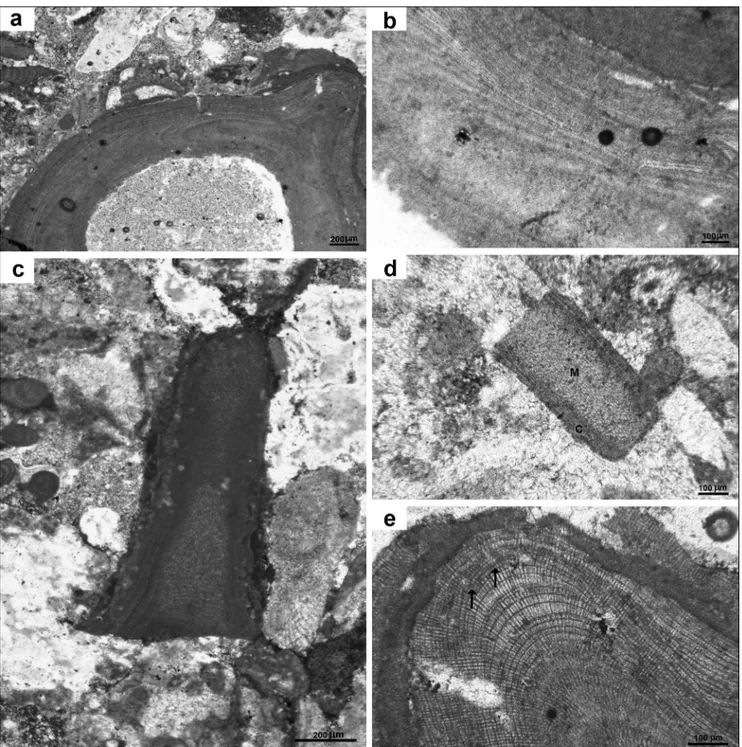
Fig. 1 - Sporolithon? diagramaticum (Johnson & Kaska) Basso & Granier comb. nov., usnM 42534 = thin section 12673B: a) lectotype specimen showing monomerous construction and a warty protuberance on the right; b) detail of a) to show the organization of cell filaments, producing the thallus elongation (evident in the “medulla”) and thickening (growing toward the periphery to form the “cortex”); c) a fragmented protuberance; d) a fragmented protuberance showing cortex (C) and medulla (M). note the different appearance of thal-lus organization due to orientation; e) magnification of the tip of a protuberance in a), showing two possible gametangial, uniporate
conceptacles on the same layer (arrows below their base). note the regular organization of the grid of cells due to uncommon cell fusions, typical of Sporolithon.
Basso D. & Granier B.
94
sub-circular to irregularly pear-shaped cavities about 100 µm in diameter opening toward the thal-lus surface, have uncertain nature (Fig. 1e). They are organised in a tier, just below the apex of a protu-berance (Fig. 1a, e).
Remarks. This species has a very variable
appearance depending on the section orientation, as Johnson & Kaska also probably noticed. some fragments (Fig. 1c, d), as well as the lectotype, show good lateral alignment of cells in the cortex, but not in the medulla. Fragment in Fig. 1d, in particular, is also very similar to that illustrated by Johnson & Kaska (1965, plate 34, fig 3). All the described frag-ments collectively correspond to the original de-scription in the protologue: “central hypothallic tis-sue with a long plumose or water jet arrangement”. The occurrence of secondary pit-connections exclude the placement of this species in the genus Lithothamnion. The aspect of the vegetative thallus and the shape and size of the possible reproductive structures correspond to those observed in some
species of extant Sporolithon (Bahia et al. 2015), the-refore we place tentatively Lithothamnium diagramati-cum Johnson & Kaska in the genus Sporolithon with the new combination Sporolithon? diagramaticum.
order Hapalidiales W.a. nelson, J.e. sutherland,
T.J. Farr & H.s. Yoon, 2015 Family Hapalidiaceae J.e. Gray, 1864 subfamily Melobesioideae Bizzozero, 1885
Genus Lithothamnion Heydrich, 1897 Type species: Lithothamnion muelleri lenormand ex rosanoff, 1866
Lithothamnion crispatum Hauck, 1878
Fig. 2
1878 Lithothamnion crispatum Hauck, p. 289, pl. 3: figs 1-4
1965 Lithothamnium toltecensum Johnson & Kaska, p. 32, pl. 21, fig. 3;
pl. 40, fig. 1
1965 Lithothamnium florea brassica - Johnson & Kaska, pp. 36-37, pl.
37, fig. 1; pl. 38, fig. 1
other synonyms are listed in Basso et al. 2011.
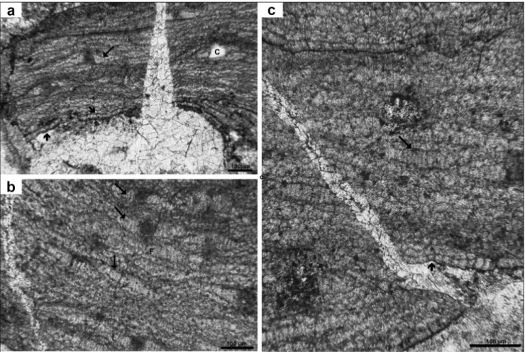
Fig. 2 - Lithothamnion crispatum Hauck: a) Holotype of L. toltecensum Johnson & Kaska 1965, usnM 42480= thin section 2205; b) Paratype of L. toltecensum Johnson & Kaska 1965: USNM 42481= thin section 2207. Note cell fusions (arrows), flattened epithallial cells (arrowhead),
and thin perithallus (brace); c, d) Specimens originally identified as L. florea-brassica (Millet) lemoine. note the diagnostic shape of
Examined material: Holotype of L. toltecensum Johnson &
Kaska 1965, pl. 21, fig. 3 (USNM 42480= thin section 2205; Fig. 2a); paratype of L. toltecensum Johnson & Kaska 1965, pl. 40, fig. 1
(usnM 42481= thin section 2207); L. florea brassica, Johnson &
Ka-ska 1965, pl. 37, fig. 1, pl. 38, fig. 1 (USNM 42551= thin section 1309H and 42530= thin sections 11260).
Derivation of name: The epithet toltecensum is dedicated to
the Toltec, the ancient Mesoamerican culture.
Age and locality: Miocene, possibly early Miocene; 4 km
sW of livingston for L. toltecensum type material; Guatemalan
loca-lities 1309H and 11260, with no further detail, for L. florea brassica
(Johnson & Kaska 1965).
Growth form and vegetative anatomy. L.
toltecensum is a crustose thallus, with a monomerous construction and a dorsiventral organisation (Figs 2a, b). Hypothallus plumose, 160-255 µm thick, made of filaments of cells L 14-33 x D 11-18 µm, that give rise to a thin perithallus of cells l 9-15 x 11-15. Cells connected by fusions (Fig. 2b). Tri-chocytes not observed. epithallial cells compressed (Fig. 2b).
Reproductive structures. The holotype of
Lithothamnium toltecensum Johnson & Kaska is sterile (Fig. 2a)
Remarks. The material of L. toltecensum is
very fragmentary and sterile, but fully corresponds to the vegetative anatomy of the living and fossil representatives of L. crispatum Hauck (Coletti et al. 2016). L. crispatum is easily recognized in the Mio-cene material from the distinctive shape of the pits, corresponding to the pore-canal opening through the multiporate conceptacle roof (Basso et al. 2011; Coletti et al. 2016, Fig. 2c-d). since a fertile Miocene specimen of L. crispatum indeed occurs in the John-son & Kaska collection (1965) under the name L.
florea brassica (Millet) Lemoine, we confidently con-sider L. toltecensum as conspecific with L. crispatum Hauck.
order Corallinales silva & Johansen, 1986
Family Corallinaceae lamouroux, 1812 subfamily Hydrolithoideae a. Kato
& M. Baba, 2011
Genus Hydrolithon (Foslie) Foslie 1909 lectotype species: Hydrolithon reinboldii (Weber van Bosse & Foslie)
Foslie
Hydrolithon guatemalaensum (Johnson & Kaska)
Basso & Granier comb. nov. Figs 3-4
Basionym: Aethesolithon guatemalaensum Johnson & Kaska
(Fossil algae from Guatemala. Professional contributions of the Co-lorado School of Mines 1, pp. 49-50, tab. 13, pl. 45, figs 1-2).
Examined material: Holotype of A. guatemalaensum
John-son & Kaska 1965, pl. 45, fig. 2 (USNM 42477= thin section 1591; Fig. 3a); paratype of A. guatemalaensum Johnson & Kaska 1965, pl. 45,
fig. 1 (USNM 42530A=thin section 11260; Fig. 3b).
Derivation of name: The name of the genus derives from aethes = unusual and lithon = stone; the specific epithet guatemalaensum
means from Guatemala.
Age and locality: early Miocene. The protologue reports
four localities numbered 1222H, 1588, 1591, 11260. out of these, only the 1222 is listed and marked in the map of Guatemala, for the area 4 km sW of livingston (Johnson & Kaska 1965: 6 and attached map).
Growth form and vegetative anatomy.
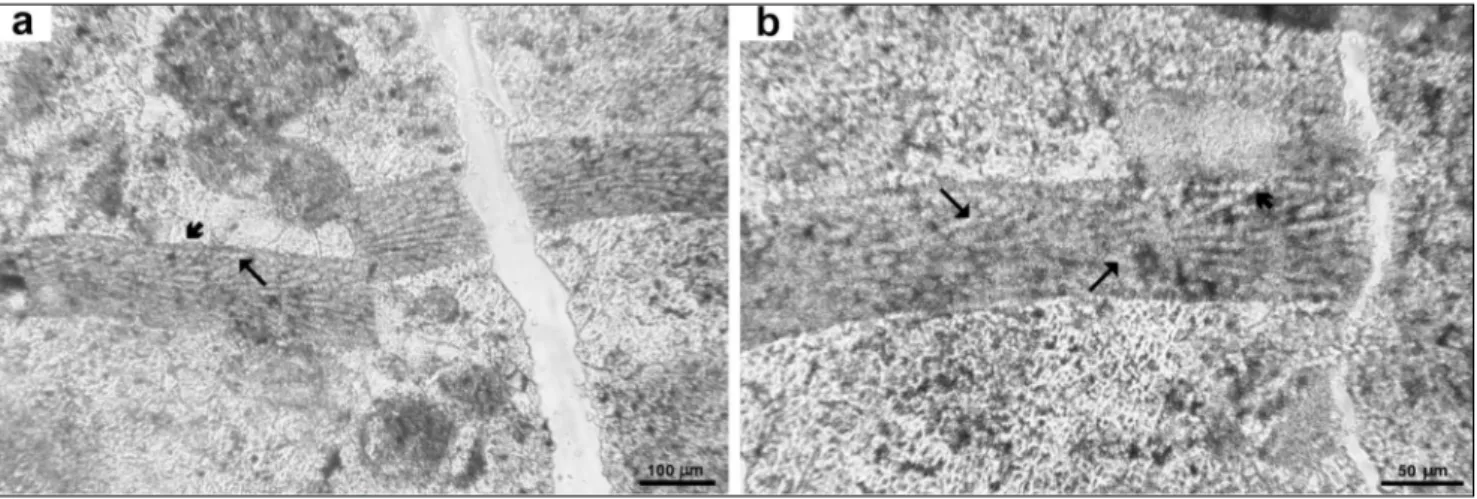
Thallus crustose, non-geniculate, protuberant, about 1200 µm thick. Thallus construction dimerous, Fig. 3 - Hydrolithon guatemalaensum (Johnson & Kaska) Basso & Granier comb. nov.: a) holotype usnM 42477= thin section 1591, C = uniporate
conceptacle chamber, T = single trichocytes, arrow = large irregular cells filling the empty conceptacles buried in the thallus, arrowhe-ad = monomerous structure with monostromatic hypothallus; b) paratype usnM 42530a= thin section 11260.
Basso D. & Granier B.
96
thin hypothallus composed of one layer of irregu-larly rectangular cells, more frequently higher than broad, L 10-20 µm x D 15-40 µm (Figs 3a-b, 4a). The perithallus is composed of cells laterally con-nected by cell fusions. The perithallial cells are very variable in size and shape, because of the oblique cut, and thus the horizontal layering is ill-defined (Fig. 4a-b). Horizontal lenses of long and broad perithallial cells up to L 50 µm x D 30 µm alternate with cells as small as L 16 µm x D 10 µm (Fig. 4b, c). large cells occur at the periphery of the numerous conceptacles (Figs 3a, 4a). sparse single trichocytes are common.
Reproductive structures. small uniporate
conceptacles buried in the perithallus are common (Figs 3, 4a). They appear in the holotype section 42477 as ovoidal chambers about D 160-270 x H 80-110 µm, with a flat to concave floor developing on a layer of very small cells (Fig. 3a). The concep-tacle chamber roof is not formed by cell filaments peripheral to the conceptacle and is compatible with a conceptacle development of Type 2
(Johan-sen 1981; Hrabovský et al. 2015). in section 42530 the conceptacles appear as lens-shaped areas 110-200 µm in diameter with an empty central area or more frequently filled by dark material (Figs 4a-b).
Remarks. The two thin sections are both
obliquely cut, therefore the microanatomical fea-tures have an improper orientation and a deformed outline in the holotype. The distortion is even more severe in section 42530. This fossil coralline indeed possesses cells of different size and shape (cells at the periphery of conceptacles, adventitious cells in-side conceptacles, and trichocytes), giving the thal-lus a distinctive chaotic appearance, but we could not ascertain the presence of the large polygonal cells, diagnostic for Aethesolithon. The occurrence of cell fusions and trichocytes, the shape and struc-ture of the uniporate conceptacles and the dimer-ous construction collectively indicate that Aetheso-lithon guatemalaensum belongs to the genus HydroAetheso-lithon (Kato et al. 2011). interestingly, published pictures of obliquely cut Hydrolithon fossil thalli show a strik-ing similarity with the chaotic perithallus of Aetheso-Fig. 4 - Hydrolithon guatemalaensum (Johnson & Kaska) Basso & Granier comb. nov.: paratype usnM 42530a: a) numerous conceptacle
cham-bers (C) buried in the thallus (arrow). note the dimerous construction and the monostromatic hypothallus (arrowhead); b-c) large
lithon guatemalaensum (e.g. Montaggioni 1979, pl. 2, fig. 3; Rösler et al. 2016, fig. 4). The disposition of the genus Aethesolithon, based on the type-species A. problematicum Johnson, 1964 would require further investigations on the relevant collection, and is out-side the aim of this contribution.
subfamily Corallinoideae (areschoug) Foslie, 1908 Genus Corallina linnaeus, 1758
lectotype species: Corallina officinalis linnaeus, 1758
Corallina matansa Johnson, 1957
Fig. 5
1957 Corallina matansa Johnson, pp. 238-239, pl. 44, figs 3-4
1965 Jania occidentalis Johnson & Kaska, pp. 56-57, pl. 26, figs 1-3. Examined material: Holotype of J. occidentalis Johnson &
Kaska, 1965, pl. 26, fig. 3 (USNM 42523= thin section 8885; Fig. 5a). Paratype of J. occidentalis Johnson & Kaska, 1965, pl. 26, fig. 2 (USNM
42519= thin section 8839; Fig. 5b). The specimens mentioned in the protologue and figured in Johnson & Kaska, 1965, pl. 26, fig. 1 and pl. 30, fig. 2 were not found. Other material: Corallina matansa,
John-son & Kaska, 1965, p. 54, pl. 21, fig. 1 (USNM 42540= thin section 15971).
Derivation of name: The specific epithet matansa derives
from the eocene Matansa limestone in saipan, the type locality.
Age and locality: Paleocene-eocene. The protologue of J. occidentalis reports the codes of five localities in Guatemala: 8839,
8840, 8885, 12673, 12673B (Johnson & Kaska 1965: 56). out of the-se, 8839 and 8840 correspond to an area 3 km nW of san luis; 12673 is on road Cadenas-san luis, 4 km before san luis; and the type locality 8885 is not indicated in the list of sample localities nor in the attached map (Johnson & Kaska 1965: 8). Corallina matansa is
reported for the lower eocene of a series of locality codes (Johnson & Kaska 1965: 54) that are not indicated in the list of sample loca-lities nor in the attached map (Johnson & Kaska, 1965: 8), with the exception of 15971, 15 km ne of Chinaja, and 12673, the same of J. occidentalis (on road Cadenas-san luis, 4 km before san luis).
Growth form and vegetative anatomy.
Ge-niculate coralline. Fragments of isolated, scattered intergenicula, cylindrical to club-shaped. one com-plete geniculum, although not properly oriented, Fig. 5 - Corallina matansa Johnson,
1957: a) holotype specimen
of Jania occidentalis Johnson
& Kaska, 1965 in usnM 42523= thin section 8885; b, c) usnM 42519= thin section 8839; b - paratype specimen of Jania occidenta-lis Johnson & Kaska, 1965,
note incipient bifurcation (arrow); c - other specimens
with preserved genicula (ar-rows); d) usnM 42540=
thin section 15971, speci-men originally identified as
Corallina matansa Johnson,
note thallus bifurcation (ar-row).
Basso D. & Granier B.
98
has been observed in a fragment occurring in thin section 42519 (Fig. 5c). The intergenicula show la-teral branching (Fig. 5b, d), are about 800-1500 µm long and 180-330 µm broad, composed of 15-22 tiers of medullary cells of similar length (l 40-60 x D 9-17 µm; Fig. 5b).
Reproductive structures. not observed. Remarks. Within the modern concept of the
genus Jania, intergenicula are unbranched or bearing two branches on broadened upper parts (Womer-sley 1996; Bressan & Babbini 2003). Therefore, the observed lateral branching of J. occidentalis Johnson & Kaska suggests that it does not belong to the genus Jania. Moreover, most of the extant species of Jania has few tiers of longer medullary cells in each intergeniculum, compared with J. occidentalis. The type material of J. occidentalis is quite poor for any certain assignment to any geniculate genus. Ho-wever, on the basis of the few characters that can be observed, we can exclude some possible alter-natives. in particular, the species cannot belong to
Arthrocardia, because this genus is characterized by species showing mostly > 40 tiers of cells in the intergeniculum, and this character is not present in J. occidentalis. in the same paper, Johnson & Kaska (1965: 54, pl. 21, fig. 1) illustrated a fragment of Corallina matansa Johnson, 1957 showing identical structure and cell size, and report a wide and over-lapping size range for J. occidentalis and C. matansa (tabs 17-18). Therefore, we consider Jania occidentalis as a heterotypic synonym of C. matansa Johnson, 1957, which has priority.
subfamily lithophylloideae setchell, 1943 Genus Amphiroa lamouroux, 1812 lectotype species: Amphiroa tribulus (ellis & solander)
lamouroux, 1812
Amphiroa guatemalense Johnson & Kaska, 1965
Figs 6-7
1965 Amphiroa guatemalense Johnson & Kaska, pp. 52-53, pl. 24, figs 1-3.
Fig. 6 - Amphiroa guatemalense Johnson & Kaska, 1965: a) lectotype specimen in usnM 42532=slide 12673a, note prostrate appearance of
the thallus; b) magnification of a to show the row of short cells alternating with one-two rows of long cells; c) specimen in usnM
42534=slide 12673B, with a preserved geniculum (arrow); d) magnification of c to show the absence of fusion between cells of
Examined material: syntype specimen of A. guatemalense
Johnson & Kaska, 1965 pl. 24, fig. 1 (USNM 42532= thin section 12673a; Fig. 6a, b); the other syntype specimen in usnM 42534= thin section 12673B (Johnson & Kaska 1965, pl. 25, fig. 1) was not found. Paratypes of A. guatemalense Johnson & Kaska, 1965, pl. 24,
figs 2-3 (USNM 42517= thin section 8802 and USNM 42538=slide 15134 respectively).
Lectotypification: The specimen of A. guatemalense
John-son & Kaska, 1965 illustrated in pl. 24, fig. 1 (USNM 42532= thin section 12673a) is here selected as lectotype (Fig. 6a-b).
Derivation of name: The specific epithet guatemalense
me-ans Guatemalan.
Age and locality: late Paleocene and early eocene. The
lectotype locality is on road Cadenas-san luis, 4 km before san luis. no information is provided in the protologue about the other localities (8802 and 15134).
Growth form and vegetative anatomy.
Geniculate coralline with intergenicula up to 1.8 mm long and 0.6 mm wide, possibly also decum-bent (Fig. 6a). structure of intergenicula with a medulla of one short–celled tier (l 13-28 x 7-10 µm) alternating with one or two long-celled tiers (commonly observed 30-40 tiers in each segment) (Fig. 6b). Cells connected by secondary pit-con-nections (Figs 6b, d). intergenicular cortex absent to thick, made of several cells L 9-12 x D 5-10 µm (Fig. 6b).
Reproductive structures. on the same thin
section usnM 42517, another specimen shows presumed uniporate conceptacles with a coarsely spherical chamber, about 100-110 µm in diameter, developed within the cortical cells (Fig. 7). The conceptacles are immersed, and open at the thal-lus surface with a pore up to 90 µm long and 30-50 µm broad (Fig. 7b).
Remarks. The conceptacle in Fig. 7 shows
well-defined boundaries, unlike bioperforations, and the cells of the periphery of the conceptacle chamber are visible inside.The uniporate concep-tacles are of unknown origin, but their shape and size are similar to the carposporangial concepta-cles described for A. rigida in Baja California, Me-xico (riosmena-rodriguez & siqueiros-Beltrones 1996, fig. 9).
Amphiroa kaskaella Johnson & Kaska, 1965
Fig. 8
1965 Amphiroa kaskaella Johnson & Kaska, pp. 53-54, pl. 25, figs
2-3
Examined material: syntypes of A. kaskaella Johnson
& Kaska 1965 pl. 25, figs 2-3 (USNM 42498= thin section 4262). USNM 42516 (thin section 8798), mentioned and figured in the protologue (p.54, pl. 26, fig. 4), is lost.
Lectotypification: Johnson & Kaska selected usnM
42498= thin section 4262 as type material, indicating that it con-tains “two good specimens”. according to articles 8.2, 8.5 and 9.15 (Melbourne Code) the type of a name of a fossil species is the specimen (or one of the specimens) on which the validating illustra-tions are based. since a specimen is a gathering of a single species
made at one time, disregarding admixtures, and since thin section 4262 contains an admixture of species, the whole thin section can-not be considered as the type specimen. out of the two fragments illustrated in the protologue, and still present in the type collection, the one figured in Johnson & Kaska (1965) pl. 25, fig. 2 is a com-plete, fertile intergeniculum. Therefore, the specimen illustrated in Johnson & Kaska (1965) p. 121, pl. 25, fig. 2, is here selected as lectotype of Amphiroa kaskaella Johnson & Kaska.
Derivation of name: The origin of the specific epithet kaskaella was not explained in the protologue. The species was
pos-sibly dedicated to a member of Harold V. Kaska’s family.
Age and locality: Paleocene. Type locality is 4262, 6 km
ne of san luis.
Fig. 7 - Amphiroa guatemalense Johnson & Kaska, 1965: a, b) two young apices in usnM 42517=thin section 8802; a - paratype specimen, detail
of the regular cell arrangement, showing one row of short cells alternating with one row of long cells; b - a small uniporate concep-tacle with ovoid chamber (C) and pore canal opening at the thallus surface (arrow).
Basso D. & Granier B.
100
Growth form and vegetative anatomy.
Geni-culate coralline. intergenicula up to 3.5 mm long and 1.2-1.4 mm wide. intergenicula composed of a me-dulla of one short–celled tier (L 18-46 x 6-13 µm) al-ternating with two or three long-celled tiers (l 53-102 x 6-13 µm, with about 50-60 tiers in each segment) (Figs 8a-b). intergenicular cortex absent to thick, made of several cells L 10-20 x D 6-11 µm. Secondary pit-connections visible in largest cells. Cortex of adjacent intergenicula partially fused at their base.
Reproductive structures. uniporate
concep-tacle chambers immersed in the intergenicular cor-tex are aligned in a row (Fig. 8a, c). The chambers are rounded to elliptical in section, D 80-170 µm x H 65-70 µm, with the largest chambers possibly resul-ting from the fusion of adjacent small ones. Pore not observed (Fig. 8c).
Remarks. Within the limits of this
investiga-tion based on few but important specimens, we have presently no elements to support a different generic attribution. Future investigation on newly collected,
abundant and well preserved material will hopefully provide full details of this fossil coralline species.
subclass corallInophycIdae le Gall
& saunders, 2007
Incertae sedis
“Lithothamnium guatemalense” Johnson & Kaska, 1965
Fig. 9
1965 Lithothamnium guatemalense Johnson & Kaska, 1965, pp. 29-30, pl.
29, figs 1-3.
Examined material: Holotype of L. guatemalense Johnson &
Kaska, 1965, pl. 29, fig. 2 (USNM 42518= thin section 8807). Paratypes of L. guatemalense Johnson & Kaska 1965, pl. 29, fig. 1 (same thin section
USNM 42518) and pl. 29, fig. 3 (USNM 42517A= thin section 8802).
Age and locality: early eocene. Johnson & Kaska (1965)
re-port four localities numbered 8796, 8802, 8807, 8798. out of these, only the 8796 was listed and marked in the map, for the area 4.5 km ne of san luis (Johnson & Kaska 1965: 8 and attached map).
Fig. 8 - Amphiroa kaskaella Johnson & Kaska, 1965, usnM 42498= thin section 4262: a) two intergenicula partially fused at the base (arrow).
The lectotype specimen is on the right. note the small conceptacles aligned at the base of the cortical thickening (arrowhead), and the alternation of one tier of short cells with two-three tiers of long cells; b) paratype specimen, with characteristic cortical thickening
Growth form and vegetative anatomy.
Thallus foliose, non protuberant, up to 110 µm thick. Thallus construction monomerous, with dor-siventral organisation. Hypothallus composing most of the total thickness (Fig. 9a-b), apparently plumo-se becauplumo-se of the oblique cut, made of long cells (mostly L 20-50 x D 7-10 µm) directed toward the thallus margin and diverging upward and downward to both thallus surfaces. Cells of adjacent filaments connected by cell fusions (Fig. 9b). Perithallus very reduced, made of few cells, or absent, terminating with an ovoidal epithallial cell (Fig. 9b). Meristem intercalary below epithallial cells, appearing as a cle-ar line of long cells, connected by cell fusions.
Reproductive structures. not observed,
the thalli are sterile.
Remarks. The specimen with perithallus of
130 µm thick mentioned in the protologue could not be found in the original material of “Lithotham-nium guatemalense”.
although Johnson & Kaska (1965) assigned the above described sterile coralline fragments to the genus Lithothamnion (at that time spelled Litho-thamnium), the occurrence of ovoidal epithallial cells and the aspect of the meristem collectively point to a different generic placement. in particular, some analogies can be found with extant species of Ne-ogoniolithon, including the growth-form, and the aspect of the intercalary meristem (Mateo-Cid et al. 2014). Following the modern concept of the genus (Penrose 1996; Kato et al. 2013), Neogoniolithon may show a coaxial or non-coaxial hypothallus, although the coaxial arrangement of the hypothallus is
consi-dered diagnostic in the fossil (Braga et al. 1993; Hra-bovský et al. 2015). Moreover, the oblique cut, also reported in Johnson & Kaska protologue (1965), may hide a coaxial hypothallus, as demonstrated in Neogoniolithon contii (Quaranta et al. 2007, pl. 1, fig. 7). nevertheless, the fragmentary material and the lack of diagnostic characters prevent a taxonomic placement of this fossil coralline, therefore we leave the species incertae sedis, and thus we report its origi-nal name in quotation marks.
order ? Rhodogorgonales Fredericq
& norris, 1995
Family elianellaceae Granier in Granier & Dias-Brito, 2016
Genus Marinella Pfender, 1939 Type species: Marinella lugeoni Pfender, 1939
Fig. 10
Marinella lugeoni Pfender, 1939, nomen cons. 1939 Marinella lugeoni Pfender nomen cons., pp. 215-216, pl. II, figs
1-2.
1965 Lithothamnium? primitiva Johnson & Kaska, pp. 30-31, pl. 6, fig. 1.
1965 Marinella lugeoni - Johnson & Kaska, p. 74, pl. 6, fig. 2.
1965 Girvanella minuta - Johnson & Kaska, p. 96, pl. 30 fig. 1.
other synonyms are listed in Granier & Dias-Brito (2016).
Examined material: Holotype of Lithothamnium? primitiva
Johnson & Kaska, 1965, pl. 6, fig. 1 (thin section USNM 42547=slide 18587); Marinella lugeoni, Johnson & Kaska, 1965, pl. 6, fig. 2 (same
thin section usnM 42547); Girvanella minuta, Johnson & Kaska,
1965, pl. 30, fig. 1 (same thin section USNM 42547).
Derivation of name: The epithet refers to the primitive
“generalized structure” of this fossil red alga.
Fig. 9 - “Lithothamnium guatemalense” Johnson & Kaska, 1965, usnM 42518= thin section 8807: a) the holotype is a fragmented laminar thallus
mostly composed of hypothallus. note the ovate epithallial cells (arrowhead) and the large, probably meristematic, subepithallial cells (arrow); b) magnification of the holotype showing cell fusions (arrows) and large meristematic cells (arrowhead). Perithallial cells are
Basso D. & Granier B.
102
Age and locality: late Jurassic to early Cretaceous. The
Guatemalan locality 18587 was not listed in the table of the inve-stigated areas, nor indicated in the attached map (Johnson & Kaska 1965: 5-10).
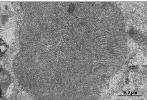
Growth form and vegetative anatomy.
a cluster of fan-shaped thalli, radiating from a sub-central empty area, looking like a “pipe puff ” (Pfender 1939). The thallus consists of juxtaposed filaments with aligned constrictions, but with non-obvious cell partitions. Filaments are 6 to 9 μm in diameter, composing a hairy fabric (Fig. 10).
Reproductive structures. not observed. Remarks. in agreement with the recent
re-vision (Granier & Dias-Brito 2016), we confirm that Johnson & Kaska misidentified Marinella lugeo-ni with two different species, although the relevant specimens occurred in the same thin section. This was a consequence of the systematic approach of the sixties, when different orientations of the sec-tioned material were not fully appreciated, and, on the contrary, great importance was given to subtle variations in cell size.
c
onclusIonsThe original collections of eight red algal species described by Johnson & Kaska (1965) from several Guatemalan localities and ages, have been examined, re-documented and critically revised. For most of the examined taxa, Johnson & Kaska (1965) clearly indicated and illustrated the type
spe-cimen on which the names of their new species were based, and these names have been accepted here (Melbourne Code art. 9.1, Mcneill et al. 2012). in three cases we had to select a lectotype. The type of fossil taxa is always a specimen (Melbourne Code art. 8.5). Moreover, the holotype (or lectotype) of a name of a fossil-species is the specimen on which the validating illustrations are based and when a type specimen is indicated but not identified among the validating illustrations, a lectotype must be de-signated from among the specimens illustrated in the protologue (art. 9.15). in agreement with the mentioned articles, we selected a lectotype for the name of the two species that were based on synt-ypes, namely Amphiroa guatemalense and A. kaskaella. in the case of Sporolithon? diagramaticum nov. comb. the holotype was lost: we selected a lectotype from the original material in agreement with Johnson & Kaska protologue, according to art. 9.2 (Mcneill et al. 2012).
We had to modify the generic placement of the revised species that resulted incorrect under modern taxonomic criteria, that is to say in most cases, with the exception of Amphiroa guatemalense and A. kaskaella. This is not surprising, as new dia-gnostic criteria at high taxonomic ranks (i.e. type of cell anastomoses) were unknown at the time of Johnson & Kaska’s contribution. some fragmentary and sterile thalli, in some cases as unique specimen, would be presently considered insufficient for the erection of a new species, and “Lithothamnium guate-malense” is emblematic of an obsolete approach to
Fig. 10 - Marinella lugeoni Pfender,
usnM 42547= thin sec-tion 18587: specimen ori-ginally selected as holotype of Lithothamnium? primitiva
Johnson & Kaska, 1965. note the lack of connection between adjacent filaments, the absence of cell layering, and the typical “pipe puff ” appearance.
systematic Palaeontology. Further studies will hope-fully be able to describe the missing, essential details of the vegetative and reproductive anatomy of this fossil alga.
Acknowledgements. DB is grateful to the uBo (université de
Bretagne occidentale) for the invitation as visiting professor in Brest during February 2014. BG benefited of a Smithsonian Fellowship for the study of the Collection Jesse Harlan Johnson conserved in the smithsonian institution in Washington, D.C. The Department of Paleobiology of the smithsonian national Museum of natural History is warmly acknowledged for the loan of the Johnson & Ka-ska collection.
RefeRences
Bahia r.G., Maneveldt G.W., amado-Filho G.M. & Yo-neshigue-Valentin Y. (2015) - new diagnostic char-acters for the order sporolithales (Corallinophycidae, rhodophyta). J. Phycol., 51: 1137-46. Doi: 10.1111/
jpy.12351.
Basso D., rodondi G. & Bressan G. (2011) - a re-description of Lithothamnion crispatum and the status of Lithothamnion superpositum (rhodophyta, Corallinales). Phycologia, 50: 144–155. Doi: 10.2216/10-20.1
Braga J.C., Bosence D.W.J. & steneck r.s. (1993) - new ana-tomical characters in fossil coralline algae and their taxo-nomic implications. Palaeontology, 36: 535-547.
Coletti G., Hrabovský J. & Basso D. (2016) - Lithothamnion crispatum: long-lasting species of non-geniculate coral-line algae (rhodophyta, Hapalidiales). Carnets Géol, 16: 27-41.
Granier B., Basso D. & Vachard D. (2017a) – les algues “cal-caires” fossiles (Permian-Miocène) du Guatémala. Cata-logue critique de la Collection J.H. Johnson. 3e partie.
Archives des Sciences 69:29-54.
Granier B., Bucur i.i. & DiasBrito D. (2017b) - about Trinoc-ladus raineri, 1922: when some Permocalculus (Gymnoco-diacean algae) reveal to be Triploporellacean algae (re-vision of the Jesse Harlan Johnson Collection. Part 5). Facies (2017) 63:27. Doi 10.1007/s10347-017-0508-x Granier B. & Dias-Brito D. (2016) - on the fossil alga
Mari-nella lugeoni Pfender, 1939, nom. cons., and its seven unfortunate avatars. revision of the Juliette Pfender Collection. Part 2. revision of the Jesse Harlan John-son Collection. Part 2. Carnets Géol, 16: 231-245. Doi : 10.4267/2042/59922
Granier B., Radoičić R. & Drobne K. (2013) - Revision of the Jesse Harlan Johnson Collection. Part 1. some fossil Dasycladales from Guatemala. Carnets Géol., article CG2013/07 (CG2013_a07): 281-301. Doi: 10.4267/2042/51824
Hrabovský J., Basso D. & Doláková n. (2015) - Diagnostic characters in fossil coralline algae (Corallinophycidae: rhodophyta) from the Miocene of southern Moravia (Carpathian Foredeep, Czech republic). J. Syst. Palaeontol., http://dx.doi.org/10.1080/14772019.2015.1071501.
Johansen H.W. (1981) - Coralline algae. A first synthesis. CRC Press, Boca raton, Florida, 239 pp.
Johnson J.H. (1957) - Geology of saipan, Mariana islands, Part 3. Paleontology. Calcareous algae. U.S. Geol. Surv. Prof. Pap. 280-e: 209-246.
Johnson J.H. (1964) - Fossil and recent Calcareous algae from Guam. Geology and hydrology of Guam, Mariana islands. U.S. Geol. Surv. Prof. Pap. 403-G: G1-G40. Johnson J.H. & Kaska H.V. (1965) - Fossil algae from
Gua-temala. Prof. Contrib. Colorado School of Mines, 1, xii + 152 p. (47 Pls).
Kato a., Baba M. & suda s. (2011) - revision of the Mas-tophoroideae (Corallinales, rhodophyta) and polyphyly in nongeniculate species widely distributed on Pacific coral reefs. J. Phycol., 47: 662-672.
Mateo-Cid l.e., Mendoza-González a.C. & Gabrielson P.W. (2014) - Neogoniolithon (Corallinales, rhodophyta) on the atlantic coast of Mexico, including N. siankanensis sp. nov. Phytotaxa, 190: 64-93.
Mateo-Cid l.e. & Pedroche F.F. (2004) - The occurrence of Neogoniolithon fosliei (Heydrich) setchell et Mason in the Mexican Caribbean and the relationship of this species to N. solubile (Foslie et Howe) setchell et Mason (Coral-linales, rhodophyta). Caribbean J. Sci., 40 (2): 182-191.
Mcneill J., Barrie F.r., Buck W.r., Demoulin V., Greuter W., Hawksworth D.l., Herendeen P.s., Knapp s., Marhold K., Prado J., Prud’homme van reine W.F., smith G.F., Wiersema J.H. & Turland n.J. (2012) - international Code of nomenclature for algae, fungi, and plants (Mel-bourne Code). Regnum Vegetabile 154. Koeltz Scientific Books, Koenigstein, xxx + 240 p.
Montaggioni L.F. (1979) - Environmental significance of rho-doliths from the Mascarene reef province, western in-dian ocean. in 2ème symposium international sur les
al-gues fossiles. Bull. Cent. Rech. Explor.-Prod. Elf-Aquitaine,
3: 713-717.
Pfender J. (1939) - sur un calcaire phytogène du lias inférieur d’espagne et l’extension dece faciès en quelques autres régions. Bull. Soc. vaudoise Sci. nat., 60(248): 213-228 (Pl.
iV).
Quaranta F., Vannucci G. & Basso D. (2007) - Neogoniolithon contii comb. nov based on the taxonomic re-assessment of Mastrorilli’s original collections from the oligocene of nW italy (Tertiary Piedmont Basin), Basin). Riv. Ital. Paleont. Strat., 113 (1): 43-55.
riosmena-rodriguez r. & siqueiros-Beltrones D.a. (1996) - Taxonomy of the genus Amphiroa (Corallinales,
rho-dophyta) in the southern Baja California Peninsula, Mexico. Phycologia, 35 (2): 135-147.
Rösler A., Perfectti F., Peña V. & Braga J.-C. (2016) - Phy-logenetic relationships of Corallinaceae (Corallinales, rhodophyta): taxonomic implications for reef-building corallines. J. Phycol. 52: 412–431.
Woelkerling W. J., Campbell s. J. & Harvey a. s. (1993) - Growth-forms in non-geniculate coralline red algae (Corallinales, rhodophyta). Aust. Syst. Bot., 6 (4): 277-293.