Dipartimento di Medicina Veterinaria
Scuola di Dottorato in Scienze Veterinarie
Indirizzo: Riproduzione, Patologia, Allevamento e
Benessere Animale
Ciclo: XXXII
Coordinatore del Corso: Prof.ssa Fiammetta Berlinguer
Effect of melatonin treatment on
reproductive activity and immune
response in Sarda sheep breed
Tutor:
Prof. Vincenzo Carcangiu
Dottorando
Dott.ssa Luisa Pulinas
La presente tesi è stata prodotta durante la frequenza del corso di dottorato in Scienze Veterinarie, Indirizzo Riproduzione, Patologia, Allevamento e Benessere animale dell’Università degli Studi di Sassari, A.A. 2018/2019 – XXXII ciclo, con il sostegno di una borsa di studio finanziata con le risorse del P.O.R. SARDEGNA F.S.E. 2014-2020 Asse III - Istruzione e Formazione - Obiettivo Tematico 10 “Investire nell’istruzione, nella formazione e nella formazione professionale per le competenze e l’apprendimento permanente”
Index
Abbreviation list Pag 4
Introduction Pag 6
Pineal gland or epiphysis Pag 6
Melatonin Pag 12
Noradrenergic regulation of MEL secretion Pag 18 MEL and seasonality (circannual / cyrcadian rhythms) Pag 25
MEL receptors (MEL-R) Pag 29
Roles of MEL Pag 34
Antioxidant / oxidant effects Pag 34
Anticancer effects Pag 35
Immunomodulatoy effects Pag 36
Reproductive effects Pag 39
Aim Pag 46
Material and methods Pag 48
Aim1a (MEL treatment in different periods of the year) Pag 48 Aim 1b (MEL treatment in different lactating period) Pag 50 Aim 2 (MEL treatment and male replacement) Pag 52 Aim 3 (influence of MEL treatment in male) Pag 55 Aim 4 (influence of different MTNR1A genotypes) Pag 57 Aim 5 (influence of the MEL treatment on milk production
and mammary immune system)
Pag 63
Results Pag 65
1a (MEL treatment in different periods of the year) Pag 65 1b (MEL treatment in different lactation period) Pag 67 2 (MEL treatment in ewes and male replacement) Pag 69
4 (influence of different MTNR1A genotypes) Pag 74 5 (influence of the MEL treatment on milk production and
mammary immune system)
Pag 80
Discussion Pag82
1a (MEL treatment in different periods of the year) Pag 82 1b (MEL treatment in different lactation period) Pag 87 2 (MEL treatment in ewes and male replacement) and 3 (influence
of MEL treatment in male)
Pag 90
4 (influence of different MTNR1A genotypes) Pag 93 5 (influence of the MEL treatment on milk production and
mammary immune system)
Pag 100
Conclusions Pag 104
Abbreviation list
5-HT: serotonin hormone 5-HTP: 5-hydroxy-tryptophan AAAD: aromatic acid decarboxylase
AANAT: arylalkylamine-N-acetyltransferase AC: adenylate cyclase
ACTH: adenocorticotropic hormone AR: adrenergic receptor
BCS: body condition score
cAMP: cyclic adenosine monophosphate CAT: catalase
cGMP: cyclic guanosine monophosphate CRE: cAMP- response element
CREB: cAMP responsive element binding proteins CREM: cAMP responsive element modulator DAG: diacylglycerol
DIML: distance in days from male introduction to lambing E: estradiol hormone
GLM: general linear model
GnRH: gonadotropin releasing hormone GPCR: G-protein-coupled-receptor GPx: glutathione peroxidase
GSH: glutathione
HIOMT = ASMT: hydroxindole-O-methyltransferase ICER: inducible cAMP early repressor
Ig: immunoglobulin IL: interleukin
LH: luteinizing hormone
MBH: mediobasal hypothalamus MEL: melatonin hormone
Mel1b = MT2: melatonin receptor type 2 ML1: high affinity melatonin receptor ML2: low affinity melatonin receptor MT1: melatonin receptor type 1 MTNR1A: MT1 gene receptor NAS: N-acetyl serotonin NE: noradrenaline NK: natural killer cells
PAPC: pituitary adenylate cyclase PCR: polymerase chain reaction
p-CREB = p-CREB-ser133: activated CREB
PKC: protein kinase C
PT: pars tuberalis of adenohypophysis QR2: quinone reductase
SCC: somatic cells count SNC: suprachiasmatic nucleus
SNPs: single nucleotide polymorphisms SOD: superoxide dismutase
STAR: steroid regulatory protein TH: tyrosine hydroxylase
TM: transmembrane α-helices (domains) of MT1 receptor TPH: tryptophan-hydroxylase
TRP: tryptophan
Introduction
Pineal gland or epiphysis
The pineal gland, or epiphysis, is a neuro-endocrine gland that in all vertebrate originates from an evagination of the ceiling of the ventricle III, in its third caudal, in the posterior margin of the corpus callosum (Figure 1). This gland is unequal and lies on a median lumen, has a conical shape that resembles a pine cone (from which derives the Latin name pinealis), its size and shape are different in the various species.
Figure 1. Sheep brain dissection and location of the pineal gland (Human Anatomy and Physiology Lab, coureses.lumenlearning.com)
The evolutionary history of the pineal gland is very long, it has undergone considerable variations during the evolution from
amphibians to mammals (Mano and Fukada, 2017).
In several animals such as amphibians and some fish, the pineal gland is called the "third eye" because same cells of it have photoreceptor characteristics, with particular photosensitivity and electrical activity. In simple vertebrates, such as lampreys, the gland is located on a peduncle near an orifice of the skull and has both endocrine and nervous properties; in these animals, since the epiphysis is tightly against the skin, it does not require interactions with eyes to record day / night rhythm and is the main biological clock (Oksche and Vaupel–von Harnack, 1965).
In birds and reptiles the epiphysis, in addition to the photoreceptor function also performs the secretory function. In birds the pineal gland is located near the cranial vault, which is very thin and transparent to light, allowing its direct excitation. By coloring the skull of migratory sparrows with black ink, it was possible to highlight a close correlation with the migratory activity, in fact the black coloring of the skull resulted in the total loss of sense of orientation, as if the pineal functioned like a sort of internal compass (Gwinner, 1996).
The pineal gland is completely absent in crocodiles and mammals belonging to the order of toothless (anteaters, sloths and armadillos), while it is present in whales and elephants even if formed only by a few cells, in the human it has size of a pea.
In general, in mammals this gland is not directly photosensitive, although a nervous connection exists with the eyes which suggests a possible influence of light on its functions (Goldman, 1991). In these animals the structure of the epiphysis is a rather homogenous tissue containing pinealocytes (mono-, bi-, or tri-polarcells), few glial cells, phagocytic cells and neurons (Simonneaux and Ribelayga 2003). The pinealocytes, deriving from the neuroepithelial matrix layer, have lost the characteristic structure of the pineal photoreceptor cells of most of the lower vertebrates and have exclusively secretory function.
Being located outside the blood-brain barrier, it can come in contact with large blood plasma molecules, thanks also to the considerable size of the interstitial spaces. It is formed by a parenchyma which is constituted of cell cords separated by capillaries with fairly large perivascular spaces (Møller e Baeres, 2002). The pineal gland is the second most perfused organ of the organism, after the kidney, the blood supply is guaranteed through a dense network of vessels. The epiphyseal capillaries originate from the posterior choroid arteries branching out and first envelop the capsule of connective tissue, then penetrate in the parenchyma of the gland. Blood drainage of the gland is guaranteed by the dorsal passage of the large cerebral vein that allows a very rapid distribution of its secretion products to the target organs.
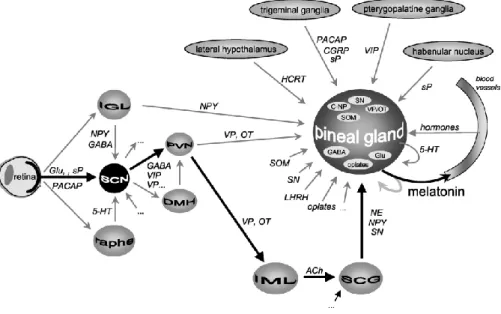
epiphysis. The light signal perceived by the photosensitive retinal cells is transformed into a nerve impulse, passing into the suprachiasmatic nucleus (SCN) and through the posterior hypothalamus and the sympathetic hypothalamic-spinal pathways, reaching the superior cervical ganglion. From the latter it branches off to post-ganglionic noradrenergic fibers that reach the epiphysis and regulate its secretory activity (Figure 2).
Figure 2. Luminous signal pathway from eye to pineal gland (Ostrin, 2019)
To enter epiphyseal cells, the nerve stimulus is converted into a chemical stimulus, through the linkage with specific receptors positioned on the pinealocytes membrane.
transducer and thus translates external light stimuli into internal hormonal messages, so that the body can synchronize its functions as environmental conditions change (Simonneaux and Ribelayga 2003). With the progress of experimentation and knowledge it has been possible to highlight how the innervation of the pineal gland is much more complex than what was initially thought. The pineal gland is innervated by nerve fibers of various origin. The main pathway consists of the retino-hypothalamus-pineal pathway, which ends with sympathetic input to the pineal parenchyma. The pineal gland also receives neural signals of central and parasympathetic origin (Figure 3) (Simonneaux and Ribelayga 2003).
Figure 3. Schematic presentation of the various neural, endocrine, paracrine inputs of the mammalian pineal gland (Simonneaux and Ribelayga 2003)
The luminous signal perceived by the retina, results in signal perception of cells that contain the pigment named melanopsin, which allows the transmission of the aforementioned signal parallel to the visual stimuli perceived by the cones and rods (Hattar et al., 2002). Neuromediators of the retinal-hypothalamus tract are mostly glutamate and PACP (pituitary adenylate cyclase activating peptide) (Hannibal et al, 1997). In addition to the light signals other nerve signals from the thalamus and the raphe reach the suprachiasmatic nucleus (SCN). In many mammals, the nucleus acts as "biological clock" that presents an oscillatory activity which is synchronized circadianly by melatonin and / or directly by the light signal (Lincoln et al. 2005). This pacemaker has an endogenous rhythm which is controlled by a series
of "clock" genes, the Per1, Per2 and Per3 called periodic genes, the Cry1 and Cry2 cryptochromic genes and the two, Clock and Bmal1 that encode some transcription factors. These genes act by activating or inhibiting the activity of the SCN (Reppert and Weaner, 2002). The function of these genes seems to be related to periodic processes of the organism, interacting with the signal coming from the epiphysis (Simonneaux et al., 2004).
The Paraventricular nuclei of the hypothalamus act as a link between the SCN and the epiphysis, in fact experimentaly induced lesions at this level have highlighted an important decrease in the melatonin secretion (Kalsbeek and Buijs, 2002). Finally, all the nerves, passing through the superior cervical ganglion innervate the epiphysis. The main innervation is of the noradrenergic type, but other fibers with different origins are also present. However, they all appear to play a role in regulating melatonin secretion (Simonneaux and Ribelayga, 2003).
Melatonin
The discovery of melatonin (MEL) dates back to 1958 and in the following year its chemical structure was revealed as N-acetyl-5-methoxytripamine (Lerner et al., 1958; 1959).
its most known activity is a hormonal transducer of photic/photoperiodic information. The pineal gland has two rhythms of secretion: a 24-h rhythm with a nocturnal peak and an annual rhythm closely dependent on seasonal variations in the photoperiod (Simonneaux and Ribelayga, 2003).
MEL is a molecule vastly present in nature, in fact, besides being produced by many invertebrates and vertebrates, it has also been found in bacteria, in unicellular eukaryotic organisms, in macro algae and in many parts of plants (Hardeland et al., 1996; 2003; 2015). In superior vertebrates its synthesis occurs to a greater or lesser extent in various parts of the body, such as in the retina, but especially in the gastrointestinal tract; as already mentioned above, the main producer is the pineal gland (Slominski et al., 2018).
Biochemically MEL is a product of amino acid tryptophan metabolism (Figure 4). Tryptophan (TRP) is an essential amino acid that once introduced with diet passes into the bloodstream and from there it is collected and transported in the pinealocytes with an active transport. Once inside, the tryptophan is converted in 5-hidroxy-TRP (5-HTP) in the pineal mitocondria by Trp-hydroxylase (TPH) (Lovenberg et al., 1967), 5-HTP is then converted into serotonin (5-HT) by the cytosolic enzyme aromatic amino acid decarboxylase (AAAD) (Lovenberg et al., 1962; Snyder and Axelrod, 1964). The gene that codes for Trp-hydroxylase has a higher nocturnal activity than in the day (Besançon
et al., 1996), unlike the AAAD activity which shows no difference in the 24 hours (King and Steinlechner, 1985). The nightly increase in TPH activity derives mainly from post-transcriptional/post-translational mechanisms (Sitaram and Lees, 1978, 1984; Ehret et al., 1991; Sun et al., 2002), this has been proven as it is more sensitive to the action of a protein synthesis inhibitor (cycloxy-oxide) than that of a transcription inhibitor (actinomycin D). AAAD is a non-specific enzyme of the pineal gland, in fact it is present in many tissues and is essential in the production of catecholamines.
The arylalkylamine-N-acetyltransferase (AANAT) considers the rate-limiting enzyme for the synthesis of MEL. It is an enzyme that catalyze the N-acetylation of 5-HT (producing N-acetyl serotonin, NAS), his gene is located on chromosome 11 in the mouse (Yoshimura et al.,1997) and present few interspecies differences in the gene sequence (Klein et al., 1997). His activity shows important diurnal/nocturnal variations, especially in the rat (Klein and Weller, 1970) and changes in its activity or structure cause changes in MEL production. In the pineal gland of the rat mRNA expression, proteins and AANAT activity are almost imperceptible during the day, while at night there is an evident increment (Borjigin et al., 1995; Klein et al., 1996; Roseboom et al., 1996; Gastel et al., 1998; Garidou et al., 2001). As for sheep, the higher nocturnal activity of AANAT with respect to the day, reflects the level of the AANAT protein in the pineal glands,
which in turn is partially controlled by the expression of the AANAT gene. This is demonstrated by a small but significant nocturnal increase of AANAT mRNA, this is proof that pre and post-translational mechanisms regulate the daily rhythm of AANAT activity (Coon et al., 1995). The main production of AANAT occurs in the pineal gland and in the retina, but this enzyme is also produced in the ovaries and testis, at very low levels. Melatonin can directly affect ovarian function. In humans, the concentration of melatonin in the preovulatory follicular fluid is significantly higher than in the peripheral serum (Brzezinski et al., 1987; Rönnberg et al., 1990; Nakamura et al., 2003).
Hydroxindole-O-Methyltransferase (HIOMT and now renamed ASMT) catalyzes the last step of the MEL synthesis process by transferring a methyl group from the S-adenosyl-methionine cofactor to the indole substrate (Baldessarini and Kopin, 1966). This enzyme also catalyzes the synthesis of other pineal 5-methoxyindoles (Figure 4) (Axelrod and Weissbach, 1961). While the enzymatic activity shows the nocturnal increase, the HIOMT gene expression is already high during the day but shows a 2-fold increase during the night (Gauer and Craft 1996; Ribelayga et al., 1999b). In vitro studies show that the nocturnal increase in pineal HIOMT activity results from noradrenergic-independent post-transcriptional mechanisms and not from nocturnal stimulation of HIOMT gene expression (Ribelayga et
al., 1997, 1999b). It is possible that the activity of HIOMT is a possible regulator of the amplitude of the night peak of melatonin (Simonneaux and Ribelayga, 2003). The nocturnal increase in AANAT and HIOMT is much more evident in rats than in sheep (Foulkes et al., 1997).
In sheep, like other species of mammals such as human (Arendt 1995), the nocturnal plasma MEL concentration has an important variability among individuals and it is clear, that this variability is under strong genetic control (Zarazaga et al., 1998).
Figure 4. Metabolism of indoleamines in the mammalian pineal gland, MEL is being the most important (Simonneaux and Ribelayga 2003)
MEL is a particular hormone because, immediately after its production, it is released into the bloodstream by passive diffusion and into cerebrospinal fluid (Reiter, 1991). Therefore, once produced it is not stored like serotonin. This is possible thanks to the dense vascular system of the gland and the lipophilic structure of MEL, which allow melatonin to pass quickly through tissues, membranes and the
blood-brain barrier. The half-life of MEL is about 20 minutes (Gibbs and Vriend, 1981), and the catabolism occurs in the liver where it is converted into 6-hydroxymelatonin by cytochrome P450 (Skene et al., 2001) and then in the kidney, where it is converted in 6-sulphatoxymelatonin and excretes through the urine. Due to its short half-life, the concentration of circulating MEL exactly reflects the pineal synthesis, showing that the main producer is the pineal gland (Pèvet at al., 2017).
Noradrenergic regulation of MEL secretion
In rats, the main neurotransmitter implicated in the mechanism of MEL secretion is noradrenaline (NE). Numerous subtypes of adrenergic receptors (AR) are expressed in the pineal gland: β1
(β1-AR) and α1 (α1-AR) in the post-synaptic pineal membrane and in the
pinealocyte membrane, α2 (α2-AR) in the presynaptic terminals and in
the membranes of the penealocytes. These ARs have been found and studied in rats (Roseboom et al., 1996), hamsters and sheep (Coon et al., 1995).
Both, density and mRNA expression of β-AR displays a circadian variation with highest levels at night time (Drijfhout et al., 1996). The activation of β1-AR alone causes an increase in cyclic adenosine
-AR and β1--AR at the same time brings the maximum increase of the cAMP (Vanecek et al., 1985). Instead the activation of only α1-AR is
not able to increase cAMP levels. As for regualtion of cAMP, also the intracellular level of cyclic guanosine monophosphate (cGMP) reaches its maximum concentration when α-AR and β-AR are activated simultaneously (Chik and Ho, 1989).
The α-AR activation leads to an increase in the Ca2+ intracellular
concentration and in the turnover of inositol triphosphate (IP3),
consequently it releases diacylglycerol (DAG) as the second messenger. The nocturnal increase of Ca2+ and much less DAG causes
the activation of protein kinase C (PKC) which improves adenylate cyclase (AC) activity and consequently the synthesis of cAMP and MEL (Figure 5) (Ho et al., 1988b). It is known that the activation of PKC alone, without the activation of AC and β-AR, is not able to increase the intracellular levels of cAMP; this increase is essential to activate AANAT and consequently the MEL synthesis (Maronde et al., 1999b).
Figure 5.
MT1 and MT2 melatonin receptor signaling. A) MEL signals through activation of th e MT1 receptor via two parallel pathways; B) signaling pathways coupled to MT 2
melatonin receptor activation (Dubocovich et al., 2010)
AANAT regulation depend on a transcriptional and post-transcriptional control and generally the post-transcriptional phase has a circadian rhythm, this trend is much more evident in the rat than in other mammals (Saha et al., 2019).
In rats the transcriptional process is linked to the phosphorylation of CREB (cAMP Response Element Binding Proteins) on Ser133 and its
activation, operated by PKA. Activated form p-CREB- Ser133
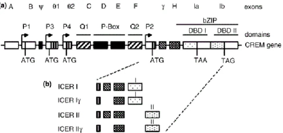
(pCREB) is a key element in the regulation of pineal gene expression, indeed pCREB improves the expression of genes that code for the enzymes responsible for MEL synthesis, which have putative cAMP- response element (CRE) sites in their promoter region such as AANAT (Baler et al., 1997; Burke et al., 1999) and HIOMT (Rodriguez et al., 1994). The cell transcription factor CREB acts in synergy or competes on CRE sites with cAMP responsive element modulator proteins (CREM). These proteins, in the pineal gland, are characterized by the fact of being strongly expressed in the form of a small transcription and, unlike the other members of this family, cMPA is able to induce its expression. The cAMP-induced transcription is strongly contrasted by inducible early repressor cAMP (ICER), (Stehle et al., 1993, 2001). ICER is a group of 4 proteins encoded by the CREM / ICER gene thanks to the presence of an internal promoter, P2, located in an intron of the CREM gene (Figure 6) (Mioduszewska et al., 2003).
Figure 6. Schematic representation of CREM / ICER gene (a) and isoform of ICER proteins (b). a): A–F, γ, H, Ia, Ib, exons; P1, P3, P4, CREM promoters; P2, ICER
promoter; TAA and TAG, indication of stop codons; DBD I and II, DNA binding domains I and II; bZIP, basic leucine zipper modified. (Mioduszewska et al., 2003)
Activation of the P2 promoter is due to activation of the transcription factor CREB (Molina et al., 1993). The ICER production is regulated by auto-negative feedback, in fact after 2-6 hours from the activation of CREB, ICER reaches a level sufficient to compete with CREB, consequently blocking its own transcription (Mioduszewska et al., 2003).
ICER acts as a strong repressor by inducing a down-regulation of the promoters of several genes that have in common a CRE sequence and their expression is induced by the intracellular increase of cAMP. The activity of the ICER consists therefore in antagonizing the pCREB that accumulates during the hours of darkness and decreases with the
increase of the ICER (which has a peak around midnight).
Therefore, the production of the key enzyme of MEL secretion, AANAT, may depend on the relationship between CREB / ICER (Stehle et al., 1993; Pfeffer et al., 1999).
Many studies have shown that nocturnal MEL secretion is managed by an increase in AANAT activity resulting from the accumulation of the same AANAT protein. However, there are interspecific differences on the accumulation and mechanism of action of AANAT. On this basis we can divide mammals into two groups: rodent species ("rat type"), in which increases AANAT gene expression and the synthesis of new AANAT molecules, and non-rodent species ("sheep type"), in which the AANAT mRNA shows high levels of day and night variation and the accumulation of AANAT proteins results from stabilization of the constantly translated protein (Figure 7). Many studies have shown that nocturnal MEL secretion is due to an increase in AANAT activity resulting from the accumulation of the same AANAT protein (Simonneaux and Ribelayga 2003).
Figure 7. Different mechanism of accumulation and action of AANAT in rodent and non-rodent species (Simonneaux and Ribelayga 2003)
In non-rodent species, the production of AANAT is regulated only at the post-transcriptional level (Simonneaux and Ribelayga 2003). The increase in cyclic AMP levels (NE-induced), is essential to inhibit the breakdown of constantly synthesized AANAT proteins by proteasomal proteolysis, leading to an elevated enzyme activity. It is clear that the different mechanisms of MEL synthesis and secretion leads to a different speed in its release from the beginning of the dark, with a long delay (several hours) in rodent species and a very short delay (few minutes) in the non-rodent species (Stehle et al., 2001).
MEL and seasonality (circannual / cyrcadian rhythms)
The seasonality of the animals was born as an adaptation to climatic conditions, which allows reproductive success and the transmission of genes to the next generation (Gerlach and Aurich, 2000). In nature the possibility of survival of the descent is mainly linked to the availability of food and water at the time of birth, so the best time to give birth is spring. In order to guarantee birth in spring, each species, based on the duration of gestation, presents different breeding seasons. This allows to divide the species in two groups: "long-day breeders", such as horses and hamsters, that cycle when days get longer (spring) and are in anestrus in fall and winter; and "short-day breeders", such as sheep, that cycle when the length of daylight shortens (fall) and are in anestrus in spring and summer (Figure 8) (Karsch et al., 1984).Figure 8. Schematic presentation of breeding seasonality in “long-day breeders” and “short-day breeders”, PP = photoperiod (Karsch et al., 1984)
It is not entirely known what is the mechanism by which MEL acts to regulate seasonal functions and how the organism is able to understand information to regulate breeding season.
The pineal gland is a neuroendocrine transducer that informs mammals about photoperiodic changes allowing them to adapt their physical state to the same annual changes, a clear example of this adaptation is the activation / inactivation of the reproductive axis as a response to photoperiodic changes. It is MEL that through the binding with the high affinity MT1 receptor, located in the suprachiasmatic nucleus (SCN), performs its chronobiotic effect. MEL is not the only endocrine output of the circadian clock, indeed other hormones, such as corticosterone, play an important role, but MEL is one of the most
stable and measurable that directly gives a fair and reliable estimate of how the clock works (Pevet et al., 2017).
Several MEL target organs have been identified, each of which is linked to specific physiological functions. The highest concentration of MEL receptors is in the pars tuberalis (PT), where MEL stimulates the secretion of prolactin (Hazlerigg et al., 2001 for review), also at the level of premammylar hypothalamus MEL controls reproductive seasonality (Malpaux et al., 1998).
However, it is not clear which aspect of the MEL secretory model (phase, duration, amplitude, or total quantity) is the most important for the control of its biological functions (Figure 9).
Duration Hypothesis: the observations of the MEL secretion model
showed that the night MEL peak duration reflects the night length (Rollag and Niswender, 1976) and that the peak duration is inversely proportional to the photoperiod (Foulkes et al., 1997). The duration changes of this peak, during the year, are the signals perceived by the reproductive neuroendocrine axis (Bittman and Karsch, 1984). This hypothesis has been demonstrated by studies conducted on different species, in which the administration of MEL during long days extends the endogenous peak of indolamine and consequently advances the reproductive activity, simulating a typical secretory rhythm of short days (Klein et al., 2002).
Coincidence hypothesis: MEL can complete its physiological effects
when its secretion coincides with a phase of sensitivity of the target organs (Simonneaux e Ribelayga, 2003).
Amplitude hypothesis: the signal perceived by the organism would be
the amplitude of the nocturnal peak of MEL.
The last theory appears the last possible, since multiple factors such as temperature, humidity and food availability can influence the amplitude of secretion, consequently modifying the metabolism of the hormone (Goridou-Boof et al., 2005).
Figure 9. Schematic presentation of different theoretical models that explain how the photoperiodic MEL signal is decoded by the target organs (Simonneaux e Ribelayga,
MEL receptors (MEL-R)
The MEL distribution in the body is very rapid thanks to its small size, lipophilic nature and an active transport mechanism (Finocchiaro and Glikin, 1998), which allows it to act independently of its receptors. The use of radio-ligand 2[125]iodomelatonin allowed the detection of high-affinity MEL receptors in different vertebrate species (Vakkuri et al., 1984).
This allowed the localization of the receptors in multiple tissues and it was thus possible to highlight a great variability both in the number and in their localization in the various species (Pévet 2003).
Several subtypes of mammalian MEL receptors have been cloned and pharmacologically characterized: one of high affinity (ML1) and one of low affinity (ML2), (Dubocovich, 1995; Reppert and Weaver, 1995b; Vanecek, 1988a).
Further three high-affinity receptors were discovered: Mel1a (later renamed MT1) and Mel 1b (later renamed MT2) are present in all vertebrates, the first mainly in the brain, the second mainly in the retina (Reppert et al.,1994; 1995a), the third called Mel 1c is present in birds (Reppert et al., 1995b). The high-affinity receptors MT1 and MT2 are members of the seven-transmembrane G-protein coupled receptor family. Whereas the low-affinity ML2 receptors (renamed to MT3 or Gpr50) use quinone reductase (QR2) as a second messenger
(Nosjean et al., 2000), and its function is not yet completely known in mammals (Dufourny et al., 2008; Hazlerigg and Loudon, 2008). The binding of MEL to the MT1 receptor causes different effects, many of which depend on cAMP inhibition, through G proteins and through increases in cytosolic calcium through Gq11 (Brydon et al., 1994). The binding of MEL to MT2 receptors in addition to determining the inhibition of cAMP also causes inhibition of cGMP (Petit et al., 1999).
The molecular structure of MT1 is composed by 350 amino acids and presents homology of 60% with the 362 amino acid MT2 (Reppert et al., 1996). Both, MT1 and MT2 receptors show the characteristic structure of the G-protein-coupled receptor (GPCR) with 7-transmembrane (TMI-TMVII) α-helices, linked by 3 alternating intracellular (ic1-ic3) and 3 extracellular (ec1-ec3) domains (Figure 10) (reviews Reppert 1997; Dubocovich et al., 2003).
Figure 10. Topology of the MT 1 receptor showing amino acids conserved in the MT 2 receptor. The gray circles indicate identical amino acids in the MT 1 and MT 2 receptors. The two glycosylation sites on the MT 1 receptor are indicated (Y) in the
N terminal (Dubocovich et al., 2010)
It was established that the presence of glycin in position 20 of TM VI, valine in position 4 of TM IV, histidine in position 7 of TM IV, serotonin in positions 8 and 12 of TM III is essential for binding with MEL (Gubitz e Reppert, 2000; Conway et al., 2000; 2001). This is because the binding of MEL with specific domains and / or amino acids defines its ability to connect, activate and modulate the receptor itself (Witt-Enderby et al., 2003). Also for MT2 any amino acid alteration in a protein can influence the interaction between the receptor and its ligand, for instance the presence of cysteine113 and cysteine190 in the second and third extracellular loop are important for
the overall structure because they are providing a disulfide linkage between these two residues (Xiao et al., 2007).
The distribution of MEL receptors is more limited in mammals and is species specific. In sheep MT1 receptors are localizzed in the hypothalamus including the area of suprachiasmatic nucleus (SCN), the site of a biological clock, adrenal glands, kidneys, B and T lymphocytes. The MT2 receptors are found at different sites in the brain, moreover in retina and small intestine. In other organs such as arteries, heart, liver, lungs and skin, both receptors are found simultaneously (Masana et al., 2002; Naji et al.,2004; Pozo et al., 1997; Richter et al., 2008; Sallinenet al., 2005; Slominski et al., 2007; Ting et al., 1999). Melatonin binding sites have been discovered in granulosa cells of human preovulatory follicles (Yie et al., 1995; Niles et al., 1999), in porcine cumulus and granulosa cells (Kang et al., 2009), in bovine oocytes and cumulus cells (El-Raey et al., 2011). Tian et al. (2017) found for the first time that the MT1 and MT2 receptors were not expressed only in sheep granulosa cells, but also in cumulus and oocyte cells.
The genes that code the MT1 and MT2 receptors are found in two different chromosomes and show similar structure. The genes structure is composed of two exons divided by a large intron, of more than 8 kb, the exon I encoding for the first cytoplasmic loop, the TM domain and the N-terminal extracellular domain, the exon II encoding for the other
parts of receptors (Reppert et al., 1996).
In sheep, the MT1 receptor has been cloned by Reppert et al. (1994) and mapped on chromosome 26 (Messer et al., 1997). Inside the MT1 gene, which encodes the corresponding receptor, two polymorphic sites (SNPs) have been identified through the use of restriction enzymes. The effect of the presence of these single nucleotide polymorphisms (SNPs) has been studied to assess the influence of this gene on the seasonality of reproductive activity (Messer et al., 1997). In fact, the studies conducted by Pelletier et al (2000) showed a correlation between the expression of reproductive seasonality and the alleles of the MT1 gene receptor (MTNR1A). This relationship was also highlighted in sheep bred in North America and this peculiarity was used by Notter et al. (2005) as a genetic marker in selection to improve reproductive activity in sheep. The candidate gene as a mediator of seasonality reproductive in sheep is therefore the MT1 and particular action is due to the structure and polymorphism of exon 2 of MTNR1A gene (Carcangiu et al., 2009; Martínez-Royo et al., 2012). This gene has been studied in different sheep breeds (Martínez-Royo et al., 2012; Meena et al., 2013; Mura et al., 2014; Luridiana et al., 2015; He et al., 2019) and specific genotypes have been associated with seasonal reproductive activity (Carcangiu et al., 2009; Saxena et al., 2015; He et al., 2019). In previous studies conducted in different sheep breeds, was found that the reproductive competence out of the normal
season is influenced by the presence of C allele at position g.17355458 and of a G allele at position g.17355452 of the MTNR1A gene exon II (according to the latest genome version Oar Ramb_v1.0). In the Rasa Aragonesa sheep breed, however only the T allele at position g.17355458 resulted in association with a greater percentage of oestrous cyclic ewes between January and August (Martínez-Royo et al., 2012). This demonstrates that the effect of these polymorphisms could be different based on several environmental and genetic factors. There is no relationship between the different genotypes found in MT2 and reproductive seasonality (Xiao et al., 2007).
MT3 has not yet been isolated in the sheep, or in other ungulates but has been found in liver, kidney, brain adipose brown tissue, skeletal muscle, lung, intestine, testis and spleen of hamster, mouse, dog and monkey (Nosejean et al., 2001). Furthermore, this receptor would also be involved in the regulation of the intraocular pressure in rabbit (Pintor et al., 2001) and in the inflammatory response in rat (Lotufo et al., 2001).
Roles of MEL
Antioxidant / oxidant effects
MEL shows antioxidant-oxidant effects, but its mechanisms of action are not yet completely known. MEL is known to be a scavenger of free
radicals with a direct effect and also increasing the efficiency of mitochondrial electron transport and promoting the activity of antioxidant enzymes (Tomás-Zapico and Coto-Montes, 2005).
Free radicals are unstable atoms that can damage cells, causing illness and aging, are linked to aging and a host of diseases. These agents include reactive oxygen species (ROS) that react and cause a molecular destruction called oxidative damage (Pierrefiche et al., 1995). ROS are responsible for the destruction of macromolecules such as proteins, DNA and lipids, leading to cell death through apoptosis (Simonneaux and Ribelayga 2003). ROS are normally formed during cellular metabolic processes, therefore the cells have developed several enzymatic and non-enzymatic antioxidant mechanisms to reduce oxidative damage. Among the enzymatic mechanisms are known: superoxide dismutase (SOD), catalase (CAT) and glutathione peroxidase (GPx). The non-enzymatic mechanisms including: vitamin E (a-tocopherol), vitamin C (ascorbate), glutathione (GSH), β-carotene and MEL. These act directly, remove ROS due to their ability to donate electrons, thus neutralizing the potential toxicity of ROS (Tomás-Zapico and Coto-Montes, 2005).
Anticancer effects
processes in normal cells and promotes pro-apoptotic signals in cancer cells (Bizzarri et al., 2013). MEL plays an important role as a non-toxic, apoptotic, oncostatic, angiogenic, differentiating and antiproliferative factor in solid and liquid tumors (Di Bella et al., 2013). It is clear that MEL cannot be used alone in tumor therapy, but it can help to amplify the cytostatic and cytotoxic effects of other conventional drugs (Bizzarri et al., 2013).
Immunomodulatoy effects
In the 1926 Berman was the first to indicate the possible link between the pineal glands and the immune system. Subsequently, several studies have shown that different cells and tissues of the immune system (like thymus and spleen, B cells) containing or / and produce MEL, express AANAT and HIOMT mRNAs (Carrillo-Vico et al., 2005). A bidirectional link between the immune system and the pineal gland is demonstrated by the fact that cytokine signaling affecting pinealocytes has resulted in a transient inhibition of melatonin synthesis and shifting MEL production to circulating macrophages (Da Silveira Cruz-Machado et al., 2010, Fernandes et al., 2006, Markus et al., 2013, Markus and Ferreira, 2011). These systems also share a common language through messengers synthesized in both systems, such as acetylcholine, adrenocorticotropic hormone (ACTH),
endorphin, vasoactive intestinal peptide (VIP), somatostatin and growth hormones (Blalock et al., 2007). The role of MEL as coordinator and modulator of the immune system is demonstrated also by a seasonal variation of immune functions in many mammals (Nelson, 2004). In fact, MEL monitors diurnal and seasonal rhythms of leukocyte proliferation (Drazen et al., 2001), cytokine production (Scheff et al., 2010) and naturals killer (NK) cell activity in mammalian bone marrow cells (Matsumoto et al., 2001).
MEL plays a dual role as an immunostimulant and an immunosuppressant, depending on the different situations. It acts as an immunostimulant in basal or immunosuppressive conditions, bringing a pre-activated state for a more effective early immune response. Contrary, it acts as an anti-inflammatory molecule (affecting the activity of lipoxygenase), in conditions of chronic or exacerbated immune response, such as septic shock (Figure 11) (Carrillo-Vico et al., 2013).
MEL acts in the immune system with an autocrine, endocrine and paracrine mechanism. It has effect on the modulation of pro-inflammatory enzymes, controls the production of pro-inflammatory mediators (cytokines and leukotrienes) and participates in the regulation of leukocyte lifetime by interfering with apoptotic processes (Herman et al., 2015).
Figure 11. Different pathways of action of MEL in the immune system (Carrillo-Vico et al., 2005)
A bidirectional pathway between the pineal gland and the immune system is known but the effect of inflammatory feedback on the functions of the gland is not yet completely known. Immune mediators such as cytokines, prostaglandins and histamine have easy access to the brain and the pineal gland, in fact they enter these during the immune response phase (Fabris, 1994). It has also been shown that antigenic stimulation (Markowska et al., 2005), histamine (Zawilska et al., 1997), cytokines (Achumnarnkul et al., 1990; Mucha et al., 1994) and prostaglandins (Voisin et al., 1993) could influence the secretory activity of the pineal gland. This influence has also been demonstrated
in sheep where treatment with IL-1β has been shown to suppress NE stimulated melatonin secretion (Herman et al., 2015).
A study shows that in goats MEL implants have improved the quality of raw milk by reducing somatic cells count (SCC) via decreasing udder oxidative stress (Yang, 2017). Also in cows, the subcutaneous injection of melatonin greatly reduces SCC in animals with subclinical mastitis, thanks to its antioxidant, anti-inflammatory and immuno-potentiation properties. In cows with mastitis, MEL works by reducing cortisol levels and increasing levels of IgG, IgM, lymphocytes and neutrophils (Yang, 2017).
Reports showed the high nightly levels of MEL in cows with low-milk SCC (Asher et al., 2015).
Reproductive effects
In temperate climates, most sheep breeds have a seasonal breeding model with a breeding season in the fall. Seasonality is stronger in primitive breeds (Hafez, 1952).
The role of MEL as a regulator of reproductive physiology is demonstrated by the synthesis and presence of this indolamine in different sites of ovaries and testes. Also in the reproductive tract MEL acts by means of the endocine, paracrine and autocrine mechanisms (Acuña-Castroviejo et al., 2014), but it is not yet fully known how the
hormone works. It is clear that MEL guarantees the quality of the oocyte and the sperm (Acuña-Castroviejo et al., 2014), moreover it was show that it is inhibiting the normal aging processes of the reproductive system in rat (Wolden-Hanson et al., 2000). Even in sheep, MEL plays its anti-aging role, indeed it can improve the functionality of the reproductive system in older animals (Forcada et al., 2007).
In the reproductive tract MEL acts through the hypothalamic neuronal systems to regulate the frequency of the GnRH pulse, this mechanism is necessary to raise the frequency secretion of LH, finally to activate the breeding period (Figure 12) (Malpaux et al., 1997).
Figure 12. Melatonin action to regulate reproductive activity (adapted from Hormone Manipulation, ansci.wisc.edu)
A study showed that only a MEL micro-implant placed in the mediobasal hypothalamus (MBH) was able to stimulate the secretion of LH (Malpaux et al., 1993). Although the higher concentration of MEL bindings is expressed in the pars tuberalis (PT) of the adenohypophysis, the placement of MEL micro-implant in this area does not modify the secretion of LH (Malpaux et al., 1995). Whereas, the micro-implants located in the MBH or in the third ventricle are able to stimulate the release of LH (Malpaux et al., 1993; 1994; 1995). Previous studies have shown that PT participates in transductions of MEL effects on seasonal change in prolactin secretion. This is demonstrated by the fact that the placement of microimplants in the PT and MBH lead to the same suppression of prolactin levels (Malpaux et al., 1995). Therefore, MEL acts directly on the pituitary gland, it may be in the PT, to control prolactin secretion (Lincoln et al., 1994). In contrast, MEL does not act directly on GnRH neurons to control the release of the hormone, in fact its action could be mediated by a complex system of interneurons. In this system the dopaminergic terminals of the median eminence play a key role (Malpaux et al., 1995). To act its strong negative feedback on GnRH secretion, in prepubescent animals and during anoestrus, estradiol (E) uses dopamine as a transducer, more precisely, the hypothalamic nucleus A15 mediates the inhibitory effects of E (Thiéry et al., 1989). Furthermore, causes an increase in tyrosine hydroxylase (TH), a
limiting step enzyme of catecholamine synthesis, (Gayrard et al., 1994).
Exposure to short days causes a decrease in dopaminergic activity in the median eminence due to both the reduction in the amount of dopamine (Thiéry et al., 1991) and the activity of the TH (Viguié et al., 1996). Similarly, MEL implants are able to increase LH secretion, reducing TH activity in the median eminence (Viguié et al., 1997). The inhibitory action of TH is evidenced by the fact that this enzyme has a higher concentration, in adult animals, during the anaestrus period (Viguié et al., 1997). Furthermore, the administration of a TH antagonist during long-days causes a pulsatile increase of LH and consequently the reactivation of reproductive activity (Tillet et al., 1997). It is therefore possible that the inhibition exercised by this enzyme is at the basis of the period preceding puberty.
It remains to be clarified what are the factors that actually trigger the onset of puberty, and in particular the concrete role of MEL. This could be essential to reduce the non-productive periods in farms. Over the years, numerous studies have been carried out using both the artificial photoperiod and exogenous MEL to improve the reproductive activity in sheep (Kennaway et al., 1984; Poulton et al., 1987) and goat (Tamanini et al., 1985; Prandi et al., 1987; Deveson et al., 1992b). To understand if and how melatonin was implicated in the mechanism of
induction of puberty in the female, experiments that included pinealectomy in ewe lambs subjected to natural photoperiod were conducted (Seamark et al., 1981). Surprisingly, these studies, conducted on Merino sheep breed, showed no abnormal seasonality despite the absence of epiphyses. This result was attributed to the possible insensitivity to the photoperiod of the Merinos, sheep with a relatively long annual fertile season and a practically indistinct anoestral period. A subsequent study, however, conducted on crossbreed of Merino X Dorset showed a considerable delay in reaching puberty following pinealectomy (Kennaway et al., 1985). In light of this, it was assumed that breed differences existed in the modulation of the photoperiod and that exogenous melatonin could, in receptive breeds, regulate the beginning of reproductive activity. The first experimental research included the administration of 2-3 mg of MEL in sheep in anoestrus, showed that was able to induce the anticipation of the reproductive season (Kennaway et al., 1982; Arendt et al., 1983). It was discovered that in order to obtain these results, the administration had to be continuous and of a duration longer than 45-50 days, since administration on alternate days seemed to have no effect (O'Callaghan et al., 1991). However, given the low practicality of daily administration, slow-release ruminal boluses and implants have been studied that are able to maintain high blood concentrations of MEL for long periods (Arendt et al., 1988; Poulton et al., 1988).
The commercial use of MEL implants in sheep is currently authorized in several European countries, such as Italy, the United Kingdom, France, Greece, Portugal and Spain. At the Mediterranean latitude, subcutaneous implants are the most used to anticipate the sexual season of the sheep. Normally, MEL treatments are accompanied by the union of sheep with males, since this triggers the "male effect" that causes the increase in LH tonic secretion and therefore the change in the state of the ovary (Martin et al., 1986; Chanvallon et al., 2011). Although chronic exposure to implants causes some damaging effects on reproductive parameters in adult Blackface (Jordan et al., 1990), their medium-term influence, associated with the male effect, in Mediterranean breeds remains to be clarified. In sheep, MEL implants can be used to control reproductive activity even if there is no uniformity in the results obtained in the various breeds (Haresign et al., 1990; Carcangiu et al., 1993, Abecia et al., 2011a). In goats, the experiences with MEL are much lower and the results obtained have not always been satisfactory (Prandi et al., 1987; Deveson et al., 1992; Abecia et al., 2011b).
In ewe lambs the administration of MEL has not shown consistent effects on the onset of reproductive activity (Kennaway et al., 1984; Foster and Olster, 1985; Sawalha et al., Mura et al., 2010; Abecia et al. 2016; Luridiana et al 2016).
sheep, had the aim to evaluate the involvement of MEL on the speed of ovarian response to the male effect (Abecia et al., 2006) or on the viability and shelf life of embryos in artificial insemination practices (Forcada et al., 2006). In both studies, MEL showed a clear influence in the improvement of the aforementioned activities.
Aim
Melatonin is a hormone that has several functions in the body, and all are of considerable importance. In fact, in addition to regulating the circadian rhythm by synchronizing the physiological activities within 24 hours, influences the reproductive seasonality and the immune response. Most of these actions are performed through specific receptors present at numerous sites in the animal body.
Concerning to the control of reproductive seasonality, although much is known about the effects of melatonin, it remains to be explained whether some physiological states of the animal may influence the advance of reproductive activity in sheep. Sometimes, melatonin treatment has reproductive responses that are not as expected, and this is often due to the fact that animals are not suitable for reproduction at that time. Therefore, understanding which factors can cause this response is very important for optimization of reproduction in sheep. In order to obtain a maximum reproductive efficiency in sheep, it is also possible to intervene by improving the male effect. In fact, managing this method in a rational way could lead to an improvement in the reproductive efficiency of sheep with a greater income for sheep farms.
receptor gene (MTNR1A) can influence reproductive resumption in sheep could provide important data that could be used in genetic selection of sheep.
Therefore, the proposal of this thesis was to evaluate the different actions of melatonin in five different objectives that can be formulated as follows:
1) to evaluate the effect of melatonin implanted a) in different periods of the year,
b) in different lactation periods;
2) to evaluate the influence of melatonin treatment in ewes and male replacement on reproductive activity;
3) to evaluate the effect of melatonin treatment in male on the onset of puberty in Sarda ewe lambs;
4) to evaluate the influence of the different MTNR1A genotypes on reproductive activity;
5) to evaluate the influence of the melatonin treatment on milk production and mammary immune system.
Material and Methods
Aim 1a (MEL treatment in different periods of the year)
Animal and experimental design
This study was conducted on 3200 Sarda sheep breed from 16 farms, located in the same area of the Northern Sardinia (40°N). The animals were reared in the same conditions on all the farms. During the day the animals grazed on leguminous and gramineous grasses and during milking they also received 300 g of commercial concentrate feed per animal every day (20.4% of raw protein and 12.5 MJ ME / kg of DM). During the night the sheep were penned and received hay (11.1% raw protein and 7.2 ME / kg DM) and water ad libitum.
For the research, 200 clinically healthy sheep were chosen from each farm, for a total of 3200 sheep. The females included in the study were lactating, 3-6 years old, with body condition score (BCS) between 2.5 and 4.0 and with a single lamb born between 1st November and 10th
December.
Treatment and registration data
On each farm, the selected animals were randomly divided into 2 groups (M and C), each containing 100 sheep. During the study the controls and the treated ewes were kept together all the time. The sheep of group M received a melatonin implant (18 mg) (Melovine,
CEVA Animal Health, Agrate Brianza, Milan) in the left retro-auricular area; group C served as a control and was not treated. In the first 4 randomly selected farms, the melatonin implants were implanted on 10th of February, in the second 4 farms on 10th of March, in the
third 4 on 10th of April and in the last 4 farms on 10th of May. 35 days
after the treatment of the ewes, adult rams (male/female ratio 1/20) were introduced into the groups for 45 days in all the studied farms. One week before the sheep, the rams were treated with 3 MEL implants in the left retroauricolar area. Sheep and rams were kept separate for three months before the rams were introduced. Starting from 45 days up to 90 days from the introduction of rams, gestation was diagnosed by transdominal ultrasound examination through the Tringa Esaote equipment (Esaote Europe BV, Maastricht, Netherlands) equipped with a 5 multi-frequency linear probe, 5 - 7.5 MHz. The date of the lambing and the number of newborn lambs of each ewe were recorded from 150 days to 190 days after the introduction of the rams.
Statistical Analysis
All statistical analysis was executed using the computing environment R (Version 3.6.1. R Core Team 2019). A logit-link Hierarchical Linear Model (HLM) suitable for binomial (lambing / not lambing) data was used to analyse the fertility rate of different treatment time. Variables considered were treatment and treatment time. To analyse the effect of treatment time on period in days from male introduction to lambing a
HLM procedure according to the following model was performed.
Yijk =μ+Tm(Pe)ij +εijk
Where Yijk is the period in days from male introduction to lambing, μ
is the overall mean, Tm is the fixed effect of treatment, Pe is the nested
time effect within treatment, and εijk is the error effect. The same model
was used to analyse the litter size. A P value < 0.05 was considered statistically significant. Multiple comparisons of the means were performed using Tukey's method.
Aim 1b (MEL treatment in different lactation periods)
Animal and experimental design
For this study two farms located in Northern Sardinia (between 39 and 40°N) were chosen. The animals were raised under the same conditions on the two farms and kept under natural photoperiod since birth. Food management included grazing leguminous and gramineous grasses during the day and the daily administration of 300 g per sheep of commercial concentrate feed (20.4% raw protein and 12.5 MJ ME/kg DM) during milking. In each farm (identified as F1 and F2) 120 lactating ewes were chosen and then divided into four groups: group 1 with 30 ewes lambed between 20th of October and 20th of
November; group 2 with 30 ewes lambed between 1st and 30th of
January and group 4 with 30 ewes lambed between 1st and 28th of
February. All animals included in the study were between 3 and 6 years old, with a body condition score (BCS) between 3.0 and 4.0 and were kept separated from the rest of the flock
Treatment and registration data
Each group of 30 ewes was divided into two subgroups (M and C) of 15 ewes each. On the 1st of April all the ewes of subgroups M
received a melatonin implant (18 mg) (Melovine, CEVA Animal Health, Agrate Brianza, Milan) in the left retro-auricular area; groups C served as a control and were not treated. In each group (M and C), 35 days after the treatment of the ewes, two adult rams of proven fertility were introduced and were kept for 70 days. Every week, from 45 days after the introduction of rams up to 45 days after the removal of rams, pregnancy diagnosing was performed. Pregnancy diagnosis was performed by transabdominal ultrasound examination through the Tringa Esaote equipment (Esaote Europe BV, Maastricht, Netherlands) equipped with 5-7.5 MHz multi-frequency linear probe. The date of the lambing and the number of newborn lambs of each ewe were recorded until 220 days after the introduction of the rams.
Statistical Analysis
R statistical software, (Version 3.6.1. R Core Team 2019) was used to perform the statistical analysis. A General Linear Model (GLM)
procedure was carry out to analyze the effect of lambing period and treatment on the litter size and on the distance in days from male introduction to lambing. To confront percentages of lambed ewes within each lambing period chi-square test was used. A P value <0.05 was considered statistically significant.
Aim 2 (MEL treatment and male replacement)
Animal and experimental design
For this study a farm located in the northern Sardinia (approximately 40°N) was chosen. All the animals raised on the farm (about 1000) have been subjected to natural photoperiod since birth. During the day the animals grazed on legumes and gramineous grasses and during milking they also received 300 g of commercial concentrate feed per animal every day (20.4% of raw protein and 12.5 MJ ME / kg of DM). During the night sheep were penned and received hay (11.1% raw protein and 7.2 ME / kg DM) and water ad libitum. The chosen sheep were between 3 and 5 years old, had a BCS between 2.5 and 4.0 and had lambed between 20th October and 1st December. In addition, the
chosen ewes were at least at third lambing. The first and second lambing ewes were excluded from the study because in the Sarda sheep breed the first lambing generally occurs between January and April, followed by a large milk production. This leads to a poor
reproductive function in the following two months and a delay in the lambing date in their second year (Carcangiu et al., 2012).
Treatment and registration data
The 400 animals chosen were divided into 4 groups with 100 sheep each. In group M the sheep were treated with one MEL implant and males were not replaced; in the MR group the sheep were treated with MEL implant and a weekly replacement of the males was performed. Group C served as a control and were not treated and males were not replaced; in the CR group the sheep was not treated but a weekly replacement of the males were performed. From the formation of the groups and during the study period, the different groups were kept separately from each other. On 20th of March the ewes of groups M
and MR received a melatonin implant (18 mg) (Melovine, CEVA Animal Health, Agrate Brianza, Milan) in the left retro-auricular area. On 24th of April 5 adult rams with proven sexual experience were
introduced into each group to induce male effect and remained in cohabitation with ewes for 40 days. Males were not treated with MEL implant. To stimulate the ram effect, the sheep were previously isolated from the males for at least 6 months, the rams were kept at a distance of 10 km, so that the auditory and olfactory stimuli coming from them could not reach the ewes. At the ram introduction every male has been provided with harnesses to paint the back of ewes for recording of mating, so detecting estrous ewes. Every day the number
of sheeps marked by rams were recorded and the color of the marking were changed weekly, Furthermore, the sexual behavior of rams was evaluated for the first five days after their introduction into the groups to assess whether rams were able to perform sexual activity. Recoded were anogenital sniffing, nudging and mounting attempts of the ewes. Starting from 45 days up to 90 days from the introduction of rams, gestation was diagnosed by transdominal ultrasound examination through the Tringa Esaote equipment (Esaote Europe BV, Maastricht, Netherlands) equipped with a 5.0-7.5 MHz multi-frequency linear probe. The date of the lambing and the number of newborn lambs of each ewe were recorded from 21st of September to 1st of November.
On the basis of the recorded data, for each group it was established: the fertility rate (as percentage of lambed ewes), the distance in days from male introduction to lambing (DIML), and the litter size (number of newborn lambs per ewe).
Statistical Analysis
The analysis of collated data was performed using the statistical software R (Version 3.6.1. R Core Team 2019). A P value < 0.05 was considered statistically significant. The fertility rate was scan using the Fisher-Freeman-Halton exact probability test for multiple group comparisons. The distance in days from male introduction to lambing (DIML) and litter size, expressed as mean values ± standard deviation (SD), were normally distributed (P > 0.05) (Shapiro–Wilk normality
test). Analysis of Variance was performed to analyze the effects of melatonin treatment (T) and male replacement (M) on reproductive activity (DIML or litter size). The BCS and age of animals did not show statistical significance and thus, were not included in the model. The following linear model was used for all dependent variables:
Yjkm = μ+ Tj + Mk + TjMk + ejkm
where Y was the variable measured (DIML or fertility or litter size), μ was the overall mean, Tj was the effect of the melatonin treatment, Mk was the effect of the male replacement, TjMk was the interplay of melatonin treatment (M) and male replacement (R) and ejkm was the error effect.
Multiple comparisons of the mean average were carryed out using Tukey's method. To compare the number of ewes lambing through all the groups a chi-square (χ2) test was used.
Aim 3 (influence of MEL treatment in male)
Animal and experimental design
For this research a farm located in the north of Sardinia was chosen. In this farm about 1.000 animals were reared, from which 200 ewe lambs were chosen. The enrolled ewe lambs were born in November and had a body weight of about 30 kg. Furthermore, on the same farm, 28
males of proven fertility were chosen, with an average age of 4.5 years. The animals were kept in the same conditions and under natural photoperiod since birth.
Treatment and registration data
On 25th May, 14 rams were treated with 3 melatonin implants each. On
20th June, the chosen ewe lambs were divided into four groups M, MS,
C, CS, each of 25 animals. On 1st of July two rams were introduced
into each ewe lamb group. In the M and MS groups treated males were introduced, while in the C and CS groups the untreated males were introduced. In addition, a weekly male replacement was performed in the MS and CS groups. The males were removed 40 days later. From 150 to 190 days after the introduction of the males, the date of lambing and the number of newborn lambs were recorded.
Statistical Analysis
The analysis was carried out by statistical software R (Version 3.6.1. R Core Team 2019). A P value < 0.05 was considered statistically significant. The fertility rate was examined using the Fisher-Freeman-Halton exact probability test for multiple group comparisons. The distance in days from male introduction to lambing (DIML) and litter size, expressed as mean values ± standard deviation (SD), were normally distributed (P > 0.05) (Shapiro–Wilk normality test). Analysis of Variance was performed to analyze the effects of
melatonin treatment (T) and male replacement (M) on reproductive activity (DIML or litter size). The following linear model was used for all dependent variables:
Yjkm = μ+ Tj + Mk + TjMk + ejkm
where Y was the variable measured (DIML or fertility or litter size), μ was the overall mean, Tj was the effect of the melatonin treatment, Mk was the effect of the male replacement, TjMk was the interaction of melatonin treatment (M) and male replacement (R) and ejkm was the error effect. Multiple comparisons of means were carryed out using Tukey's method. To compare the number of ewes lambing across all the groups a chi-square (χ2) test were used.
Aim 4 (influence of different MTNR1A genotypes)
Animal and experimental design
For this study 8 farms were selected, all located in the north of Sardinia (on the 40°N). Animals were reared with similar nutritional and management regimes and kept under natural photoperiod since birth in all the farms. Animals grazed on leguminous and gramineous grasses during the day and during milking they also received 300 g of commercial concentrate feed per animal every day (20.4% of raw protein and 12.5 MJ ME / kg of DM). In each farm (identified F1-F8) about 400 ewes were genotyped in position g.17355452 G > A of exon
II of the MTNR1A gene. In each farm, 150 sheep were selected (50 for each genotype A / A, A / G, G / G). All animals included in the study were between 3 and 5 years old and, were multiparous (at their third parturition) and lambed between 20th of October and 1st of December.
The chosen animals were kept away from the rest of the flock. In each group of 150 sheep, eight adult rams (3 to 6 years old) of proven fertility (male/female ratio 1/20) were introduced to the sheep at different times (see table 1). After 70 days the rams were removed.
Table 1: Time of ram introduction to ewes on eight farms
Time Period Data of male introduction Farms
T1 25 March F1 25 March F2 T2 15 April F3 15 April F4 T3 5 May F5 5 May F6 T4 1 June F7 1 June F8
Before the introduction of rams, sheep and rams were kept separate for 90 days. Every week, from 45 days after male joining with ewes up to 45 days after the removal of rams, gestation diagnosing was done. Pregnancy diagnosis was performed by transabdominal ultrasound examination through the Tringa Esaote equipment (Esaote Europe BV,