ARTICLE NO. 0484
Construction of a YAC Contig Covering Human Chromosome 6p22
P
ATRIZIAM
ALASPINA, A
NTONELLAR
OETTO,* F
LAVIAT
RETTEL, C
ARLAJ
ODICE, P
AOLAB
LASI,
M
ARINAF
RONTALI,† M
ASSIMOC
ARELLA,‡ B
RUNELLAF
RANCO,§
C
LARAC
AMASCHELLA,*
,Ø ANDA
NDREAN
OVELLETTO1Dipartimento di Biologia, Universita` di Roma Tor Vergata, 00133 Rome, Italy; *Dipartimento di Scienze Biomediche ed Oncologia Umana, Ospedale San Luigi, Orbassano, 10100 Turin, Italy; †Istituto di Medicina Sperimentale, C.N.R., 00137 Rome, Italy;
‡IRCCS-Ospedale CSS, 71013 San Giovanni Rotondo, Italy; §TIGEM, San Raffaele Biomedical Science Park, 20132 Milan, Italy; andØ
C.I.O.S., C.N.R., 10100 Turin, Italy Received March 21, 1996; accepted June 19, 1996
et al. (1995) it is still represented by two separate paths A contig covering human chromosome 6p22 that con- of levels 3 and 1, respectively. In the STS-based map
sists of 134 YAC clones aligned based on the presence/ of the human genome (Hudson et al., 1995) two ‘‘doubly
absence of 52 DNA markers is presented. This contig linked’’ contigs overlap this region.
overlaps with the 6p23 contig at its telomeric end and Several genes have been assigned to 6p22. By using with the 6p21.3 contig at its centromeric end. The or- panels of somatic cell hybrids, Albig et al. (1993) as-der of loci within the contig resolves the relative posi- signed to this band the cluster of H1 histone genes; tions of several genetically mapped markers. Among Abbott and Povey (1991) assigned the prolactin gene; the additional markers used here, there are eight Durkin et al. (1994) assigned brain-specific ESTs by novel PCR assays. The 12 known genes and anonymous PCR; and Pappas et al. (1995) found six ESTs mapping ESTs located within the contig establish a first step
to 6p21.3 – p23, one of which was identified as a zinc
toward a transcriptional map of this region. The
insta-finger protein. A number of disease causing genes or
bility of YAC clones observed during this work is also
cDNAs linked to the MHC are reported in OMIM with
discussed. q 1996 Academic Press, Inc.
a possible location overlapping 6p22. These include the methionine-tRNA-i gene 1 (TRMI1), the transcription factor ID4, the COL11A2 gene involved in Stickler
syn-INTRODUCTION
drome and otospondylomegaephyseal dysplasia, the branched-chain alpha-keto acid dehydrogenase E1-The cytogenetic band 6p22 is a Giemsa positive band
beta gene (BCKDHB), and one of the processed pseu-(GTG/; Holmquist, 1992) spanning approximately from
dogenes of the dihydrofolate reductase (DHFRP2). The genetic positions 34 to 46, or 5 – 6% of the entire genetic
location for the Sry-related gene SOX4 (Farr et al., length of chromosome 6 (Gyapay et al., 1994).
Cytoge-1993) has been shown to overlap 6p22 by somatic cell netically it has been measured to span 21% of the
hybrid analysis. Recently, the gene for hereditary idio-length of the chromosome 6 short arm (Francke, 1994).
pathic hemochromatosis (HFE) has been shown to be This band is delimited centromerically by 6p21, a very
closely linked to markers in 6p22 (Raha-Chowdhury et gene-rich region containing the MHC, and
telomeri-cally by 6p23. Both regions have been subjected to ex- al., 1995). Moreover, evidence from studies in
indepen-tensive physical analysis (Campbell et al., 1993; Ne- dent family sets has accumulated (reviewed by Pelto-mani et al., 1994; Olavesen et al., 1995; Burt et al., nen, 1995) in favor of a major locus predisposing to 1996; Totaro et al., 1996). The intensive effort to gener- schizophrenia in a region overlapping 6p22. Finally, ate new genetic markers (Gyapay, 1994) has identified Foulkes et al. (1993), based on the finding of loss of several polymorphic and nonpolymorphic microsatel- heterozygosity, suggested the presence of a tumor sup-lites that have been later anchored to 6p22 by FISH. presor gene relevant to ovarian carcinoma whose puta-However, in the first-generation physical map of the tive position may overlap 6p22.
human genome (Cohen et al., 1993) this band was To provide complete physical coverage of 6p22, we poorly covered, and in the recent report by Chumakov constructed a single YAC contig spanning from D6S306 centromerically to D6S109 telomerically, juxtaposing with the contiguous resource of YACs extended up to
P.M. and A.R. contributed equally to this study and should be
re-garded as joint first authors. B.F., C.C., and A.N. are senior authors. 6p24 (Olavesen et al., 1995). In this work, the content
1To whom correspondence should be addressed at Dipartimento
of the same set of YAC clones was checked for an
inte-di Biologia, Universita` inte-di Roma Tor Vergata, Via della Ricerca
Scien-grated assemblage of polymorphic genetic markers,
tifica, 00133 Rome, Italy. Telephone: 72594321. Fax:
/39-6-2023500. E-mail: [email protected]. anonymous STSs and ESTs, known genes, and
anony-399
0888-7543/96 $18.00 Copyrightq 1996 by Academic Press, Inc. All rights of reproduction in any form reserved.
in a final volume of 20ml, and the same PCR thermal profile with
mous subcloned PCR products. Both PCR screening
the exception of the annealing conditions and number of cycles. The
and hybridization probes were used to determine the
standard conditions were 4 min at 947C, then the appropriate
num-relationship between the YAC clones. ber of cycles of 45 s at 947C, annealing, and 45 s at 727C, followed
by a final extension of 5 min at 727C. In all experiments, distilled water and yeast strain AB1380 DNA were used as negative controls,
MATERIALS AND METHODS and genomic DNA was used as a positive control. The markers taken
from the literature and the corresponding PCR annealing and cycling conditions are listed in Table 2.
Isolation and preparation of YAC DNA. YAC clones from the CEPH (Albertsen et al., 1990; Chumakov et al., 1995) and ICI (Anand et al., 1991) libraries were obtained from the CEPH (Paris), ICRF
RESULTS
(London), and DIBIT-HSR (Milan) resource centers. The original cul-tures and culcul-tures obtained from single colonies were grown in AHC
medium and resuspended in 80ml LMP agarose plugs containing 7 Initial Assembly of Contigs 1 107 cells. Plugs were treated according to standard techniques.
Two microliters of molten plug was used in PCR assays, whereas An initial set of 154 YACs identified from the first an entire plug was used for restriction enzyme digestion, agarose release of Ge´ne´thon data (Cohen et al., 1993) to reside electrophoresis, and Southern transfer. Probe labeling and
hybrid-potentially in 6p22 was assembled. Clonal cultures
ization were performed according to standard techniques. Filter
were grown and DNA prepared to retest YACs
individ-washing was performed at high stringency.
ually with the markers used to construct the genetic
Development of new markers. We generated new STSs and probes
map, D6S422, D6S299, D6S461, D6S464, D6S306,
by using three different methods: (1) Alu-PCR products were
gener-ated from selected YACs. PCR with 250 ng of one of the primers D6S258, D6S276, D6S265, and D6S273. Among the described by Nelson et al. (1989) was carried out for 35 cycles with YACs detected by tridimensional screening at Ge´ne´-1.5 mM MgCl2and an annealing temperature of 657C. After agarose
thon to contain these microsatellite markers, 24% were
electrophoresis, bands distinctive for recombinant yeast were eluted
not confirmed by individual testing . On the other hand,
and reamplified under the same conditions to reduce contamination
by nonspecific products. Some of these bands were subcloned we found 33 YACs positive with at least one of these
(BTVC1.5, BTVD1.4, BTVG2.9), while others were eluted from 1% markers and not reported as positive in Ge´ne´thon files. LMP agarose gels and used as hybridization probes to filters con- We then added individual analyses for markers taining either Alu-PCR products or digested DNA of individual YACs
D6S109, SOX4, D6S506, D6S507, D6S242, D6S105,
in the region. Hybridization was carried out as described by
Wapen-H1.5, and TRMI1.
aar et al. (1994) in the presence of total human DNA to suppress
signals from repetitive sequences (Sealey et al., 1985). (2) PCR prod- The 76 YACs positive for at least one of the markers
ucts were generated with a pool of primers designed on splicing donor listed above could be arranged in four separate contigs: and acceptor site consensus sequences (Pritchard et al., 1994;
Sena-the first one contained D6S109, D6S422, SOX4,
pathy et al., 1990) (BTV9F, SSADH). PCR was performed with 25
D6S507, and D6S506; the second one contained
pmol each of 3 primers with 1.5 mM MgCl2for 35 cycles with an
D6S276, D6S461, D6S299, and D6S242; the third one
annealing temperature of 457C. After agarose electrophoresis, bands
not observed in wildtype yeast were eluted and reamplified under contained D6S464, D6S306, H1.5, and D6S105; and the the same conditions. (3) The third method relied on the observation fourth one contained D6S273, D6S258, and D6S265. that nonspecific PCR products were generated by some markers on
These results showed that the STS content of the YACs
certain sets of YACs. In some cases the presence/absence pattern of
could provide a finer resolution than the genetic map.
such bands was consistent with a location of the corresponding target
In fact, Chumakov et al. (1995) reported that the loci
sequences within the contig. The bands were then excised, subcloned,
and sequenced, and specific primers were developed (BTVOL506). D6S464, D6S306, D6S258, D6S276, and D6S265 repre-Products obtained with the above methods were subcloned in sent the single position 0.46 together with the HLA pUC18 as blunt-ended inserts (Sure Clone kit, Pharmacia) and
se-cluster. On the other hand, our analysis grouped
quenced with universal primers with the dideoxy chain termination
D6S276 in a cluster mapped 4 cM telomerically and
method by Taq polymerase (AmpliCycle kit, Perkin – Elmer). The
sequences were then checked for their repetitive content by compari- placed D6S258 and D6S265 in the proximity of
son with human repetitive elements (Jurka et al., 1992). Primers D6S273. When our pool of YACs was assayed by hy-were designed to obtain PCR products in the 100- to 500-bp range bridization with a probe specific for the telomeric MHC from the nonrepetitive regions. The genomic origin of the STSs was
gene HLA-F (Amadou et al., 1995), only YACs of the
checked on a somatic cell hybrid retaining the sole human
chromo-fourth contig produced a positive signal. This placed
some 6 on hamster background (GM10629A; NIGMS Human Genetic
Mutant Cell Repository). the latter subgroup of YACs centromeric to the others.
A specific assay for human butyrophilin (BT) was developed: the This region was not considered for further analysis. PCR product obtained using bovine primers (Amadou et al., 1995)
on human genomic DNA was directly sequenced to design
human-Filling Gaps
specific primers. PCR conditions of the above-mentioned markers are reported in Table 1.
To fill the gaps between the contigs and to extend
Alu-vector probes were generated from individual YACs by using
the coverage to the 6p22 – p23 boundary (Olavesen et
Alu and vector primers as previously described (Nelson et al., 1989;
Zoghbi and Chinault, 1995). The bands were eluted from agarose al., 1995), we used different methods to generate addi-gels and used as hybridization probes to digested DNA of individual tional STS markers in the form of PCR assays. This YACs using the protocol for suppression of signals from repetitive
search was directed toward single-copy loci to avoid
sequences (Sealey et al., 1985).
the problems associated with contig assembly in the
Probes, PCR primers, and conditions. All STS markers used the
presence of repeated or highly homologous loci (Foote
buffer recommended by the manufacturer (Promega), 0.2 mM
TABLE 1
New Markers Developed
Name Annealing
(original YAC) Size (bp) Primers conditions MgCl2(mM) Cycles
BTVC1.5 105 CAGATGCTATGCTTCAC 527C, 30 s 1.5 30 (738 b 5) CCAACAGATGGGTTTAG BTVG2.9 160 ACTCAGTATAATTCCATAG 527C, 30 s 1.5 30 (927 e 10) AACTGTATTGCTTGTACAC BTVD1.4 450 CTTTAATTCAAGAAGAGAAC 497C, 30 s 1.5 30 (934 d 11) GTGAAGCATAGGTGATTG BTV9F 264 CCTGATTTAAAGCACATGGC 537C, 30 s 1.5 35 (830 b 5) GCATTTACTGTCTTCTTGCC BTVOL506 230 CCAACATGCAGACACACTAC 557C, 30 s 1.5 35 GACTATAAGGGGGAAAGGC SSADH 203 GTGTCTTCGGCAGGCTTC 557C, 30 s 1.5 35 (775 g 10) GGTTTGTCAATCAGTTGTGC L34820 94 TATGTGTGTTACGGGGGC 547C, 30 s 1.5 35 AATAATGGATGGCATGTACC BT 198 CAGAGAGATTTGACTCCTGG 657C, 45 s 2.5 30 CAGTACCCATTTCCATACAAC
YAC fragments: preliminary amplification of inter-Alu The description of the butyrophilin locus (Amadou et DNA or amplification with a pool of primers potentially al., 1995; Vernet et al., 1993) in a region of the mouse
specific for coding sequences (see Materials and Meth- genome showing homology with the human 6p22 led ods). Of 67 bands identified in YAC-bearing but not us to develop a specific PCR assay for the human BT. wildtype yeast, 29 (43%) were successfully picked and These novel markers were used to rescreen the Mega-reamplified in a second PCR run under the same condi- CEPH and ICI libraries. Additional YACs were also tions. The 16 products cloned and fully sequenced were included in our analysis based on the results of screen-compared with a compilation of human repetitive ele- ing reported by the MIT Genome Center with CHLC ments with FASTA. Six of them still contained repeti- and newly developed STSs (10 markers).
tive sequences that prevented a specific assay, 1 was The set of YACs, now totaling 243, was analyzed with an artifact resulting from primer multimerization dur- all of the above-mentioned markers as well as a series ing the repeated amplifications, and 9 did not show any of ESTs and known genes mapped or possibly mapping relevant homology with repetitive elements (optimal to 6p22. One hundred thirty-four YAC clones turned out alignment scores 49 – 99) and were considered for PCR to contain at least one marker and could be now arranged development. The check for repetitive content by com- in a single contig reaching at its telomeric end a region puter analysis alone turned out to be highly effective. already covered in 6p23 (Olavesen et al., 1995).
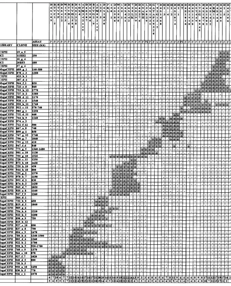
When PCR was performed under stringent conditions Figure 1 shows a simplified version of the contig com-(Table 1), in 9 of 9 cases a sharp band of the expected prising 65 YACs. The remaining YACs were omitted molecular weight was obtained. When tested as probes for simplicity of presentation but the overall number against digested human genomic DNA, all of these of YACs positive for each marker is reported. A variable products hybridized to a single band, as expected for redundancy is apparent, but it is not possible to deter-single-copy sequences. By performing specific PCR mine whether this is the result of an unequal represen-assays on a hamster hybrid containing only human tation in the original libraries or is due to a still incom-chromosome 6, four of nine STSs were excluded from plete detection during the screening procedure. this chromosome, thus confirming that material from The order of markers was established by the pres-chromosomes other than chromosome 6 is abundant ence of two or more markers within a single YAC (Fig. in the YACs used. By comparison with sequences in 1, last row) and by building up the number of overlap-GenBank, one of the clones contained a 244-bp insert ping clones. In addition, inter-Alu band probes were that shared a stretch of identity with the human used as hybridization probes to reinforce the link be-SSADH gene (Chambliss et al., 1995). We then de- tween adjacent loci further.
signed an additional PCR assay (L34820) to amplify
the 3*-end of coding and part of the 3*-untranslated Resolving Ambiguities regions of human SSADH cDNA specifically.
The order of markers presented in Fig. 1 generated We observed a nonspecific PCR product generated by
the minimum number of internal deletions irrespective marker D6S506 on genomic DNA. A group of adjacent
of the number of markers missing in each case. The YACs replicated this spurious band, suggesting the
relative positions of D6S242 vs D6S461 and AF-presence of a second locus homologous to D6S506 in
M342xe5 vs SOX4 could not be resolved because the 6p22. Subcloning and sequencing of this band led to
TABLE 2
Previously Described Markers Used in 6p22 Physical Mapping
PCR conditions
Name Annealing MgCl2(mM) Cycles References
Histone gene H1.1 507C, 60 s 1.5 30 Albig et al., 1993 Histone gene H1.2 567C, 45 s 1.5 30 Albig et al., 1993 Histone gene H1.3 637C, 30 s 1.5 30 Albig et al., 1993 Histone gene H1.4 637C, 30 s 1.5 30 Albig et al., 1993 Histone gene H1.5 587C, 45 s 1.5 35 Albig et al., 1993
D6S422 527C, 45 s 1.5 35 Gyapay et al., 1994
AFM342xe5 477C, 45 s 1.5 30 Gyapay et al., 1994
AFM082tel 597C, 45 s 1.5 30 Gyapay et al., 1994
D6S461 557C, 30 s 1.5 35 Gyapay et al., 1994 D6S299 527C, 45 s 1.5 35 Gyapay et al., 1994 D6S276 557C, 45 s 1.5 30 Gyapay et al., 1994 D6S464 527C, 45 s 1.5 35 Gyapay et al., 1994 D6S265 557C, 30 s 1.5 35 Gyapay et al., 1994 D6S258 557C, 30 s 1.5 35 Gyapay et al., 1994 D6S273 537C, 45 s 1.5 30 Gyapay et al., 1994 D6S306 527C, 45 s 1.5 35 Gyapay et al., 1994
EST449 527C, 45 s 1.5 35 Pappas et al., 1995
EST2534 577C, 30 s 1.5 30 Pappas et al., 1995
EST798 547C, 30 s 1.5 35 Pappas et al., 1995
SOX4 657C, 30 s 1.5 35 Farr et al., 1993
WI9755 587C, 45 s 1.5 35 Hudson et al., 1995
D6S1078 627C, 45 s 1.5 30 Hudson et al., 1995
D6S1074 607C, 60 s 1.5 30 Hudson et al., 1995
WI7513 627C, 45 s 1.5 30 Hudson et al., 1995
D6S1328 527C, 45 s 1.5 30 Hudson et al., 1995
WI3998 567C, 45 s 1.5 30 Hudson et al., 1995
D6S1921 527C, 45 s 1.5 30 Hudson et al., 1995 D6S1468 567C, 45 s 1.5 30 Hudson et al., 1995 D6S1918 587C, 90 s 1.5 30 Hudson et al., 1995 D6S1422 587C, 45 s 1.5 30 Hudson et al., 1995 125 5 527C, 45 s 1.5 30 Hudson et al., 1995 150 1 527C, 45 s 1.5 30 Hudson et al., 1995 100 1 587C, 30 s 1.5 30 Hudson et al., 1995
GATAP19326 567C, 45 s 1.5 30 Hudson et al., 1995
STS G9945 587C, 90 s 2.5 30 Hudson et al., 1995 D6S1558 657C, 45 s 1.5 30 Hudson et al., 1995 D6S1016 607C, 45 s 1.5 30 Buetow et al., 1994 D6S1050 607C, 45 s 1.5 30 Buetow et al., 1994 D6S1002 607C, 60 s 1.5 30 Stone et al., 1994 D6S109 657C, 30 s 1.5 35 Ranum et al., 1991 D6S506 607C, 60 s 1.5 30 GDB D6S507 677C, 60 s 1.5 30 GDB D6S986 677C, 120 s 1.5 30 GDB D6S242 587C, 45 s 1.5 35 GDB
PRL 667C, 60 s 1.5 30 Abbott and Povey, 1991
Amadou et al., 1995
TRMI1 507C, 60 s 1.5 30 Polymeropoulos et al., 1991
PGC 557C, 45 s 1.5 30 Hayano et al., 1988
DSP 577C, 45 s 1.5 30 Arnemann et al., 1991
DHFRP2 557C, 45 s 1.5 30 Anagnou et al., 1985
D6S346E 627C, 60 s 1.5 30 Durkin et al., 1994
D6S105 547C, 60 s 1.5 30 Weber et al., 1992
729LK 657C, 30 s 2.5 28 H. Zoghbi, pers. comm., 1994, Houston, TX
correlation for their presence/absence in YACs (Fig. 1, On the other hand, YAC deletions in the H1 histone cluster generated ambiguities to order the correspond-last two rows). This was also true for SSADH vs
L34820. However, we were able to determine their rela- ing markers. Among several best-fitting orders, Fig. 1 shows that obtained by Volz et al. (1994) on the basis tive order by hybridizing the flanking YACs with a
cDNA probe overlapping the 3*-end of the gene. This of long-range restriction mapping.
The entire set of markers is ‘‘doubly linked’’ to adja-result further confirmed the assignment of the SSADH
FIG. 1. Results of YAC alignment based on marker content of clones. A subset of 65 YACs is shown. Presence (/) or absence (0) of DNA markers in YAC clones is indicated. The second row reports the method of marker ascertainment: P, PCR; S, Southern blotting. The penultimate row reports the overall number of YACs containing the corresponding marker. The last row reports the number of YACs containing simultaneously each marker and the marker immediately to its right.
(Fig. 1, last row). The first ‘‘singly linked’’ interval is Instability of YACs
between markers WI7513 and BTVD1.4, which are
Thirty cultures of the Mega-YAC library were exam-both contained only in YAC 966_e_10; the second is
ined in double or triple replicates by two laboratories between D6S276 and SSADH (755_g_10), and the third
that obtained them through different resource centers, is between BT and D6S1016 (905_g_1).
i.e., after several rounds of mitotic propagation. Of the An estimate of the size of the YAC contig can be
YACs in this group, 15 gave identical positive results made by summing the sizes of the smallest overlapping
for both members of each pair, 1 gave identical negative and nonoverlapping YACs with no known deletion
results, while 5, 4, 2, and 3 exhibited a loss of one, two, across the contig. By these means the region covered
three or four markers, respectively (47% of YACs with is 8.0 – 9.0 Mb.
losses). The finding of such a high rate of marker loss The order of markers in the contig refines the relative
raises questions as to whether it may affect the order-positions of loci reported in the CEPH genetic map
ing of loci. Forty-one markers were examined in at least (Chumakov et al., 1995). In particular, D6S285 and
a pair of subcultures of the same original YAC clone, D6S422 (3 cM) are separated by 9 markers, as
deter-and 155 markers 1 pair assays were performed. Over-mined by our results and those by Olavesen et al.
all 31 markers showed loss in one culture among the (1995); D6S422 and D6S461 (5 cM) are separated by examined pairs (10% rate of loss). Thus there is a 90% 17 markers. D6S461 and D6S299, not genetically
re-chance for a given marker to be retained in a YAC, solved, are now oriented based on the discordant re- ensuring that, with the number of clones examined in sults of YACs 720_c_11, 944_h_10, 970_b_7, and this work and found positive (Fig. 1, last two rows), the 947_f_9 (Fig. 1). In agreement with the MIT data (con- order of loci can be reliably inferred. Indeed, visual tig WC140), D6S276, genetically mapped 4 cM from inspection of the contig showed that the ordering of loci [D6S461 – D6S299], turns out to be adjacent to D6S299, involved in losses in each pair is supported by several with its position being defined by discordant results in other YAC clones. An exception is represented by the several YACs. D6S299 and D6S464 (4 cM) are sepa- H1 cluster-containing region, where the order of loci rated by 22 markers and 2 additional markers separate was drawn from long-range restriction mapping (Volz the latter from D6S306. Our contig also supports the et al., 1994). Recurrent marker loss and the low effi-consensus map reported by Volz et al. (1994) with odds ciency in obtaining YACs from this region (Fig. 1, pen-of only 14:1. YAC 423_b_1 places D6S105 telomerically ultimate row) might be the result of enhanced mitotic although very close to H1.5. The finding that the HLA- instability of YACs containing portions of this region F probe does not hybridize with YACs containing of the genome.
D6S464, D6S306, and D6S276 is in agreement with the location of HLA further centromeric to the H1 histone
DISCUSSION
cluster and, by extension, to the above-mentioned loci (Amadou et al., 1995; Volz et al., 1994). This is in line
We have constructed an integrated map of chromo-with the observation that among the 42 YACs reported
some 6p22 consisting of YACs, polymorphic genetic by Chumakov et al. (1995) to contain genetic position markers, anonymous STS and ESTs, anonymous sub-0.46, not one scored simultaneously positive for any of cloned Alu-PCR products, and known genes. We pri-the three microsatellite markers and pri-the MHC cluster marily used PCR screening to determine the relation-markers. The order of markers here reported confirms ships between the YAC clones, but hybridization prob-that of the doubly linked MIT contig WC140 and estab- ing was also used to confirm them. We developed novel lishes a link between WC140 and WC1316. In the inte- STS markers and integrated their analysis with pre-grated map (Hudson et al., 1995), however, D6S1422 viously described markers in the characterization of is placed telomerically to D6S1918, and AFM342xe5 is the same set of YAC clones.
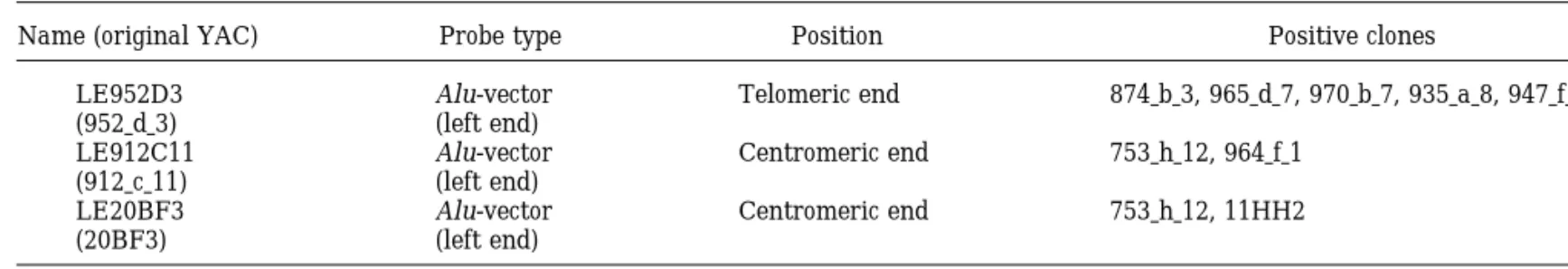
placed telomerically to D6S1328, an order not sup- We resolved the relative positions of nine markers ported in our contig. In addition, the same authors re- used to construct the Ge´ne´thon genetic map (Gyapay port AFM342xe5 and WI7513 to comap in the same et al., 1994), confirmed the presence of six of them in radiation hybrid, whereas we resolve their order with this region, and assigned the remaining three to the four intervening markers. more proximal MHC-containing region. In fact, in their Inter-Alu bands and anonymous probes from the recent report, Burt et al. (1996) physically mapped insert-vector boundary of selected YACs were gener- D6S265 and D6S258 in the proximity of HLA-A and ated to reinforce the link between adjacent loci fur- HLA-F genes. D6S276, one of the markers used in the ther. 792G12_2 is a specific Alu band obtained by Alu- recent radiation hybrid framework map (Gyapay et al., PCR of YAC 792_g_12 (Fig. 1). LE952D3, LE912C11, 1996), is retained in many of the YACs harboring and LE20BF3, obtained by the corresponding YAC D6S299 and in none of those harboring either D6S464 clones by using Alu-vector protocols, when used as or D6S306. Its position is thus considered more te-hybridization probes on a subset of YACs demon- lomeric than previously reported, perhaps as a result strated that the corresponding inserts are oriented as of a limited number of recombination events with re-spect to neighboring markers. The repositioning of reported in Table 3.
TABLE 3 Hybridization Probes
Name (original YAC) Probe type Position Positive clones
LE952D3 Alu-vector Telomeric end 874 b 3, 965 d 7, 970 b 7, 935 a 8, 947 f 9 (952 d 3) (left end)
LE912C11 Alu-vector Centromeric end 753 h 12, 964 f 1 (912 c 11) (left end)
LE20BF3 Alu-vector Centromeric end 753 h 12, 11HH2
(20BF3) (left end)
D6S276 would imply the breakdown of the level 1 path assignment of YACs 878_e_3 and 753_h_12 by FISH (Bray-Ward et al., 1996). These clones are found on between genetic positions 0.42 and 0.46 (Chumakov et
al., 1995). the proximal side of our contig (Fig. 1) and produced
hybridization signals on proximal 6p22 and the We superimposed on this map 46 additional markers.
These include the SOX4, prolactin, butyrophilin, and 6p21.3 – p22 boundary, respectively.
methionine-t-RNA genes as well as the H1 histone clus- Centromerically, our contig reaches the contig re-ter containing several genes (Albig et al., 1993). We ported by Burt et al. (1996), sharing with it four YACs could physically locate this cluster for the first time and three markers, and ensuring that 6p22 is physi-between D6S1468 and D6S1016. In addition, we re- cally covered up to its proximal end. In addition, we fined the location of two of six ESTs with a 6p22 – p23 could orient and order several markers reported to lie assignment (Pappas et al., 1995) by positioning them in the proximity of the 6p21.3 – p22 boundary (Amadou within the contig. We assigned for the first time the et al., 1995; Raha-Chowdhury et al., 1995; Vernet et al.,
gene for NAD/
-dependent succinic semialdehyde dehy- 1993). Based on the volume of metaphase chromosome drogenase (SSADH) to this portion of the genome and (Heslop-Harrison et al., 1989) and the relative length found it to be oriented from telomere to centromere. of bands (Franke, 1994), the 6p22 band can be esti-The number of markers per genetic interval corre- mated to span 13 Mb. However, this measurement is lates fairly well with the genetic distances reported by hardly comparable with the length of our contig, due Chumakov et al. (1995), providing no evidence for a to variations in the extent of chromosome condensa-biased distribution. By virtue of the new markers we tion. To its telomeric end, our contig reaches that pub-were able to bring all paths between adjacent loci to lished by Olavesen et al. (1995), sharing with it five level 1. YACs and three markers. The continuous resource of When comparing our results with those of the MIT aligned YAC clones now spans from 6p21.3 to distal Genome Center, the STS WI9755 was not found in any 6p24.3 (Davies et al., 1995).
of our YACs. In particular, we examined individually Our work enabled us to evaluate the instability of YACs 787_d_1, 847_c_8, 927_f_10, and 844_h_10, re- YAC clones upon several runs of mitotic propagations ported with three ‘‘definite’’ YAC/STS hits and one ‘‘am- and revealed a remarkable proportion of YACs with biguous’’ YAC/STS hit in the contig WC140. The first marker losses. This might considerably affect the use-two were indeed confirmed to share markers at the fulness of YACs in constructing contigs based on STS telomeric end of 6p22. With this exception, we observed content. We used a large number of independent clones a better agreement with the individual doubly linked to increase internal consistency and found reproducible contigs rather than the integrated map, since in some links between adjacent loci in most cases.
instances it reports an alternative order of markers. In The expressed genes localized within the contig and this work we examined YACs individually, thus exclud- the reagents utilized in this work provide the first core ing the imprecision introduced by the tridimensional of a transcriptional map of this chromosomal region. screening scheme (Hudson et al., 1995). Recurrent loss
of markers in independent YACs or complex
rearrange-ments in radiation hybrids may contribute to the ob- ACKNOWLEDGMENTS served discrepancies with the integrated map.
We excluded from this region the DHFRP2 and DSP
We are grateful to Drs. J. Ragoussis, M. Olavesen, and H. Cann
genes and the EST D6S346E, reported in the literature
for their continuous support, sharing of data, and help in obtaining
with 6pter-qter, 6pter-p21, and 6p22.1 – p21.3 assign- reagents and to Professor L. Terrenato for his invaluable advice. We ments, respectively. Moreover, we excluded the are also indebted to Professor A. Ballabio for support and discussion. The collaboration of Drs. D. Toniolo and C. Sala for the screening of
EST798 (Pappas et al., 1995), an expressed sequence
the CEPH and ICI libraries is gratefully acknowledged. We thank
assigned to 6p22 – p23. Olavesen et al. (1995) excluded
C. Grottoli for technical assistance. This work was supported by E.U.
its presence from 6p23 as well, thus leaving the precise BIOMED Contracts GENE-CT93-0075, GENE-CT93-0101 (Euro-position of this sequence still to be determined. gem), and BMH4-CT96-0994, MURST 60%, and the Italian Telethon
Foundation.
of a locus for orofacial clefting on human chromosome 6p24 and
REFERENCES
STS content map of the region. Hum. Mol. Genet. 4: 121 – 128. Durkin, A. S., Nierman, W. C., Zoghbi, H. Y., Jones, C., Kozak, Abbott, C., and Povey, S. (1991). Development of human
chromo-C. A., and Maglott, D. R. (1994). Chromosome assignment of hu-some-specific PCR primers for characterization of somatic cell
hy-man brain expressed sequence tags (ESTs) by analysing fluores-brids. Genomics 9: 73 – 77.
cently labeled PCR products from hybrid cell panels. Cytogenet. Albertsen, H. M., Adberrahim, H., Cann, H. M., Dausset, J., Le
Pas-Cell Genet. 65: 86 – 91. lier, D., and Cohen, D. (1990). Construction and characterization
Farr, C. J., Easty, D. J., Ragoussis, J., Collignon, J., Lovell-Badge, of a yeast artificial chromosome library containing seven haploid
R., and Goodfellow, P. N. (1993). Characterization and mapping human genome equivalents. Proc. Natl. Acad. Sci. USA 87: 4256 –
of human SOX4 gene. Mamm. Genome 4: 577 – 584. 4260.
Foote, S., Vollrath, D., Hilton, A., and Page, D. C. (1992). The human Albig, W., Drabent, B., Kunz, J., Kalff-Suske, M., Grzeschik, K., and
Y chromosome: Overlapping DNA clones spanning the euchromatic Doenecke, D. (1993). All known human H1 histone genes except
region. Science 258: 60 – 66. the H17 gene are clustered on chromosome 6. Genomics 16: 949–
654. Foulkes, W. D., Ragoussis, J., Stamp, G. W. H., Allan, G. J., and Trowsdale, J. (1993). Frequent loss of heterozygosity on chromo-Amadou, C., Ribouchon, M. T., Mattei, M. G., Jenkins, N. A., Gilbert,
some 6 in human ovarian carcinoma. Br. J. Cancer 67: 551 – 559. D. J., Copeland, N. G., Avoustin, P., and Pontarotti, P. (1995).
Localization of new genes and markers to the distal part of the Francke, U. (1994). Digitized and differentially shared human chro-human major histocompatibility complex (MHC) region and com- mosome ideograms for genomic application. Cytogenet. Cell Genet. parison with the mouse: New insights into the evolution of mam- 65: 206 – 219.
malian genomes. Genomics 26: 9 – 20. Gyapay, G., Morissette, J., Vignal, A., Dib, C., Fizames, C., Millas-Anagnou, N. P., Antonarakis, S. E., O’Brien, S. J., and Nienhuis, seau, P., Marc, S., Bernardi, G., Lathrop, M., and Weissenbach, J. A. W. (1985). Chromosomal localization and racial distribution of (1994). The 1993 – 94 Ge´ne´thon human genetic linkage map. Na-the polymorphic hDHFR-psi-1 pseudogene. Clin. Res. 33: 328A. ture Genet. 7: 246 – 339.
Anand, R., Ogilvie, D. J., Butler, R., Riley, J. H., Finniear, R. S., Gyapay, G., Schmitt, K., Fizames, C., Jones, H., Vega-Czarny, N., Powell, S. J., Smith, J. C., and Markham, A. F. (1991). A yeast Spillett, D., Muselet, D., Prud’Homme, J., Dib, C., Auffray, C., artificial chromosome contig encompassing the cystic fibrosis locus. Morissette, J., Weissenbach, J., and Goodfellow, P. N. (1996). A Genomics 9: 124 – 130. radiation hybrid map of the human genome. Hum. Mol. Genet. 5:
339 – 346. Arnemann, J., Spurr, N. K., Wheeler, G. N., Parker, A. E., and
Bux-ton, R. S. (1991). Chromosomal assignment of the human genes Hayano, T., Sogowa, K., Ikihara, Y., Fuji-Quriyama, Y., and Taka-coding for the major proteins of the desmosome junction, Des- hashi, K. (1988). Primary structure of human pepsinogen C gene. moglein DGI (DSG), desmocollins DGII/III (DSC), desmoplakins J. Biol. Chem. 263: 1382 – 1385.
DPI/II (DSP), and plakoglobin DPIII (JUP). Genomics 10: 640 – Heslop-Harrison, J. S., Leitch, A. R., Schwarzacher, T., Smith, J. B.,
645. Atkinson, M. D., and Bennett, M. D. (1989). The volumes and
Bray-Ward, P., Menninger, J., Liemann, J., Desai, T., Mokady, N., morphology of human chromosomes in mitotic reconstructions. Banks, A., and Ward, D. C. (1996). Integration of the cytogenetic, Hum. Genet. 84: 27 – 34.
genetic, and physical maps of the human genome by FISH mapping Holmquist, G. P. (1992). Chromosome bands, their chromatine fla-of CEPH YAC clones. Genomics 32: 1 – 14. vors, and their functional features. Am. J. Hum. Genet. 51: 17 – Buetow, K. H., Weber, J. L., Ludwigsen, S., Scherpbier-Heddema, 37.
T., Duyk, G. M., Sheffield, V. C., Wuang, Z., and Murray, J. C. Hudson, T. J., Stein, L. D., Gerety, S. S., Ma, J., Castle, A. B., Silva, (1994). Integrated human genome-wide maps constructed using J., Slonim, D. K., Baptista, R., Kruglyak, L., Xu, S., Hu, X., Colbert, the CEPH reference panel. Nature Genet. 6: 391 – 394. A. M. E., Rosenberg, C., Reeve-Daly, M. P., Rozen, S., Hui, L., Wu, X., Vestergaard, C., Wilson, K. M., Bae, J. S., Maitra, S., Ganiatsas, Burt, M. J., Smit, D. J., Pyper, W. R., Powell, L. W., and Jazwinska,
E. C. (1996). A 4.5-megabase YAC contig and physical map over S., Evans, C. A., DeAngelis, M. M., Ingalls, K. A., Nahf, R. W., Horton, L. T., Jr., Anderson, M. O., Collymore, A. J., Ye, W., Kou-the hemochromatosis gene region. Genomics 33: 153 – 155.
youmjian, V., Zemsteva, I. S., Tam, J., Devine, R., Courtney, Campbell, R. D., and Trowsdale, J. (1993). Map of the human MHC.
D. F., Renaud, M. T., Nguyen, H., O’Connor, T. J., Fizames, C., Immunol. Today 14: 349 – 352.
Faure´, S., Gyapay, G., Dib, C., Morissette, J., Orlin, J. B., Birren, Chambliss, K. L., Caudle, D. L., Hinson, D. D., Moomaw, C. R.,
B. W., Goodman, N., Weissenbach, J., Hawkins, T. L., Foote, S., Slaughter, C. A., Jakobs, C., and Gibson, K. M. (1995). Molecular
Page, D. C., and Lander, E. S. (1995). An STS-based map of the cloning of the mature NAD/-dependent succinic semialdehyde
de-human genome. Science 270: 1945 – 1954. hydrogenase from rat and human. J. Biol. Chem. 270: 461 – 467.
Jurka, J., Walichiewicz, J., and Milosavljevic, A. (1992). Prototypic Chumakov, I. M., Rigault, P., Le Gall, I., Bellanne`-Chantelot, C.,
sequences for human repetitive DNA. J. Mol. Evol. 35: 286 – 291. Billault, A., Guillou, S., Soularue, P., Guasconi, G., Poullier, E.,
Nelson, D. L., Ledbetter, S. A., Corbo, L., Maureen, F. W., Ramirez-Gros, I., Belova, M., Sambucy, J. L., Susini, L., Gervy, P., Glibert,
Solis, R., Webster, T. D., Ledbetter, D. H., and Caskey, T. (1989) F., Beaufils, S., Bui, H., Massart, C., De Tand, M. F., Dukasz, F.,
Alu polymerase chain reaction: A method for rapid isolation of Lecoulant, S., Ougen, P., Perrot, V., Saumier, M., Soravito, C.,
human-specific sequences from complex DNA sources. Proc. Natl. Bahouayila, R., Cohen-Akenine, A., Barillot, E., Bertrand, S.,
Co-Acad. Sci. USA 86: 6686 – 6690. dani, J. J., Caterina, D., Georges, I., Lacroix, B., Lucotte, G.,
Sahba-tou, M., Schmit, G., Sangouard, M., Tubacher, E., Dib, C., Faure´, Nemani, M., Cherif, D., Chesne, H., Pelandakis, M., Ougen, P., Berger, R., Weissenbach, J., Le Paslier, D., Choen, D., and Cann, S., Fizames, C., Gyapay, G., Millasseau, P., NGuyen, S., Muselet,
D., Vignal, A., Morissette, J., Menninger, J., Lieman, J., Desai, T., H. M. (1994). A YAC contig in 6p23 based on sequence tagged sites. Genomics 22: 388 – 396.
Banks, A., Bray-Ward, P., Ward, D., Hudson, T., Gerety, T., Foote,
S., Stein, L., Page, D. C., Lander, E. S., Weissenbach, J., Le Paslier, Olavesen, M. G., Davies, A. F., Broxholme, S. J., Wixson, J. L., D., and Cohen, D. (1995). A YAC contig map of the human genome. Senger, G., Nizetic, D., Campbell, R. D., and Ragoussis, J. (1995). Nature 377: 175 – 183. An integrated map of human chromosome 6p23. Genome Res. 5:
342 – 358. Cohen, D., Chumakov, I., and Weissenbach, J. (1993). A
first-genera-tion physical map of the human genome. Nature 366: 698 – 701. Pappas, G., Polymeropoulos, M. H., Boyle, J. M., and Trent, J. M. (1995). Regional assignment by hybrid mapping of 36 expressed Davies, A. F., Stephens, R. J., Olavesen, M. G., Heather, L., Dixon,
Peltonen, L. (1995). All out for chromosome six. Nature 378: 665 – Isolation of the CA dinucleotide repeats close to D6S105; linkage disequilibrium with hemocromatosis. Hum. Mol. Genet. 3: 2043 – 666.
2046. Polymeropoulos, M. H., Xiao, H., Rath, D. S., and Merril, C. R. (1991).
Totaro, A., Rommens, J. M., Grifa, A., Lunardi, C., Carella, M., Trinucleotide repeat polymorphism at the human Met-tRNA-i gene
Huizenga, J. J., Roetto, A., Camachella, C., De Sandre, G., and 1 (TRMI). Nucleic Acids Res. 19: 4306.
Gasparini, P. (1996). Hereditary hemochromatosis: Generation of Pritchard, M., Fuentes, J. J., Bosch, A., and Estivill, X. (1994).
To-a trTo-anscription mTo-ap within To-a refined To-and extended mTo-ap of the HLA wards a transcriptional map of human chromosome 21q22.2 – 22.3.
class I region. Genomics 31: 319 – 326. Third EUROGEM meeting, Montpellier, France. [Abstract.]
Vernet, C., Boretto, J., Mattei, M., Takahashi, M., Jack, L. J. W., Raha-Chowdhury, R., Bowen, D. J., Stone, C., Pointon, J. J.,
Terwil-Mather, I. H., Rouquier, S., and Pontarotti, P. (1993). Evolutionary liger, J. D., Shearman, J. D., Robson, K. J. H., Bomford, A., and
study of multigenic family mapping close to the human MHC class Worwood, M. (1995). New polymorphic microsatellite markers
I region. J. Mol. Evol. 37: 600 – 612. place the hemochromatosis gene telomeric to D6S105. Hum. Mol.
Volz, A., Boyle, J. M., Cann, H. M., Cottingham, R. W., Orr, H. T., and Genet. 4: 1869 – 1874.
Ziegler, A. (1994). Report of the Second International Workshop on Ranum, L. P., Chung, M. Y., Duvick, L. A., Zoghbi, H. Y., and Orr, human chromosome 6. Genomics 21: 464 – 472.
H. T. (1991). Dinucleotide repeat polymorphism at the D6S109
Wapenaar, M. C., Schiaffino, M. V., Bassi, M. T., Scaefer, L., Chi-locus. Nucleic Acids Res. 19: 1171.
nault, A. C., Zoghbi, H. Y., and Ballabio, A. (1994). A YAC based Sealey, P. G., Whittaker, P. A., and Southern, E. M. (1985). Removal binning strategy facilitating assembly of cosmid contig: 1.6 mega-of repeated sequences from hybridization probes. Nucleic Acids bases of overlapping cosmid in Xp22. Hum. Mol. Genet. 3: 1155 –
Res. 13: 1905 – 1922. 1161.
Senapathy, P., Shapiro, M. B., and Harris, N. L. (1990). Splice junc- Weber, J. L., Kwitkok, A. E., May, P. E., and Zoghbi, H. Y. (1992). tions, branchpoint size and exons: Sequence statistics, identifica- Dinucleotide repeat polymorphism at the D6S105 locus. Nucleic tion and applications to genome project. Methods Enzymol. 183: Acids Res. 19: 968.
252 – 278. Zoghbi, H. Y., and Chinault, A. C. (1995). Generation of YAC contigs. In ‘‘YACs: A User Guide’’ (B. H. Brownstein and D. L. Nelson, Stone, C., Pointon, J. J., Jazwinska, E. C., Halliday, J. W., Powell,