Trivandrum-695 023 Kerala, India
Biochemical Aspects of Human Nutrition, 2010: 295-312 ISBN: 978-81-7895-478-3 Editors: Luciana Avigliano and Luisa Rossi
16. Redox bases underlying the anti-tumor
activity of garlic-contained organo-sulfur
compounds: Implication in chemoprevention
and chemotherapy
Giuseppe Filomeni1, Giuseppe Rotilio1,2 and Maria R. Ciriolo1,2
1Department of Biology, University of Rome “Tor Vergata”, via della Ricerca Scientifica
00133 Rome, Italy; 2Research Centre I.R.C.C.S. San Raffaele - Pisana
Via dei Bonacolsi, 00163, Rome, Italy
Abstract. The beneficial effects of phytochemicals on human health have been
extensively addressed. The majority of this outcome derives from their capability to function as antioxidants, thus the consumption of foods rich in these compounds is considered an advisable preventive therapy in slowing oxidative stress-mediated degenerative processes, such as those occurring during aging. Nevertheless, high concentrations of redox-active compounds could switch the antioxidant property to a pro-oxidant action leading to cell cycle arrest and death. This aspect place phytochemicals as promising therapeutics particularly for cancer prevention or treatment. Although their beneficial properties are known from ancient times, only during the recent years the molecular mechanisms underlying the anti-proliferative effects mediated by garlic-derived organo-sulfur compounds (OSC) are going to be clarified, with particular regard to what their pro-apoptotic features concerns. This chapter discusses the main findings that have contributed to the comprehension of OSC-mediated redox-dependent events governing growth arrest and apoptosis. Particularly, we report the mechanisms through which OSC have been suggested to generate reactive oxygen species and to modulate the redox state of specific reactive cysteines. Both processes will be argued as necessary events in inducing either irreversible damage to cellular macromolecules (e.g. DNA and
cytoskeleton proteins), or waves of signaling finally resulting in the activation of
Correspondence/Reprint request: Dr. Maria Rosa Ciriolo, Department of Biology, University of Rome “Tor Vergata”, via della Ricerca Scientifica, 00133 Rome, Italy. E-mail: [email protected]
the apoptotic program. In this perspective, the classes of proteins which have been indicated to represent the targets of OSC-mediated oxidative modifications, and to have a role in cellular redox response will be discussed.
Introduction
The use of garlic (Allium sativum) and its derivatives has long history: from the time of Egyptians, passing through the ancient Romans up to the Middle Ages, when garlic was often used as a remedy for intestinal disorders, respiratory infections, skin and heart diseases [1, 2]. More recently a huge amount of publications have confirmed that the consumption of garlic preparations, including garlic extracts have beneficial effects for human health. The biological responses induced by garlic and its derivatives include reduction of blood cholesterol [3] and other risk factors for cardiovascular diseases [4, 5]; modulatory effects of immune function [reviewed in 6]; protection against Helicobacter
pylori and other bacterial infection [reviewed in 7]; enhanced xenobiotics detoxification
[8-10], and many other effects. Moreover, epidemiology studies pointed out an inverse correlation between garlic consumption and the onset of cancer [11-13]. These latest results were further corroborated by several papers in which an anti-proliferative and anti-tumor activity of garlic and its derivatives have been suggested both in in vitro and in vivo models.
The mechanisms by which garlic and its derivatives exert the above mentioned beneficial effects, especially those regarding their anti-tumor activity, have been often ascribed to their antioxidant activity [14-17]. However, during the last decade a novel idea regarding a pro-oxidant function for garlic and garlic-derived organosulfur compounds (OSC) has been arising, allowing to draw a more general scheme of action for these molecules able to reconcile results from literature only apparently conflicting.
OSC geneteration and redox bases underlying their anti-proliferative
activity
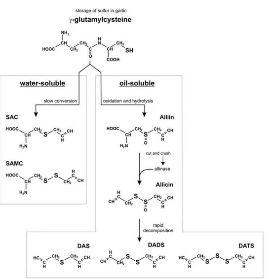
The formation of the OSC contained in garlic extracts relies on a relatively complex chemistry, which was likely developing as a protective mechanism against host insults. Garlic contains a huge amount of molecules ranging from vitamins (vitamin B1, B2, B6, C, E, biotin and several polyphenolic compounds) to essential amino acids [1, 18]; from lipids to steroidal glycosides [19]. However, the high amount of sulfur-containing molecules makes garlic unique among all vegetables. The primary constituents of crude garlic, serving as storage of sulfur, is γ-glutamylcysteine. It can be hydrolyzed and oxidized to form γ-glutamyl-S-alk(en)yl-cysteines and S-alk(en)yl-cysteine sulfoxides, among which alliin (S-allyl-cysteine sulfoxide) represents the most abundant, accounting for by 1% of OSC [1, 20]. During storage of garlic bulbs at cool temperatures, alliin accumulates naturally. On average, a garlic bulb contains up to 0.9% γ-glutamylcysteines and up to 1.8% alliin. After processing, such as cutting and crushing, the vacuolar enzyme alliinase rapidly transforms alliin into the smelling alkyl alkane-thiosulfinates, including allicin (diallyl thiosulfinate) [21]. Allicin and other thiosulfinates are highly unstable and immediately decompose to generate the so-called oil soluble OSC, such as diallyl sulfide (DAS), diallyl disulfide (DADS), diallyl trisulfide (DATS), methyl allyl disulfide, methyl allyl trisulfide, dithiins and E,Z-ajoene [1, 18, 22]. At the same time, another pathway, completely independent on allicin formation, leads to the conversion of γ-glutamylcysteine in the so-called water-soluble OSC, such as S-allyl-cysteine (SAC) and S-allyl-mercaptocysteine (SAMC) [1] (Figure 1).
Figure 1. Generation of water and oil-soluble OSC from γ-glutamyl cysteine in garlic. It has been
demonstrated that several OSC have anti-proliferative activity, nevertheless some distinctions should be made in order to classify and distinguish their different effectiveness as putative anti-tumor compounds. Normally, the oil-soluble OSC exert a higher cytostatic and cytotoxic activity with respect to the water-soluble counterparts; moreover, these effects directly correlate with the number of sulfur atoms present within the molecule; hence, DATS is more efficient than DADS, which in turn is more active than DAS in inducing cell cycle arrest and death of tumor cells. The functional groups seem to play a pivotal role in the occurrence of such events as well, with the allyl substituent more effective than methyl, ethyl or propyl groups in showing anti-tumor properties [1, 13].
A huge amount of data reported until late 1990s indicate that one of the main effects of OSC relied on their anti-oxidant and detoxifying activity. In fact, the use of OSC or garlic crude extracts, in combination with carcinogens or xenobiotics, decreases, and sometimes completely abolishes, the mutagenic effects induced both in in vitro and in
in vivo systems [23, 24]. Therefore, the belief that OSC could function as anti-oxidants
became a common opinion, although these beneficial properties did not well correlate with the cytotoxic/anti-proliferative effects that OSC showed at pharmacological doses.
From an exquisitely redox perspective, not all the molecules able to assist the cell against pro-oxidant conditions are necessarily reductants. In fact, it is widely accepted that treatments with low concentration of pro-oxidant molecules transcriptionally induce sets of gene required for the anti-oxidant response: a phenomenon known as “pre-conditioning”, which makes the cell more resistant towards the occurrence of further oxidative challenges. In this context, one of the well known examples of transcription factor able to induce resistance to oxidative condition and to activate detoxifying response is the nuclear erythroid factor 2-related factor 2 (Nrf2). Normally, Nrf2 is sequestered in the cytoplasm, particularly at the level of actin filaments, where it is strongly associated with Keap1. Keap1 binding to Nrf2 maintains Nrf2 unable to translocate towards nucleus and to recognize the electrophylic responsive elements (EpRE, or antioxidant responsive elements, ARE) [25]. In fact, it is a redox-sensitive anti-apoptotic transcription factor that, in response to reactive oxygen species (ROS) production or alteration in the ratio between the reduced and the disulfide forms of glutathione (GSH/GSSG), induces transcription of genes for antioxidant defense and detoxification. Keap1 represents the real redox switch for Nrf2 activation, since, upon oxidation of specific cysteines onto its surface, Keap1 changes its conformation leading Nrf2 free to translocate into the nucleus [26]. Therefore, although Nrf2 is the final effector, Keap1 represents the real inducer of antioxidant response indicating for the Nrf2/Keap1 dimer a characteristic two-module redox-sensitive system. Nrf2 modulation seems more complex with respect to a single component, such as p53, activator protein 1 (AP1) or nuclear factor (NF)-κB, where the redox modification affects the transcription factor itself; however, the Nrf2/Keap1 couple allows the system to be highly safe against irreversible oxidative insults. This can explain why this mode of signaling has been successfully exploited also by redox-activated protein kinases, which mediates apoptotic response.
Few years ago it was demonstrated that oil-soluble OSC were able to rapidly activate Nrf2-dependent transcription of the antioxidant genes glutathione-S-transferase (GST), heme oxygenase 1 (HO-1) and NAD(P)H:quinone oxidoreductase 1 (NQO1), and that this event was indispensable for cellular anti-oxidant response [27, 28]. These papers were among the first clear indications that OSC are not reductant per se but, instead, possess pro-oxidant properties able to induce cellular anti-oxidant response. Therefore, cell death or anti-oxidant response downstream of OSC treatment are just “the two side of the coin”, whose difference only relied upon the concentration of OSC employed. Actually, this dual role seems to be shared among several redox-active molecules, especially those deriving from foods. For example, polyphenols, such as quercetin, have been shown to have anti-oxidant as well as pro-oxidant characteristics [29, 30]. The same behavior has been suggested also for ascorbate, for which it has been shown to increase some oxidative markers in human plasma [31]. Two of the main determinants of this double redox-nature are: i) the concentration; ii) the presence of transition metals. The most relevant consequence of such a feature, is that these redox-active molecules, such as OSC themselves, can be of help in cancer treatment, because they could play a role in: prevention, at low doses, such as those reaching from diet; or therapy, at high doses, such as those employed pharmacologically. In the former case, the anti-oxidant response, reasonably induced by Nrf2, produces a pre-conditioning state, which counteracts cell transformation. In the latter, the resulting oxidative stress could be of help in inducing death of cancer cells. Indeed, many of the efforts in cancer research are dealing with the identification of more specific treatments, which selectively induce cell death in tumors. In this context, the use of oxidative stress as tool to induce apoptosis is one of the
undertaken routes. Recent studies have shown that cancer cells produce higher levels of ROS than normal cells, owing to both intense metabolic activity and mitochondrial defects [32]. These conditions leads to an intrinsic oxidative stress, which selectively targets malignant cells for therapeutic strategies based on further ROS production and consequent irreversible oxidative insult [33]. Several anticancer drugs commonly used in chemotherapy (e.g., adriamycin and etoposide), besides their well know property to affect DNA integrity, are able to induce a site-directed burst of ROS as a part of their mechanism of action [34].
OSC induce apoptosis of tumor cells through a ROS-dependent
activation of MAP kinases
In 2002 Kwon and coworkers indicated for the first time the association between the occurrence of caspase-3 dependent apoptosis and ROS generation in human leukemia cells HL-60 treated with DADS. Although no functional relationship was suggested, the relevance of the very early burst of ROS in apoptosis execution was underlined by the incubation with antioxidants, such as N-acetylcysteine (NAC) and catalase, which were able to completely counteract caspase-3 cleavage [35]. Later on, our laboratory demonstrated the molecular mechanisms underlying ROS-dependent cell cycle arrest and apoptosis. Indeed, treatments with DADS induced ROS production in neuroblastoma SH-SY5Y cells, and caused oxidative damage to proteins and lipids, which accumulated time-dependently up to the appearance of apoptotic markers [36]. Both overexpression of the Cu,Zn containing superoxide dismutase (SOD1) and incubation with the radical scavenger 5,5’-dimethylpyrroline-N-oxide were able to induce a delay of cell cycle arrest and apoptosis, but not a complete rescue of cell viability. From a mechanistic viewpoint, we demonstrated that ROS increase was tightly associated with the detachment of GST from the member of the mitogen activated protein (MAP) kinase, c-Jun-NH2-terminal kinase (JNK): the key event in the
final induction of apoptosis (Figure 2).
In fact, JNK activity is normally maintained at low levels, even in the presence of high concentrations of growth factors and in unstressed cells seems to be inhibited by its association with GST [37]. Conversely, under stress conditions, such as those occurring upon treatment with DADS, GST/JNK complex dissociates, leading to the phospho-activation of JNK and the induction of the downstream apoptotic events [38]. In support of the pivotal role of JNK in ROS-mediated apoptosis, its inhibition in DADS-treated cells, induced a significant decrease of apoptotic extent; however, also in this case, we were not able to completely restore cell survival, but instead an increase of the percentage of G2/M blocked cells was induced [36].
The results obtained allowed us to hypothesize that the molecular mechanisms underlying DADS-mediated antiproliferative effects as well as cognate OSC could be of general application to different tumor histotypes. On the contrary, when we extended the results obtained to other cell lines, we surprisingly observed that not all tumor histotypes underwent apoptosis when incubated with DADS, even at much higher concentrations. Particularly, the adenocarcinoma gastric cell line AGS responded to DADS by a transient arrest of cell growth without commitment to apoptosis, and re-started to proliferate later on when the conditions became permissive for cell survival [39]. Nevertheless, the “ROS-based theory”, as rationale for OSC-induced cytotoxicity, did not fail but was further confirmed when we found that AGS were equipped with an efficient anti-oxidant defense, mainly dependent on GSH and GSH peroxidase (GPx). Particularly, the capacity
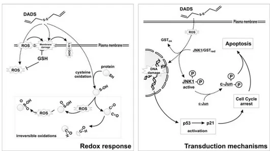
Figure 2. Sensitivity to pro-oxidant and pro-apoptotic effects of DADS. DADS can induce ROS
production and yield lipids and protein oxidation. For the latter class of macromolecules, the formation of carbonyl groups and/or (ir)reversible oxidation of cysteine residues can occur (left). The resulting oxidative unbalance induces p53/p21 activation downstream of DNA damage and GST-mediated dissociation/activation of JNK/c-Jun pathway. Both these events concur in the execution of caspase-mediated cell death (right).
Figure 3. Resistance to pro-oxidant and pro-apoptotic effects of DADS. Cells rich in glutathione
peroxidase (GPx) can counteract DADS-produced ROS, as well as the increase of lipid peroxides. Moreover, S-glutathionylation processes can help in protecting reactive cysteines of proteins from irreversible oxidation (left). The mild oxidative unbalance resulting from such a buffer response mediates the de-activation of ERK1/2 that, along with the increase of p53/p21 system, is responsible for a transient cell cycle blockage. This event allows the repair of cellular structures and, when completed, cell cycle re-entry (right).
in forming reversible mixed disulfides between GSH and protein thiols, together with the
high expression levels of the gastroinstestinal isoform of GPx (GI-GPx), allowed AGS cells to buffer ROS insults and prevent JNK-mediated phosphorylative cascades. Moreover, the concomitant de-activation of the extracellular-related kinase 1/2 (ERK1/2), which is normally implicated in cell cycle progression, represented the event needed to inhibit cell growth. This allowed the cells to repair possible damages and, then, start to proliferate [39] (Figure 3).
Interestingly, we speculated that the transient modulation of ERK1/2 could be a redox-regulated event as well, thus underlying the oxidative nature of DADS-mediated effects; but no further characterization of the phenomenon was made at that time. In support to our observations, few months later a novel redox-modulation of the activity of the ERK1/2-specific protein phosphatase, MAP kinase phosphatase-3 (MKP3) was demonstrated. In particular, the reversible oxidation of Cys-293 of MPK3 was indicated as redox switch able to regulate MPK3 function and in turn, the activity of the downstream effector ERK1/2 [40].
On the basis of what reported, the pivotal role of ROS and different members of MAP kinases in apoptosis induced by OSC became certain, and many other papers confirmed this assumption afterward [41, 42]. However the question of how and where ROS could be generated remained still unsolved.
Redox chemistry of sulfur-containing allyl compounds
DADS and DATS, once in solution, could reasonably produce the allyl-thiyl radical and/or allyl-disulfide radical anion, respectively. These reactions are facilitated by the presence of metals and thiols, such as GSH, which physiologically accounts almost for the whole available sulphydryls [43]. In line with this, and taking into account the physiological high concentration of oxygen, superoxide (O2.-) and hydrogen peroxide
(H2O2) represent some of the main products deriving from DADS and DATS
decomposition. This chain of reactions provides a chemical basis for ROS production after OSC treatment and a rationale for the protective role played by SOD1 [36] and catalase [35] against DADS-mediated cytotoxicity. In accordance with this knowledge and with the chemical structure of OSC, the mono-sulfide compound DAS should not be able to generate ROS and, in fact, no evidence of the occurrence of oxidative stress has been never provided for this compound.
The main source of sulfides and polysulfides is represented by the oil-soluble allyl compounds. Among them, DADS is the most abundant, being about 50% of the total oil-soluble allyl sulfides, with DATS (20%), diallyl tetrasulfide (15%) and diallyl pentasulfide (6%) accounting for most of the remaining part [44]. Jacob and coworkers dissected in depth the effects of garlic-derived sulfides and polysulfides, by rigorously supplying the chemical bases of the reaction mechanisms [45]. While we refer to this literature for a deeper understanding of the sulfur chemistry, here we will give a brief overview of how allyl sulfides and polysulfides impose oxidative conditions in living cells. The chemistry of sulfides and polysulfides is peculiar and relevant to the broad range of effects mediated by several garlic-derived OSC in patho-physiological processes. Actually, some of the possible oxidative reactions hypothesized to occur
in vitro, have recently been evidenced to take place intracellularly, and many other
reactions have been postulated. On the knowledge available it is possible to arrange the chemical reactions carried out by these compounds in four different class.
Thiol/disulfide exchange
Contrarily to DAS, which contains only one sulfur atom, DADS, DATS and other allyl polysulfides can react with intracellular thiols, oxidizing them and giving rise to thiol- (e.g. CH2-CH-CH2-SH) or perthiol-allyl derivatives (e.g. CH2-CH-CH2-SnH, where
n ≥ 2). These reactions are fueled by GSH (reaction 1 in Figure 4a) and protein thiols (reactions 2 and 3 in Figure 4a) because they are present at high intracellular concentration. Therefore, thiol/disulfide exchange mostly determines decrease of GSH and thiolation of reactive cysteine residues on proteins. Exhaustive oxidation of GSH could be responsible for the occurrence of oxidative unbalance, while thiol-protein oxidation is responsible of (ir)reversible alterations of protein function. Recently, it has been demonstrated that DADS reversibly modifies cysteine residues of the non-selective cation channel TRPA1 of sensory nerve endings, thus inducing the acute pain underlying the pungent effects of DADS [46].
ROS production
Allyl sulfides and polysulfides, with the exception of DAS, can produce ROS by different reactions that mainly rely on homolytic cleavage of the disulfide bond. In fact, the S-S group of DADS, DATS and other allyl polysulfides represents not only the position for thiol/disulfide exchange, but also for dissociation, leading to the formation of allyl-thiyl (CH2-CH-CH2-S.) or allyl-perthiyl (CH2-CH-CH2-S-S.) radicals (reaction 5 in
Figure 4b). Since the latter species is stabilized by partial double or π-bond character, the dissociation energy for polysulfides is lower than for disulfides [45]; however, it can be
Figure 4. Redox reactions mediated by OSC. a. DATS, that has been taken as example of OSC,
can rapidly react with GSH (1) or protein thiols (2), thus generating allyl-GSH or ally-protein mixed disulfides and allyl-perthiol species. These reactions establish pro-oxidant conditions, because a decrease of GSH levels and thiolation of proteins (2, 3) are induced. b. DATS can also
directly produce ROS (O2.- and H2O2) by the reaction of its allyl-perthiol derivatives with
protein-bound O2 [e.g. that bound to hemoglobin, (4)]. This reaction, along with the homolytic cleavage of
DATS (5), generates the allyl-perthiyl radical that, in the presence of GSH, produces a (di)sulfide
radical anion (6). This is a very unstable and reactive compound which can directly reduce O2 to
assumed that the homolytic cleavage of both allyl sulfides and polysulfides occurs intracellularly. Once generated, and depending on their relative concentrations, (per)thiyl radicals can either dimerize to regenerate a polysulfide, or react with GSH (reaction 6 in Figure 4b). This latter reaction is very fast and leads to the formation of sulfide or polysulfide radical anions, which cause further loss of GSH and, on the other hand, may reduce oxygen to produce ROS along with the regeneration of mixed sulfides or polysulfides with glutathione (reaction 7 in Figure 4b). O2.- and H2O2 can be also
produced as by-products of the reaction between perthiol and oxygen (e.g. O2 bound to
hemoglobin, reaction 4 in Figure 4b); this concomitantly increases thiyl or perthiyl radical levels and contributes doubly in creating an intracellular pro-oxidant state. The intracellular production of ROS during the early time of exposure to DADS and DATS is one of the most established phenomenon reported as causative of the detrimental effects mediated by these compounds [39, 42].
Metal binding
Thiols (e.g. GSH and metallothioneins) are good ligands for transition metals [47, 48]; owing to their lower pKa, perthiols should be at least just as good as thiols in
coordinating metal ions. Unluckily, only little evidence suggests a role for perthiols in metal coordination [49], and this aspect of their chemistry still remains speculative. However, it has been demonstrated that sulfide and polysulfides are able to form metal complexes in a way resembling multi-dentate ligands [50], suggesting that OSC from garlic, with their thiol and perthiol derivatives, might have a role in chelating both free and protein-bound metals. For instance, it has been reported that dimethyl disulfide (an allyl compound from leek) can inhibit cytochrome c oxidase activity through copper chelation [51] This aspect deserves to be further investigated since it could provide evidence for additional (indirect) pro-oxidant activity of garlic-derived allyl sulfides, taking into account also that the antioxidant superoxide dismutase and catalase are metallo-enzymes.
Inorganic sulfide generation
Perthiols, sulfides and polysulfides from garlic by reacting with GSH can also generate partially protonated sulfide anions (Sn2-) [52]. In the last years the importance of
such compounds in biology has emerged, especially the role of hydrogen sulfide (H2S) as
new gaseous signaling molecule. It has been demonstrated that H2S deriving from DADS
and DATS is involved in vasorelaxation [53, 54], and that H2S induces a reversible
lethargic-like state, the so-called “suspended animation state”, in non-hibernating animals by decreasing their metabolic rate [55]. As previously described for thiols and perthiols, H2S, or its thiolate form (HS-), as well as other forms of Sn2- can function as metal
ligands, ROS producers, and potential modifiers of cysteine residues of proteins. In particular, HS- has been shown to inhibit the metallo-enzyme carbonic anydrase by acting
as adventious ligand to the zinc ion located at the enzyme active site [45, 46].
Recently, Antosiewicz and coworkers indicated another possible pathway through which DATS produce ROS [56]. In particular the authors showed that the incubation with DATS caused a marked increase in the level of labile iron that was associated with the degradation of ferritin light chain. This event was suggested to increase the labile pool of iron, which represent the key event in the production of ROS and the following cell cycle arrest. Interestingly, the inhibition of both upstream kinases of JNK, the JNK kinase 2
(JNKK2) and the stress activated protein kinase 1 (SEK1), significantly attenuated the phenomenon, thus providing evidence for the existence of a novel DATS-mediated pathway involving JNK in the generation of ROS [56]. These data pointed out the inadequacy of the cause-effect relationship between ROS and JNK previously established; actually, in this circumstance, phosphorylative cascades hold the role of an indirect ROS generator. Although this hypothesis could be of particular interest, it leaves some questions unsolved. For instance, if ROS are no more the primum movens, what is the ROS-independent mechanism/pathway able to activate JNK? Unraveling this problem would be fundamental, especially because this putative mechanism must be quite fast in order to assume that JNK could somehow activate the degradation of ferritin thus inducing a sustained release of iron and allowing the generation of ROS in few minutes. On the basis of these observations, it is likely that a sum of events concur in ROS production upon OSC treatment; however, the possibility that this depends on JNK or other specific signaling pathways, remains still elusive and needs to be deeply dissected.
OSC affect cell cycle by redox-dependent mechanisms
Several years ago Knowles and Milner identified the capability of some OSC to mediate cell cycle arrest in G2/M phase of the cell cycle [57-59]. The proposed idea was that allyl sulfides induced a significant decrease in the phosphatase activity of Cdc25C, which, in turn, resulted in the stabilization of the inactive form of cyclin dependent kinase 1 (Cdk1). The proposed model presumed that the hyper-phosphorylated form of Cdk1 remained bound to cyclin B1 and no progression in M phase was then promoted. Such a model is a milestone in the research of OSC-mediated cell cycle effects, however, only recently it has been proposed a mechanism for such phenomenon, where ROS play the principal role in affecting Cdc25C activity. In particular the group of Singh demonstrated that ROS, produced upon DATS treatment, caused a decrease in the total level of Cdc25C and a concomitant raise of its Ser216-phosphorylated form [60], which has been indicated to be sequestered within the cytosol by the interaction with 14-3-3 proteins and to increase in response to DNA damage [61]. They also reported the concomitant activation of other pathways, which concur in the induction of cell cycle arrest, such as those mediated by the ataxia-telangiectasia mutated/Rad3 related factor (ATR) and the checkpoint kinase 1 (Chk1). The cascade of events downstream of ATR/Chk1 activation was proposed to be responsible for cell cycle arrest in pro-metaphase, suggesting to represent, also, the result of double strand breakes (DSB)-induced DNA damage [62, 63]. In line with these observations, it has been recently reported that DADS induces the phospho-activation of the DSB-sensitive histone H2A.X, also in this case as consequence of DNA damage [64]; therefore, the occurrence of a genotoxic stress downstream of OSC treatment can be assumed to be ROS-dependent as well, and allows hypothesizing that OSC-mediated alteration of DNA integrity could be responsible for cell cycle arrest.
Results so far reported indicate a causal relationship between ROS and cell cycle regulating proteins with OSC-mediated detrimental effects on DNA structure being the principal link. However, this relationship could be even closer considering the role of Cdc25C. In fact, Cdc25C is a member of the protein tyrosine phosphatase family, which catalyzes dephosphorylation reactions by means of a cysteine residue (Cys377). In the free enzyme, Cys377 exists as thiolate anion, the chemistry of which provides not only the catalytic centre to all Cdc25s, but also the means for reversible regulation. This critical cysteine could be oxidized via direct reaction with ROS to sulfenic acid derivative, an unstable specie that, in the presence of excess of ROS, can be further
oxidized to generate the sulfinate form of the cysteine [65]. Alternatively, at low concentrations of ROS, sulfenic specie has been shown to form an intramolecular disulfide with the so-called backdoor cysteine (Cys330), which slows further oxidation to irreversibly modified forms [66]. Although all oxidative modifications of the active-site cysteines lead to a complete loss of catalytic activity, the intramolecular disulfide specie of Cdc25C appears to be functional for the selective interaction between Cdc25C and 14-3-3 proteins [67]. On the basis of these pieces of evidence, the question arising is: “Could OSC modulate Cdc25C activity by a thiol-oxidizing mechanism?” Interestingly, the effects of the redox-inactivation of Cdc25C strongly resembles those previously reported to occur upon OSC treatment [60]. Indeed, it is likely that OSC can modify both Cys337 and Cys330 redox state, and that it can occur by two distinct mechanisms: the first relies on thiol oxidation catalyzed by OSC-produced ROS; the second is being characterized, and could involve a direct conjugation of the OSC-deriving mercapto-allyl group to a reactive cysteine residue. This putative novel mode of redox modifying protein thiols, that we can refer to S-allylation (for homology with the well known S-glutathionylation or S-nitrosylation processes), has been demonstrated to occur for Cys12 and Cys354 of β-tubulin (see below), and could be also effective for other proteins, such as Cdc25C. Work is in progress in our laboratory in order to verify these assumptions and whether redox modulation of Cdc25C should be responsible for OSC-dependent cell cycle effects.
The role of cytoskeleton in OSC-mediated toxicity
In searching for the mechanisms underlying OSC-mediated apoptosis, the group of Weinstein focused on the capability of SAMC to interfere with microtubule polymerization [68]. They demonstrated that this water-soluble garlic derivative induced the disruption of microtubule network and the formation of monopolar and multipolar spindles in mitotic cells [69]. They provided in vitro evidence for a putative thiol-oxidizing activity of SAMC, and further generalized this property to other OSC, such as DADS [70]. They confirmed that JNK had a pivotal role in the induction of apoptosis induced by SAMC, however it seemed to be functional only as early-phase inducer of the apoptotic program. In fact, G2/M phase arrest due to SAMC interference with microtubule assembly, presumably triggered spindle checkpoint and other responses, which subsequently determined a late-phase apoptosis completely independent on JNK activation [69]. These results pointed out that a bifurcated pathway could be drawn downstream of OSC treatment: i) from one hand ROS production triggers JNK/c-Jun apoptotic signal, which was further demonstrated to be Bax/Bak-dependent [71]; ii) from the other hand, tubulin depolymerization and spindle disassembly, which concomitantly occur in a thiol-dependent manner, confer to the apoptogenic stimulus a feature of irreversibility that can not be further recovered. This evidence perfectly matched with previous results in which the inhibition of JNK was not sufficient per se to completely rescue from cell death, but rather induced an increase of cells blocked in G2/M phase [36].
In the light of these results, it was emerging that OSC exerted a dual redox effect, in which the oxidation of thiols seemed to have a predominant role, thus relegating MAP kinases activation to a secondary process for the final execution of apoptosis. To definitively clarify the mechanism through which OSC induced redox-dependent tubulin depolymerization, Hosono and coworkers demonstrated, by mass spectrometry, that DATS induced an increase of 71.2 Da of tubulin, which corresponded to the mass of a fragment deriving from DATS, as well as from SAMC. These results supported the
occurrence of oxidation reaction to Cys12β and Cys354β in forming S-allyl adducts, which has been suggested to represent the main event in triggering microtubule network disassembly and inducing interphase arrest [72]. The authors also proposed that
S-allylation is a reversible process and that this feature was guaranteed by cellular redoxins
(i.e. glutaredoxin); however, the substrate specificity of these enzymes necessary for the reduction of S-glutathionylated proteins, suggests that this reaction should not occur via glutaredoxin-mediated catalysis.
On the basis of these recent data regarding the role of tubulin sulphydryls in OSC-mediated cytotoxic effects, the last question arising is whether microfilaments, could be as much affected by OSC as reported for microtubules. It is now well established that actin could be regulated in polymerization processes by redox modification on different cysteine residues [73]. Moreover, the ratio between globular and filamentous actin relies on reversible oxidation process on Cys374, which has been demonstrated to mediate cell adhesion [74, 75]. Since 2008, only one evidence was reported about the involvement of OSC in the perturbation of cortical actin polymerization [76]. However, very recently we reported that DADS induces an early ROS-mediated oxidation and disassembly of both
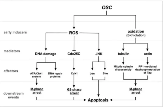
Figure 5. Scheme of action of OSC-mediated cell cycle arrest and apoptosis in tumor cell. OSC can
induce ROS production or directly oxidize cysteine residues on protein surface. These are the early events occurring after OSC administration, which result in the modulation of secondary mediators. For instance, ROS can yield DNA damage, as well as activate JNK-dependent phosphorylative pathways, which culminate on c-Jun and Bim activation, and inhibit Cdc25C activity via oxidation and/or phosphorylation. Concomitantly, the direct S-thiolation of cytoskeleton components gives rise to mitotic spindle disassembly, as well as stress fibers alteration, which finally lead to a widespread dysfunction of cytoskeleton-associated proteins (e.g. Tau). This series of events converge on the induction of cell cycle arrest in different phases of the cell cycle and, depending on the success of the tumor cell repair machinery, on different cell responses: cell survival (resistance) or apoptosis (sensitivity).
microfilaments and microtubules in neuroblastoma cells. This event has been associated with Tau dephosphorylation by means of the activation of the redox-sensitive protein phosphatase 1 (PP1) [77]. Tau is considered the main microtubules-associated protein responsible for microtubules assembly and stabilization in cells of neuronal origin [78]. Indeed, deregulation of its function through an altered phosphorylation on Ser or Thr residues has been claimed as the principal cause of cytoskeleton disruption in a number of neurodegenerative disease named tauopathies [79]. Tau cleavage and/or change in its phosphorylation state are the principal events in the failure of the microtubule network occurring upon treatment with microtubules poisons commonly used in cancer therapy [80-82]. Therefore, the involvement of Tau in DADS-induced neuroblastoma cell death from one hand adds a novel potential target of OSC and, from the other, strengthens the damaging role of OSC towards cytoskeleton dynamics.
The results that are rapidly coming out will be fundamental to wholly comprehend the redox effects of OSC on cell viability and how they concur in the induction of downstream signaling pathways leading to apoptosis. In this context, cytoskeleton is assuming a pivotal role, since the increasing knowledge on apoptotic mechanisms allows speculating that it could represent a good candidate in linking OSC-mediated damages to the mitochondrial pathway of apoptosis. Along this line, recent results indicated that JNK mediates hyper-phosphorylation of Bim, a BH3-only pro-apoptotic member of the Bcl-2 superfamily, and that this represents one of the earlier events of garlic-induced cell cycle arrest and apoptosis [83]. Indeed, Bim has been often indicated to be involved in JNK-dependent apoptosis when oxidative stress induces alteration of microtubule architecture [84].
On the basis of such results, we can reasonably speculate that there is no hierarchy among the apoptotic mechanisms induced by OSC, but JNK activation and cytoskeleton disassembly could act in concert in inducing cell response. The decision between proliferation, arrest or cell death represents the result of the relative contribution of the antioxidants and MAP kinase equipment, as well as the efficiency of DNA repairing machinery, which will channel the final cell response (Figure 5).
Lessons from epidemiological studies: Is garlic a panacea or a
promising chemotherapeutic agent?
Progress in establishing systemic pharmacological effects for fresh, crushed garlic or OSC in humans is hindered by several difficulties mostly related to the inability to measure allicin bio-availability, as well as to the lack of a direct evidence that allicin has significant systemic activity at doses of garlic normally consumed. Moreover, the knowledge reported so far on the widespread anti-proliferative effects of allyl compounds from garlic are related to cells that replicate at very high rate, such as tumor cells, and directly set against the cytotoxic effects of those compounds. Therefore, there is a gap between what we know till now about the pharmacological potentialities of garlic and its derivatives as chemotherapeutic agents and the paucity of epidemiological studies on humans. The absence of straightforward and consistent results in vivo does not allow yet to propose a therapeutic approach with allyl compounds in cancer treatment, or to define recommended dietary intake for a successful prevention of cancer.
The German Kommission E monograph (1988) proposed a daily intake of up to two cloves of garlic per day for health benefits, but no epidemiological or clinical studies have been performed afterwards [1]. Ngo et al. in 2007 published a systematic
review analyzing the available literature studies performed either in human or animal on the effects of garlic intake and colorectal cancer. The authors stated that due to great heterogeneity of measures of intake among case control and cohort studies, it is not possible to determine the minimum intake of garlic necessary to exhibit a protective effect in human. However, overall, the case control and the cohort studies demonstrated a consistent inverse association between a high garlic intake and colorectal cancer [85] as well as digestive tract cancers [86] development. On the other hand, animal studies were more systematic and the results pointed out that both oil- and water- soluble allyl sulfides, as well as aged or fresh garlic suspension or powder were effective in suppression of colonic carcinogenesis in a dose- and time-dependent manner. Moreover, colorectal tumors did not respond equally to different garlic allyl sulfur constituents, with DATS being 10 times more effective than DADS in suppressing colonic tumors [72, 87].
Studies of the efficacy of garlic or garlic-derived OSC on other types of human or animal cancer are more contradictory, or not even undertaken. Because of the peculiar chemistry of OSC, this lack of data could rely on possible not yet identified in vivo active components of garlic. In fact, despite a multitude of studies examining biological and chemical properties of garlic and its OCS, little is known about their metabolism. Lawson and Wang provide two methods for the evaluation of allicin bio-availability from garlic and garlic supplements in human [88]. This study presents important information on the metabolic pathway and the metabolic effect of allicin and allicin-derived compounds containing a dithioallyl groups. In particular, they demonstrated that, in humans after consumption of standardized garlic preparations, allicin is the solely responsible for breath allyl methyl sulfide (AMS). At isomolar concentrations of dithioallyl groups, DATS, DADS, ajoene, and SAMC, showed the same quantitative effects as allicin. Consumption of isomolar allyl AMS also gave the same effects as allicin, indicating that AMS was the main active metabolite of allicin. Therefore, breath AMS provides a new tool to assess allicin bio-availability.
Another aspect that deserves to be discussed is related to the quantity of garlic recommended for chemoprevention. Assuming that each fresh clove weights about 2 g, we can roughly estimate an average of 10-20 mg of allyl compounds per clove, the majority of which would be alliin and allicin. This, in theory, would be sufficient to reach blood concentrations of sulfur-containing allyl compounds of about 10-20 µmol/L after clove ingestion, but many factors make this hypothetical value diverge from the real blood or tissue concentrations, which are generally one or two orders of magnitude lower. This is due to the high reactivity of garlic-contained allyl compounds with intracellular thiols, particularly GSH, and explain why allicin is not found in blood or urine of human and rats even after ingestion of high quantities [88-91]. Another feature discouraging the use of purified allicin, DADS or other oil-soluble organo-sulfur compounds is related to their toxicity. For instance, hepatocytes, normally deputed to metabolize xenobiotics, are more resistant than epithelial cells of intestinal mucosa, which normally replicate at high rates. It has been demonstrated that raw garlic juice, as well as allicin or high doses of commercially available garlic preparations, causes severe damages to the stomach and the intestinal mucosa of rats, resulting in ulcers, shrinkage and bleeding [1, 92]. Instead, it is worth noting that water-soluble allyl sulfides, administered at high doses, do not show cytotoxicity and are able to induce cell responses through the activation of the antioxidant/detoxifying systems [93-95], or by triggering death of tumor cells [68, 96]. Finally, the latest knowledge available from nutrigenomic and nutrigenetic studies is that: “diet/genome interaction” determines a selective response of each individual to dietary
intake of garlic, and in turn, in the modulation of its effects on human health. In fact, it appears that dietary constituents (vitamins, micronutrients, antioxidants, phytochemicals etc.) affect metabolic pathways and homeostatic control by activating particular set of genes, and that the incidence of gene variants can influence such response to nutrients. This holistic approach to the nutritional aspects of biology is still in the phase of development; however, no particular polymorphism associated with apoptotic or antioxidant genes has yet been shown to be involved in garlic-derived allyl sulfides metabolism. However, we should consider that OSC derived from garlic can inhibit experimental cancer in various animal models through modification of carcinogen-detoxifying enzymes, such as cytochrome P450 or GST. It was reported that both DAS and DADS efficiently inhibit CYP2E1, one of the isoenzymes of cytochrome P450, which is responsible for the activation of nitrosamine, hydrazine and benzene [97], whereas DAS and AMS increased hepatic level of CYP1A family enzymes [98]. Moreover, it was shown that mice fed with DADS and allicin over-expressed GST in stomach and small intestine, particularly the α and µ isoforms of the enzyme [99]. From these observations, it seems plausible that the in vivo metabolism of sulfur-containing allyl compounds, and their effects on cancer – mostly in the initiation and promotion phases – would be strongly influenced by polymorphism of both cytochrome P450 and GST genes [100]. While information on this aspect has not been provided yet, it certainly deserves particular attention in order to plan an appropriate nutritional approach based on garlic consumption in cancer treatment.
In conclusion, garlic and its OSC appear to exert their anticarcinogenic effects through multiple mechanisms that include: i) modulation of carcinogen metabolism; ii) upregulation of antioxidant defences and DNA repair systems; iii) arrest of cell proliferation and iv) induction of apoptosis. Since multiple signaling pathways are dysfunctional in cancer and new oncogenic mutations accumulate with carcinogenic progression, dietary agents, such as garlic with its rich array of bioactive OSC that modulates cancer cascades, offer promise as potential chemopreventive and chemotherapeutic agents. Therefore, it seems advisable to include garlic and other allium plants in regular diet.
Acknowledgement
This work was partially supported by grants from Ministero dell’Università e della Ricerca Scientifica (MIUR) and Ministero della Salute.
References
1. Amagase, H., Petesch, B.L., Matsuura, H., Kasuga, S., and Itakura, Y. 2001, J. Nutr., 131, 955S.
2. Rivlin, R.S. 2001, J. Nutr., 131, 951S.
3. Yeh, Y.Y., and Liu, L. 2001, J. Nutr., 131, 989S. 4. Neil, A., and Silagy, C. 1994, Curr. Opin. Lipidol., 5, 6. 5. Steiner, M., and Li, W. 2001, J. Nutr., 131, 980S.
6. Kyo, E., Uda, N., Kasuga, S., and Itakura, Y. 2001, J. Nutr., 131, 1075S. 7. Sivam, G.P. 2001. J. Nutr. 2001, 131, 1106S.
8. Yang, C.S., Chhabra, S.K., Hong, J.Y., and Smith, T.J. 2001, J. Nutr., 131, 1041S.
9. Guyonnet, D., Belloir, C., Suschetet, M., Siess, M.H., and Le Bon, A.M. 2001, Mutat. Res., 495, 135.
10. Demeule, M., Brossard, M., Turcotte, .S, Regina, A., Jodoin, J., and Béliveau, R. 2004, Biochem. Biophys. Res. Commun., 324, 937.
11. Steinmetz, K.A., Kushi, L.H., Bostick, R.M., Folsom. A.R., and Potter, J.D. 1994, J. Epidemiol., 139, 1.
12. Fleischauer, A.T., and Arab, L. 2001, J. Nutr, 131, 1032S. 13. Milner, J.A. 2001, J. Nutr., 131, 1027S.
14. Ide, N., and Lau, B.H. 1999, Phytomedicine, 6, 125. 15. Ide, N., and Lau, B.H. 2001, J. Nutr., 131, 1020S.
16. Numagami, Y., and Ohnishi, S.T. 2001, J. Nutr., 131, 1100S. 17. Borek, C. 2001, J. Nutr., 131, 1010S.
18. Fenwick, G.R., and Hanley, A.B. 1985, Crit. Rev. Food Sci. Nutr., 22, 273.
19. Matsuura, H., Ushiroguchi, T., Itakura, Y., Hayashi, H., and Fuwa, T. 1988, Chem. Pharm. Bull., 36, 3659.
20. Songh, K., and Milner, J.A. 2001, J. Nutr., 131, 1054S. 21. Lawson, L.D. 1993, Chem. Abst., 119, 216.
22. Block, E., Ahmad, S., Jain, M.K., Crecely, R.W., Apitz-Castro, R., and Cruz, M.R. 1984, J. Am. Chem. Soc., 106, 8295.
23. Shukla, Y., and Kalra, N. 2007, Cancer Lett. 247, 167.
24. Hernandez, L.G., and Forkert, P.G. 2007, Carcinogenesis. 28, 1824.
25. Kensler, T.W., Wakabayashi, N., and Biswal, S. 2007, Annu. Rev. Pharmacol. Toxicol., 47, 89.
26. Motohashi, H., and Yamamoto, M. 2004, Trends Mol. Med., 10, 549. 27. Chen, C., and Kong, A.N. 2004, Free Rad. Biol. Med., 36, 1505.
28. Chen, C., Pung, D., Leong, V., Hebbar, V., Shen, G., Nair, S., Li, W., and Kong, A.N. 2004, Free Rad. Biol. Med., 37, 1578.
29. Hadi, S.M., Asad, S.F., Singh, S., and Ahmad, A. 2000, IUBMB Life, 50, 167. 30. Williams, R.J., Spencer, J.P., and Rice-Evans, C. 2004, Free Radic. Biol. Med., 36, 838. 31. Podmore, I.D., Griffiths, H.R., Herbert, K.E., Mistry, N., Mistry, P., and Lunec, J. 1998,
Nature, 392, 559.
32. Behrend, L., Henderson, G., and Zwacka, R.M. 2003, Biochem. Soc. Trans., 31, 1441. 33. Pelicano, H., Carney, D., and Huang, P. 2004, Drug Resist. Updat., 7, 97.
34. Rotilio, G., Mavelli, I., Rossi, L., Ciriolo, M.R. 1985, Environ. Health Perspect., 64, 259. 35. Kwon, K.B., Yoo, S.J., Ryu, D.G., Yang, J.Y., Rho, H.W., Kim, J.S., Park, J.W., Kim, H.R.,
and Park, B.H. 2002, Biochem. Pharmacol., 63, 41.
36. Filomeni, G., Aquilano, K., Rotilio, G., and Ciriolo, M.R. 2003, Cancer Res., 63, 5940. 37. Adler, V., Yin, Z., Fuchs, S.Y., Benezra, M., Rosario, L., Tew, K.D., Pincus, M.R., Sardana,
M., Henderson, C.J., Wolf, C.R., Davis, R.J., and Ronai, Z. 1999, EMBO J. 18, 1321. 38. Adler, V., Yin, Z., Tew, K.D., and Ronai, Z. 1999, Oncogene, 18, 6104.
39. Filomeni, G., Aquilano, K., Rotilio, G., and Ciriolo, M.R. 2005, Cancer Res., 65, 11735. 40. Levinthal, D.J., and Defranco, D.B. 2005, J. Biol. Chem., 280, 5875.
41. Sriram, N., Kalayarasan, S., Ashokkumar, P., Sureshkumar, A., and Sudhandiran, G. 2008, Mol. Cell. Biochem., 311, 157.
42. Antosiewicz, J., Ziolkowski, W., Kar, S., Powolny, A.A., and Singh, S.V. 2008, Planta Med. 74, 1570.
43. Munday, R., Munday, J.S., and Munday, C.M. 2003, Free Radic. Biol. Med., 34, 1200. 44. Lawson, L.D., Wood, S.G., and Huges, B.G. 1991, Planta Medica, 57, 263.
45. Münchberg, U., Anwar, A., Mecklenburg, S., and Jacob, C. 2007, Org. Biomol. Chem., 5, 1505.
46. Bautista, D.M., Movahed, P., Hinman, A., Axelsson, H.E., Sterner, O., Högestätt, E.D., Julius, D., Jordt, S.E., and Zygmunt, P.M. 2005, Proc. Natl. Acad. Sci. USA., 102, 12248.
47. Hamer, D.H. 1986, Annu. Rev. Biochem., 55, 913.
48. Freedman, J.H., Ciriolo, M.R., and Peisach, J. 1989, J. Biol. Chem. 264, 5598. 49. Sawahata, T., and Neal, R.A. 1982, Mol. Pharmacol., 21, 464.
51. Dugravot, S., Grolleau, F., Macherel, D., Rochetaing, A., Hue, B., Stankiewicz, M., Huignard, J., and Lapied, B. 2003, J. Neurophysiol., 90, 259.
52. Chatterji, T., and Gates, K.S. 2003, Bioorg. Med. Chem. Lett., 13, 1349.
53. Benavides, G.A., Squadrito, G.L., Mills, R.W., Patel, H.D., Isbell, T.S., Patel, R.P., Darley-Usmar, V.M., Doeller, J.E., and Kraus, D.W. 2007, Proc. Natl. Acad. Sci. USA., 104, 17977. 54. Lefer, D.J. 2007, Proc. Natl. Acad. Sci. USA., 104, 17907.
55. Blackstone, E., Morrison, M., and Roth, M.B. 2005, Science, 308, 518.
56. Antosiewicz, J., Herman-Antosiewicz, A., Marynowski, S.W., and Singh, S.V. 2006, Cancer Res., 66, 5379.
57. Knowles, L.M., and Milner J.A. 1998, Nutr. Cancer., 30, 169. 58. Knowles, L.M., and Milner, J.A. 2000, Carcinogenesis, 21, 1129. 59. Knowles, L.M., and Milner, J.A. 2001, J. Nutr., 131, 1061S.
60. Xiao, D., Herman-Antosiewicz, A., Antosiewicz, J., Xiao, H., Brisson, M., Lazo, J.S., and Singh, S.V. 2005, Oncogene, 24, 6256.
61. Sanchez, Y., Wong, C., Thoma, R.S., Richman, R., Wu, Z., Piwnica-Worms, H., and Elledge, S.J. 1997, Science., 277, 1497.
62. Herman-Antosiewicz, A., and Singh, S.V. 2005, J. Biol. Chem. 280, 28519.
63. Herman-Antosiewicz, A., Stan, S.D., Hahm, E.R., Xiao, D., and Singh, S.V. 2007, Mol. Cancer Ther., 6, 1249.
64. Aquilano, K., Filomeni, G., Baldelli, S., Piccirillo, S., De Martino, A., Rotilio, G., and Ciriolo, M.R. 2007, J. Neurochem., 101, 1327.
65. Rudolph, J. 2005, Antioxid. Redox Signal., 7, 761.
66. Fauman, E.B., Cogswell, J.P., Lovejoy, B., Rocque, W.J., Holmes, W., Montana, V.G., Piwnica-Worms, H., Rink, M.J., and Saper, M.A. 1998, Cell, 93, 617.
67. Savitsky, P.A., and Finkel, T. 2002, J. Biol. Chem., 277, 20535.
68. Shirin, H., Pinto, J.T., Kawabata, Y., Soh, J.W., Delohery, T., Moss, S.F., Murty, V., Rivlin, R.S., Holt, P.R., and Weinstein, I.B. 2001, Cancer Res., 61, 725.
69. Xiao, D., Pinto, J.T., Soh, J-W., Deguchi, A., Gundersen, G.G., Palazzo, A.F., Yoon, J-T., Shirin, H., and Weinstein, I.B. 2003, Cancer Res., 63, 6825.
70. Xiao, D., Pinto, J.T., Gundersen, G.G., and Weinstein, I.B. 2005, Mol. Cancer Ther., 4, 1388. 71. Xiao, D., Lew, K.L., Kim, Y.A., Zeng, Y., Hahm, E.R., Dhir, R., and Singh, S.V. 2006, Clin.
Cancer Res., 12, 6836.
72. Hosono, T., Fukao, T., Ogihara, J., Ito, Y., Shiba, H., Seki, T., and Ariga, T. 2005, J. Biol. Chem., 280, 41487.
73. Dalle-Donne, I., Rossi, R., Milzani, A., Di Simplicio, P., and Colombo, R. Free Radic. Biol. Med., 31, 1624.
74. Fiaschi, T., Cozzi, G., Raugei, G., Formigli, L., Ramponi, G., and Chiarugi, P. 2006, J. Biol. Chem., 281, 22983.
75. Chiarugi, P., and Fiaschi, T. 2007, Cell Signal., 19, 672.
76. Sela, U., Ganor, S., Hecht, I., Brill, A., Miron, T., Rabinkov, A., Wilchek, M., Mirelman, D., Lider, O., and Hershkoviz, R. 2004, Immunology, 111, 391.
77. Aquilano, K., Vigilanza, P., Filomeni, G., Rotilio, G., and Ciriolo, M.R. 2008, J. Cell. Mol. Med., 6, 1.
78. Binder, L.I., Frankfurter, A., and Rebhun, L.I. 1985, J. Cell. Biol., 101, 1371. 79. Lee, V.M., Goedert, M., and Trojanowski, J.Q. 2001, Annu. Rev. Neurosci., 24, 1121. 80. Mattson, M.P. 1992, Brain Res., 582, 107.
81. Davis, D.R., Brion, J.P., Couck, A.M., Gallo, J.M., Hanger, D.P., Ladhani, K., Lewis, C., Miller, C.C., Rupniak, T., Smith, C., and Anderton, B.H. 1995, Biochem. J., 309, 941. 82. Merrick, S.E., Demoise, D.C., and Lee, V.M. 1996, J. Biol. Chem., 271, 5589. 83. Lund, T., Stokke, T., Olsen, Ø.E., and Fodstad, Ø. 2005, Br. J. Cancer., 92, 1773. 84. Lei, K., and Davis, R.J. 2003, Proc. Natl. Acad. Sci. USA, 100, 2432.
85. Ngo, S.N.T., Williams, D.B., Cobiac, L., and Head, R.J. 2007, J. Nutr., 137, 2264. 86. Iciek, M., Kwiecien, I., and Wlodek, L. 2009, Environ. Mol. Mutagen., 50, 247. 87. Milner, J.A. 2006, J Nutr., 136 S827.
88. Lawson, L.D., and Wang, Z.J. 2005, J. Agric. Food Chem., 53, 1974. 89. Lawson, L.D., Ransom, D.K., and Hughes, B.G. 1992, Thromb. Res., 65, 141.
90. Egen-Schwind, C., Eckard, R., Jekat, F.W., and Wirterhoff, H. 1992, Planta Med., 58, 83. 91. Pushpendran, C.K., Devasagayam, T.P.A., Chintalwar, G.J., Banerji, A., and Eapen, J. 1980,
Experientia, 36, 1000.
92. Shashikanth, K.N., Basappa, S.C., and Murthy, V. 1985, J. Food. Sci. Technol., 22, 110. 93. Ide, N., Nelson, A.B., and Lau, B.H.S. 1997, Planta Med., 63, 263.
94. Imai, J., Ide, N., Nagae, S., Moriguchi, T., Matsuura, H., and Itakura, Y. 1994, Planta Med., 60, 417.
95. Amagase, H., and Milner, J. 1993, Carcinogenesis, 14, 1627.
96. Li, G., Qiao, C.H., Lin, R.I., Pinto, J., Osborne, M.P., and Tiwari, R.K. 1995, Oncol. Rep., 2, 787.
97. Wargovich, M.J. 2006, J. Nutr., 136, 832S.
98. Davenport, D.M., and Wargovich, M.J. 2005, Food Chem. Toxicol. 43, 1753. 99. Andorfer, J.H., Tchaikovskaya, T., and Listowsky, I. 2004, Carcinogenesis, 25, 359. 100. Reszka, E., Wasowicz, W., and Gromadzinska, J. 2006, Br. J. Nutr., 96, 609.