B-cell chronic lymphocytic leukaemia
in an African lion (Panthera leo)
R. Meoli
1, C. Eleni
1, P. Cavicchio
2, M.C. Tonnicchia
2, B. Biancani
3,
L. Galosi
4, G. Rossi
4*
1Experimental Zooprophylactic Institute of Lazio and Tuscany “M. Aleandri”, Rome, Italy 2Zoological Society of Pistoia, Pistoia, Italy
3Park Oltremare, Riccione, Italy
4School of Biosciences and Veterinary Medicine, University of Camerino, Matelica, Italy
*Corresponding author: [email protected]
ABSTRACT: A 15-year-old female African lion (Panthera leo) maintained at the Zoological Garden of Pistoia, Tuscany (Italy), showed signs of malaise, dyspnoea, tremors, pale mucous membranes and ataxia for two days prior to death. Complete blood count showed anaemia, thrombocytopaenia and severe lymphocytosis. At autopsy, the most relevant finding was severe, generalised splenomegaly. Histologically, high numbers of neoplastic lympho-cytes diffusely infiltrated the spleen, liver, heart, pancreas, kidney and lungs. The lymphoid cells were positive for CD79a and negative for CD3 on immunohistochemistry. The histologic features of the neoplastic cells and their immunophenotype are consistent with B-cell lymphocytes. Based on surface membrane co-expression of immunoglobulin M and immunoglobulin D in most neoplastic cells, which indicated that they were naïve, antigen-inexperienced and mature circulating resting B-cells, and consistent with their appearance as small lymphocytes with high nuclear-to-cytoplasmic ratios, a diagnosis of B-cell chronic lymphocytic leukaemia was made.
Keywords: lymphocytosis; feline, neoplastic disease
Neoplasms of the haematopoietic system are commonly seen in domestic animals; malignant lymphomas have been extensively studied in do-mestic canids and felids, but there is limited infor-mation on malignant lymphomas in non-domestic animals (Gabor et al. 1998; Harrison et al. 2010).
Exotic felids in captivity, particularly African li-ons (Panthera leo), are rarely diagnosed with ma-lignant lymphoma and only three cases of T-cell chronic lymphocytic leukaemia have been report-ed so far (Harrison et al. 2010). These animals were males and had weight loss, anaemia, lymphocytosis and splenomegaly. Although lymphomas in domes-tic felids have been associated with feline leukae-mia virus and feline immunodeficiency virus, there is only one documented case of lymphoma in a captive African lion that was concurrently infected with feline lentivirus (Harrison et al. 2010; Helfand and Kisseberth 2010; Vezzali et al. 2010).
In veterinary medicine, as in humans, haemat-opoietic neoplasms are classified as myeloid or lym-phoid on the basis of histogenesis and are divided into acute and chronic forms, based on clinical pres-entation and the proliferative rate of the neoplastic cell clone (Tordiffe et al. 2013). Acute leukaemias are clonal proliferations of a progenitor cell with limited differentiation; therefore, identification of cellular markers is often necessary for definitive identifi-cation of the cell of origin. In chronic leukaemias, neoplastic cells are terminally differentiated, and the cellular origin is more readily identifiable (Gabor et al. 1998). The primary laboratory finding in chronic lymphocytic leukaemia is leucocytosis caused by an absolute lymphocytosis. In domestic cats, lympho-cyte counts varying from 36 000 to 250 000/l have been reported and haematological findings could include anaemia, thrombocytopaenia, neutropaenia and neutrophilia. Animals with thrombocytopaenia
are predisposed to haemorrhage owing to decreased platelet numbers and abnormal cell function (Valli et al. 2000).
Case description
We describe herein a case of B-cell chronic lym-phocytic leukaemia in a 15-year-old female African lion (Panthera leo), maintained at the Zoological Garden of Pistoia, Tuscany (Italy). In October 2015, the lioness displayed signs of malaise, dyspnoea, tremors, pale mucus membranes and ataxia for two days. The lioness was anaesthetised with a com-bination of 500 mg of tiletamine and zolazepam (Zoletil®, Virbac, France) with addition of 7 mg
of detomidine (Domosedan®, Pfizer, USA) for
di-agnostic evaluation. Full anaesthesia was reached 25 minutes after injection and lasted for one hour. Ultrasonographic examination of the abdomen re-vealed diffuse, uniform and severe splenomegaly. Complete blood cell count results were anaemia and severe lymphocytosis. Biochemical indicators were elevated concentrations of urea and alanine aminotransferase, while albumin and glucose con-centrations were low (Table 1). ELISA tests for fe-line immunodeficiency virus, fefe-line leukaemia virus (SNAP® FIV/FeLV Combo Plus Test, IDEXX, USA)
and Dirofilaria immitis (SNAP® 4Dx® Test, IDEXX,
USA) were negative. Two days after the investiga-tion, the lioness died.
Pathological findings. An autopsy was
per-formed at the Zoological Garden of Pistoia. Samples of various organs were fixed in 10% buffered for-malin and sent for histologic examination at the Experimental Zooprophylactic Institute of Lazio and Tuscany. The samples were embedded in par-affin, sectioned at 5 µm, and stained with haema-toxylin-eosin (H&E). Immunohistochemistry was performed with CD3 (Dako, UK), CD79a (Dako, UK) and CD20 (thermo Fisher Scientific, USA) an-tibodies at a dilution of 1 : 600 to establish wheth-er the neoplastic cells wwheth-ere of T- or B-cell origin. Furthermore, immunoglobulin M (goat anti-cat IgM, Biorad, USA) and anti- immunoglobulin D (rabbit polyclonal anti-IgD, Zeta Corporation, USA) antibodies were both used at a dilution of 1 : 200 in immunohistochemistry staining to evalu-ate the stevalu-ate of eventual activation of the B-cells, necessary to make a more precise diagnosis of the neoplastic form. Positive immunohistochemical
controls included a normal feline lymph node. For negative controls, the primary antibodies were re-placed with homologous non-immune sera.
The most relevant finding during gross examina-tion was a severe generalised splenomegaly. The spleen had rounded edges, was dark purple and was very dense. It weighed 7.5 kg (nearly 20 times its normal size) and was 110 cm high × 50 cm long × 20 cm wide. Other findings included pale mucos-ae, pale kidneys, pulmonary congestion associated with areas of lung parenchyma consolidation, mild catarrhal enteritis and numerous ovarian cysts; a small mass of increased toughness was observed in uterus. All lymph nodes appeared normal.
Histologically, large numbers of neoplastic lym-phocytes diffusely infiltrated the spleen, liver, heart, pancreas, kidney and lung. The most prominent infiltrates were seen in the spleen (60% involve-ment), concentrated in the red pulp, with atrophy of lymphoid follicles (Figure 1). Massive infiltration of neoplastic cells was observed in liver sinusoids and, to a lesser extent, between the cardiac muscle fibres and around the pancreatic acini. Neoplastic cells consisted of small atypical lymphoid cells (6.5/7.5 µm) with small nuclei (high nuclear-to-cy-toplasm ratio), an absence of nucleoli and coarsely
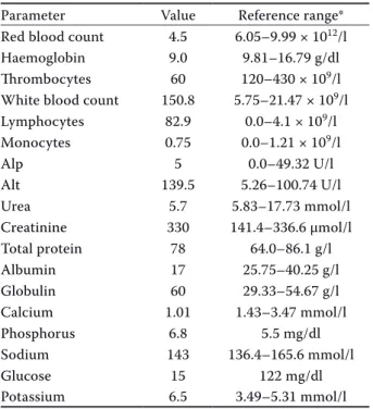
Table 1. Haematological and biochemical values
Parameter Value Reference range* Red blood count 4.5 6.05–9.99 × 10¹²/l Haemoglobin 9.0 9.81–16.79 g/dl
Thrombocytes 60 120–430 × 109/l
White blood count 150.8 5.75–21.47 × 10⁹/l Lymphocytes 82.9 0.0–4.1 × 10⁹/l Monocytes 0.75 0.0–1.21 × 10⁹/l Alp 5 0.0–49.32 U/l Alt 139.5 5.26–100.74 U/l Urea 5.7 5.83–17.73 mmol/l Creatinine 330 141.4–336.6 µmol/l Total protein 78 64.0–86.1 g/l Albumin 17 25.75–40.25 g/l Globulin 60 29.33–54.67 g/l Calcium 1.01 1.43–3.47 mmol/l Phosphorus 6.8 5.5 mg/dl Sodium 143 136.4–165.6 mmol/l Glucose 15 122 mg/dl Potassium 6.5 3.49–5.31 mmol/l
*Reference ranges were calculated using the International Species Information System (ISIS) database values (Teare 1999) for lions
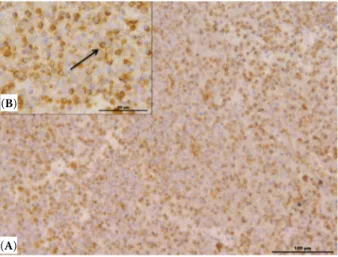
90% of the neoplastic cells were positive for CD79a (Figure 2). The neoplastic lymphoid proliferation was classified using the current veterinary and human WHO classification of haematopoietic neoplasms (Valli et al. 2000; Vezzali et al. 2010).
In this case, the lioness showed typical signs of chronic lymphocytic leukaemia, including weight loss, splenomegaly, anaemia and marked lympho-cytosis. Unfortunately, we were unable to obtain the bone marrow for evaluation. A massive infiltration of neoplastic cells was observed in liver sinusoids. Despite not having bone marrow, this favours clas-sification as a leukaemia. Moreover, thrombocyto-paenia indicates an important presence of neoplastic cells in the bone marrow. The massive infiltration of the spleen and other organs, and the features of the neoplastic cells, suggests that the lioness was affect-ed by chronic lymphocytic leukaemia. The CD79a-positivity and CD3-negativity provide evidence that the leukaemia was primarily B-cell in origin. In particular, according to reports (Caligaris-Cappio et al. 1993), the accumulation in different organs of naïve, antigen-inexperienced, well-differentiated B lymphocytes that circulate in the resting state, is considered a typical mark of B-CLL.
DISCUSSION AND CONCLUSIONS
Malignant lymphoma is the most common malig-nant neoplasm diagnosed in domestic cats, while aggregated chromatin and moderate amounts of pale
cytoplasm. These cells, in many areas of organ infil-tration, resembled normal lymphocytes, although slightly smaller. These neoplastic cells showed a low mitotic index (0 and 1 mitoses/HPF). In addition, membranoproliferative glomerulonephritis and chronic interstitial pneumonia, with areas of scleros-ing alveolitis, were observed, along with lymphop-lasmacytic enteritis and leiomyoma in the uterus. In lymph nodes, depletion of lymphoid follicles was observed. Immunohistochemically, most of the lym-phoid cells showed characteristic cell membrane CD79a positivity and negativity for CD3. The B-cell marker CD20, used in this study, does not cross-react with African lion lymphocytes. Furthermore, B-cells showed a constant co-expression of immunoglobulin M and immunoglobulin D on their surface mem-branes, which usually marks naïve B-cells. Indeed, the view that B-cell chronic lymphocytic leukaemia (B-CLL) cells are antigen-naïve and resting but ma-ture, is consistent with their appearance in tissue infiltrates, as small lymphocytes with a high nuclear-to-cytoplasmic ratio (Bennett et al. 1989). These im-munohistochemistry findings were observed in all organs, with particular evidence in the spleen, where
Figure 1. Spleen of an African lion with chronic lympho-cytic leukaemia. (A) Neoplastic lymphocytes diffusely replacing most of the normal splenic cell population. Haematoxylin and eosin (H&E). Bar = 200 μm. (B) Higher magnification; a detailed view of the charac-teristic features of atypical lymphocytes. Neoplastic cells consisted of small atypical lymphoid cells (6.5/7.5 µm) with small nuclei (moderate N/C ratio), absence of nucleoli and coarsely aggregated chromatin and moder-ate amounts of pale cytoplasm. Haematoxylin and eosin (H&E). Bar = 20 μm
Figure 2. Spleen of an African lion with chronic lym-phocytic leukaemia. (A) Neoplastic lymphocytes are immunoreactive for CD79a antibody. Bar = 100 μm. (B) At higher magnification, characteristic cell membrane CD79a positivity was observed (arrow). Bar = 50 μm
(A) (B)
(A) (B)
chronic lymphocytic leukaemia (CLL) is rare (Tebb et al. 2004). Exotic felids in captivity, particularly African lions, are rarely diagnosed with malignant lymphomas or CLL (Hruban et al. 1992; Owston et al. 2008; Valli 2010). Eleven cases of malignant lym-phoma in African lions were described, and only one case was described as a diffuse large B-cell lym-phoma (Harrison et al. 2010). The literature sug-gests that chronic lymphocytic leukaemia is most commonly of T-cell origin in exotic felids; the aeti-ology and pathogenesis in humans, dogs, cats and lions remains unknown. Neither feline leukaemia virus nor feline immunodeficiency virus, important causes of malignant lymphoma in domestic cats, are likely to be significant in the pathogenesis of malignant lymphoma in African lions (Poli et al. 1995; Callanan et al. 1996; Harrison et al. 2010).
Chronic lymphocytic leukaemia (CLL) is a com-mon condition that typically affects geriatric human beings (Binet et al. 1981). In T-CLL cells, now called T-cell prolymphocytic leukaemia (Catovsky et al. 2001), a co-occurrence of high levels of CD3 antigen together with low levels of the B-cell surface anti-gens CD19, CD20 and CD23 is described (Moreau et al. 1997). The levels of the surface immunoglobulin CD20 were characteristically very low compared with those found on normal B-cells (Ginaldi et al. 1998).
The CD79a protein is present on the surface of B-cells throughout their life cycle and is also pre-sent in virtually all B-cell neoplasms; however, it is absent on all other healthy cells, making it a highly reliable marker for B-cells in immunohistochem-istry. The B-cell marker CD20 is more commonly retained on mature B-cell lymphomas, so that the two are often used together in immunohistochem-istry panels (Leong et al. 2003). In contrast, B-CLL cells typically express high levels of CD20, CD79a and surface Ig (Catovsky et al. 2001). Regarding surface Ig expression, B-CLL cells are generally na-ïve, antigen-inexperienced mature B lymphocytes that circulate in the resting state (Caligaris-Cappio et al. 1993), and their characteristic surface mem-brane co-expression of immunoglobulin M and immunoglobulin D, indicates their status as “vir-gin” B-cells (Coffman and Cohn 1977). Also, in the present case, B-CLL was diagnosed on the basis of ubiquitous B-cell marker CD79a immunochemistry expression, and surface membrane co-expression of immunoglobulin M and immunoglobulin D; unfortunately, the B-cell marker CD20 does not cross-react with lioness lymphocytes.
The numerous statistics published on CLL have revealed a certain number of prognostic factors. The importance of sex, age, the degree of periph-eral lymphocytosis, the degree of associated anaemia or thrombocytopaenia, cutaneous manifestations, signs of inflammation and the onset of a sarcoma have all been stressed but it has not been possible to determine the relative importance of each of these (Binet et al. 1981). In humans, CLL is a neoplasm composed of monomorphic small B lymphocytes in the peripheral blood, bone marrow, spleen and lymph nodes that form proliferation centres in tissue infiltrates (Helfand and Kisseberth 2010). In CLL, B-cells exhibit uncontrolled proliferation and ac-cumulate in the bone marrow and blood, crowding out healthy red or white blood cells and platelets. Asymptomatic people are often diagnosed after lym-phocytosis is noted in a routine complete blood cell count; a marrow aspirate and biopsy generally are not required for the diagnosis (Hallek et al. 2008).
CLL in cats, where the disease appears most of-ten, is characterised by small cells with round nuclei typically 1 to 1.5 red blood count in diameter with a very narrow rim of cytoplasm. Leukaemia would be expected to involve multiple tissues and in those interpreted to be true leukaemia, both acute lympho-cytic leukaemia and CLL were more likely widely dis-tributed, which attests to the utility of liver aspiration as an adjunct to examination of blood and marrow in the diagnosis of leukaemia (Vezzali et al. 2010).
Although CLL is a systemic disease, collections of pale cells known as proliferation centres, which are newly formed structures, and have not been described in reactive lymphadenitis or other lym-phoproliferative conditions, can be found. The cells in these collections are also malignant cells but are somewhat larger and mitotically active. In the current case, no lymph node involvement was ob-served, like in domestic cats, and the lioness was negative for feline leukaemia virus and feline im-munodeficiency virus; therefore, the aetiology of the leukaemia is unknown.
To our knowledge, this is the first case of B-cell chronic lymphocytic leukaemia reported in an African lion (Panthera leo).
Acknowledgements
The authors express their thanks to Professor Susan Cork from the University of Calgary, Canada,
for her valuable assistance in writing up this case, and Dr Paul Gatenby for his revision of the text.
REFERENCES
Bennett JM, Catovsky D, Daniel MT, Flandrin G, Galton DA, Gralnick HR, Sultac C (1989): Proposals for the clas-sification of chronic (mature) B and T lymphoid leukae-mias. French-American-British (FAB) Cooperative Group. Journal of Clinical Pathology 42, 567–584. Binet JL, Auquier A, Dighiero G, Chastang C, Piquet H,
Goasquen J, Vaugier G, Potron G, Colona P, Oberling F, Thomas M, Tchernia G, Jacquillat C, Boivin P, Lesty C, Duault MT, Monconduit M, Belabbes S, Gremy F (1981): A new prognostic classification of chronic lymphocytic leukemia derived from a multivariate survival analysis. Cancer 48, 198–206.
Caligaris-Cappio F, Gottardi D, Alfarano A, Stacchini A, Gregoretti MG, Ghia P, Bertero MT, Novarino A, Bergui L (1993): The nature of the B lymphocyte in B-chronic lymphocytic leukemia. Blood Cells 19, 601–613. Callanan JJ, Jones BA, Irvine J, Willett BJ, McCandlish IA,
Jarrett O (1996): Histologic classification and immu-nophenotype of lymphosarcomas in cats with naturally and experimentally acquired feline immunodeficiency virus infection. Veterinary Pathology 33, 264–272. Catovsky D, Ralfkiaer E, Muller-Hermelink HK (2001):
T-cell prolymphocytic leukaemia. In: Jaffe ES, Harris NL, Stein H, Vardiman JW (eds): World Health Organization Classification of Tumours: Pathology and Genetics of Tumours of Haematopoietic and Lymphoid Tissues. Lyon, France: IARC Press. 195–196.
Coffman RL, Cohn M (1977): The class of surface immu-noglobulin on virgin and memory B lymphocytes. The Journal of Immunology 118, 1806–1815.
Gabor LJ, Malik R, Canfield PJ (1998): Clinical and ana-tomical features of lymphosarcoma in 118 cats. Austral-ian Veterinary Journal 76, 725–732.
Ginaldi L, De Martinis M, Matutes E, Farahat N, Morilla R, Catovsky D (1998): Levels of expression of CD19 and CD20 in chronic B cell leukaemias. Journal of Clinical Pathology 51, 364–369.
Hallek M, Cheson BD, Catovsky D, Caligaris-Cappio F, Dighiero G, Dohner H, Hillmen P, Keating MJ, Montser-rat E, Rai KR, Kipps TJ (2008): Guidelines for the diag-nosis and treatment of chronic lymphocytic leukemia: a report from the International Workshop on Chronic Lymphocytic Leukemia updating the National Cancer Institute-Working Group 1996 guidelines. Blood 15, 5446–5456.
Harrison TM, McKnight CA, Sikarskie JG, Kitchell BE, Garner MM, Raymond JT, Fitzgerald SD, Valli VE, Agnew D, Kiupel M (2010): Malignant lymphoma in African lions (Panthera leo). Veterinary Pathology 47, 952–957. Helfand SC, Kisseberth WC (2010): General features of
leukemia and lymphoma. In: Weiss DJ, Wardrop KJ (eds): Schalm’s Veterinary Hematology. 6th edn. Wiley-Black-well, Ames. 455–466.
Hruban Z, Vardiman J, Meehan T, Frye F, Carter WE (1992): Haematopoietic malignancies in zoo animals. Journal of Comparative Pathology 106, 15–24.
Leong ASY, Cooper K, Leong F, Joel WM (eds) (2003): Manual of Diagnostic Cytology. 2nd edn. Greenwich Medical Media. 83–84.
Moreau EJ, Matutes E, A’Hern RP, Morilla AM, Morilla RM, Owusu-Ankomah KA, Seon BK, Catovsky D (1997): Im-provement of the chronic lymphocytic leukemia scoring system with the monoclonal antibody SN8 (CD79b). American Journal of Clinical Pathology 108, 378–382. Owston MA, Ramsay EC, Rotstein DS (2008): Neoplasia in
felids at the Knoxville Zoological Gardens, 1979–2003. Journal of Zoo and Wildlife Medicine 39, 608–613. Poli A, Abramo F, Cavicchio P, Bandecchi P, Ghepardi E,
Pistello M (1995): Lentivirus infection in an African lion: A clinical, pathologic and virologic study. Journal of Wild-life Disease 31, 70–74.
Teare AJ (ed.) (1999): International Species Information Sys-tem: Physiological Data Reference Values. Apple Valley. Tebb AJ, Cave T, Barron R, Brown AL, Martineau HM,
Wil-lett BJ, Hosie MJ (2004): Diagnosis and management of B cell chronic lymphocytic leukaemia in a cat. Veterinary Record 154, 430–433.
Tordiffe ASW, Cassel N, Lane EP, Reyers F (2013): Multiple myeloma in a captive lion (Panthera leo). Journal of the South African Veterinary Association 84, 1–5.
Valli VE (2010): B-cell Tumors. In: Weiss DJ, Wardrop KJ (eds): Schalm’s Veterinary Hematology. 6th edn. Wiley-Blackwell, Ames. 491–585.
Valli VE, Jacobs RM, Norris A, Couto CG, Morrison WB, McCaw D, Cotter S, Ogilvie G, Moore A (2000): The his-tologic classification of 602 cases of lymphoproliferative disease using the National Cancer Institute Working For-mulation. Veterinary Journal of Diagnostic Investigation 12, 295–306.
Vezzali E, Parodi AL, Marcato PS, Bettini G (2010): Histo-pathologic classification of 171 cases of canine and feline non-Hodgkin lymphoma according to the WHO. Vet-erinary and Comparative Oncology 8, 38–49.
Received: November 16, 2017 Accepted after corrections: May 30, 2018