Osteogenic differentiation of mesenchymal stem cells from dental bud:
Role of integrins and cadherins
Adriana Di Benedetto
a,⁎
, Giacomina Brunetti
b, Francesca Posa
a, Andrea Ballini
c, Felice Roberto Grassi
c,
Graziana Colaianni
b, Silvia Colucci
b, Enzo Rossi
e, Elisabetta A. Cavalcanti-Adam
d, Lorenzo Lo Muzio
a,
Maria Grano
b, Giorgio Mori
aaDepartment of Clinical and Experimental Medicine, Medical School, University of Foggia, Italy
bSection of Human Anatomy and Histology, Department of Basic and Medical Sciences, Neurosciences and Sense Organs, University of Bari, Italy c
Department of Basic and Medical Sciences, Neurosciences and Sense Organs, University of Bari, Italy
d
Institute of Physical Chemistry, Department of Biophysical Chemistry, |University of Heidelberg AND Max Planck Institute for Intelligent Systems, Stuttgart, Germany
e
Private Practice, Oral and Maxillofacial Surgery, Poggio a Caiano, Florence, Italy
a b s t r a c t
a r t i c l e i n f o
Article history: Received 21 May 2015
Received in revised form 25 July 2015 Accepted 24 September 2015 Available online 30 September 2015 Keywords:
Mesenchymal stem cells Dental tissues Dental bud stem cells Osteogenic differentiation Cadherin
Integrin
Regenerative medicine
Several studies have reported the beneficial effects of mesenchymal stem cells (MSCs) in tissue repair and regen-eration. New sources of stem cells in adult organisms are continuously emerging; dental tissues have been iden-tified as a source of postnatal MSCs. Dental bud is the immature precursor of the tooth, is easy to access and we show in this study that it can yield a high number of cells with≥95% expression of mesenchymal stemness makers and osteogenic capacity. Thus, these cells can be defined as Dental Bud Stem Cells (DBSCs) representing a promising source for bone regeneration of stomatognathic as well as other systems. Cell interactions with the extracellular matrix (ECM) and neighboring cells are critical for tissue morphogenesis and architecture; such in-teractions are mediated by integrins and cadherins respectively. We characterized DBSCs for the expression of these adhesion receptors and examined their pattern during osteogenic differentiation. Our data indicate that N-cadherin and cadherin-11 were expressed in undifferentiated DBSCs and their expression underwent changes during the osteogenic process (decreasing and increasing respectively), while expression of E-cadherin and P-cadherin was very low in DBSCs and did not change during the differentiation steps. Such expression pattern reflected the mesenchymal origin of DBSCs and confirmed their osteoblast-like features. On the other hand, os-teogenic stimulation induced the upregulation of single subunits,αV, β3, α5, and the formation of integrin recep-torsα5β1 and αVβ3. DBSCs differentiation toward osteoblastic lineage was enhanced when cells were grown on fibronectin (FN), vitronectin (VTN), and osteopontin (OPN), ECM glycoproteins which contain an integrin-bind-ing sequence, the RGD motif. In addition we established that integrinαVβ3 plays a crucial role during the com-mitment of MSCs to osteoblast lineage, whereas integrinα5β1 seems to be dispensable. These data suggest that functionalization of biomaterials with such ECM proteins would improve bone reconstruction therapies starting from dental stem cells.
© 2015 The Authors. Published by Elsevier B.V. This is an open access article under the CC BY-NC-ND license (http://creativecommons.org/licenses/by-nc-nd/4.0/).
1. Introduction
Numerous studies have reported beneficial effects of multipotent MSCs in tissue repair and regeneration (Marx and Harrell, 2014; Otte et al., 2013; Yamaguchi, 2014). These cells can be isolated from many
different adult tissues, are self-renewable and can differentiate into all cell lineages that form mesenchymal and connective tissues (Yamaguchi, 2014; Barthes et al., 2014; Paschos and Brown, 2014).
The most well characterized source for MSCs is still the bone marrow, however in the past decade, populations of stem cells have been isolated from different dental tissues (Gronthos et al., 2000; Miura et al., 2003). A population of adult stem cells isolated from dental pulp (DP) tissues and designed as dental pulp stem cells (DPSCs), has been identified as a promising source of MSCs for tissue engineering (Gronthos et al., 2000; Daltoe et al., 2014; Mori et al., 2011; Mori et al., 2010; Spath et al., 2010; d'Aquino et al., 2009; Mangano et al., 2010; Galli et al., 2011; Mangano et al., 2011).
Abbreviations: MSCs, mesenchymal stem cells; DBSCs, dental bud stem cells; ECM, extracellular matrix; FN,fibronectin; VTN, vitronectin; OPN, osteopontin; BMMSCs, bone marrow MSCs; DP, dental pulp; DPSCs, dental pulp stem cells; DFSCs, dental follicle stem cells; ALP, alkaline phosphatase; ARS, Alizarin red staining.
⁎ Corresponding author at: Department of Clinical and Experimental Medicine, Medical School, University of Foggia, V.le L. Pinto 1, 71100 Foggia, Italy.
E-mail address:[email protected](A. Di Benedetto).
http://dx.doi.org/10.1016/j.scr.2015.09.011
1873-5061/© 2015 The Authors. Published by Elsevier B.V. This is an open access article under the CC BY-NC-ND license (http://creativecommons.org/licenses/by-nc-nd/4.0/).
Contents lists available atScienceDirect
Stem Cell Research
MSCs have also been identified in human Dental Follicle (DF) and Dental Bud (DB). DF is the loose connective tissue sac that surrounds the dental bud, which is the unerupted deciduous tooth. These tissues are more undifferentiated than mature Dental Pulp, thus dental bud is a more productive tissue of MSCs than mature DP and would have alter-native applications in bone and periodontal tissue engineering (Huang et al., 2009; Shi et al., 2001).
DBSCs give rise to all the tissues present in the mature tooth: enamel, dentin, pulp, cement, and periodontal ligament. We show in the present paper that the central part of the bud, corresponding to the dental papilla, contains the MSCs which can differentiate into osteoblast-like cells. To obtain these postnatal stem cells, the ex-traction of a tooth before its eruption is required; however, the early removal (age 7–12) of the dental bud of third molars yields excellent stem cells. Furthermore, patients selected for the extraction are often subjects that will undergo dental crowding which is worsened by wisdom teeth eruption, thus they will not lose a functional tooth. Since the stemness and osteogenic commitment of DFSCs has been previously established (Mori et al., 2012), these cells represent an excellent source for tissue engineering therapy and we can speculate on their possible use for bone regeneration of stomatognathic and other systems. In order to obtain an efficacious reconstruction of bone tissue, stem cells must grow and differentiate in osteogenic conditions on biomaterial scaffolds to be subsequently implanted in vivo. However the new bone formation or the osteointegration with the resident bone tissue sometimes result to be ineffective due to a failed recruitment and adhesion of stem cells on the scaffold. To achieve the correct tissue architecture during morphogenesis, cells must interact each other and with ECM. These interactions are mediated by two classes of adhesion receptors, respectively cadherins and integrins (Weber et al., 2011; Chen and Gumbiner, 2006; Berrier and Yamada, 2007). Cadherins are single chain trans-membrane glycoproteins that mediate cell–cell adhesion and inter-fere with intracellular signaling, in particular the Wnt/b-catenin pathway (Nelson and Nusse, 2004; MacDonald et al., 2009). They control proliferation, differentiation and survival of MSCs, which ex-press low levels of multiple cadherins and their pattern changes as stem cells are committed to a differentiated cell lineage. Several in vivo and in vitro studies have established a main role of cadherins, in particular N-cadherin and cadherin-11, in osteoblastogenesis and bone formation either by controlling cell-cell adhesion or interacting with Wnt intracellular signaling (Schambony et al., 2004; Bienz, 2005; Brembeck et al., 2006). Expanding the knowledge on cadherin expression in MSCs of dental origin would be fundamen-tal to promote the possible use of these cells in bone reconstruction therapies. Thus, as already demonstrated for DFSCs (Mori et al., 2012), the stemness and osteogenic commitment of DBSCs was con-firmed. Further, these cells were characterized for the expression of cadherins and their pattern examined during osteogenic differentia-tion. To achieve the goal of bone regeneration and osteointegration, it would be also important to examine cell–matrix interaction medi-ated by integrins. These are a family of cell surface receptors that pri-marily mediate adhesion of cells to the ECM proteins. Integrins are heterodimeric transmembrane proteins, consisting of associatedα andβ subunits, with a large extracellular domain and a short cyto-plasmic tail. Differently combinedα and β subunits result in a variety of integrin heterodimers that can bind a specific repertoire of ECM proteins. Among others arefibronectin, laminin, collagens, tenascin, vitronectin, osteopontin, bone sialoprotein, and dentin matrix protein-1 (Docheva et al., 2007). Besides, integrins transmit crucial signals inside the cells. Adhesion of MSCs to ECM is still poorly characterized, as well as protein matrix role in regulating osteogenic differentiation. Thus we characterized our cell model of DBSCs for the expression of four integrin subunits and receptor heterodimers and then analyzed the effect of DBSC adhesion on three ECM glycoproteins containing the integrin-binding sequence RGD, namely FN, VTN, and OPN.
2. Results
2.1. Immunophenotype of DBSCs
DBSCs from 20 donors were tested for the criteria to be called MSCs (Dominici et al., 2006), as was done for DFSCs (Mori et al., 2012), in order to ensure that these cells had actually stem and mesenchymal fea-ture. Flow cytometric analysis of research accepted MSC surface markers was performed and all populations exhibited≥95% expression of CD73, CD90, and CD105 while were negative for CD45, a common leukocyte antigen. DBSC samples from three donors were selected to perform the experiments, since they showed higher and similar per-centage values of stemness marker expression.Fig. 1upper panel, shows flow cytometry histograms from one representative DBSC culture.
2.2. Cadherin expression profile in DBSCs reflected their mesenchymal origin
Expression of“classical” cadherins was investigated in undifferenti-ated DBSCs and during their osteogenic differentiation. The cells were kept in favoring conditions for MSC maintenance and were character-ized for cadherin expression. Subcellular localization showed a high ex-pression of N-cadherin, as well as for cadherin-11 (Fig. 1A–B). E-cadherin was expressed to a lesser extent and P-E-cadherin was poorly expressed (Fig. 1C–D). Notably, only N-cadherin was markedly local-ized at the cell–cell contact, perhaps contributing to maintain the stem cells niche in the dental bud.
Thus cadherin expression profile was evaluated during osteogenic differentiation. As already demonstrated for DFSCs, (Mori et al., 2012) DBSCs cultured under osteogenic conditions differentiated into osteo-blast-like cells producing mineralized matrix nodules and expressed the typical osteoblastic markers Runx-2, alkaline phosphatase (ALP), and Collagen I (Coll I) (data not shown). Cadherin expression profile was analyzed by Western blotting during the different steps of osteo-genic differentiation. As shown in Fig. 1panel E, N-cadherin was expressed in undifferentiated DBSCs, thus confirming the subcellular observation; its expression remained constant after 7 days of osteogenic differentiation, but a slight decrease initiated after 14 days keeping a low expression of differentiation at 21 and 28 days. Similarly, cadherin-11 was expressed in undifferentiated DBSCs and its expression did not change after 7 days of differentiation, however after 2 weeks cadherin-11 strongly increased and remained highly expressed also at the later stages. Thus, while N-cadherin and cadherin-11 were present in undifferentiated DBSCs and their expression underwent changes dur-ing the osteogenic process, expression of E-cadherin and P-cadherin was low in DBSCs and did not change during the differentiation steps. Such expression profile of cadherins in DBSCs, and more precisely the changes undergoing cadherin-11 and N-cadherin (increasing and de-creasing respectively), confirmed that these stem cells can acquire an osteoblastic phenotype under appropriate osteogenic conditions. The subcellular localization of cadherins was observed only during the early phases of osteogenic differentiation (3–7 days), since these cells usually reach the confluence in few days and grow in multilayers, thus preventing their microscopic observation. E-cadherin and P-cadherin were poorly expressed and homogenously distributed in DBSCs and their localization did not change after 3–7 days of osteogenic trigger (data not shown) as their expression until 28 days of differentiation (Fig. 1panel E). Conversely N-cadherin and cadherin-11 changed their localization in response to osteogenic stimulation. InFig. 1(bis) F on time zero (T0) N-cadherin was localized at the cell–cell adhesion sites, being probably involved in the formation of adherens junction com-plexes as revealed by the“red bridges” merging with actin cytoskeleton marked by Phalloidin and resulting in an intense yellow staining. After 3 days N-cadherin remained localized in the adherens junction com-plex, while after 7 days N-cadherin lost the characteristic bridge-like
Fig. 1. Upper panel: immunophenotype of DFSCs. The expression of the indicated mesenchymal stem cell markers on DBSCs was analyzed usingflow cytometry. Results from one repre-sentative DBSCs culture are shown. The black histograms signify staining with isotype controls, and the white histograms represent staining with the specified surface marker antibody. Lower panel: Cadherin expression profile in DBSCs. Midsection confocal microscopy images of DBSCs cultured in basal conditions, counterstained for different type of Cadherins (red) and Phalloindin (green). Subcellular localization shows high expression of N-Cadherin (markedly localized at the cell–cell contact) and Cadherin-11 (A–B). E-Cadherin and P-Cadherin are expressed to a lesser extent (C–D). Results are depicted for one donor but are representative of three different donors. (E) Immunoblots showing the cadherin expression profile during the osteogenic differentiation process (0–28 days). Data are presented as mean ± SEM of 3 independent donors *P b 0.01 and #P b 0.05 compared to T0. Student's t-test was used for single comparison. Fig. 1 (bis): Confocal images show that N-Cadherin and Cadherin-11 change their localization in response to osteogenic stimulation (F–K). On time zero (F) and after 3 days (G), N-Cadherin is localized at the cell–cell adhesion sites forming “red bridges”, while after 7 days N-Cadherin loses the characteristic bridge-like appearance (H). Cadherin-11 accumu-lates and localizes at adherens junction sites after 3–7 days of differentiation (J–K).
appearance although in part remaining at the cell–cell boundaries not changing its expression. On the contrary cadherin-11 seemed to have an opposite trend. Indeed on T0, DBSCs expressed Cadherin-11, which was not localized at the cell–cell adhesion sites, but homogenously dis-tributed in multiple sites. After 7 days of osteogenic differentiation, cadherin-11 accumulated at the cell periphery and localized at adherens junction sites acquiring the bridge-like trend observed for N-cadherin in less differentiated cells.
2.3.αV and β3 integrin subunits increase during DBSCs osteogenic differen-tiation and form the functional integrin
Integrins trigger tissues morphogenesis, thus we investigated the subunits expressed in mesenchymal tissues (Shin et al., 2004; Takada et al., 2007; Schwab et al., 2013).
Again, due to the rapid propensity of the cells to form a multilayer, the presence of single integrin subunitsαV and β3 was analyzed by con-focal microscopy in DBSCs undifferentiated and after 7 days of osteo-genic differentiation. As shown inFig. 2 A–C both subunits were localized on the surface of undifferentiated cells, distributed in multiple sites and after 7 days of differentiation, each subunit seemed to reorga-nize and localize at the focal adhesion sites (Fig. 2B–D). The expression profile of αV and β3 was analyzed by Western blotting during 28 days of osteogenic differentiation (Fig. 2, panel E) confirming that both subunits were expressed at T0, increased after 7 days and progressively raised. Since integrin receptors are heterodimeric molecules, and alpha sub-units form complexes with various beta subsub-units, we verified if αV andβ3 were associated in DBSCs and formed the integrin complex. DBSCs co-stained forαV (red) and β3 (green) (Fig. 2F–G), showed some dots of colocalization (yellow staining) in osteogenic condition (7 days) (Fig. 2G), suggesting that the subunits could be associated. To assess whether the subunitsαV and β3 were associated to form the integrin receptor, whole cell lysates of DBSCs in basal and osteogenic condition for 7–14–21–28 days were immunoprecipitated with LM609 antibody (which recognizes only the complex) and western blotted withαV and β3 antibodies (Fig. 2H). This experiment failed to reveal the association ofαV and β3 in undifferentiated DBSCs, while showed that the presence of the receptor dimer was strictly time depending
during the differentiation process, reaching the higher expression after 21 days in DBSCs that turned in osteoblast-like cells. The subcellular distri-bution of integrinαVβ3, observed by confocal microscopy, revealed a scarce presence of the receptor in undifferentiated DBSCs (possibly not de-tected by the biochemical assay), however its localization appeared strong-ly augmented on the focal contacts in differentiated cells. (Fig. 2I–J; K–L). 2.4. Increased expression ofα5 integrin subunits in DBSCs osteogenic differ-entiation and association withβ1 subunit
The expression ofα5 and β1 subunits was detected by confocal mi-croscopy in undifferentiated DBSCs showing that both were present and homogenously localized on the cell surface (Fig. 3A–C). After 7 days in osteogenic conditions, expression ofα5 seemed to increase and accu-mulate in large peripheral clusters apparently localized on the base of the cell. The same trend was observed forβ1 suggesting that following the osteogenic trigger the subunits were recruited to form the integrin receptor (Fig. 3B–D). Furthermore, the expression trend of both sub-units was analyzed by Western blotting during 28 days of osteogenic differentiation.α5 was upregulated mainly at 7–14–28 days of differen-tiation (Fig. 3panel E), whileβ1 did not exhibit significant expression changes. As shown inFig. 3F–G, where α5 was stained in green and β1 in red, the two subunits appeared mainly separated in undifferenti-ated cells, with only few merged yellow dots, whereas they co-localized in differentiated cells and accumulated on the focal contacts. Immuno-precipitation experiments confirmed that α5 and β1 were physically as-sociated essentially in differentiated cells.
2.5. Interaction of integrins with correspondent extracellular matrix pro-teins enhances osteogenic differentiation and mineralization of DBSCs
We found that DBSCs expressed integrin subunitsαV, β3, α5, and β1 and that osteogenic trigger enhanced the expression ofαV, β3, and α5. Additionally, the osteogenic differentiation induced the association of single subunits to form the integrin heterodimersαVβ3 and α5β1, that localized at the focal adhesions. Integrin binding to ECM proteins affects intracellular signaling cascades involved in cell adhesion and mi-gration, as well as in stem cell commitment and differentiation. Thus we
investigated DBSCs osteogenic differentiation in the presence of three different ECM proteins, FN (which bindsα5β1 and to a lesser extent αVβ3), VTN (which binds only αVβ3), and OPN (which binds αVβ3
and to a lesser extentα5β1). The cells were seeded on the three protein coatings and on poly-L-lysine (PLL) as control (CTR), and cultured in osteogenic conditions. Western blot analysis was performed to
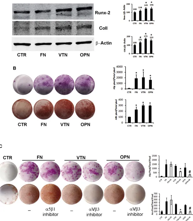
investigate the expression of two early markers of osteogenic differenti-ation, Runx-2 and Collagen I (Col I), that were found to increase during osteogenic differentiation of DFSCs (Mori et al., 2012). Moreover, using histochemical assays, we evaluated the expression of the osteoblast marker ALP after 7 days and the mineralization capacity with ARS after 21 days.
The differentiation pattern of DBSCs on all 3 surfaces was enhanced in comparison to the control (Fig. 4). Indeed we detected higher abun-dance of Runx2 and Col1 protein in DBSCs differentiated for 7 days on FN, VTN, and OPN, relative to CTR conditions (Fig. 4A). In particular cells grown on FN showed a 18% and 30% increase of Runx2 and Col1 re-spectively, while the increase of both markers was more accentuated on VTN and OPN (VTN: 45% for Runx2, 73% for Col1; OPN: 46% for Runx2, 52% for Col1). This suggests that integrin interaction with ECM proteins promoted intracellular responses essential for the commitment of DBSCs to osteogenic phenotype. Therefore, we applied a histochemical assay to assess the ability to differentiate in osteoblast-like cells evaluat-ing ALP expression and found that after 7 day interaction with the all three ECM proteins highly increased ALP staining, as revealed by pixel quantification. Accordingly, mineral matrix nodule deposition capacity assayed after 21 days by ARS staining, was significantly higher in cells cultured on all three coating surfaces compared to control coating (Fig. 4. B). Differentiation on FN was apparently less accentuated as re-vealed especially by Runx2 and Col1 expression and mineralization, al-though the differences observed among the different ECM proteins were not significant. This might be due to the observation that both integrins,α5β1 and αVβ3 had a similar expression trend during osteo-genic differentiation of DBSCs and interaction with their ECM partners activated signaling cascades that gave positive cues to osteogenic differ-entiation. To establish if such interactions were essential for the com-mitment of DBSCs to osteogenic phenotype, we performed the same culture conditions in the presence of neutralizing antibodies directed against integrinsα5β1 and αVβ3. More precisely the antibody against α5β1 was added to the FN coating cultures, while the antibody against αVβ3 to VTN and OPN coating cultures. We showed that the treatment with anti-αVβ3 completely reversed the increase of ALP and ARS stain-ing in cultures of DBSCs differentiated on VTN and OPN, while, the inhi-bition of interaction betweenα5β1 and FN failed to reach similar results (Fig. 4C). Thisfinding demonstrated that enhanced differentiation of DBSCs to osteoblast phenotype achieved by culturing these cells on VTN and OPN is strictly dependent by the adhesion viaαVβ3. By con-trast, the increased differentiation on FN coating dispensed from inter-action with integrinα5β1.
3. Discussion
Dental tissues represent an alternative and promising source of post-natal MSCs for tissue engineering (Gronthos et al., 2000; Ballini et al., 2007; Mori et al., 2013; Di Benedetto et al., 2014). Indeed, despite other sources of MSCs in the adult body (bone marrow, adipose tissue, skin, brain, liver, etc.), dental tissues are formed at a later stage and therefore with a high amount of stem cells, due to late completion of odontogenesis process and tooth eruption. Furthermore DPSCs and DBSCs can be obtained from the wisdom tooth (mature and at the stage of germ respectively) not essential for human masticatory func-tion and frequently extracted for orthodontic reasons or dysodontiasis. In addition, the tooth extraction, especially by piezo-surgery technique,
is less invasive compared to bone marrow or other tissue biopsy. The challenge of new bone formation and graft integration is strictly depen-dent on recruitment and adhesion of stem cells on the scaffolds, to ob-tain a successful cell commitment and differentiation. The cell adhesion molecules cadherins and integrins, address tissue morpho-genesis and architecture, but also act intracellularly by modulating cru-cial pathways of cell commitment and differentiation. A main role of some cadherins has been established in the control of mesenchymal cells survival, proliferation, and differentiation (Larue et al., 1996; Wheelock and Johnson, 2003; Gumbiner, 2005) and in osteoblastogen-esis and bone formation (Cheng et al., 1998; Ferrari et al., 2000; Kawaguchi et al., 2001; Lemonnier et al., 2001). In this work, expression of“classical” cadherins was characterized in undifferentiated DBSCs and during their osteogenic differentiation. Our data indicated that N-cadherin and N-cadherin-11 were expressed in undifferentiated DBSCs and their expression underwent changes during the osteogenic process (decreasing and increasing respectively), while expression of E-cadherin and P-E-cadherin was low in DBSCs and did not change during the differentiation steps. Similar expression pattern was reported in various cell lines of the mesenchymal lineage (Cheng et al., 1998; Shin et al., 2000; Marie et al., 2014; Marie and Hay, 2013).Kawaguchi et al., 2001found that N-Cad was expressed in different lines of mesenchymal cells, Cad-11 in preosteoblasts, preadipocytes, and in differentiated os-teoblast while decreased in adipocyte cells, while P-Cad was only poor expressed in calvaria OBs. More recently,Alimperti et al. (2014) report-ed that human hair follicle derivreport-ed MSCs (HF-MSCs) expressreport-ed both N-Cadherin and N-Cadherin 11, that localized at the cell–cell contact sites. In addition our data indicated different subcellular localization, with N-cadherin forming adherens junctions in undifferentiated cells, while cadherin-11 only in differentiated cells. The cadherin results reflected the mesenchymal origin of DBSCs and confirmed their osteoblast-like features. Indeed literature data support that both N-cadherin and cadherin-11 prompt the osteogenic commitment of precursor cells, but to proceed in the osteogenic program and terminal differentiation N-cadherin must be down regulated and Cadherin-11 highly expressed in fully differentiated osteoblasts (Lai et al., 2006; Greenbaum et al., 2012; Di Benedetto et al., 2010). Furthermore a recent review from Alimperti (Alimperti and Andreadis, 2015) elegantly summarized the body of evidences that N-Cad and Cad-11 are present in all mesenchy-mal cells but their expression pattern may vary as they commit to differ-ent lineage, or during the transition from one cellular type to another. For example it is accepted that the epithelial to mesenchymal transition (EMT) is characterized by augmented expression of N-Cad and Cad11 and diminished expression of E-Cad. The latter observation is also in agreement with ourfinding showing a very low expression E-Cadherin in DBSCs. This supported the issue to address a possible use of DBSCs as bone forming cells, of stomatognathic and other systems. The effective-ness of bone regeneration depends on cell adhesion on biomaterial scaf-folds, and several studies suggested that cues from ECM regulated cell adhesion and migration but also differentiation by activating specific integrin subunits and heterodimers (Cheng et al., 2001; El-Amin et al., 2002; Schneider et al., 2001; Hamidouche et al., 2009). Our data indicat-ed that osteogenic trigger in DBSCs, inducindicat-ed the upregulation of single subunits,αV, β3, α5, and the formation of integrin receptors α5β1 andαVβ3, localized in the adhesion sites of the cells to the underlying ECM, well known as focal adhesions. MSCs adhere mainly via α5β1-integrin binding to FN (Ode et al., 2010) and viaαVβ3-integrin to OPN
Fig. 2. Expression ofαV and β3 subunits and formation of the functional Integrin in DBSCs; midsection confocal microscopy images show the expression of single integrin subunits αV and β3 (red) in basal and differentiated conditions, cytoskeleton was counterstained with green Phalloidin. A–C show that both single subunits were distributed in multiple sites in undiffer-entiated cells. After 7 days of differentiation, each subunit seems to localize on the periphery of the cell where are the focal adhesion sites (B–D). (E) Immunoblots showing the trend ex-pression ofαV and β3 during the osteogenic differentiation process (0–28 days) of DBSCs. Each graph represents means + SE of 3 independent donors. *P b 0.01 and #P b 0.05 compared to T0. Student's t-test was used for single comparison. (F–G) DBSCs co-stained for αV (red), β3 (green), show some dots of colocalization (yellow staining) in osteogenic condition (7 days) (G). (H) Protein were immunoprecipitated with LM609 antibody (which recognizes only theαVβ3 complex) and blotted with antibodies for the single subunits αV and β3. (I–L) LM609 antibody was also used to detect by confocal microscopy the subcellular distribution of integrinαVβ3 (red). Actin cytoskeleton was counterstained with green Phalloidin. Undifferentiated cells show a very mild presence of the receptor (I-L), perhaps not detected by the immunoprecipitation assay. Differentiated cells, showed strongly augmented localization of integrin αVβ3 in correspondence of the focal contacts (J–L). Results are depicted for one donor but are representative of three different donors.
and VTN (Shin et al., 2004; Takada et al., 2007) and when BMSCs were cultured under osteogenic conditions, their commitment to the osteo-blastic lineage and their ability to form a mineralized matrix increased in the presence of FN and OPN (Schwab et al., 2013). We cultured DBSCs on FN, VTN, and OPN coated surfaces and studied the effects of these interactions during osteogenic differentiation. DBSCs uniformly colonized all the three coated surfaces after 2–3 days while in the con-trol the cells maintained the typical colony-forming unit (CFU) appear-ance (not shown). This observation is in agreement with other data demonstrating a strong adhesive interaction between MSCs and RGD-containing glycoproteins (Schwab et al., 2013; Ode et al., 2010; Klees et al., 2005; Lavenus et al., 2011; Gronthos et al., 2001). The CFU appear-ance in the control would suggest a major cell retention in the“niche” stem status compared to the cells on coated surfaces and thus a less committed state of the cells. This idea was supported by DBSC expres-sion of two early markers of osteoblast commitment and differentiation, Runx-2 and Col I, that were strongly increased in the cells cultured on VTN and OPN and to a lesser extent on FN. Accordingly, the higher ex-pression of these genes was accompanied by increased exex-pression of ALP and enhanced mineral deposition, with no significant difference
among the three glycoproteins. So far these data strengthen the idea that MSCs from dental tissues could be a good promise for bone tissue regeneration. Indeed we demonstrated for thefirst time that DBSCs not only differentiated into osteoblast-like cells depositing mineral ma-trix (Mori et al., 2012) but also expressed a pattern of adhesion recep-tors as cadherins very similar to BM-MSCs, switching to the typical osteoblast phenotype as they were maintained in osteogenic conditions. A main role for cadherins has been established in the control of mesen-chymal cells survival and osteoblastogenesis, although still remaining a matter of debate, thus a deeper knowledge of cadherins behavior in these cells would help to promote their possible use in bone reconstruc-tion. Furthermore we found that the expression pattern of integrins would determine the optimal extracellular environment to achieve an efficacious osteogenic differentiation of these cells. Indeed DBSC differ-entiation toward osteoblastic lineage was enhanced when cells were grown on FN, VTN, and OPN. The interaction betweenαVβ3 and their ECM partners VTN and OPN appeared to be crucial in DBSC commit-ment. Indeed the accentuated ALP expression and mineralized matrix formation obtained growing the cells on VTN and OPN was completely abolished when cultures were treated withαVβ3 neutralizing antibody.
Fig. 3. Expression ofα5 and β1 subunits and formation of the functional integrin in DBSCs: Midsection confocal microscopy images show the expression of single integrin subunits αV and β3 (red) in basal and differentiated conditions (7 days), Phalloidin is green. A–C show that both subunits were homogenously localized on the cell surface. After 7 days in osteogenic con-ditions, expression of both subunits accumulates in large peripheral clusters (B–D). (E) Immunoblots showing the trend expression of αV and β3 during the osteogenic differentiation process (0–28 days). Each graph represents means + SE of 3 independent donors. *P b 0.01 compared to T0. Student's t-test was used for single comparison. (F–G) DBSCs co-stained forα5 (green) and β1 (red), show some dots of colocalization (yellow staining) in osteogenic condition (7 days) (G). (H) Immunoblots of protein immunoprecipitated with antibody JBS5 (which recognizes only theα5β1 complex) and blotted with α5 and β1 antibodies, show that subunits are physically associated essentially in differentiated cells. Results are depicted for one donor but are representative of the three different donors.
Conversely, by usingα5β1 neutralizing antibody we did not have any inhibition of ALP and ARS staining in cultures performed on FN coating. Although MSCs adhere to FN via α5β1, it is known that multiple integrins can bind to a single ECM protein and multiple ECM proteins can bind a single integrin (Miranti and Brugge, 2002), thus a possible explanation of these results is that some other integrin could
compensate for inactivatedα5β1 in the binding to FN and give evenly positive cues to osteogenic differentiation. On the contrary, the results onαVβ3 inhibition demonstrated that accentuated differentiation gained on VTN and OPN is mostly exclusively mediated by integrin αVβ3. Few previous studies reported an involvement of αVβ3 in the regulation of osteoblastic differentiation, although most of them were
Fig. 4. Integrin interaction with ECM proteins enhances osteogenic differentiation and mineralization of DBSCs; (A) immunoblots show the expression of two early markers of osteogenic differentiation, Runx-2 and Collagen I (Col I), in DBSCs seeded and differentiated for 7 days on FN, VTN, OPN coatings and on PLL as control (CTR). Higher levels of both markers are de-tectable in cells cultured on ECM proteins compared to control. One among three representative experiments was chosen for Image J quantification of reactive bands. (B) ALP histochemical assay (purple staining) performed on DBSCs seeded and differentiated for 7 days on all three surfaces and CTR. Mineral matrix deposition was assayed after 21 days by ARS (red staining). The graphs show quantification of positive staining as percentage compared to CTR (*P b 0.001) and are representative for 3 independent donors. Data are presented as mean ± SEM. Student's t-test was used for single comparisons. (C) ALP staining performed after 7 days and ARS staining after 14 days on DBSCs seeded and differentiated on FN +α5β1 blocking an-tibody, VTN +αVβ3 blocking antibody, OCN + αVβ3 blocking antibody and CTR (*P b 0.001 vs control culture without antibody). Data are representative for 3 independent donors, and are presented as mean ± SEM. Student's t-test was used for single comparisons.
conducted on committed osteoblast or osteoblast cell lines (Schneider et al., 2001; Weyts et al., 2002) and some of those demonstrated essen-tially a synergy with other signaling pathways (Lai and Cheng, 2005; Kim et al., 2007; Su et al., 2010). However, little is known about the role ofαVβ3 in MSCs commitment to osteoblast lineage. We demon-strated in DBSCs, which are a good cell model of MSCs, that integrin αVβ3 is indispensable for the commitment to osteoblast lineage and its interaction with ECM ligands VTN and OPN enhanced this process. In conclusion the profile expression of adhesion receptors, and their ex-pression changing during osteogenic differentiation, reflected the mes-enchymal origin of DBSCs and confirmed their capacity for committing to osteoblast-like cells. Furthermore these data suggest that strati fica-tion of biomaterials with ECM protein as FN, VTN, and OPN would im-prove the bone reconstruction therapies starting from dental stem cells. In addition we established that IntegrinαVβ3 plays a crucial role during the commitment of MSCs to osteoblast lineage, while Integrin α5β1 seems to be dispensable.
4. Materials and methods 4.1. Materials
Antibody anti N-Cadherin, E-Cadherin, P-Cadherin, Integrinα5, αV, β1, and β3 were from BD Bioscience; antibody anti Cadherin-11 was from Zymed; Antibody anti-αVβ3 clone LM609 and antibody anti-α5β1 clone JBS5 were from Millipore. Ascorbic acid, β-glicerophosphate, dexa-methasone, Alizarin red powder and alkaline phosphatase (ALP) staining kit, poly-L-Lysine, FN, OPN, and VTN were from Sigma Aldrich. 4.2. Patients and dental bud stem cells cultures
Third molar tooth buds were extracted with piezo-surgery from 20 healthy pediatric donors, aged 8–12 years undergoing to extractions for orthodontics reasons, after obtaining informed consent from each patients' parents. The study was approved by the Institutional Review Board of the Department of Clinical and Experimental Medicine, Univer-sity of Foggia and patients' parents gave written informed consent. Den-tal buds (DBs) were dissected, and the peripheral component corresponding to the enamel organ and to the dental follicle were elim-inated. The remaining dental papillae were digested for 1 h at 37 °C in agitation in a solution of 3 mg/ml type I collagenase plus 4 mg/ml dispase (Gibco Ltd., Uxbridge, UK). Single cell suspensions were obtain-ed by passing the cells through a 70μm BD Falcon strainer (Falcon) (Becton & Dickinson, Sunnyvale, CA, USA). Afterfiltration, single cell suspensions were centrifuged at 1300 rpm and resuspended in mesen-chymal stem cell culture medium supplemented with 5% fetal bovine serum (FBS), 100 U/ml penicillin-G, 100μg/ml streptomycin (Gibco Limited, Uxbridge, United Kingdom) at 5 × 10³cells/cm2. Flasks were incubated at 37 °C and 5% CO2and the medium was changed every
3 days. For induction of osteogenic differentiation, the cells were seeded 3000/cm2inα-MEM supplemented with 2% FBS, 10−8M
dexametha-sone and 50μg/ml ascorbic acid. To evaluate the ability to form miner-alized nodules in vitro, 10 mMβ-glycerophosphate was added to the previous medium.
4.3. Immunophenotypic analysis
The followingfluorescein isothiocyanate (FITC)-conjugated or phyco-erythrin (PE)-conjugated mAbs were used for immunofluorescent stain-ing of DBSCs: PE-CD73, PE-CD90, sotype control (all BD-Pharmstain-ingen, Milano, Italy), as well as FITC-CD105, and FITC-CD45 (Beckman Coulter, Inc., Milano, Italy). Cells from each donor were washed in FACS buffer (phosphate-buffered saline pH 7.2, 0.2% bovine serum albumin, and 0.02% sodium azide) containing 3% human serum, incubated with fluoro-chrome-conjugated mAbs for 30 min and then washed with the same buffer beforeflow cytometric analysis. Data were acquired using a
MACSQuant® Analyzer 10 (Miltenyi Biotec, Inc., Milan, Italy)flow cytometer. The area of positivity was determined using an isotype-matched mAb.
4.4. Immunofluorescence
Cells were counted and seeded on glass coverslips, grown in the ap-propriate culture medium, and thenfixed in 4% (PFA)/PBS. The fixed cells, were washed with PBS, and blocked in 1% BSA, 5% normal goat serum in PBS for 20 min. The samples were incubated with the follow-ing antibodies:αV, β3, α5, β1, and αVβ3 (clone LM609 antibody). After washing, bound antibodies were detected using 2μg/ml of fluorescently labeled goat anti-mouse or anti-rabbit secondary antibody (Alexa Fluor 488, Alexa Fluor 568, Invitrogen); cytoskeleton was counterstained with Phalloidin (Invitrogen). The cells were then visualized and photographed using a multispectrum confocal microscope Leica TCS SP5.
4.5. Western blot
The proteins of interest were identified by Western blot. The cell ly-sates were cleared by centrifugation at 13,000 rpm for 15′ at 4 °C. The supernatant was recovered and the protein concentration was assessed by protein assay (BIORAD). Equal amounts of proteins for each sample were separated by SDS-PAGE, transferred to nitrocellulose membranes (Amersham, UK) using the Trans-Blot (Biorad, USA). The membranes were incubated overnight at 4 °C with primary antibodies, followed by the incubation with IRDye-labeled secondary antibodies at room tem-perature for 90′. The reaction was displayed by the Odyssey Infrared Im-aging System of LI-COR (LI-COR Biotechnology Lincoln, Nebraska, USA).
4.6. Immunoprecipitation
Cells were lysed and the lysates were cleared by centrifugation at 13,000 rpm for 15′ at 4 °C. The supernatant was recovered and the pro-tein concentration was assessed by propro-tein assay (BIORAD). The prima-ry antibodies, LM609 and JBS5, which recognize respectively only the complexαVβ3 and α5β1 were added to the cell lysates. They were in-cubated with gentle rocking (put on ice into agitation) for 2 h at 4 °C. Protein G was added and incubated with a gentle rocking at 4 °C over-night. The next day the samples were centrifuged, washed, andfinally western blotted withαV and β3 antibodies, α5 and β1 antibodies.
4.7. ECM glycoproteins and coating procedure
Tissue culture treated polystyrene surfaces were coated with vitronectin (VN, human plasma, 0.5μg/cm2
, Sigma, Steinheim, Germa-ny),fibronectin (FN, human plasma, 4 μg/cm2, Sigma) and osteopontin
(OPN, human recombinant, 0.5μg/cm2, Sigma) diluted in 1 × phosphate
buffered saline (PBS, pH 7.2, PAA, Coelbe, Germany) or water according to the manufacturers' recommendations. Poly-L-lysine (PLL, 2μg/cm2,
Sigma) was used as control. The different amounts ensured the com-plete coating of the surface for each type of ECM protein. Surface were coated with the ECM protein solution for 30 min at 37 °C, washed twice with PBS, blocked with 1% bovine serum albumin (BSA, Sigma) in PBS for 10 min at room temperature and sterilized with UV light for 30 min. To confirm the coating adsorption, the protein content in the coating solution was measured using a micro BCA assay. The amount of adsorbed protein was determined by calculating the difference in the coating solutions pre- and post-coating. Additionally, the coatings were validated byfluorescence microscopy using fluorescently labeled proteins (data not shown).
4.8. Alkaline phosphatase (ALP)
The expression of ALP, a biochemical marker for the osteoblastic ac-tivity, was assessed in DBSCs, grown in osteogenic medium, with a com-mercial kit: Leukocyte Alkaline Phosphatase Kit (Sigma Aldrich). Cells werefixed with a fixative solution provided from the kit for 30″ at room temperature. After being gently rinsed with distilled water, cells were stained in the dark with ALP solution (a mixture of FRV-Alkaline Solution, Naphthol AS-BI Alkaline Solution, NaNO2) for 15′, washed
with water, air dried and then analyzed under the microscope. Osteo-blasts positive for ALP show a purple color.
4.9. Alizarin red staining (ARS)
ARS is a method to detect calcium-rich deposits in cell cultures. The cell culture medium was removed before the staining procedure, the cells were rinsed gently with the same volume or more of PBS and fixed by adding 0.5 ml of 10% formalin per well at room temperature for 10 min. Fixative residues were removed by washing twice with de-ionized water, with the same volume used tofix. 1% ARS solution was added, 0.5 ml per well, and incubated at room temperature for 10 min. ARS solution was discarded, the wells were rinsed twice with deionized water and air dried. The monolayer appeared red stained. 4.10. Statistical analyses
Statistical analyses were performed by Student's t-test with the Sta-tistical Package for the Social Sciences (spssx/pc) software (SPSS, Chica-go, IL, USA). The results were considered statistically significant for pb 0.05.
Conflict of interest
The authors have declared that no competing interest exists. Acknowledgments
Funding for this work was awarded from Ministero dell'Istruzione, dell'Università e della Ricerca— PRIN 20098KM9RN Mori (Progetto di Ricerca d'Interesse Nazionale— Grant 2009); EACA is grateful for the support from the DFG (SFB TRR 79 TPB5).
References
Alimperti, S., Andreadis, S.T., 2015.CDH2 and CDH11 act as regulators of stem cell fate de-cisions. Stem Cell Res. 14 (3), 270–282.
Alimperti, S., You, H., George, T., Agarwal, S.K., Andreadis, S.T., 2014.Cadherin-11 regu-lates both mesenchymal stem cell differentiation into smooth muscle cells and the development of contractile function in vivo. J. Cell Sci. 127 (Pt 12), 2627–2638.
Ballini, A., De Frenza, G., Cantore, S., Papa, F., Grano, M., Mastrangelo, F., et al., 2007.In vitro stem cell cultures from human dental pulp and periodontal ligament: new pros-pects in dentistry. Int. J. Immunopathol. Pharmacol. 20 (1), 9–16.
Barthes, J., Ozcelik, H., Hindie, M., Ndreu-Halili, A., Hasan, A., Vrana, N.E., 2014.Cell micro-environment engineering and monitoring for tissue engineering and regenerative medicine: the recent advances. BioMed. Res. Int. 2014, 921905.
Berrier, A.L., Yamada, K.M., 2007.Cell-matrix adhesion. J. Cell. Physiol. 213 (3), 565–573.
Bienz, M., 2005.Beta-catenin: a pivot between cell adhesion and Wnt signalling. Curr. Biol. 15 (2), R64–R67.
Brembeck, F.H., Rosario, M., Birchmeier, W., 2006.Balancing cell adhesion and Wnt signal-ing, the key role of beta-catenin. Curr. Opin. Genet. Dev. 16 (1), 51–59.
Chen, X., Gumbiner, B.M., 2006.Crosstalk between different adhesion molecules. Curr. Opin. Cell Biol. 18 (5), 572–578.
Cheng, S.L., Lecanda, F., Davidson, M.K., Warlow, P.M., Zhang, S.F., Zhang, L., et al., 1998.
Human osteoblasts express a repertoire of cadherins, which are critical for BMP-2-in-duced osteogenic differentiation. J. Bone Miner. Res. Off. J. Am. Soc. Bone Miner. Res. 13 (4), 633–644.
Cheng, S.L., Lai, C.F., Blystone, S.D., Avioli, L.V., 2001.Bone mineralization and osteoblast differentiation are negatively modulated by integrin alpha(v)beta3. J. Bone Miner. Res. Off. J. Am. Soc. Bone Miner. Res. 16 (2), 277–288.
Daltoe, F.P., Mendonca, P.P., Mantesso, A., Deboni, M.C., 2014.Can SHED or DPSCs be used to repair/regenerate non-dental tissues? A systematic review of in vivo studies. Braz. Oral Res. 28 (1), 1–7.
d'Aquino, R., De Rosa, A., Lanza, V., Tirino, V., Laino, L., Graziano, A., et al., 2009.Human mandible bone defect repair by the grafting of dental pulp stem/progenitor cells and collagen sponge biocomplexes. Eur. Cell. Mater. 18, 75–83.
Di Benedetto, A., Watkins, M., Grimston, S., Salazar, V., Donsante, C., Mbalaviele, G., et al., 2010.N-cadherin and cadherin 11 modulate postnatal bone growth and osteoblast differentiation by distinct mechanisms. J. Cell Sci. 123 (Pt 15), 2640–2648.
Di Benedetto, A., Carbone, C., Mori, G., 2014.Dental pulp stem cells isolation and osteo-genic differentiation: a good promise for tissue engineering. Methods Mol. Biol. 1210, 117–130.
Docheva, D., Popov, C., Mutschler, W., Schieker, M., 2007.Human mesenchymal stem cells in contact with their environment: surface characteristics and the integrin system. J. Cell. Mol. Med. 11 (1), 21–38.
Dominici, M., Le Blanc, K., Mueller, I., Slaper-Cortenbach, I., Marini, F., Krause, D., et al., 2006.Minimal criteria for defining multipotent mesenchymal stromal cells. The In-ternational Society for Cellular Therapy position statement. Cytotherapy 8 (4), 315–317.
El-Amin, S.F., Attawia, M., Lu, H.H., Shah, A.K., Chang, R., Hickok, N.J., et al., 2002.Integrin expression by human osteoblasts cultured on degradable polymeric materials appli-cable for tissue engineered bone. Journal of Orthopaedic Research: Official Publication of the Orthopaedic Research Society. 20, pp. 20–28.
Ferrari, S.L., Traianedes, K., Thorne, M., Lafage-Proust, M.H., Genever, P., Cecchini, M.G., et al., 2000.A role for N-cadherin in the development of the differentiated osteoblastic phenotype. J. Bone Miner. Res. Off. J. Am. Soc. Bone Miner. Res. 15 (2), 198–208.
Galli, D., Benedetti, L., Bongio, M., Maliardi, V., Silvani, G., Ceccarelli, G., et al., 2011.In vitro osteoblastic differentiation of human mesenchymal stem cells and human dental pulp stem cells on poly-L-lysine-treated titanium-6-aluminium-4-vanadium. J. Biomed. Mater. Res. A 97 (2), 118–126.
Greenbaum, A.M., Revollo, L.D., Woloszynek, J.R., Civitelli, R., Link, D.C., 2012.N-cadherin in osteolineage cells is not required for maintenance of hematopoietic stem cells. Blood 120 (2), 295–302.
Gronthos, S., Mankani, M., Brahim, J., Robey, P.G., Shi, S., 2000.Postnatal human dental pulp stem cells (DPSCs) in vitro and in vivo. Proc. Natl. Acad. Sci. U. S. A. 97 (25), 13625–13630.
Gronthos, S., Simmons, P.J., Graves, S.E., Robey, P.G., 2001.Integrin-mediated interactions between human bone marrow stromal precursor cells and the extracellular matrix. Bone 28 (2), 174–181.
Gumbiner, B.M., 2005.Regulation of cadherin-mediated adhesion in morphogenesis. Nat. Rev. Mol. Cell Biol. 6 (8), 622–634.
Hamidouche, Z., Fromigue, O., Ringe, J., Haupl, T., Vaudin, P., Pages, J.C., et al., 2009. Prim-ing integrin alpha5 promotes human mesenchymal stromal cell osteoblast differenti-ation and osteogenesis. Proc. Natl. Acad. Sci. U. S. A. 106 (44), 18587–18591.
Huang, G.T., Gronthos, S., Shi, S., 2009.Mesenchymal stem cells derived from dental tis-sues vs. those from other sources: their biology and role in regenerative medicine. J. Dent. Res. 88 (9), 792–806.
Kawaguchi, J., Kii, I., Sugiyama, Y., Takeshita, S., Kudo, A., 2001.The transition of cadherin expression in osteoblast differentiation from mesenchymal cells: consistent expres-sion of cadherin-11 in osteoblast lineage. J. Bone Miner. Res. Off. J. Am. Soc. Bone Miner. Res. 16 (2), 260–269.
Kim, S.K., Kwon, J.Y., Nam, T.J., 2007.Involvement of ligand occupancy in insulin-like growth factor-I (IGF-I) induced cell growth in osteoblast like MC3T3-E1 cells. Biofactors 29 (4), 187–202.
Klees, R.F., Salasznyk, R.M., Kingsley, K., Williams, W.A., Boskey, A., Plopper, G.E., 2005.
Laminin-5 induces osteogenic gene expression in human mesenchymal stem cells through an ERK-dependent pathway. Mol. Biol. Cell 16 (2), 881–890.
Lai, C.F., Cheng, S.L., 2005.Alphavbeta integrins play an essential role in BMP-2 induction of osteoblast differentiation. J. Bone Miner. Res. Off. J. Am. Soc. Bone Miner. Res. 20 (2), 330–340.
Lai, C.F., Cheng, S.L., Mbalaviele, G., Donsante, C., Watkins, M., Radice, G.L., et al., 2006. Ac-centuated ovariectomy-induced bone loss and altered osteogenesis in heterozygous N-cadherin null mice. J. Bone Miner. Res. Off. J. Am. Soc. Bone Miner. Res. 21 (12), 1897–1906.
Larue, L., Antos, C., Butz, S., Huber, O., Delmas, V., Dominis, M., et al., 1996.A role for cadherins in tissue formation. Development 122 (10), 3185–3194.
Lavenus, S., Pilet, P., Guicheux, J., Weiss, P., Louarn, G., Layrolle, P., 2011.Behaviour of mes-enchymal stem cells,fibroblasts and osteoblasts on smooth surfaces. Acta Biomater. 7 (4), 1525–1534.
Lemonnier, J., Hay, E., Delannoy, P., Lomri, A., Modrowski, D., Caverzasio, J., et al., 2001.
Role of N-cadherin and protein kinase C in osteoblast gene activation induced by the S252Wfibroblast growth factor receptor 2 mutation in Apert craniosynostosis. J. Bone Miner. Res. Off. J. Am. Soc. Bone Miner. Res. 16 (5), 832–845.
MacDonald, B.T., Tamai, K., He, X., 2009.Wnt/beta-catenin signaling: components, mech-anisms, and diseases. Dev. Cell 17 (1), 9–26.
Mangano, C., De Rosa, A., Desiderio, V., d'Aquino, R., Piattelli, A., De Francesco, F., et al., 2010.The osteoblastic differentiation of dental pulp stem cells and bone formation on different titanium surface textures. Biomaterials 31 (13), 3543–3551.
Mangano, C., Paino, F., d'Aquino, R., De Rosa, A., Iezzi, G., Piattelli, A., et al., 2011.Human dental pulp stem cells hook into biocoral scaffold forming an engineered biocomplex. PLoS One 6 (4), e18721.
Marie, P.J., Hay, E., 2013.Cadherins and Wnt signalling: a functional link controlling bone formation. BoneKEy Rep. 2, 330.
Marie, P.J., Hay, E., Modrowski, D., Revollo, L., Mbalaviele, G., Civitelli, R., 2014. Cadherin-mediated cell-cell adhesion and signaling in the skeleton. Calcif. Tissue Int. 94 (1), 46–54.
Marx, R.E., Harrell, D.B., 2014.Translational research: the CD34+ cell is crucial for large-volume bone regeneration from the milieu of bone marrow progenitor cells in craniomandibular reconstruction. Int. J. Oral Maxillofac. Implants 29 (2), e201–e209.
Miranti, C.K., Brugge, J.S., 2002.Sensing the environment: a historical perspective on integrin signal transduction. Nat. Cell Biol. 4 (4), E83–E90.
Miura, M., Gronthos, S., Zhao, M., Lu, B., Fisher, L.W., Robey, P.G., et al., 2003.SHED: stem cells from human exfoliated deciduous teeth. Proc. Natl. Acad. Sci. U. S. A. 100 (10), 5807–5812.
Mori, G., Centonze, M., Brunetti, G., Ballini, A., Oranger, A., Mori, C., et al., 2010.Osteogenic properties of human dental pulp stem cells. J. Biol. Regul. Homeost. Agents 24 (2), 167–175.
Mori, G., Brunetti, G., Oranger, A., Carbone, C., Ballini, A., Lo Muzio, L., et al., 2011.Dental pulp stem cells: osteogenic differentiation and gene expression. Ann. N. Y. Acad. Sci. 1237, 47–52.
Mori, G., Ballini, A., Carbone, C., Oranger, A., Brunetti, G., Di Benedetto, A., et al., 2012. Os-teogenic differentiation of dental follicle stem cells. Int. J. Med. Sci. 9 (6), 480–487.
Mori, G., Brunetti, G., Ballini, A., Di Benedetto, A., Tarantino, U., Colucci, S., 2013.Biological characteristics of dental stem cells for tissue engineering. Key Eng. Mater. 541, 51–59.
Nelson, W.J., Nusse, R., 2004.Convergence of Wnt, beta-catenin, and cadherin pathways. Science 303 (5663), 1483–1487.
Ode, A., Duda, G.N., Glaeser, J.D., Matziolis, G., Frauenschuh, S., Perka, C., et al., 2010. To-ward biomimetic materials in bone regeneration: functional behavior of mesenchy-mal stem cells on a broad spectrum of extracellular matrix components. J. Biomed. Mater. Res. A 95 (4), 1114–1124.
Otte, A., Bucan, V., Reimers, K., Hass, R., 2013.Mesenchymal stem cells maintain long-term in vitro stemness during explant culture. Tissue Eng. Part C Methods 19 (12), 937–948.
Paschos, N.K., Brown, W.E., Eswaramoorthy, R., Hu, J.C., Athanasiou, K.A., 2014.Advances in tissue engineering through stem cell-based co-culture. J. Tissue Eng. Regen. Med.
Schambony, A., Kunz, M., Gradl, D., 2004.Cross-regulation of Wnt signaling and cell adhe-sion. Differ. Res. Biol. Divers. 72 (7), 307–318.
Schneider, G.B., Zaharias, R., Stanford, C., 2001.Osteoblast integrin adhesion and signaling regulate mineralization. J. Dent. Res. 80 (6), 1540–1544.
Schwab, E.H., Halbig, M., Glenske, K., Wagner, A.S., Wenisch, S., Cavalcanti-Adam, E.A., 2013.Distinct effects of RGD-glycoproteins on integrin-mediated adhesion and oste-ogenic differentiation of human mesenchymal stem cells. Int. J. Med. Sci. 10 (13), 1846–1859.
Shi, S., Robey, P.G., Gronthos, S., 2001.Comparison of human dental pulp and bone mar-row stromal stem cells by cDNA microarray analysis. Bone 29 (6), 532–539.
Shin, C.S., Lecanda, F., Sheikh, S., Weitzmann, L., Cheng, S.L., Civitelli, R., 2000.Relative abundance of different cadherins defines differentiation of mesenchymal precursors into osteogenic, myogenic, or adipogenic pathways. J. Cell. Biochem. 78 (4), 566–577.
Shin, H., Zygourakis, K., Farach-Carson, M.C., Yaszemski, M.J., Mikos, A.G., 2004. Attach-ment, proliferation, and migration of marrow stromal osteoblasts cultured on biomi-metic hydrogels modified with an osteopontin-derived peptide. Biomaterials 25 (5), 895–906.
Spath, L., Rotilio, V., Alessandrini, M., Gambara, G., De Angelis, L., Mancini, M., et al., 2010.
Explant-derived human dental pulp stem cells enhance differentiation and prolifera-tion potentials. J. Cell. Mol. Med. 14 (6B), 1635–1644.
Su, J.L., Chiou, J., Tang, C.H., Zhao, M., Tsai, C.H., Chen, P.S., et al., 2010.CYR61 regulates BMP-2-dependent osteoblast differentiation through the {alpha}v{beta}3 integrin/ integrin-linked kinase/ERK pathway. J. Biol. Chem. 285 (41), 31325–31336.
Takada, Y., Ye, X., Simon, S., 2007.The integrins. Genome Biol. 8 (5), 215.
Weber, G.F., Bjerke, M.A., DeSimone, D.W., 2011.Integrins and cadherins join forces to form adhesive networks. J. Cell Sci. 124 (Pt 8), 1183–1193.
Weyts, F.A., Li, Y.S., van Leeuwen, J., Weinans, H., Chien, S., 2002.ERK activation and alpha v beta 3 integrin signaling through Shc recruitment in response to mechanical stim-ulation in human osteoblasts. J. Cell. Biochem. 87 (1), 85–92.
Wheelock, M.J., Johnson, K.R., 2003.Cadherin-mediated cellular signaling. Curr. Opin. Cell Biol. 15 (5), 509–514.
Yamaguchi, D.T., 2014.“Ins” and “outs” of mesenchymal stem cell osteogenesis in regen-erative medicine. World J. Stem Cells 6 (2), 94–110.