Plasma Homocysteine Concentration, C677T MTHFR
Genotype, and 844ins68bp CBS Genotype in Young Adults
With Spontaneous Cervical Artery Dissection and
Atherothrombotic Stroke
Alessandro Pezzini, MD; Elisabetta Del Zotto, MD; Silvana Archetti, PhD; Riccardo Negrini, MD;
Paolo Bani, MD; Alberto Albertini, MD; Mario Grassi, PhD; Deodato Assanelli, MD;
Roberto Gasparotti, MD; Luigi Amedeo Vignolo, MD; Mauro Magoni, MD;
Alessandro Padovani, MD, PhD
Background and Purpose—The role of mild hyperhomocysteinemia as a risk factor for cerebral ischemia may depend on
stroke subtype. To test this hypothesis, we undertook a prospective case-control study of a group of patients with
spontaneous cervical artery dissection (sCAD), a group of patients with atherothrombotic stroke (non-CAD), and a
group of control subjects.
Methods—Fasting total plasma homocysteine (tHcy) concentration, C677T MTHFR genotype, and 844ins68bp CBS
genotype were determined in 25 patients with sCAD, 31 patients
⬍45 years of age with non-CAD ischemic stroke, and
36 control subjects. Biochemical data in the patient groups were obtained within the first 72 hours of stroke onset.
Results—Median tHcy levels were significantly higher in patients with sCAD (13.2
mol/L; range, 7 to 32.8 mol/L)
compared with control subjects (8.9
mol/L; range, 5 to 17.3 mol/L; 95% CI, 1.05 to 1.52; P⫽0.006). Cases with tHcy
concentration above the cutoff level of 12
mol/L were significantly more represented in the group of patients with
sCAD compared with control subjects (64% versus 13.9%; 95% CI, 2.25 to 44.23; P
⫽0.003); a significant association
between the MTHFR TT genotype and sCAD was also observed (36% versus 11.1%; 95% CI, 1.10 to 19.23; P
⫽0.045).
No significant difference in tHcy levels and in the prevalence of thermolabile MTHFR was found between patients with
non-CAD ischemic stroke and control subjects and between patients with sCAD and non-CAD ischemic stroke. The
distribution of the 844ins68bp CBS genotype and the prevalence of subjects carrying both the TT MTHFR and
844ins68bp CBS genotypes were not significantly different among the 3 groups.
Conclusions—Our results are consistent with the hypothesis that increased plasma homocysteine levels and the TT MTHFR
genotype may represent risk factors for sCAD. In contrast, their role in atherothrombotic strokes remains a contentious
issue. (Stroke. 2002;33:664-669.)
Key Words: cervical artery dissection
䡲 cystathionine -synthase 䡲 homocysteine
䡲 methylenetetrahydrofolate reductase
C
ervical artery dissection (CAD) accounts for up to one
fifth of ischemic strokes occurring in young and
middle-aged patients.
1Despite the increasing clinical awareness and
the development of noninvasive investigational tools such as
ultrasound techniques and MRI, the pathogenesis of this
condition remains unclear, and the potential role of common
risk factors for vascular disease is still controversial.
How-ever, epidemiological observations suggest that some
as-yet-unrecognized predisposing factors could be heritable
2– 4and
that CAD might be the result of coexisting genetic and
environmental factors.
In the past decade, raised plasma total homocysteine (tHcy)
level has emerged as a potential risk factor for the
develop-ment of vascular disease.
5,6Mild hyperhomocysteinemia may
result from both nutritional and genetic influences. The most
common genetic defect associated with raised Hcy is the
C-to-T substitution at nucleotide 677 in the coding region of
the gene for methylenetetrahydrofolate reductase (MTHFR),
which is associated with a thermolabile variant of the enzyme
with about half-normal activity. Mutations in the
cystathi-onine
-synthase (CBS) gene, the key enzyme in the
trans-sulfuration of Hcy to cystathionine, may also be associated
Received August 6, 2001; final revision received November 21, 2001; accepted November 21, 2001.
From Clinica Neurologica (A.P., E. Del Z., L.A.V., M.M., A.P.); III Laboratorio di Analisi, Biotecnologie (S.A., R.N., P.B., A.A.); Clinica Cardiologica (D.A.); and Dipartimento di Radiologia, Neuroradiologia (R.G.), Università degli Studi di Brescia, Brescia, Italia, and Istituto di Statistica Medica e Biometria, Università degli Studi di Pavia (M.G.), Pavia, Italia.
Correspondence to Alessandro Pezzini, Clinica Neurologica, Università degli Studi di Brescia, P. le Spedali Civili, 1, 25100, Brescia, Italia. E-mail [email protected]
© 2002 American Heart Association, Inc.
Stroke is available at http://www.strokeaha.org
with altered Hcy metabolism and premature vascular
disease.
7Whether mild hyperhomocysteinemia can be a causative
risk factor for ischemic stroke is still controversial. Most
retrospective studies found higher mean Hcy levels in
pa-tients with arterial thrombosis,
8,9whereas results from
pro-spective studies indicated smaller or no association. In
particular, several findings indicate that Hcy concentrations
are not significantly increased during the acute phase of
stroke but rise after the event,
10,11thus prompting speculation
that mild hyperhomocysteinemia does not predate ischemic
stroke but could be the consequence of tissue damage as such.
A possible limitation of most studies is that they have
correlated Hcy concentrations with all kinds of strokes
without considering the specific subtypes. In line with these
observations, Gallai and coworkers
12recently suggested that
mild hyperhomocysteinemia might represent a predisposing
condition for spontaneous CAD (sCAD).
To test the hypothesis that raised Hcy concentration and its
genetic determinants are associated with sCAD but not with
ischemic stroke of different pathogenesis, we undertook a
prospective case-control study of consecutive patients
admit-ted to our department. Fasting levels of tHcy, genotyping for
the C677T mutation of the MTHFR gene and for the
844ins68bp mutation of the CBS gene, were determined in a
series of patients with sCAD and compared with those of a
group of patients with atherothrombotic stroke (non-CAD)
and with those of a group of control subjects.
Subjects and Methods
Consecutive patients admitted to our department between January 1997 and December 2000 were prospectively considered for partic-ipation. All subjects in whom clinical and/or duplex ultrasound findings were consistent with the hypothesis of acute CAD under-went neuroradiological investigations to confirm the diagnosis.
Four-vessel conventional angiography and/or MRI (Siemens Mag-netom, 1.5 T) and magnetic resonance angiography (MRA; 3-dimensional time of flight) of the brain and neck were included in the diagnostic workup.
The presence of the double-lumen sign (a false lumen or an intimal flap), luminal narrowing with the “string sign,” and gradual tapering ending in total occlusion of the lumen (flamelike occlusion) were considered reliable angiographic findings of CAD, whereas a nar-rowed lumen surrounded by a semilunar-shaped intramural hema-toma on axial T1-weighted images was considered the pathogno-monic MR sign. Dissections occurring as an immediate consequence of a major trauma were labeled “traumatic” and excluded.
The group of subjects with non-CAD ischemic stroke was selected from a population of consecutive young patients (⬍45 years of age) suffering first-ever acute cerebral ischemia, after exclusion of the subgroup with CAD-related infarction. Stroke was defined as a sudden loss of global or focal cerebral function that persisted for
⬎24 hours with a probable vascular cause. Patients with CT- and/or
MRI-proven cerebral infarction were included. For the purpose of the present study, only subjects with infarction caused by large-vessel atherosclerotic vasculopathy or small-large-vessel disease were included. This allowed us to restrict the analysis to patients whose stroke occurred as a consequence of similar pathological processes and to focus on the primary role of the arterial wall in the events leading to cerebral infarction. Actually, the hypothesis that both microangiopathy and large-artery atherothrombosis may share com-mon pathophysiological mechanisms is supported by the character-istics of the 2 small-vessel pathologies of major pathogenetic significance to lacune formation. The first one, “lipohyalinosis,” is characterized by a distal subintimal degeneration of the small
penetrating arterioles, with evidence of mural foam cells and fibrinoid vessel wall necrosis. The second one affects perforating arteries larger than lipohyalinosis and is pathologically similar to the atherosclerotic process of the large cervicocranial arteries.13–15
Extensive ancillary examinations were performed in each subject to assess the mechanism of infarction. All patients underwent a diagnostic protocol, including complete blood cell count, biochem-ical profile, urinalysis, ECG, and chest roentgenography. Coagula-tion testing, including prothrombin and activated partial thrombo-plastin times, antiphospholipid antibodies, fibrinogen, protein C, protein S, activated protein C resistance, and antithrombin III, was carried out at admission in all subjects and at least 3 months later in all except 5 who died after the acute event. Genotyping to detect the G1691A mutation in the factor V gene (factor V Leiden) and the G20210A mutation in the prothrombin gene was also performed, according to a standardized multiplex polymerase chain reaction method.16Doppler ultrasonography with frequency spectral analysis and B-mode echotomography of the cervical arteries and transcranial Doppler ultrasonography were also performed in all patients on admission. Conventional angiography and/or MRA were used to investigate extracranial and intracranial vessels. Transthoracic and/or transesophageal echocardiography with intravenous injections of agitated saline with the patient at rest and during the Valsalva maneuver was performed to rule out cardiac sources of emboli.
On the basis of such investigations, patients were classified according to a classification based on the Trial of Org 10172 in Acute Stroke Treatment (TOAST) criteria, accommodated and val-idated for the cause of stroke in the young.17
In both sCAD and non-CAD ischemic stroke groups, only the subjects in whom clinical evaluation and collection of blood samples were performed within 72 hours from signs and/or symptoms onset were eligible for the study. Reference control subjects matched to the cases by age in 3-year bands and by ethnic and geographic origin were selected from the list of local general practitioners by random-digit dialing after exclusion of individuals with known history of vascular disease. The study was designed and carried out in obser-vance of the ethical principles established by the local Institutional Guidelines on Clinical Investigation. Written, informed consent was provided by all study participants.
Demographic data (age, sex) and history of conventional vascular risk factors, including hypertension, diabetes mellitus, cigarette smoking, and hypercholesterolemia, were obtained from each sub-ject. Hypertension was considered present if systolic blood pressure was ⬎160 mm Hg and diastolic pressure was ⬎95 mm Hg in 2 separate measurements after the acute phase or if the subject was under treatment with antihypertensive drugs before recruitment. The diagnosis of diabetes mellitus was established according to World Health Organization criteria.18Cigarette smokers were categorized as current smokers or nonsmokers (the latter included former smokers who had quit smoking for at least 6 months before the study). Hypercholesterolemia was considered present if cholesterol serum levels were⬎220 mg/dL or if the subject was under treatment with cholesterol-lowering drugs.
In all subjects, we measured fasting plasma tHcy concentration, serum levels of cobalamin (vitamin B12), and folate and carried out genetic analysis of the C677T mutation in the MTHFR gene and of the 844ins68bp mutation in the CBS gene.
Biochemical and Genetic Analyses
For determination of plasma tHcy levels, venous blood sampling took place in the early morning (before 7 AM) in subjects after overnight fasting. tHcy was measured photometrically after separa-tion with a reversed-phase column. The interassay and intra-assay coefficients of variation for this assay were⬍7%. Hyperhomocys-teinemia was defined as fasting plasma tHcy levels⬎12mol/L.19 Folate and vitamin B12concentrations were determined in heparin-ized plasma by routine hospital assays.
Genomic DNA was isolated from ⫺20°C frozen samples of EDTA-anticoagulated whole blood by use of standard DNA extrac-tion. Genotyping for the CBS 844ins68bp was performed according to the method of Sebastiao and coworkers.20 MTHFR genotypes
were determined according to the method of Frosst and coworkers21 using polymerase chain reaction amplification and restriction diges-tion with HinfI to distinguish mutant from wild-type alleles.
Statistical Analysis
Data were analyzed with the SPSS (version 10.1) program. A value of P⬍0.05 on 2-sided tests was considered significant. A multino-mial logistic regression model that included age, sex, hypertension, smoking status, hypercholesterolemia, B12, folate, 844ins68bp muta-tion in the CBS gene, TT MTHFR genotype, and tHcy was used to examine the effect of these variables in the prediction of sCAD or non-CAD ischemic strokes. A second model that included the categorical variable hyperhomocysteinemia instead of the continu-ous variable plasma tHcy was also performed. Finally, the effect of genotypes on the prediction of sCAD or non-CAD ischemic strokes was tested in a further multinomial logistic regression model that included age, sex, hypertension, smoking status, hypercholesterol-emia, and TT MTHFR or 844ins68bp CBS genotype. The variables tHcy, B12, and folate were not considered genotype predictors and thus were not entered into this model. Diabetes mellitus was also not entered into the multiple regression equations because of the low frequency of this condition in the present series. Results are given as 95% CI.
Study Group
Thirty-eight patients with CAD were recruited. Thirteen were ex-cluded from final analysis: 3 owing to traumatic damage of the arterial wall as a consequence of either car accident (n⫽2) or fight (n⫽1), 8 owing to late admission (after the first 72 hours from symptoms onset), and 2 because of an unwillingness to participate. Thus, a total number of 25 unrelated patients were included. Intravenous heparin was administered for 7 to 10 days and then changed to oral anticoagulant therapy in all patients admitted in the acute phase. Fasting blood samples were collected within 24 hours of symptoms onset in 12 patients (48%), within 48 hours in 6 (24%), and within 72 hours in the remaining 7 (28%).
Cases with non-CAD ischemic stroke were recruited from a series of 115 unrelated patients after exclusion of subjects with the following mechanisms of infarct: cardioembolism (n⫽38), thrombo-philic disorders (n⫽14), oral contraceptive use (n⫽4), nonathero-sclerotic vasculopathy other than arterial dissection (n⫽2; 1 patient with meningovascular syphilis22and 1 patient with MELAS, respec-tively), and drug-related infarct (n⫽1). Twenty-four patients with stroke of undetermined origin were also excluded. Atherothrombotic vasculopathy and small-vessel disease were the presumed mecha-nism of infarct in the remaining 32 patients. One of them was referred to our department after the acute phase and thus excluded. Hence, a total number of 31 unrelated patients were enrolled for analysis. Atherothrombotic vasculopathy was considered the cause of infarction in 18 patients. The atherothrombotic mechanism was classified as probable in 10 patients with a stenosis of⬎60% in the carotid arteries and possible in the remaining 8 patients showing atherosclerotic plaques with no signs of flow abnormalities. Thirteen patients met the criteria for lacunar infarct (probable in 10 and possible in 3). One had signs of previous silent ischemic lesions on brain MRI. Low-dose aspirin as an antiplatelet agent was the treatment of choice for all patients with a presumed atherothrombotic or lacunar infarction. Of these 31 subjects, 17 (54.8%) provided a fasting blood sample within 24 hours, 10 (32.2%) within 48 hours, and 4 (13%) within 72 hours of symptoms onset.
All the subjects in whom the acute-phase biochemical determina-tions revealed high fasting levels of tHcy were treated orally with folate supplementation (7.5 mg/d).
Thirty-six subjects were included in the group of control subjects. General characteristics and personal risk factors of the study popu-lation are summarized in Table 1. The patient groups more often had hypertension and more often were smokers compared with control subjects. Serum concentration levels of B12 and folate were not significantly different among the 3 groups (data not shown). None of the subjects included in the study group had coexisting conditions
associated with mild hyperhomocysteinemia (ie, renal impairment, hypothyroidism, and drug therapy).
Among the 25 patients with sCAD, 34 extracranial dissected vessels (20 internal carotid artery dissections and 14 vertebral artery dissections) were found. Seven patients had bilateral dissection, involving the internal carotid artery (2 patients), the vertebral artery (4 patients), and both the internal carotid and vertebral arteries (1 patient). Diagnosis relied on conventional angiography in 19 patients (combined with MRI/MRA in 13) and on MRI/MRA in 6 patients. Five patients had fibromuscular dysplasia. Nineteen patients had cerebral infarction, whereas the remainder had isolated Horner’s syndrome caused by internal carotid artery dissection with no evidence of cerebral ischemia. Two patients reported “trivial” trauma such as chiropractic manipulation of the cervical spine (1 patient) and sustained rotation of the head (1 patient) within 1 week preceding the onset of symptoms. The acute event was associated with upper respiratory tract infection in 2 patients and with wind instrument playing in 1 patient.
Results
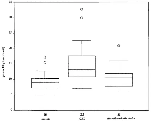
Data on Hcy concentration are summarized in the Figure. The
fasting median plasma tHcy levels were higher in patients
with sCAD (13.2
mol/L; range, 7 to 32.8 mol/L) compared
with control subjects (8.9
mol/L; range, 5 to 17.3 mol/L)
and patients with non-CAD ischemic stroke (10.9
mol/L;
range, 6 to 30.2
mol/L). There was a positive relationship
between tHcy concentration and sCAD. In particular, a
significant difference was found compared with control
subjects (95% CI, 1.05 to 1.52; P
⫽0.006). This difference
remained significant (95% CI, 1.02 to 1.48; P
⫽0.038) even
after the exclusion of the sCAD cases with no evidence of
structural ischemic cerebral lesions (median plasma tHcy
concentration in the subgroup with cerebral infarction, 13.5
mol/L; range, 7 to 22.7 mol/L). In contrast, although there
was a tendency toward a higher median concentration in
patients with non-CAD ischemic stroke compared with
con-trol subjects, the difference was not statistically significant
(95% CI, 0.95 to 1.35). No significant difference was found
between the group of patients with sCAD and the group of
patients with non-CAD ischemic stroke (95% CI, 0.80 to
1.56).
Similar results were obtained comparing the prevalence of
subjects whose plasma tHcy levels were above the normal
values in each group. A significant difference (95% CI, 2.25
to 44.23; P
⫽0.003) was observed between the group of
patients with sCAD (n
⫽16 subjects with abnormal tHcy
values; 64%) and the group of control subjects (n
⫽5 subjects
with abnormal tHcy values; 13.9%), whereas there was no
significant difference between the group of patients with
TABLE 1. General Characteristics of the Study Population
Characteristic sCAD (n⫽25) Non-CAD Ischemic Stroke (n⫽31) Control Subjects (n⫽36) Age, y 42.3⫾11.2 39.9⫾4.2 40.9⫾9.9 Male sex, n (%) 15 (60) 20 (64.5) 23 (63.9) Current smoker, n (%) 10 (40) 13 (41.9) 11 (30.6) Hypertension, n (%) 4 (16) 4 (12.9) 3 (8.3) Diabetes mellitus, n (%) 1 (4) 2 (6.5) 0 Hypercholesterolemia, n (%) 5 (25) 6 (19.4) 7 (19.4)
non-CAD ischemic stroke (n
⫽9 subjects with abnormal tHcy
values; 29%) and the group of control subjects (95% CI, 0.50
to 8.77), as well as between the group of patients with sCAD
and the group of patients with non-CAD ischemic stroke
(95% CI, 0.36 to 63.27). None of the other variables that were
entered into the multiple regression equation were significant
risk factors for sCAD.
We next investigated the prevalence of the TT MTHFR
genotype in each group. The homozygous TT transition was
present in 9 of 25 patients (36%) with sCAD, in 4 of 31
patients (12.9%) with non-CAD ischemic stroke, and in 4 of
36 control subjects (11.1%; Table 2).
There was a significant association between the TT
geno-type and the group of patients with sCAD compared with
control subjects (95% CI, 1.10 to 19.23; P
⫽0.045). No
significant difference in TT frequency was observed between
the group of patients with non-CAD ischemic stroke and the
group of control subjects (95% CI, 0.23 to 5.15) or between
the group of patients with sCAD and the group of patients
with non-CAD ischemic stroke (95% CI, 0.30 to 58.7).
As opposed to the TT MTHFR genotype, the frequency of
the 844ins68bp CBS mutation (12% in the group of patients
with sCAD, 13.9% in the group of controls, and 25.8% in the
group of patients with non-CAD ischemic stroke) was not
statistically different among the 3 groups after multiple
regression analysis (P
⫽0.411). Similarly, there was no
dif-ference in the frequency of subjects carrying the combination
of both the 844ins68bp CBS mutation and the MTHFR TT
genotype (1 in each group).
Discussion
The main finding in the present study is the evidence that both
high plasma levels of tHcy and the homozygous MTHFR TT
genotype are strongly associated with sCAD. In contrast,
neither fasting tHcy levels nor the frequency of the
thermo-labile form of MTHFR significantly differed between patients
with non-CAD ischemic stroke and control subjects.
To the best of our knowledge, there have been only sparse
reports
23,24and 1 prospective case-control study
12of the
relationship between fasting tHcy and sCAD. The evidence
derived from such study agrees with our results arguing for a
strong relationship between high levels of tHcy and arterial
dissection. However, that study failed to detect any
associa-tion between genetic abnormalities in Hcy metabolism and
risk of sCAD. No previous studies compared fasting tHcy
level and its genetic determinants in stroke patients with
primary vascular disorders of different pathogenesis, such as
arterial dissection and atherothrombosis of large and small
vessels. The raised concentrations of tHcy we found in sCAD
patients immediately after the acute event are suggestive of a
causal association, whereas, in agreement with the results of
previous prospective studies, we observed no evidence of
such a correlation in cases of stroke caused by a presumed
atherothrombotic process. Taken together, these findings are
consistent with the hypothesis that the role played by Hcy in
the pathophysiology of cerebral ischemia may depend on the
stroke subtypes and the specific underlying mechanism.
TABLE 2. Distribution of the MTHFR Genotypes in the 3 Groups sCAD (n⫽25) Non-CAD Ischemic Stroke (n⫽31) Control Subjects (n⫽36) MTHFR genotype, % (n) TT 36% (9) 12.9% (4) 11.1% (4) Tt 36% (9) 58.1% (18) 38.9% (14) tt 28% (7) 29% (9) 50% (18) T-t allele ratio 0.54:0.46 0.42:0.58 0.31:0.69 Box plot of fasting total plasma Hcy con-centration in the group of patients with sCAD, group of patients with non-CAD ischemic stroke, and group of control subjects.
Although the exact process leading to sCAD remains
elusive, a structural defect of the arterial wall is thought to
represent a predisposing condition. Recently, Brandt and
coworkers
25found irregular collagen fibrils and elastic fiber
fragmentation in skin biopsies of the majority of subjects
from a series of sCAD patients, providing further evidence of
a plausible relationship between sCAD and generalized
con-nective tissue disorders and supporting the hypothesis that
potential defects in the extracellular matrix of the vessel wall
may play a key role in the pathogenesis of arterial dissection.
Indirect evidence of an underlying structural defect of the
arterial wall is also suggested by the association of sCAD
with other vascular disorders.
26 –29Several biological mechanisms might explain the
associa-tion between increased levels of plasma tHcy and arterial
dissection. Among these, a link between
hyperhomocysteine-mia and abnormalities in the elastic components of the
arterial wall has been reported. In vitro studies demonstrate
that high levels of plasma Hcy result in a decrease in the
elastin content of the arterial wall as a direct or an indirect
consequence of the Hcy-induced activation of
metallopro-teases
30and serine elastases.
31This increased elastolytic
activity may result in an opening and enlargement of
fenestrae in the medial elastic laminae, leading to a premature
fragmentation of the arterial elastic fibers and degradation of
the extracellular matrix.
30 –32Hcy has been shown to block
aldehydic groups in elastin, thereby inhibiting the
cross-linking necessary to stabilize elastin.
33The cross-linking of
collagen may also be impaired.
34Thus, Hcy may have an
influence on the elastic properties of the arterial wall. These
findings lead to the hypothesis that the toxic effect of Hcy
might be more pronounced in the carotid artery and in the
aorta with their many elastic laminas compared with the
femoral and brachial arteries in which the muscular layer is
thicker. The high frequency of carotid and aortic dissection
compared with the low frequency in the femoral and brachial
arteries is in line with these findings and indirectly
strength-ens the hypothesis of a link between increased levels of tHcy
and sCAD.
A possible limitation of our study is inherent in the
different therapeutic approaches to sCAD with respect to
atherothrombotic strokes. However, there is no evidence that
either antiplatelets or anticoagulants agents may influence
plasma tHcy levels.
35Furthermore, we measured tHcy
con-centration in the very acute phase of the vascular event, which
is likely to reduce the impact of this potential bias. This also
allows us to refute the hypothesis that the raised
concentra-tions of tHcy we observed might be the result of the increase
in methylation reactions after tissue injury, which has been
suggested to explain the high tHcy levels in patients sampled
after the acute phase of stroke.
36The association between sCAD and the homozygous
MTHFR TT genotype is also worth mentioning. It has been
reported that
⬇5% of patients with spontaneous arterial
dissection have at least 1 family member who has had the
same disorder.
4This finding suggests that the underlying
structural defects of the arterial wall might be heritable in
some cases. As to the 844ins68bp mutation in the CBS gene,
it has been observed that the heterozygous state, combined
with the thermolabile MTHFR, may increase the risk of
venous and arterial occlusive disease.
37However, our
find-ings do not support this hypothesis and a relationship between
the 844ins68bp mutation in the CBS gene, alone or in
combination with the thermolabile MTHFR, and sCAD seems
unlikely.
In conclusion, our data suggest that Hcy may play a
differential role in the pathogenesis of vascular disorders,
according to the specific mechanism of vascular injury. In
particular, increased concentration of plasma tHcy seems to
represent a predisposing condition for sCAD, whereas its role
in atherothrombotic damage remains a contentious issue.
Taking into account the relatively small number of subjects in
the present study, we cannot exclude that the lack of
signif-icant difference between patients with atherothrombotic
stroke and control subjects may be the result of an
under-powered comparison. Furthermore, our data point to a
rela-tionship between genetic abnormalities in Hcy metabolism
and risk of sCAD and suggest a potential association with
polymorphisms in other genes.
References
1. Schievink WI. Spontaneous dissection of the carotid and vertebral arteries. N Engl J Med. 2001;344:898 –906.
2. Schievink WI, Michels VV, Mokri B, Piepgras DG, Perry HO. A familial syndrome of arterial dissection with lentiginosis. N Engl J Med. 1995; 332:576 –579.
3. Majamaa K, Portimojarvi H, Sotaniemi KA, Myllyla V. Familial aggre-gation of cervical artery dissection and cerebral aneurysm. Stroke. 1994; 25:1704 –1705.
4. Schievink WI, Mokri B, Piepgras DG, Kuiper JD. Recurrent spontaneous arterial dissection: risk in familial versus nonfamilial disease. Stroke. 1996;27:622– 624.
5. Hankey GJ, Eikelboom JW. Homocysteine and vascular disease. Lancet. 1999;354:407– 413.
6. Stehouwer CDA, Weijenberg MP, van den Berg M, Jakobs C, Feskens EJM, Kromhout D. Serum homocysteine and risk of coronary heart disease and cerebrovascular disease in elderly men: a 10-year follow-up. Arterioscler Thromb Vasc Biol. 1998;18:1895–1901.
7. Langman LJ, Cole DEC. Homocysteine. Crit Rev Clin Lab Sci 1999;36: 365– 406.
8. Graham IM, Daly LE, Refsum HM, Robinson K, Brattstrom LE, Ueland PM, Palma-Reis RJ, Boers GH, Sheahan RG, Israelsson B, Uiterwaal CS, Meleady R, McMaster D, Verhoef P, Witteman J, Rubba P, Bellet H, Wautrecht JC, de Valk HW, Sales Luis AC, Parrot-Rouland FM, Tan KS, Higgins I, Garcon D, Andria G, et al. Plasma homocysteine as a risk factor for vascular disease: the European Concerted Action Project. JAMA. 1997;277:1775–1781.
9. den Heijer M, Koster T, Blom HJ, Bos GM, Briet E, Reitsma PH, Vandenbroucke JP, Rosendaal FR. Hyperhomocysteinemia as a risk factor for deep-vein thrombosis. N Engl J Med. 1996;334:759 –762. 10. Lindgren A, Brattstrom L, Norving B, Hultberg B, Anderson A,
Johansson BB. Plasma homocysteine in the acute and convalescent phases after stroke. Stroke. 1995;26:795– 800.
11. Meiklejohn DJ, Vikers MA, Dijkhuisen R, Greaves M. Plasma homo-cysteine concentrations in the acute and convalescent periods of athero-thrombotic stroke. Stroke. 2001;32:57– 62.
12. Gallai V, Caso V, Paciaroni M, Cardaioli G, Arning E, Bottiglieri T, Parnetti L. Mild hyperhomocyst(e)inemia: a possible risk factor for cervical artery dissection. Stroke. 2001;32:714 –718.
13. Bamford JM, Warlaw CP. Evolution and testing of the lacunar hypothesis. Stroke. 1988;19:1074 –1082.
14. Fischer CM. Lacunar strokes and infarcts: a review. Neurology. 1982;32: 871– 876.
15. Lammie GA. Pathology of small vessel stroke. Brit Med Bull. 2000;56: 296 –306.
16. Ripoll L, Paulin D, Drouet LO. Multiplex PCR-mediated site-directed mutagenesis for one-step determination of factor V Leiden and G20210A transition of the prothrombin gene. Thromb Haemost. 1997;78:960 –961.
17. Johnson CJ, Kittner SJ, McCarter RJ, Sloan MA, Stern BJ, Buchholz D, Price TR. Interrater reliability of an etiologic classification of ischemic stroke. Stroke. 1995;26:46 –51.
18. WHO Study Group on Diabetes Mellitus. World Health Organization Technical Report, Series 727. Geneva, Switzerland: World Health Orga-nization; 1985.
19. Ubbink JB, Delport R. Reference ranges for homocysteine concen-trations. In: Robinson K, ed. Homocysteine and Vascular Disease. Norwell, Mass: Kluwer Academic Publishers; 2000:41–57.
20. Sebastiao G, Sperandeo MP, Panico M, De Franchis R, Kraus JP, Andria G. The molecular basis of homocystinuria due to cystathionine beta-synthase deficiency in Italian families, and report of four novel mutations. Am J Hum Genet. 1995;56:1324 –1333.
21. Frosst P, Blom HJ, Milos R, Goyette P, Sheppard CA, Matthews RG, Boers GJH, den Heijer M, Kluijtmans LAJ, van den Heuvel LP, Rozen R. A candidate genetic risk factor for vascular disease: a common mutation in methylenetetrahydrofolate reductase. Nat Genet. 1995;10:111–113. 22. Pezzini A, Gulletta M, Pinelli L, Marangoni A, El-Hamad I, Gasparotti R,
Padovani A. Meningovascular syphilis: a vascular syndrome with typical features? Cerebrovasc Dis. 2001;11:352–353.
23. Caso V, Cardaioli G, Gallai V, Parnetti L. Vertebral artery dissection and hyperhomocysteinemia: a case report. Cerebrovasc Dis. 2000;10(suppl 4):9 –11.
24. Cardaioli G, Parnetti L, Caso V, Paciaroni M, Venti M, Bottiglieri T, Gallai V. Homocysteine (Hcy) and cervical artery dissections (CAD): a possible marker of arterial damage? Cerebrovasc Dis. 1999;9(suppl 1):16. Abstract.
25. Brandt T, Hausser I, Orberk E, Grau A, Hartschuh W, Anton-Lamprecht I, Hacke W. Ultrastructural connective tissue abnormalities in patients with spontaneous cervicocerebral artery dissections. Ann Neurol. 1998; 44:281–285.
26. Schievink WI, Mokri B, Piepgras DG. Angiographic frequency of saccular intracranial aneurysms in patients with spontaneous cervical artery dissection. J Neurosurg. 1992;76:62– 66.
27. Tzourio C, Cohen A, Lamisse N, Biousse V, Bousser MG. Aortic root dilatation in patients with spontaneous cervical artery dissection. Circu-lation. 1997;95:2351–2353.
28. Barbour PJ, Castaldo JE, Rac-Grant AD. Internal carotid artery redundancy is significantly associated with dissection. Stroke. 1994;25: 1201–1206.
29. Guillon B, Tzourio C, Biousse V, Adrai V, Bousser MG, Touboul PJ. Arterial wall properties in carotid artery dissection: an ultrasound study. Neurology. 2000;55:663– 666.
30. Charpiot P, Bescond A, Augier T, Chareyre C, Fraterno M, Rolland PH, Garcon D. Hyperhomocysteinemia induces elastolysis in minipig arteries: structural consequences, arterial site specificity and effect of captopril-hydrochlorothiazide. Matrix Biol. 1998;17:559 –574.
31. Rahmani DJ, Rolland PH, Rosset E, Branchereau A, Garcon D. Homo-cysteine induces synthesis of a serine elastase in arterial smooth muscle cells from multi-organ donors. Cardiovasc Res. 1997;34:597– 602. 32. Rolland PH, Friggi A, Barlatier A, Piquet P, Latrille V, Faye MM,
Guillou J, Charpiot P, Bodard H, Ghiringhelli O, Calaf R, Luccioni R, Garcon D. Hyperhomocysteinemia-induced vascular damage in the minipig: captopril-hydrochlorothiazide combination prevents elastic alterations. Circulation. 1995;91:1161–1174.
33. Jakson SH. The reaction of homocysteine with aldehyde: an explanation of the collagen defects in homocystinuria. Clin Chim Acta. 1973;45: 215–217.
34. Kang AH, Trelstad RL. A collagen defect in homocystinuria. J Clin Invest. 1973;52:2571–2578.
35. Ueland PM, Refsum H, Schneede J. Determinants of plasma homo-cysteine. In: Robinson K, ed. Homocysteine and Vascular Disease. Norwell, Mass: Kluwer Academic Publishers; 2000:59 – 84.
36. Dudman NPB. An alternative view of homocysteine. Lancet. 1999;354: 2072–2074.
37. de Franchi R, Fermo I, Mazzola G, Sebastio G, Di Minno G, Coppola A, Andria G, D’Angelo A. Contribution of the cystathionine beta-synthase gene (844ins68) polymorphism to the risk of early-onset venous and arterial occlusive disease and of fasting hyperhomocysteinemia. Thromb Haemost. 2000;84:576 –582.