U
U
N
N
I
I
V
V
E
E
R
R
S
S
I
I
T
T
A
A
’
’
D
D
E
E
G
G
L
L
I
I
S
S
T
T
U
U
D
D
I
I
D
D
I
I
P
P
I
I
S
S
A
A
F
F
A
A
C
C
O
O
L
L
T
T
A
A
’
’
D
D
I
I
M
M
E
E
D
D
I
I
C
C
I
I
N
N
A
A
E
E
C
C
H
H
I
I
R
R
U
U
R
R
G
G
I
I
A
A
D
D
i
i
p
p
a
a
r
r
t
t
i
i
m
m
e
e
n
n
t
t
o
o
C
C
a
a
r
r
d
d
i
i
o
o
T
T
o
o
r
r
a
a
c
c
i
i
c
c
o
o
e
e
V
V
a
a
s
s
c
c
o
o
l
l
a
a
r
r
e
e
D
D
o
o
t
t
t
t
o
o
r
r
a
a
t
t
o
o
d
d
i
i
R
R
i
i
c
c
e
e
r
r
c
c
a
a
i
i
n
n
F
F
i
i
s
s
i
i
o
o
p
p
a
a
t
t
o
o
l
l
o
o
g
g
i
i
a
a
e
e
C
C
l
l
i
i
n
n
i
i
c
c
a
a
d
d
e
e
l
l
l
l
’
’
A
A
p
p
p
p
a
a
r
r
a
a
t
t
o
o
C
C
a
a
r
r
d
d
i
i
o
o
v
v
a
a
s
s
c
c
o
o
l
l
a
a
r
r
e
e
e
e
R
R
e
e
s
s
p
p
i
i
r
r
a
a
t
t
o
o
r
r
i
i
o
o
T
T
e
e
s
s
i
i
d
d
i
i
D
D
o
o
t
t
t
t
o
o
r
r
a
a
t
t
o
o
P
P
r
r
e
e
v
v
a
a
l
l
e
e
n
n
c
c
e
e
,
,
C
C
l
l
i
i
n
n
i
i
c
c
a
a
l
l
S
S
i
i
g
g
n
n
i
i
f
f
i
i
c
c
a
a
n
n
c
c
e
e
a
a
n
n
d
d
T
T
i
i
m
m
e
e
-
-
C
C
o
o
u
u
r
r
s
s
e
e
o
o
f
f
N
N
e
e
o
o
n
n
a
a
t
t
a
a
l
l
E
E
C
C
G
G
A
A
b
b
n
n
o
o
r
r
m
m
a
a
l
l
i
i
t
t
i
i
e
e
s
s
a
a
s
s
s
s
o
o
c
c
i
i
a
a
t
t
e
e
d
d
w
w
i
i
t
t
h
h
A
A
r
r
r
r
h
h
y
y
t
t
h
h
m
m
i
i
a
a
s
s
d
d
u
u
r
r
i
i
n
n
g
g
I
I
n
n
f
f
a
a
n
n
c
c
y
y
:
:
t
t
h
h
e
e
e
e
x
x
p
p
e
e
r
r
i
i
e
e
n
n
c
c
e
e
o
o
f
f
B
B
r
r
i
i
n
n
d
d
i
i
s
s
i
i
R
Re
e
la
l
at
to
or
re
e
:
:
D
D
o
o
tt
t
to
o
ra
r
an
n
da
d
a
:
:
C
Ch
h
ia
i
ar
r.
.m
mo
o
P
P
ro
r
o
f.
f
.
A
A
le
l
e
ss
s
sa
a
nd
n
d
ro
r
o
D
D
IS
I
S
T
T
AN
A
N
TE
T
E
Do
D
o
tt
t
t.
.s
ss
sa
a
G
G
io
i
ov
va
an
n
na
n
a
C
CH
H
IT
I
T
A
A
NO
N
O
A
AN
NN
NO
O
AC
A
CC
CA
AD
DE
EM
MI
IC
CO
O
2
20
0
05
0
5
-2
-
20
0
06
0
6
CONTENTS
Abstract 3 Introduction 4
Aim 6
ECG and Arrhythmia 7
The normal neonatal ECG 8
The abnormal neonatal ECG 12
Methods 27
Study Population 27
Data acquisition 27
Statistical analysis 29
Results 30
Results of ECG screening 30
Follow-up results 31 Discussion 33 Conclusions 37 References 38 Tables 49 Figures 54 Appendix 61
A
A
b
b
s
s
t
t
r
r
a
a
c
c
t
t
Background: Some rhythm disturbances may be detected on the surface
electrocardiogram even during the neonatal period. Most of them are transitory and benign arrhythmias, others can constitute the first sign of cardiac disease or may become malignant and life-threatening. Furthermore, arrhythmogenic syndromes such as the Long QT syndrome and the Brugada syndrome can cause sudden death in infancy and may also contribute to SIDS (Sudden Infant Death Syndrome). The risk of fatal arrhythmias - which often manifest themselves in infancy, childhood or even at later age - justifies preventive measures. Since diagnosis in symptom-free patients requires electrocardiography, such a non invasive methodology must be available to provide early diagnosis and therapy. The Italian Ministry of Health has funded a prospective study in 50.000 neonates, involving 16 Italian centers, to assess the feasibility of a nationwide neonatal ECG screening.
Aim: The purpose of this project was to assess prevalence and clinical significance of
neonatal ECG abnormalities. This paper considers the data collected in Brindisi, at the “Perrino” Hospital, with the participation of ISBEM (Euro Mediterranean Scientific Biomedical Institute).
Methods: From 2001 to 2006, electrocardiograms were recorded and the demographic
data of newborns and of their parents were collected, as well as the clinical history of the newborn babies and of their mothers. All electrocardiographic parameters were measured according to the guidelines for the interpretation of the neonatal ECG. Data of all newborns enlisted in the study were entered into the neonatal ECG database. Newborns with clinically relevant ECG abnormalities were followed-up, according to the guidelines work-up section for clinical management of pathologies related to ECG anomalies.
Results: Of the 2619 ECGs recorded, 418 (16%) presented abnormalities. Among the
abnormalities 5 (0.2%) had a major clinical significance such as prolonged QT syndrome, Wolff-Parkinson-White syndrome and congenital heart disease.
Conclusions: Although life-threatening arrhythmias are rare in infants, they are very
serious and must be sought to avoid both short- and long-term morbidity and mortality. The electrocardiographic screening for neonates, as an additional measure of prevention, especially for the early identification of infants at risk of life-threatening arrhythmias, is available and relatively at low cost, and can also help detecting complex cardiac malformations.
I
I
n
n
t
t
r
r
o
o
d
d
u
u
c
c
t
t
i
i
o
o
n
n
Some rhythm disturbances (arrhythmias) may be detected on the surface electrocardiogram (ECG) even during the neonatal period. Cardiac arrhythmias are difficult to define epidemiologically, therefore there are limited data on their prevalence [1]. It is widely recognized that pediatric patients commonly experience arrhythmias both early and late after cardiac surgical procedures [2]. Hospital based studies provide a more accurate assessment of arrhythmias, but it is difficult to apply this information to the general population, as not all arrhythmias require hospitalization either because they are short-lived or asymptomatic. Arrhythmias are found in 1% to 5% of newborns during the first 10 days of life. Most are premature supraventricular beats that will disappear over the first month of life [3]. On the other hand, there are some arrhythmias that can constitute the first sign of cardiac disease, or they can become malignant and life-threatening. Many congenital heart diseases and acquired cardiac diseases, such as myocarditis or cardiomyopathy, are associated with the occurrence of rhythm disturbances [4-6]. Over the past decade, molecular genetic research has established a link between a number of inherited lethal cardiac arrhythmias and mutations in genes encoding for ion channels or other membrane components. Among these inherited cardiac arrhythmias there are the congenital and acquired long QT syndrome (LQTS) [7,8] and the Brugada syndrome [9]. These arrhythmogenic syndromes pass unnoticed because they occur in children or young individuals completely asymptomatic and assumingly healthy, who can conduct a normal life. They are often diagnosed only after the appearance of the first symptoms. Long QT syndrome is characterized by the occurrence of syncopal episodes and by a high risk of sudden cardiac death among untreated patients due to a particular type of ventricular tachycardia, also known as torsades de pointes. In 12% of patients with LQTS, sudden death was the first manifestation of the disease, whilst in 4% this happened in the first year of life [10]. Results of a prospective study in 34,000 newborn babies showed that 50% of children, who died from Sudden Infant Death Syndrome (SIDS), had a QTc interval on ECG that was higher than normal and that a prolonged QT interval increased the risk of SIDS by 41 times [11]. Also with regard to the Brugada syndrome there exist documented cases of
malignant arrhythmias in babies of few months of life [12,13]. Such arrhythmogenic disorders of genetic origin, that can cause sudden death in infancy and also contribute to SIDS, are identifiable by ECG and molecular screening. The risk of fatal arrhythmias which often shows in infancy, childhood or even later justifies preventive measures. Since diagnosis in symptom-free patients requires electrocardiography, such non invasive methodology must be available, so as to provide early diagnosis and therapy.
On the basis of different studies on this subject, the Italian Ministry of Health has suggested offering, free of charge, electrocardiographic screening to all newborns [14], as part of a cardiovascular screening program. Moreover, it has funded a prospective study in 50.000 newborns (more or less 10% of the births that occur in Italy every year) to assess the feasibility of a nationwide neonatal ECG screening. The project, under the scientific direction of Peter J. Schwartz, has involved 16 Italian centers.
A
A
i
i
m
m
The purpose of the multi-center project ”Prospective study on prevalence, clinical significance and time-course of neonatal ECG abnormalities associated with arrhythmias during infancy” was to assess prevalence and clinical significance of neonatal ECG abnormalities, but also to appraise if an ECG done in neonatal age (15-25 days of life) can contribute to the prevention of Sudden Infant Death Syndrome (SIDS), as well as to an early diagnosis of Multiple Cardiac Morbid Disorders and to somehow pave the way to potential treatments. Such disorders include: cardiac arrhythmias, Wolff-Parkinson-White syndrome, some forms of congenital heart disease not yet clinically evident, Brugada syndrome, QT prolongation of unknown origin but potentially associated with increased risk of fatal arrhythmias, and long QT syndrome. This paper considers the data collected in Brindisi, at the “Perrino” Hospital, with the participation of ISBEM (Euro Mediterranean Scientific Biomedical Institute).
E
E
C
C
G
G
a
a
n
n
d
d
A
A
r
r
r
r
h
h
y
y
t
t
h
h
m
m
i
i
a
a
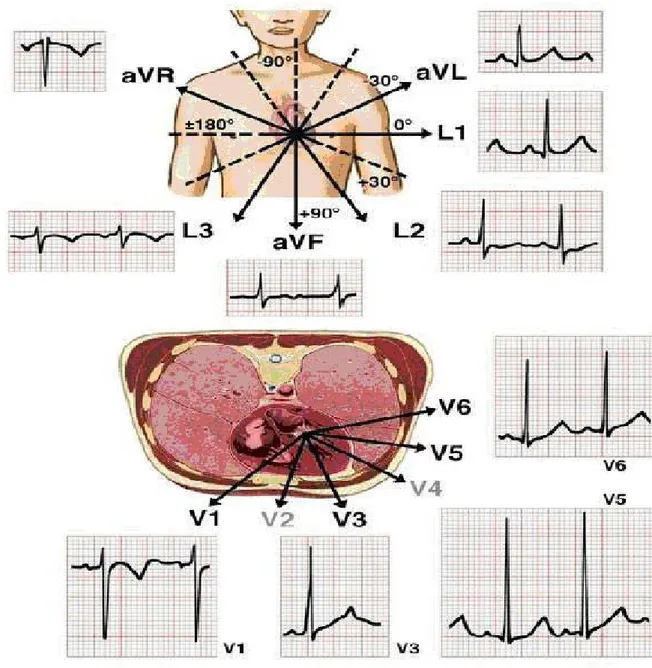
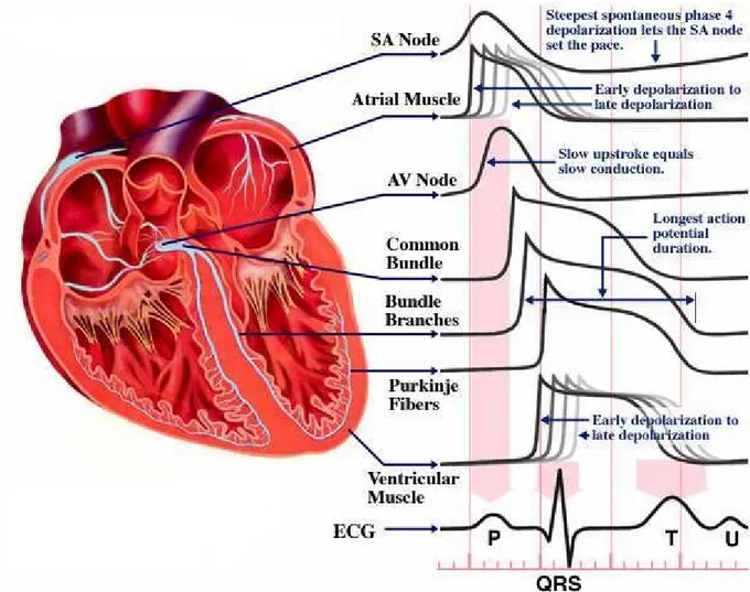
The contraction (depolarization) and relaxation (repolarization) of the heart muscle is caused by a flow of electrolytes (sodium, potassium, and calcium) in and out of each heart muscle cell through the surrounding membrane. This flow generates a weak electrical current. Through an amplifier (the electrocardiograph) capable of picking up this weak current from the surface of the body, it is possible to record the electrical potential generated by the heart muscle cells during each heartbeat. This electrical potential displayed continuously on a roll of graph paper, is the standard electrocardiogram (ECG). It consists of 12 leads recorded with electrodes placed on various points on the chest, both arms, and the left leg, thus providing electrical "views" of the heart from various angles (fig. 1). Any deviation from the horizontal line, or baseline, whether above (positive potential) or below (negative potential), denotes electrical activity. Each change of potential is called a wave [15]. The normal electrocardiogram shows a sequence of evenly spaced complexes produced by each heartbeat. The electrical activity, that triggers each heartbeat, begins in the atria and then travels into the ventricles. It starts in the sino-atrial node, the heart’s natural pacemaker, which is localized in the upper part of the right atrium, and it is conveyed to all atrial myocytes, leading to coordinated depolarization and contraction of the atria. On the ECG, atrial depolarization can be visualized as a P wave. The electrical activity of the pacemaker is also conveyed to specialized cells that connect the atria and the ventricles, known as the atrio-ventricular node [16]. From there, it is transmitted to the ventricles via specialized fibers known as the bundle of His, which is subdivided into the right and left bundle and which runs along the right and the left side of the interventricular septum, respectively. Each of the bundle branches has many bifurcations, which finally create the Purkinje fibers network [17] leading to the rapid depolarization of all ventricular myocytes and to the coordinated contraction of the ventricles. Depolarization of the ventricles can be visualized on the ECG as the QRS complex. Ventricular myocytes then slowly repolarize - on the ECG this is denoted as the T wave - thus leading to cardiac relaxation and completion of one cardiac cycle [16] (fig. 2). The healthy heart has a regular sequence of action. The regularity of the heartbeat, its rate, and the origin and
spread of impulses can all deviate from normality. These abnormalities are called arrhythmia, or disturbance of the heart rhythm. The term is used even if the rhythm, or regular sequence of beats, is undisturbed, but the rate is too slow or too fast (bradycardia, tachycardia) [15]. In general, the abnormality that causes an arrhythmia may be one of the following: 1) abnormal function of the cells in the sinoatrial node, such that these cells are either not firing or not transmitting impulses properly; 2) delayed or improperly produced/conducted impulses through the atrioventricular node (which could cause heart block) or through the ventricles (which could cause bundle branch block); 3) an extra pathway in the conduction system, causing additional heartbeats; 4) electrical impulses arising from places in the heart other than the sinoatrial node. Furthermore, there are a number of heart-related factors associated with arrhythmias, including: congenital heart disease; arrhythmogenic syndrome (e.g., long QT syndrome, Brugada syndrome); abnormalities of the heart structure or function (cardiomyopathy, valvular heart disease); side effects from medications. The electrocardiogram is the principal tool for diagnosing cardiac arrhythmias, as it shows the rate and regularity of heartbeats; the shape, direction, and relationship of the waves to the other complexes, including the absence of waves; and deviations in the contour of the complexes and waves in the abnormally spaced beats. An equally important role of the electrocardiogram is that to provide information concerning the state of the heart muscle on the basis of certain alterations in the electrocardiographic complexes. However, this feature of the electrocardiogram requires more cautious interpretation because the alterations produced by changes in the heart muscle are less specific than those produced by arrhythmias. Experience has established the limits of normal variation in the height and direction of the waves and the baseline segments among them, in healthy persons according to age group [15].
T
T
h
h
e
e
n
n
o
o
r
r
m
m
a
a
l
l
n
n
e
e
o
o
n
n
a
a
t
t
a
a
l
l
E
E
C
C
G
G
Growth and development result in major changes of body size and shape, and at the same time in the changes of size and position of the heart relative to the body and cardiac physiology [18]. These changes eventually lead to the
variability of findings of normal ECGs in children. The normal infant ECG varies rapidly over the first few weeks of life. At 3 years of age it begins to resemble characteristics of an adult. Significant differences, however, persist [18]. There are many normal variables with a wide overlap from normal to abnormal. The less than 35 week gestation premature infant will have a different ECG from the full term infant [19]. In order to interpret pediatric electrocardiograms, the age of the infant and a table of normal values are essential [18]. Normal electrocardiographic values between the 2nd and 98th percentile in the first year of life traditionally derive from those published by Davignon et al. [20] (see appendix ).
Heart rate
In children, cardiac output is determined primarily by heart rate, as opposed to stroke volume. With age, the heart rate decreases as the ventricles mature and stroke volume plays a larger role in cardiac output. The average resting heart rate varies with age: it increases from the first day of life and peaks between the first and the second month, then declines, returning at the sixth month to the values recorded at birth. After one year of age, due to maturation of the sinus node vagal innervation, it slowly declines [21]. The heartbeats of newborns can range from 90 to 160 beats per minute (bpm), without clinically significant gender differences.
P wave
The P wave represents depolarization of the two atria. It is generally pointed in lead II and aVF and more rounded in other leads, and may be diphasic in V1. The location of the P wave axis indicates the site of origin of the rhythm.
PR interval
The PR interval, measured in lead II from the onset of the P wave to the beginning of the QRS, corresponds to the time in which electrical impulses travel through the atria and the atrio-ventricular node. Here, the electrical activity will be delayed for a few milliseconds, giving the atria time to pump blood into the ventricles [16]. The PR interval, increases with age and
decreases with heart rate. The normal neonatal PR interval ranges from a minimum of 70 ms to a maximum of 140 ms, with a mean of 100 ms [19].
QRS complex
Three waves together constitute the QRS complex. This complex shows depolarization of the two ventricles and coincides with the onset of ventricular contraction. QRS morphology in the newborn may have more notches and direction changes than seen in older children or adults [19]. In utero, blood is shunted away from the lungs by the patent ductus arteriosus, and the right ventricle provides most of the systemic blood flow. As a result, the right ventricle is the dominant chamber in the newborn infant. In the neonate and young infant (up to 2 months), the ECG shows right ventricular dominance and right QRS axis deviation. In a normal full-term newborn a QRS axis between 55° and 200° is a normal finding, with upper limit that falls to 160° or less by 1 month. If right axis deviation is seen in normal neonates, instead left axis deviation is seen in a variety of abnormalities including atrioventricular septal defect, ventricular septal defect, tricuspid atresia, and WPW syndrome.
The first wave of the complex, small and negative, called Q wave, indicates the direction of septal depolarization in the precordial leads. Normally, there is a Q wave in leads V5–V6 indicating depolarization from left to right. Normal values of Q wave amplitudes vary with lead and age. Q wave duration >30 ms is abnormal. The second one is the tall and positive R wave. The appearance of secondary r waves (r’ or R’) in the right chest leads is frequent in normal neonates. Thomaidis et al. published normal voltages from healthy term and premature neonates [22]. The S wave is the last of the complex; it is negative during the first year of life and has elevated amplitude in left leads. The total amplitude of R+S in each limb lead ≤ 0.5 mV may be indicative of myocarditis or cardiomyopathy [19]. In the following years the Q wave does not show substantial modifications, whereas the amplitude of R wave progressively reduces in the right precordial leads and increases in the left precordial leads, and opposite modifications are shown by the S wave. Such changes are due to the progressive reduction of the right ventricular mass and to the relative increase of the left ventricular mass. QRS duration, from the beginning of the Q wave to the end of the S wave, increases with age, in neonate it measures
30-80 ms. A QRS duration exceeding 30-80 ms in newborn and infant may indicate delayed intraventricular activation, while QRS duration ≥ 120ms is called bundle branch block [23].
T wave
The T wave expresses the recovery of the excitability of the ventricular myocytes. In the first week of life this wave is normally rather variable. After the first week of life through the age of 8 years, T waves are usually inverted in leads V1 through V3 and positive in V5–V6, as the so called “juvenile T-wave pattern”. T wave positive in V1 and V2 leads during childhood can be a sign of systolic overload of the right ventricle, as seen in right ventricular hypertrophy; inverted T wave in V4-V6 indicates left ventricle suffering and is found in aortic stenosis [23].
ST segment
The ST segment is isoelectric and joins the QRS complex to the T wave at the baseline (or slightly above or below the baseline). This segment shows the beginning of ventricular repolarization. In neonates and infants it is better to consider as the isoelectric line the TP segment instead of the PQ segment [19], because the Q portion of the QRS complex is frequently indiscernible. ST segment elevation in infancy has several causes, the most frequent is pericarditis. Less frequent causes of ST segment elevation, with or without T wave abnormalities are hyperkalaemia, intracranial hemorrhage, pneumothorax and pneumopericardium, subepicardial injury due to anomalous left coronary artery or to Kawasaki disease with cardiac involvement. ST segment elevation with other ECG features is also seen in the Brugada syndrome.
QT interval
The QT interval on ECG, between the beginning of the QRS complex and the end of the T wave, represents the time from onset of depolarization to fulfill of repolarization. Due to the fast heart rate of infants the P wave may be superimposed on the T wave, particularly when the QT interval is prolonged. The QT interval duration changes with heart rate and is usually corrected. The Bazett formula is the most commonly used manual method to measure the
corrected QT interval (QTc) [24]. This equation accounts for the normal physiologic shortening of the QT interval that occurs with increasing heart rate. The mean QTc on the 4th day of life is 400 ± 20 ms [11] and, at variance with the adult, no gender differences are present [25]. Therefore, the upper normal limit of QTc (2 standard deviations above the mean, corresponding to the 97.5 percentile) is 440 ms. By definition, 2.5% of normal newborns are expected to have a QTc greater than 440 ms [19]. In healthy infants there is a physiological prolongation of QTc by the second month (mean 410 ms), followed by a progressive decline [26], so that by the sixth month QTc returns to the values recorded in the first week. QT interval prolongation may often be caused by electrolyte imbalance: hypocalcaemia increases the ST segment length, instead hypokalaemia and hypomagnesaemia usually decrease T wave amplitude. Also the use of widespread drugs during the neonatal period or infancy, as macrolide [27], antibiotics or prokinetics, may induce QT interval prolongation, as these block the channel of ionic current involved in the control of ventricular repolarization. Transient QT interval prolongation is found in babies positive for the anti-Ro/SSA antibodies from mothers with autoimmune diseases. Prolongation disappears concomitantly with the disappearance of the anti Ro/SSA antibodies. Central nervous system abnormalities can produce QT prolongation and T wave inversion. Moreover QT interval prolongation may indicate the congenital LQTS.
T
T
h
h
e
e
a
a
b
b
n
n
o
o
r
r
m
m
a
a
l
l
n
n
e
e
o
o
n
n
a
a
t
t
a
a
l
l
E
E
C
C
G
G
Heart rate
A sinus rhythm with a heart rate above or below the normal limit for age is defined respectively sinus tachycardia and sinus bradycardia. There are several causes of non-cardiac origin associated with sinus tachycardia and bradycardia. Autonomic nervous system dysfunction with vagal overactivity in infants may provoke sinus bradycardia and even significant sinus pauses. Sinus pauses produce electrical pauses in the rhythm. These may last 800-1000 ms. Pauses greater than 2 s are abnormal. Escape beats from atria or AV-junction may follow sinus pauses. Junctional escape rhythms occur when the atrial rate slows to the point where the AV node automaticity takes over. Apparent
life-threatening events (ALTE) have been associated to sinus pauses and sudden bradycardia. These events have also been described in infants affected by Long QT syndrome, an arrhythmogenic syndrome which is evident in the neonatal period [28]. Slow heart rates may predispose to certain tachyarrhythmias, such as atrial flutter [29]. Neonates show a more regular rhythm than young children and adolescents, due to their fast heart rate, particularly in the first week of life. No pathologic features are associated with wandering pacemaker. Such irregularity of the rhythm is entirely due to a shift of the pacemaker from the sinoatrial node to the atrium and the atrioventricular (AV) junction, that is indicated by a gradual change of P wave axis and morphology.
Atrial enlargement
Abnormal P waves may be sign of atrial enlargement and/or hypertrophy. If this occurs in the right atrium, it typically produces increased P wave amplitude (greater than 2 mV in infants) with a normal P wave duration, usually more evident in lead II. Conversely, if this occurs in the left atrium, it typically produces an increased and prolonged negative terminal deflection of the P wave in lead V1 [19]. Abnormal P waves may also be seen in infants with non-sinus origin of the P wave, with ectopic atrial rhythms whose origin is in other sites of the right or left atrium.
Atrioventricular conduction abnormalities: Atrioventricular blocks
If the PR interval is greater than the upper limit for corresponding age, atrioventricular conduction abnormalities are present. Atrioventricular (AV) block describes delayed or incomplete conduction of impulses through the AV node. Three degrees of AV block are recognized. First-degree AV block is defined as prolonged conduction through the AV node; this produces a prolonged PR interval on the ECG, but there is consistent 1:1 AV conduction. Second-degree AV block has two forms: Mobitz I and Mobitz II. Mobitz type I AV block (see appendix), also known as Wenckebach, is recognized on the ECG by its typical pattern of a gradually lengthening PR interval followed by a nonconducted P wave. In Mobitz type II AV block, there is intermittent failure of conduction of P wave, but wherever measurable PR intervals occur, they are consistent and do not lengthen. Generally, Mobitz II AV block produces a fixed ratio of P waves to
QRS complexes (2:1 or 3:1), but occasionally the AV block is variable [29]. First or second degree AV heart block can be observed in neonates and rarely it has been found progression to complete AV block after birth [30]. Functional AV block can be observed in neonates because they have a fast atrial rate and the P wave falls into the very prolonged T wave [19]. Long QT syndrome is occasionally complicated by impaired atrioventricular conduction, mostly 2:1 AV block [31,32]. Third-degree AV block (see appendix) is defined as complete failure of the conduction of atrial impulses to the ventricles. On the ECG, AV dissociation is seen when the atrial rate is faster than the ventricular rate [29]. AV block may be congenital or acquired. Congenital AV block is estimated to have an incidence of 1 to 22,000 births [33]. Congenital complete block can be observed in complex congenital heart malformations [34]. Mortality rate in patients with neonatal AV block is still high, especially during the first 3 months of life [35]. Acquired complete AV block is rare in neonates, and it is mainly due to viral myocarditis or may be related to tumours. In addition, it appears to be a strong association between SS-A/Ro or SS-B/La autoantibodies (present in collagen vascular diseases such as lupus erythematosis) of the mothers and the development of isolated neonatal AV block [36].
Intraventricular conduction abnormalities: Bundle Branch Block
Congenital isolated complete right and left bundle branch blocks are very rare in neonates. Only one case of complete RBBB in a population of 3383 apparently healthy newborn infants has been reported [37], RBBB with increased PR interval are seen in Ebstein’s anomaly of the tricuspid valve. Left anterior fascicular block is found in association with congenital heart malformations, such as atrio-ventricular canal defects and tricuspid atresia. Interruption of the left bundle, which results from the involvement of the left ventricle and/or its conduction system, has been reported in severe cardiomyopathy with poor prognosis [38]. Bundle branch block may also be inherited as an autosomal dominant trait [39,40]; when this happens, the subjects may show various combinations of conduction defects, from RBBB to AV block.
Sensitivity and specificity of the ECG in recognition of ventricular hypertrophy has not been specifically tested in the neonate, besides recognition of left ventricular hypertrophy is poorer than generally recognized. Right ventricular hypertrophy may be suspected from a QR complex in V1, an upright T wave in V1 (normal in the first week of life), increased R wave amplitude in V1, and increased S wave amplitude in V6 (according to the Davignon criteria [20]). QR patterns are commonly seen with pressure overload congenital lesions, rSR’ patterns are seen in volume overload lesions. Left ventricular hypertrophy ECG signs in children have been described as T wave abnormalities in leads V5 and V6, increased R wave amplitude in V6, increased S wave amplitude in V1 (according to the Davignon criteria [20]), and a combination of these last two variables. Left to right shunt lesions may manifest as biventricular hypertrophy. Left ventricular hypertrophy in the newborn may be attenuated by the normal right-sided predominance of the newborn.
Premature beats
Other pacemakers than the S-A node, that are generally activated only if an impulse from above fails to arrive, may assume an active role and discharge an electrical impulse out of turn, before the expected stimulation from above arrives. Such premature single impulses are called ectopic beats or premature beats. They may originate at any point of the conducting system or even in abnormally stimulated heart muscle cells. Atrial or junctional (A-V nodal) impulses are called supraventricular beats. Ectopic impulses originating in the lower portion of the conducting system are called ventricular beats [15]. A premature atrial beat is a premature P wave, that usually have a different morphology and mean vector from sinus P waves and that may be conducted to the ventricles normally, or with ventricular aberration or not conducted or ‘blocked’. The latter some times occur in a bigeminal sequence, so-called ‘blocked atrial bigeminy’. Premature atrial beats conducted normally, aberrantly and blocked may occur together. In infants, premature atrial beats may be conducted with either RBBB or left bundle branch aberration. If the QRS complex has a different morphology from the sinus, and is not preceded by a premature P wave, is a premature ventricular beat. Both premature atrial beats
and premature ventricular beats, which are manifest as premature P wave preceding a wide QRS, rarely occur in infants [19].
Supraventricular tachycardia
Supraventricular tachycardia (SVT) refers to a sustained tachyarrhythmia that originates above the bundle of His. SVT is the most common abnormal tachyarrhythmia in children, with an estimated incidence in the pediatric population of 0.1% to 0.4% [41] In infants the rhythm typically appears with an extremely regular R-R interval after the first 10–20 beats. Rates vary from 260 to 300 bpm depending on the patient’s age and the SVT mechanism. On the ECG, in 60% of cases, P waves are visible and have always a different morphology from sinus. In over 90% of infants and children with SVT, the QRS complex is usually normal in configuration, but in less than 10% of cases the QRS is wide due to aberrant conduction to the ventricles [41]. Several SVT mechanisms have been identified. The vast majority of SVT rhythms are either due to a re-entrant circuit, with or without an accessory pathway, or to an automatic ectopic focus. Triggered activity accounts for a very small percentage of SVT. The relative incidence of different types of SVT varies with age. Atrio-Ventricular Reciprocating Tachycardia (AVRT) has a relatively high incidence among newborns and infants, with male patients affected more than female patients. AVRT is a common type of SVT that relies on at least two electrical connections between the atria and ventricles: the AV node and an accessory pathway (more than one accessory pathway may be present). Conduction proceeds antegrade down one pathway and retrograde up the other to form a re-entrant circuit. Nearly 75% of the SVT rhythms in children are mediated by accessory pathways. The most common form of AVRT is orthodromic reciprocating tachycardia, which involves antegrade conduction down the AV node to the ventricles and retrograde conduction up the accessory pathway to the atria. Antidromic reciprocating tachycardia (down the accessory pathway, up the AV node) occurs in less than 10% of patients [29]. In many of these infants SVT will spontaneously resolve by 6 to 12 months of age, but over 30% will have recurrence later in life [42]. Among patients with AVRT, about half of them have a concealed accessory pathway, meaning that pre-excitation is not evident on the 12-lead ECG [43]. AV nodal re-entrant tachycardia is virtually unseen in
neonates, but occurs more frequently with increasing age, and represents over 50% of the SVT in adults. Other types of supraventricular arrhythmias, such as atrial fibrillation or multifocal tachycardia, are extremely rare in the neonate [19].
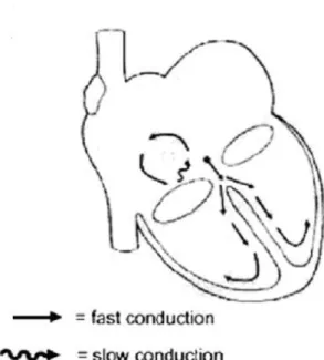
Atrial flutter
Intra-atrial re-entrant tachycardia (IART), commonly referred to as atrial flutter, is a reentrant tachycardia that is confined to the atria (fig. 3). Propagation of IART relies on an electrical pathway within the atria with both an area of slow conduction and an anatomic obstruction that results in unidirectional block. This milieu for IART exists after atrial surgery for congenital heart disease and in fact, almost 95% of atrial flutter diagnosed beyond infancy is associated with structural heart disease [43]. On infant ECG (see appendix), a regular appearance of atrial depolarization, characteristic saw-tooth waves (flutter waves) are seen at rates of 300 to 500 bpm with variable AV conduction. Occasionally atrial flutter is associated with WPW syndrome [19].
Atrial fibrillation
Atrial fibrillation (AF) demonstrates a rapid atrial rate with a very chaotic pattern, and an irregular ventricular rhythm, caused by chaotic electrical impulses in the atria with loss off coordination between the atria and ventricles. A typical ECG in atrial fibrillation shows a rapid irregular tachycardia in which recognizable P waves are absent. QRS complex are generally normal. The chaotic electrical activity produces deflections on the ECG, referred as fibrillatory waves. They vary in size and shape and are irregular in rhythm. AF is extremely rare in childhood and extremely uncommon before adolescence, but has been occasionally reported in all pediatric age groups and in fetuses [44,45]. It has been described in adult patients as a possible manifestation of sinoatrial disease, but this disorder accounts only for a minor portion of atrial fibrillation in childhood. AF has usually been associated with structural heart disease such as rheumatic heart disease, congenital heart disease, and various form of cardiomyopathy [46]. There are a few patients with atrial fibrillation without evidence of an underlying structural cardiac defect, or a disorder of the cardiac conduction system. This so called idiopathic or lone atrial fibrillation reported mainly in adults, has been documented also in very few children [44].
Ventricular tachycardia
Ventricular tachycardia (VT) is defined as three or more consecutive premature ventricular complexes at a rate greater than 120 bpm but usually less than 250 bpm [29]. The QRS complexes are typically with a prolonged duration for the age group with AV dissociation (sinus P waves continuing unrelated to VT). There may be also retrograde P waves or no visible P waves. VT in infants is called a “wide QRS” if complex duration is greater than 90 ms [19]. Abnormal QRS complex originate from ventricles, but the specific morphology of the QRS is generally not helpful to distinguish VT from SVT with aberration. In infants, the rate of VT may be from 200–500 bpm and it is possible a slight variation in the R-R interval over several beats. VT that occurs at a rate less than 200 bpm and alternating to sinus rate is called “slow VT” or Accelerated Ventricular Rhythm [19]. VT may be unsustained (lasting less than 10 seconds) or sustained (lasting 10 seconds or longer). Monomorphic VT refers to tachycardia in which all the QRS complexes have a similar morphology, in contrast to polymorphic VT with multiform complexes. Torsades de pointes (“twisting of the points”) is a type of polymorphic VT characterized by rapid, wide, undulating QRS complexes that appear to be spiraling around an axis. Mechanisms for VT are not as well described as for SVT, but appear to include re-entry (similar to IART), abnormal automaticity, and triggered activity [29]. The incidence of VT in the pediatric population is unknown, but accounts for about 6% of patients followed for tachyarrhythmia [47]. VT may be associated with severe electrolyte or metabolic abnormalities, hypoxia, hypothermia, or drug toxicity. Cardiac conditions such as cardiomyopathy, myocarditis, arrhythmogenic right ventricular dysplasia, ventricular tumors, surgery for congenital heart disease may create a substrate for VT. Another recognized cause of VT is congenital or acquired long QT syndrome [29].
Ventricular fibrillation
Ventricular fibrillation (VF) displays unidentifiable QRS complexes due to an uncoordinated state of ventricular depolarization, resulting in a state of poor cardiac output. Significant ventricular dysrhythmias, such as VF, in the pediatric age range are most commonly encountered in the setting of congenital heart
diseases, myocarditis, cardiomyopthies, hypoxia, acidosis and electrolyte abnormalities [23].
Brugada syndrome
The Brugada syndrome is a disorder associated with increased susceptibility to ventricular fibrillation and sudden cardiac death in the absence of cardiac structural abnormalities [48] and it is believed to be responsible for 4% to 12% of all sudden deaths [49]. Brugada syndrome may cause sudden death in children even in the first months of life when it may be misdiagnosed as SIDS. Priori and colleagues [12], in a single family study, demonstrated that an unusual form of Brugada syndrome with extremely severe manifestation in early childhood caused familial sudden death after unexplained cardiac arrest in five children from the same family. Brugada syndrome, that was suspected based on the transient manifestation of the typical electrocardiogram pattern in one of these children, was confirmed by genetic analysis. Incomplete penetrance, where a parent with a normal ECG may still carry and transmit the mutation, has been reported in Brugada syndrome [50] and has been seen in this family, where three gene carriers were completely asymptomatic with normal ECG. Suzuki et al. [13] reported the youngest patient documented to have Brugada syndrome, a 6-month-old Japanese infant with a malignant form of Brugada syndrome, who had frequent episodes of ventricular fibrillation (VF) and unsustained polymorphic ventricular tachycardia (VT). Moreover, his twin died suddenly during sleep at the age of 4 months. The most typical electrocardiographic features of the Brugada syndrome include an accentuated J wave appearing principally in the right precordial leads (V1 to V3) and taking the form of an ST-segment elevation, often followed by a negative T wave. In most patients, however, the typical widened S wave in the left lateral leads is absent, suggesting that this is not true RBBB. Early high takeoff of the ST segment in the right precordial leads (the “J wave”) can mimic RBBB [51]. Two ST-segment morphologies have been described in leads V1, V2, and V3: convex curved or “coved” and “saddle shaped”–type ST-segment elevation [52-54]. In individual patients, both morphologies may subsequently be present. The presence of coved ST-segment elevation has been suggested to have a stronger arrhythmogenic potential [52,54] but this relation could not be
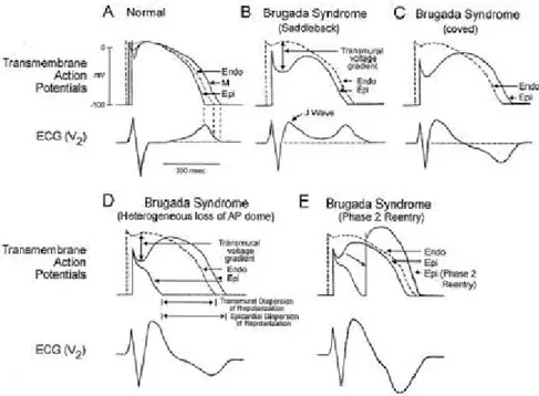
confirmed by others [53]. ECG abnormalities are inconsistently present [9;48,55-57]. The intermittent nature of this electrocardiogram pattern may obscure correct clinical diagnosis and therefore genetic screening contributes to the diagnosis of the disease [9,58]. Like the long-QT syndrome, the Brugada syndrome has both congenital and acquired forms; the latter is just beginning to be recognized [49]. The hereditary nature of the syndrome, characterized by an autosomal dominant mode of transmission, is well established. Chen et al. [9] were the first in 1998 to link the syndrome to the alfa subunit of the cardiac sodium channel gene, SCN5A. Several dozen SCN5A mutations have been linked to the syndrome [59-61] but unlike those causing the LQT3 (where there is an abnormality in Na-channel inactivation, resulting in a gain of function mutation and a prolonged QT interval), SCN5A mutations leading to Brugada syndrome are invariably associated with a loss-of-function. Loss-of-function SCN5A mutations also lead to conduction defects, which are often part of the clinical picture in Brugada syndrome [9,62,63]. For example the syndrome may be unmasked during a febrile state [64] because of the premature inactivation of the mutant channel which is exaggerated at temperatures above the physiological range, and those patients with the Brugada syndrome may be at an increased risk. Another locus on chromosome 3, close, but distinct from SCN5A, was recently linked to the syndrome [65]. Only 20% of Brugada syndrome cases have been linked to SCN5A mutations. A long list of candidate genes encoding for a variety of ion channels and other proteins have been proposed [60,63]. Heterogeneity of repolarization across the wall of the RV outflow tract (RVOT) contribute to the ECG patterns and the genesis of extrasystoles and ventricular tachycardia in the Brugada syndrome. Action potentials (APs) of epicardial cells display a pronounced phase 1 (fig. 4A), referred to as spike-and-dome morphology or notch [66], which is the result of at least 3 different currents: INa, Ito, and L-type calcium current. Perturbations in these currents can lead to striking abbreviation of the epicardial AP, with the resultant potential for reexcitation based on epicardial-endocardial heterogeneity of repolarization. It has been hypothesized that, because of the thinness of the RV wall, the relative contribution of epicardial APs to the surface ECG is more prominent in right than left precordial leads. Hence, the impact of changes in epicardial AP morphology will be most pronounced in precordial
leads V1 and V2 facing the RVOT [62]. Under normal conditions, the ST segment is isoelectric because of the absence of transmural voltage gradients at the level of the action potential plateau. A reduction in the density of the sodium channel current, as occurs with the inherited mutations discussed above, is known to accentuate the epicardial action potential notch that leads to exaggeration of transmural voltage gradients and thus to accentuation of the J wave, causing an apparent ST-segment elevation as a result of transmural current flow from endocardium to epicardium [60,67]. If the epicardial repolarization precedes repolarization of the M-cells and endocardial regions, the T wave will remain positive and the result will be a saddleback form of ST segment elevation (fig. 4B). Further accentuation of the notch as a consequence of additional reduction of INa may be accompanied by a prolongation of the epicardial action potential such that the direction of the transmural voltage gradient is reversed, thus leading to the development of a coved-type of ST segment elevation and inversion of the T wave (fig. 4C), typically observed in the electrocardiograms (ECG) of patients with Brugada syndrome. The arrhythmogenic substrate is believed to arise when a further shift in the balance of currents leads to loss of the action potential dome at some epicardial sites, but not at endocardium, that results in the development of a marked transmural dispersion of repolarization and refractoriness, which is responsible for the development of a vulnerable window during which a premature impulse or extrasystole can induce a reentrant arrhythmia. Moreover, loss of the epicardial action potential dome at some sites but not others creates a dispersion of repolarization within epicardium (fig. 4D) [68]. Propagation of the action potential dome from sites at which it is maintained to sites at which it is lost causes local re-excitation by means of a phase 2–reentry mechanism, leading to the development of a very closely coupled extrasystole, which captures the vulnerable window across the wall, thus triggering a circus movement reentry (fig. 4E) in the form of VT/VF [66,68]. Despite substantial progress in the identification and characterization of the Brugada syndrome over the past decade, relatively little progress has been made in the approach to therapy. Pharmacological treatment does not protect effectively against recurrent events, and currently, implantation of an a cardioverter-defibrillator (ICD) is the only effective therapy to prevent sudden death [48,69-71]. This,
however, is not an adequate solution for infants and young children. The pharmacological approach to therapy is focused on a rebalancing of currents active in the early phases of the RV epicardial action potential so as to reduce the magnitude of the action potential notch and/or restore the action potential dome [49].
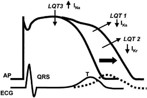
Long QT syndrome
The LQTSs are phenotypycally and genotypically diverse [72] but have in common the appearance of long QT interval on the electrocardiogram (ECG) [73-75], an polymorphic VT, and in many but not all cases, a relatively high risk for sudden cardiac death [72]. Other ECG abnormalities in LQTSs include prominent U waves, a T-U complex with an indistinct termination of the T wave, and broad T waves, which may be notched, biphasic, or inverted. T-wave alternans, an alternating amplitude and polarity of the T waves, also may be present [76]. All these changes in the electrocardiogram reflect differences in repolarizing currents across the ventricular wall (fig. 5). In LQT conditions, a disproportionate prolongation of action potential duration of the Purkinje fiber and the cells of the deep subendocardium/midmyocardium (the M cell) has been thought to be responsible for the development of LQT intervals, the different T-wave morphologies, and the U waves. The resulting dispersion of repolarization have also been shown to contribute to the malignant ventricular dysrhythmias [77]. Although other dysrhythmias can occur, the hallmark dysrhythmia is torsades de pointes (“twisting of the points”) (see appendix) [78, 79]. In 1975 [73] two hereditary variants were included under the unifying name of ‘Long QT Syndrome’: a more common autosomal dominant form with a pure cardiac phenotype [80,81] and a rare autosomal recessive form characterized by the coexistence of cardiac abnormalities and congenital deafness [82]. They are referred to as the Romano–Ward syndrome and the Jervell and Lange– Nielsen syndrome (J-LN) respectively. Since 1995, when the first two genes responsible for LQTS were identified [83,84], molecular genetic studies have revealed a total of eight forms of congenital LQTS caused by mutations in genes of the potassium, sodium and calcium channels or membrane adapter located on chromosomes 3, 4, 7, 11, 12, 17 and 21 [85-88] (see appendix). Mutations in KCNQ1 and KCNE1, the α and β subunits of the potassium
channel gene, respectively, are responsible for defects (loss of function) in the slowly activating component of the delayed rectifier potassium current (IKs) underlying the LQT1 and LQT5 forms of LQTS [89,90]. Mutations in KCNH2 and KCNE2 cause defects in the rapidly activating component of the delayed rectifier potassium current (IKr) responsible for the LQT2 and LQT6 forms [83,91]. Mutations in SCN5A, the gene that encodes the a subunit of the sodium channel, result in an increase (gain of function) in the late sodium current (INa) responsible for LQT3 [84]. Most recently, a mutation in CACNA1C was reported to be responsible for the defect in the L-type calcium current (ICa-L) underlying the LQT8 form, an arrhythmia disorder associated with dysfunction in multiple organ systems, including congenital heart disease, syndactyly, immune deficiency, and autism [85]. Mutations in KCNQ1 [92] and KCNE1 [89,90] are responsible also for the J-LN syndrome [93] and are present as homozygous or compound heterozygous defects [94]. It is thought by several authors [58,95] that the so-called LQT4 [87] and LQT7 [96] associated with a mutation in Ankyrin-B, a membrane adapter, that leads to the intracellular Ca2+ overload [87] and mutations in KCNJ2 encoding for the inward rectifier potassium current (IK1) [86] respectively, represent disorders different from the classic LQTS; because the fact that they can be associated with some degree of QT prolongation, usually rather modest, does not justify to consider them part of LQTS, a disease with very specific clinical manifestations. Approximately 10% of children with LQTS will present with sudden death, and younger children are more likely to die suddenly [78,79,97]. Ventricular tachyarrhythmias stemming from congenital long QT syndrome (LQTS) specifically have been postulated to account for some cases of sudden infant death syndrome (SIDS) [11,98-100]. The suggestion that the same mechanisms operant in LQTS could contribute to some of the SIDS victims was advanced in 1974 to 1976 [101,102] but the first evidence, that a prolonged QT interval was associated with a significantly higher risk of SIDS was provided in 1998 from the large cohort study by Schwartz et al. [11]. They documented strong clinical evidence of an association between prolongation of the QT interval and SIDS [103] in a 19-year prospective collection of day 3 or 4 of life screening 12-lead ECG on 34,442 neonates. From this original cohort, 34 infants died within their first year with 24 secondary to SIDS. The average QTc among the 24 SIDS was 435±45 ms.
Further, 12/24 of the SIDS decedents had manifest a QTc exceeding 440 ms [11] This study demonstrated QTc as a significant risk factor for SIDS in day 3/day 4 of life, QTc>440 ms having a 41.3 odds ratio for SIDS, an odds ratio far greater than other established risk factors for SIDS such as prone sleep position and environmental exposure to smoke. This study was followed by molecular demonstrations that “de novo” LQTS mutations created an arrhythmia-prone substrate responsible for cardiac arrest, as a result of documented ventricular fibrillation, in a typical case of near-miss for SIDS and another one caused by SIDS [99,104]. These findings provided the necessary “proof of concept” that LQTS could cause SIDS but their limit was represented by their anecdotal nature. A postmortem molecular screening of a cohort of 93 SIDS victims showed a prevalence of LQTS mutations of 5.2% among the 58 white infants [100]. The prevalence of LQTS gene variants in sudden infant death are also assessed recently through a molecular study in a cohort of 201 Norwegian SIDS victims. Arnestad et al. [105] identified 8 mutations and 7 rare variants in 9.5% of SIDS victims which were not seen in controls. This last study strengthens the molecular evidence that some SIDS cases result from cardiac electrical diseases, such as congenital LQTS [106]. These findings have direct clinical implications and the treatment of all those diagnosed as affected is even if there are no symptoms [19]. Therapy is aimed at reducing sympathetic activity to the heart, either pharmacologically or surgically. Beta-blockers are generally recommended as the initial therapy of choice, as they have been shown to significantly reduce episodes of syncope and sudden death with a decrease in mortality from 71% in untreated patients to 6% in those treated with beta-blockade [107]. All beta-blockers seem to be effective, but propanolol and nadolol are most commonly used [108] Furthermore, beta-blockers have been shown to effectively eradicate dysrhythmias in 60% of patients [97]. Although many patients with congenital LQTS have a resting bradycardia, most tolerate high dose beta-blockade [76]. Some patients may require a pacemaker because of drug-induced bradycardia superimposed on their already slow heartbeat [79]. Approximately 20% of patients remain refractory to treatment with betablockers and continue to experience recurrent syncope [107]. A direct link between mutations in the ion channel genes and each genotype has made
possible the advent of genotype-specific treatments for each LQTS genotype as reported first by Schwartz et al. in 1995 [109].
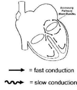
Wolff-Parkinson-White syndrome
The Wolff-Parkinson-White (WPW) syndrome is a frequently encountered type of AVRT. Orthodromic reciprocating tachycardia (fig. 6) results from reentry antegradely through the atrioventricular node and retrogradely through the accessory pathway (a direct muscular connection between the atria and ventricles). Conduction through the atrioventricular node and the accessory pathway (Kent bundle) results in collision of two electrical wavefronts at the ventricular level that appears as a delta wave and a fusion QRS complex with prolonged duration, along with a shortened PR interval. The diagnosis of ventricular preexcitation is solely based on the findings of the surface ECG (see appendix). In newborns and infants intermittent preexcitaton are common [19]. A study in newborns reported a high prevalence of WPW syndrome when 2 of 4 following characteristics were noted: PR interval ≤ 100 ms, QRS complex duration ≥ 80 ms, lack of a Q wave in V6 and left axis deviation [110]. WPW syndrome prevalence in the pediatric population has been estimated at 0.15 to 0.3% [111] which arrives to 0.5% in children with structural heart disease [42]. There is an increased incidence of WPW in patients with Ebstein’s anomaly, tricuspid atresia and hypertrophy cardiomyopathy [112]. Tachycardias associated with the syndrome are usually paroxysmal and may produce symptoms of presyncope, syncope, and cause sudden death [113]. Presence of the accessory pathway predispose to the atrial fibrillation (AF) (observed in 40% of cases with the WPW syndrome) with very fast ventricular response (fast conducting via the accessory pathway). Extremely fast ventricular rate may induce ventricular fibrillation. The risk of sudden cardiac death is 0.15% per year [114], however during the whole life it is greater than 10%. The incidence of sudden death in preexcitation syndrome during childhood has been estimated to be as high as 0.5% [115] and cardiac arrest may be the initial presentation in children with preexcitation [116]. However, data on newborns and infants are lacking. One study on a series of 90 newborns and infants with WPW syndrome and supraventricular tachycardia reported sudden death in two patients with a normal heart during follow-up. Both infants, however, had been
treated with digoxin [117]. As digoxin shortens the antegrade effective refractory period of the accessory pathway and promotes rapid atrioventricular conduction during atrial flutter or atrial fibrillation over the pathway, the use of digoxin is contraindicated at any age [118,119]. Verapamil should also be avoided as it may increase the ventricular response rate during atrial fibrillation in those patients, and may cause cardiovascular collapse in infants and young children [19]. A recent study by Gollob et al. [113] examined members of 2 families in whom the condition occurred as a familial autosomal-dominant disorder with complete penetrance and variable degrees of expression. They identified a mutation in the gene that encodes the gamma-2 regulatory subunit of the AMP-activated protein kinase. The molecular defect found may, in some way, inhibit the normal regression of muscle fibers during atrioventricular septation.
M
M
e
e
t
t
h
h
o
o
d
d
s
s
Study Population
From October 2001 to February 2006, all mothers of newborns discharged from the nursery of the “Perrino” Hospital in Brindisi, were invited to bring back their babies for the electrocardiographic screening, between the fifteenth and twenty-fifth day of life of the infant, as recommended by the Guidelines of the European Society of Cardiology [19]. At discharge, mothers were given an informative leaflet with recommendations on behaviors to adopt in order to reduce the risk of SIDS (see appendix), thus also improving the medical education in the overall population.
Electrocardiograms were recorded in a total of 2619 neonates. They were all healthy and full-term babies, as the very premature and sick newborns, who had been transferred to intensive care units, were excluded from the study.
Data acquisition
Demographic and clinical data
The demographic data of newborns and of their parents were collected, as well as the clinical history of the newborn babies and of their mothers.
Electrocardiography
ECGs were carried out by pediatric nurses trained to make ECG on newborns. Twelve-lead electrocardiograms were recorded at paper speed of 25 mm per second with Cardioline Delta 60 Plus recorder. The amplitude and duration of the waves and the intervals length in the newborn babies’ ECGs were hand measured, as the computerized systems are often inadequate in the newborns [19]. The electrocardiograms were analyzed by a neonatologist, expert in pediatric cardiology, who for the interpretation of the neonatal electrocardiogram referred to the guidelines [19], created by the Task Force of the European Society of Cardiology (ESC).
All electrocardiographic parameters were measured according to the guidelines for the interpretation of neonatal ECG [19]. In particular, RR and QT intervals
were measured in lead II in five nonconsecutive beats, using the longest value. The corrected QT interval (QTc) was calculated by dividing the QT interval by the square root of the RR interval (Bazett’s formula) [24].
Database
Every participating center has been provided with a username and password to access the online database (www.hyperphar.com) created for the multi-center project. Demographic, clinical and electrocardiographic data of all newborns enlisted in the study were entered into the neonatal ECG database. ECG tracings were transmitted from the participating center to the coordinating center in order to build a database of neonatal ECG traces.
Clinical management and follow-up
Newborns with clinically relevant ECG abnormalities were followed-up, according to the guidelines work-up section for clinical management of pathologies related to ECG anomalies [19] with the support of the Istituto Auxologico Italiano. The clinical outcome at 12 months was obtained via telephone interview to the parents and the referring physician.
S
S
t
t
a
a
t
t
i
i
s
s
t
t
i
i
c
c
a
a
l
l
a
a
n
n
a
a
l
l
y
y
s
s
i
i
s
s
Statistical analysis was performed using the SAS statistical software (SAS Institute Inc, Cary, NC) versions 8.2 for Microsoft Windows. Continuous variables were presented as the mean ± SD and as minimum and maximum value. Data were processed using SAS procedures as PROC FREQ for frequency tables; PROC MEANS and PROC UNIVARIATE for percentile distribution graphs of data.
R
R
e
e
s
s
u
u
l
l
t
t
s
s
Results of ECG screening
During the study period 2619 infants have been enrolled. The age range of the infants was from 7 to 94 days (mean age 31days). 1317 (50.3%) babies were male and 1302 (49.7%) were female. To define abnormalities findings on the electrocardiographic trace we referred to tables of normal electrocardiographic values in the paediatric population published in 1979 by Davignon et al. [20], and also to values and criteria indicated in the guidelines for the interpretation of neonatal ECG [19].
Of the 2619 ECGs 1610 (61.5%) were of infants in the first month of life (821 male and 789 female), and 1009 (38.5%) were of infants from one to three months (496 male and 513 female). The percentile distribution of electrocardiographic variables of the 2619 ECGs was obtained with the SAS statistical software and the results are shown in the tables.
The mean heart rate was 157±15 bpm in the whole population. Sinus tachycardia was discovered in 207 babies (7.9%) whilst sinus bradycardia in 27 babies (1%). No newborns presented sinus pauses. Findings of supraventricular ectopic beats were detected in 3 cases (0.1%), whilst only one case showed ventricular ectopic beats.
The mean PR interval was 100±10 ms; short PR intervals occurred in 25 cases (PR≤ 70 ms) and only in one were they associated with delta waves (Wolff-Parkinson-White syndrome). No atrial enlargement was found, as no infant presented abnormal P wave amplitude(1.2±0.4 mV) or duration (41±6 ms), however, there were 3 (0.1%) cases of ectopic atrial rhythm as in those infants the P wave showed a non-sinus origin (P wave axis from 0 to –90°).
The mean duration of the QRS complex was 64±6 ms; normal values were found in all ECGs, although 60 (2.3%) of them presented typical signs of incomplete RBBB. The mean QRS axis was 98°±39°. There were 8 (3%) cases of Left Axial Deviation (LAD) (from 0°to –90°) and 8 (3%) cases of extreme Right Axial Deviation (RAD) (from -180° to -90°).
The mean QT and RR intervals were 248±17 ms and 386±40 ms respectively. The mean QTc in the whole population was 400±21 ms, (399±20 and 402±21