Abstract
Negli ultimi anni è emersa l’ipotesi che le alterazioni nel sistema immunitario durante la gravidanza possano partecipare, insieme con altri fattori (ad esempio genetici e ambientali), alla generazione di disfunzioni neurologiche caratteristiche di diversi disturbi del neuro-sviluppo. Queste ipotesi sono state supportate da numerosi dati epidemiologici in cui si è vista una correlazione tra l’attivazione del sistema immunitario materno e il rischio di sviluppare i disturbi del neuro-sviluppo. È stato dimostrato che l’attivazione del sistema immunitario materno determina alterazioni comportamentali e funzionali nella prole, sebbene i meccanismi alla base di queste alterazioni rimangano ancora da chiarire. Data la difficoltà di poter applicare questi studi direttamente sull’uomo, legati a motivi sia etici che tecnici, molti studi sono stati condotti sugli animali. Diversi modelli animali (dai roditori ai primati) sono stati utilizzati per indurre l’attivazione immunitaria materna, i così detti modelli di “maternal immune activation” (MIA). Tali modelli hanno dimostrato una stretta correlazione tra attivazione del sistema immunitario materno e il rischio di sviluppare i disturbi del neuro-sviluppo.
In questo contesto, i miei studi di dottorato si sono focalizzati nello studiare come l’attivazione del sistema immunitario materno possa indurre i disturbi del neuro-sviluppo e trovare un potenziale trattamento farmacologico che sia in grado di prevenire questi disturbi.
Nella prima parte del mio dottorato ho studiato come l’attivazione del sistema immunitario materno influenza lo sviluppo cerebrale nella prole, e nello specifico ho studiato il ruolo di NF-κB in queste malattie. Per far ciò, ho utilizzato due modelli animali di MIA, uno genetico, topi mancanti della subunità p50 (topi p50-/-) e un modello indotto dove, come stimolo pro-infiammatorio è stato utilizzato il lipopolisaccaride (LPS). Nella seconda parte del mio dottorato ho focalizzato i miei studi sugli effetti comportamentali e funzionali, che si possono manifestare nella prole murina di topi esposti durante tutto il periodo della gravidanza ad un agente pro-infiammatorio e se queste alterazioni possano essere prevenute da un trattamento farmacologico condotto sulle madri.
Per capire il ruolo di NF-κB nei processi del neuro-sviluppo, la prole di entrambi i modelli è stata valutata dal punto di vista dell’infiammazione (sia periferica che centrale), della citoarchitettura corticale (analisi delle minicolonne) e comportamentale (il comportamento della prole è stato studiato durante le tre fasi principali di vita dei topi: infanzia, adolescenza ed età adulta). Sia la prole del modello genetico (topi p50-/-) che del modello indotto (topi LPS) hanno mostrato un’alterazione nell’infiammazione periferica e centrale: è stato trovato un aumento significativo nei livelli sierici di interluchina-1β e un aumento significativo di marcatori dell’infiammazione centrale, glial fibrillary
acidic protein (GFAP)- and ionized calcium-binding adapter molecule 1 (Iba1)-positive cells nella corteccia della prole adulta. Inoltre, la prole adulta di entrambi i modelli ha mostrato significative alterazioni nella citoarchitettura corticale, e nello specifico nell’area somato-sensoriale e del cingolo anteriore. Anche dal punto di vista comportamentale la prole ha mostrato significative alterazioni sia nel periodo dell’infanzia, dell’adolescenza che nell’età adulta. Questi risultati hanno dimostrato che l’infiammazione durante la gravidanza, sia che sia legata a fattori genetici che ambientali, induce alterazioni funzionali e comportamentali permanenti nella prole.
Successivamente, il modello indotto con LPS è stato utilizzato per testare se un preventivo trattamento farmacologico con un classico antinfiammatorio, il meloxicam, fosse in grado di prevenire lo sviluppo delle alterazioni osservate nella prole. Il meloxicam è stato in grado di prevenire l’infiammazione periferica e centrale nella prole adulta, ma non è stato in grado di prevenire le alterazioni comportamentali e strutturali osservate nella prole.
Index
CHAPTER 1 ... 4
Introduction ... 4
1. Neurodevelopmental disorders ... 4
2. Maternal immune activation (MIA) ... 5
2.1. MIA and Schizophrenia ... 6
2.2. MIA and autism spectrum disorders ... 7
2.3. Possible mechanisms mediating the effects of MIA on the offspring ... 8
3. Animal models of MIA ... 10
3.1. Viral model of infection: polyriboinosinic:polyribocytidylic acid ... 14
3.2. Bacterial model of infection: lipopolysaccharide ... 16
4. Genetic risk factor in SZ and ASDs ... 19
5. p50-/- mice ... 20
5.1. p50-/- mouse as model of NDDs ... 22
6. Possible pharmacological treatments ... 22
CHAPTER 2... 25
COMPARISON BETWEEN AN LPS-INDUCED AND A GENETIC (p50-/- ) MOUSE MODEL OF NEURODEVELOPMENTAL DISORDERS ... 25
7. Materials and methods ... 25
7.1. Animals ... 25
7.2. MIA model ... 26
7.3. Offspring ... 26
7.4. Immunohistochemistry ... 26
7.5. Cortical column analysis ... 27
7.6. ELISA test ... 28
7.7. Behavioral testing ... 28
7.8. Mother-pups interaction ... 28
7.9. Pups behavior ... 28
7.9.1. Ultrasonic vocalizations in pups... 28
7.9.2. Homing test ... 28
7.10. Juvenile mice behavior ... 29
7.10.1. Olfactory habituation/dishabituation test ... 29
7.11. Adult mice behavior ... 29
7.11.2. Reciprocal social interaction test ... 29
7.11.3. Home cage observation test ... 30
7.11.4. Marble burying test ... 30
7.12. Statistical analysis ... 30
8. Results ... 31
8.1. Both adult p50-/- mice and WT-LPS mice showed a long-lasting activation of peripheral and central inflammation ... 31
8.2. Abnormal organization in the cortex of adult p50-/- mice and WT-LPS mice ... 32
8.3. Reduced maternal care in p50-/- mice ... 33
8.4. Altered ultrasonic vocalizations and reduced body weight in p50-/- pups ... 34
8.5. p50-/- pups and WT-LPS does not show alteration in social recognition and motility test ... 36
8.6. Olfactory deficit in LPS-induced model ... 37
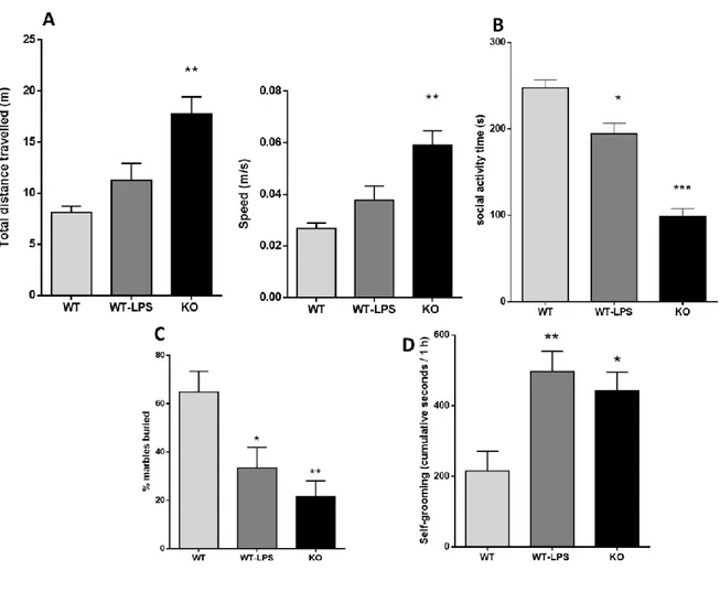
8.7. Behavioral alterations in adult p50-/- mice ... 39
9. Discussion and conclusion ... 41
CHAPTER 3... 44
ANALYSIS OF CHRONIC LPS EXPOSURE THROUGHOUT PREGNANCY IN WT MICE AND POSSIBLE PREVENTIVE PHARMACOLOGICAL TREATMENTS ... 44
10. Materials and Methods ... 45
10.1. MIA model ... 45
10.2. Drug treatment ... 45
10.3. Offspring ... 45
10.4. Immunohistochemistry ... 45
10.5. Cortical column analysis ... 46
10.6. ELISA test ... 46
10.7. Behavioral testing ... 46
10.8. Pups behavior ... 46
10.8.1. Ultrasonic vocalizations in pups... 46
10.8.2. Homing test ... 46
10.8.3. Olfactory habituation/dishabituation test ... 46
11. Adult mice behavior ... 46
12. Statistical analysis ... 47
13. Results ... 47
13.1. LPS induced both peripheral and central inflammation, partially prevented by meloxicam .. 47
13.2. A preventive pharmacological treatment with meloxicam does not rescue from the columnar alteration observed in the cortex of adult LPS-mice ... 48
3
13.4. Prenatal LPS exposure and meloxicam treatment do not affect social recognition and motility
in pups 51
13.5. The olfactory deficit in juvenile LPS-mice is partially prevented by meloxicam ... 52
13.6. Effect of prenatal LPS exposure and of preventive treatment with meloxicam on adult mice behavior 54 14. Discussion ... 55
15. Conclusion and future prospective ... 58
Bibliography ... 60
CHAPTER 1
Introduction
1. Neurodevelopmental disorders
Neurodevelopmental disorders (NDDs) are a heterogeneous group of disorders associated with impaired brain development. Generally, NDDs manifest in the early childhood and they are characterized by specific behavioral deficits including social, cognition, language and motor deficits. These deficits can change from very specific limitations of learning or control of executive functions to global impairments of social skills or intelligence [1]. However, NDDs frequently co-occur; for instance, individuals with Autism Spectrum Disorder (ASD) often have intellectual disability and many children with attention-deficit/hyperactivity disorder (ADHD) also have a specific learning disorder.
Diagnostic and Statistical Manual version 5 (DSM-5) classifies NDDs as follows: Intellectual disabilities (ID), Communication disorders, Autism spectrum disorder (ASD), attention deficit hyperactivity disorders (ADHD), neurodevelopmental motor disorders, specific learning disorder [2]. NDDs can also include pathologies associate to a genetic, environmental or medical specific condition such as fragile X syndrome, tuberous sclerosis, and Rett syndrome (genetic disorders), epilepsy (medical conditions) and very low birth weight and fetal alcohol exposure (environmental factors) (Fig.1). Due to the involvement of several factors including genetic, environmental, metabolic and immune factors, the etiopathogenesis of NDDs remains unknown. In recent years, the hypothesis has emerged that prenatal environmental factors, including maternal immune activation, may be implicated in the etiology of NDDs [3-7]. Indeed, maternal infection and the inflammation generated are plausible risk factors for these outcomes [5, 8-11].
Fig.1 The concepts of Gene x Environment interactions (GxE) and Gene-Environment correlations (G-E), as well as their potential implications for clinical NDDs and their experimental animal models [2].
2. Maternal immune activation (MIA)
Several epidemiological studies have shown a correlation between maternal immune activation (MIA) and NDDs [4, 9, 12, 13], but not without controversy. After the 1964 rubella pandemic, the incidence of two neurodevelopmental disorders, ASD and schizophrenia (SZ), rose from less than 1% in the unexposed population to about 13 and 20%, respectively [7]. Other studies revelated an association among flu [14], measles [15], chickenpox [16], polio [17] and SZ, ASD and mood disorders. However not all epidemiological studies, based on ecological data, have replicated these associations, probably due to a difference in the estimated population exposed [18]. Successively, these studies have been supplanted by several prospective studies following birth cohorts [3, 13, 19]. These studies used serological biomarkers of infection or inflammation in individual pregnancies to support the association between viral infections and NDDs in offspring [20-26]. Moreover, they added new potential pathogens to the list: bacterial infections, including pneumonia and others respiratory infections [27-29], genital/reproductive infections [26] and toxoplasma gondii infection [30-32]. All these studies strengthen the hypothesis that activation of maternal immune system can lead to NDDs. Furthermore, the presence of communicable diseases combined with an increase in autoimmune disorders [19], could explain the recent increase in incidence of NDDs [33]. Therefore, currently, prenatal stage is considered as a critical window which, if disrupted, could have long- lasting influences on neurodevelopment [34]. In fact, considering orchestrated processes that occur during foetal development, such as neurogenesis, migration, differentiation, synapse formation and apoptosis, perturbation in one or more of these processes during pregnancy can cause neurodevelopmental disorders [13, 19, 34-37].
The strength of the association between maternal immune activation and NDDs appears to be critically influenced by the precise timing of prenatal exposure. Although the exact timing remains unknown, it appears to be crucial for inducing disease-specific neurodevelopmental processes. Indeed, some retrospective epidemiological studies found a significant association between prenatal infection during the second semester of pregnancy and SZ [14, 38, 39] or during mid and late gestational periods and ASD [40-43]. However, recent epidemiologic findings have shown that contract an infection in early gestation is associated with the highest risk to develop SZ [15, 26, 27, 44]. It follows that the vulnerability to infection-induced neurodevelopmental alterations differs between distinct stages of foetal development and that the exact time of infection contraction could become crucial in the development of different neurodevelopmental disorders.
2.1. MIA and Schizophrenia
Schizophrenia (SZ) is a chronic form of psychotic illness that affects approximately 1% of population worldwide [45, 46]. It is characterized by an alteration in mental functions, emotions and behaviour. Several epidemiological studies have shown that the etiology of SZ involves aberrant neurodevelopmental processes that can occur during early brain development long before its clinical manifestation [47-50].
In recent years, several studies have shown that there is a high risk of developing SZ following prenatal exposure to infection [3, 51-53]. Key epidemiologic results included the association between maternal infectious pathogens and SZ. For instance, in the Child Health and Development study (CHDS) based on a large birth cohort in northern California, it showed an increased risk of developing SZ in children whose mothers were exposed to influenza during early to mid-gestation [54]. An elevated maternal T.gondii IgG levels and maternal reproductive infections was associated with a twofold increase in SZ risk [26, 30]. Maternal exposure to herpes simplex type 2 virus (HSV-2) was associated with nonaffective and affective psychoses in the National Collaborative Perinatal Project but not in the birth cohorts of the CHDS [55] or of the Finnish prenatal studies [56]. Neonatal antibodies to toxoplasma gondii and cytomegalovirus have been associated with non-affective psychosis in adulthood [31]. Other studies showed a correlation between an increase in IL-8 or maternal C-reactive protein levels and risk of SZ [23, 54]. All these findings suggest that various infections can lead to SZ, probably following different pathogenic pathways.
Many additional investigations based on maternal immune activation (MIA) animal models have produced a large amount of new data supporting the predictive potential of epidemiological studies, such as behavioral, neurochemical, morphological and transcriptional changes [11, 57-61].
including Polyriboinosinic:polyribocytidylic acid (poly I:C), lipopolysaccharide (LPS), influenza virus and select inflammatory cytokines support the epidemiological findings suggesting that the association between MIA and SZ is not limited to a single infectious or inflammatory condition [3, 5]. Despite the similarities between MIA models, there are some notable differences between the models with respect to the morphological and behavioural changes. For instance, whereas prenatal poly I:C exposure in rodents have been shown to induce cellular, neurochemical, and behavioural phenotypes that are characteristic of a hyperdopaminergic state [62-64], prenatal LPS exposure induces a hypodopaminergic state in adult rodents. Prenatal LPS exposure in the rhesus monkey causes a significant increase in global white matter volume [65]. An opposite pattern with a decreased in white matter volume was observed in rhesus monkey offspring born to mothers infected with influenza [66]. These differences can be explained both by prenatal timing of exposure to pathogen and the genetic background, but also because different immunogens can induce a different neuroimmune abnormalities across brain development and, consequently, may lead to differing long- term deficits in brain structures and functions [12]. These results support epidemiological findings which suggest that not all immunogens have the same potential to increase neuropsychiatric risk [3, 67].
2.2. MIA and autism spectrum disorders
Autism spectrum disorders (ASDs) are a group of neurodevelopmental disorders characterised by impairments in social interaction and communication and repetitive behaviour [68, 69]. The estimated prevalence of ASDs has recently increased: in 1992, it was estimated that 1 in 500 children had ASDs but currently 1 in 68 children (and 1 in 42 boys) are estimated [19, 70]. This increase could be explained by the presence of environmental factors that increased the likelihood of developing ASDs. Consistent with this idea, several studies have shown that an alteration in immune response during prenatal or early postnatal development can contribute to development of ASDs [25, 42, 43]. For instance, some case studies have reported increased risk of ASDs in children following prenatal exposure to immunogens such as rubella or cytomegalovirus [71-76]. Recently, a Danish birth cohort study found a nearly threefold increased risk for ASDs following hospitalization for viral infection in the first trimester, as well as an increased risk following hospitalization for bacterial infections in the second trimester [43]. A study from Kaiser Permanente Research Northern California showed that episodes of fever during pregnancy, particularly with no anti-fever medication, were associated with increased risk of ASDs [77]. However, a study of a large Swedish cohort reported that the association between maternal infection and ASDs was independent of both infectious agent and time of pregnancy [42]. A recent study of the Swedish cohort found an association between increased risk of
ASDs and severe infections during pregnancy, such as sepsis, pneumonia, meningitis, pyelonephritis and influenza [78]. Further recent studies have also shown that risk of ASDs could increase if the infection occurs during the second trimester of pregnancy [79]. Collectively these studies suggest that the more severe infection is accompanied by a robust inflammatory response, the higher the risk of ASDs in children increases. This phenomenon appears to be related to the maternal response and the time during pregnancy rather than the pathogen itself [34]. Quantification of cytokines, chemokines, interleukins, maternal C-reactive protein and other inflammatory markers obtained from maternal serum samples and amniotic fluid, support the link between maternal infection and increased ASDs risk [44, 79-82].
Animal models further support this hypothesis; prenatal exposure to poly I:C, LPS or allergies and asthma can induce behavioural abnormalities such as social interaction deficit, alteration in ultrasonic communication and high level of repetitive behaviours [5, 83-86]. These models also showed morphological and cellular anomalies in offspring’s brain implicated in ASDs, including abnormal cerebellar development, impaired expression of the extracellular matrix protein reelin, and altered synapse density and neural connectivity [4, 5, 61]. Notably, some of these rodent findings have been extended to rhesus monkeys, both at behaviour and brain morphology [34, 65, 87, 88].
2.3. Possible mechanisms mediating the effects of MIA on the offspring
The precise mechanisms responsible for the pathological effects of maternal infection on the offspring neurodevelopment remain to be determined. Exposure to a harmful pathogen during pregnancy provokes in the maternal environment a systemic inflammatory response with activation of different immune molecules, including cytokines, chemokines, prostaglandins, leukotrienes, and hypothalamic-pituitary-adrenal (HPA) axis [89-94]. Consequently, this inflammatory response can cause fever, lethargy, reduction of motivation in food and social contact in mothers [95-97]. The effects in the offspring are still more complex and the final outcome is still not clear. Different mechanisms have been speculated, but the prevailing hypothesis suggests that alteration in the expression of maternal immune molecules may be the key mediating factor for the observed change in offspring [98-103].
This hypothesis is based on the emerging discovery that there is a crosstalk between immune molecules and brain development through which these molecules acquire a pivotal role in the processes of neurodevelopment [4, 9, 37, 104, 105]. Normally immune molecules, in particular cytokines and glia cells, have an important role in brain development, including neurogenesis, synapse formation and plasticity [7, 19, 106-109]. In presence of inflammatory response placental
provoking a placental dysfunction that could induce changes in brain function [9, 110-112]. Successively, all these factors may induce the activation of inflammatory response in the foetal blood, with the activation of cytokines, chemokines, prostaglandins and other foetal pro-inflammatory factors that can lead to permanent changes in the foetal brain development [106-109] (Fig.2). Thus, the perturbation in the balance between immune molecules and brain development could explain the increased risk of developing NDDs when there is exposure to harmful pathogens.
Moreover, maternal infection can induce oxidative stress increasing the level of oxygen species (ROS) and nitrogen species (RNS), that can participate in the alteration of foetal neurodevelopment [9, 113, 114]. It changes also the maternal and foetal availability of several micronutrients, including iron and zinc, both of which are highly important for the normal development of peripheral and central organs [115-117]. All these factors, taken together, could increase the risk to develop neurodevelopment disorders [6, 118-123].
3. Animal models of MIA
Animal models of MIA are necessary to understand the causal relationship between MIA and the risk of developing NDDs. In addition, to direct research on human subjects, basic animal research represents a convenient approach to study the neurobiological basis of brain and behavioral alterations in NDDs and to develop novel pharmacological therapies. Indeed, animal models, not only allow a rigorous experimental control of subjects in genetically homogenous populations and facilitate the identification of neurobiological factors, but also provide indispensable tools for testing hypotheses that cannot be directly tested on human subjects for technical and ethical reasons, including the verification of casual relationship in epidemiological studies. Moreover, clinical research on pregnant women is very complicated and in most cases the offspring are born healthy and the symptoms appear many years after birth [4, 124, 125]. However, the use of animal models to reproduce human psychiatric conditions, such as hallucinations and delirium, have always been met with some skepticism because they are specific to humans and cannot clearly be identified in animals [70, 126, 127]. Therefore, to avoid the inability to reproduce the entire syndrome in animals, the experimental approach focuses on individual behaviors using specific tests [128-130], such as prepulse inhibition of the acoustic startle reflex, social interaction and recognition tests.
Due to these challenges of studying MIA in humans, animal research remains essential for identifying the biological mechanisms and developing new therapies for NDDs. A plethora of animal studies, not only on rodents but also on non-human primates, conduced during the years, have clearly demonstrated a correlation between MIA and NDDs (Fig.3) [4, 9, 11, 65, 66, 88, 131]. In these models, animals are exposed to an immunological stimulus at a specific gestational stage. One class of animals is exposed to live pathogens, such as influenza virus [58-61] and Toxoplasma gondii [30,31] (table 1). These models are mainly used to verify causal relationships in epidemiologic studies evaluating the role of specific infectious pathogens. Another class of animal models makes use of immune-activating agents that stimulate the innate immune system, such as the bacterial endotoxin lipopolysaccharide (LPS) and the synthetic double-stranded RNA analogue polyriboinosinic:polyribocytidylic acid (poly I:C) [5, 11, 57, 61, 62, 132, 133], and more recently staphylococcal enterotoxin [134]. These models were initially developed to test whether alteration in maternal or fetal immune cytokines may be the critical mediator between prenatal infection and postnatal brain pathology [135-137]. An improvement of this second class of models was the application of specific cytokines as immune-activating agents, such as IL-1β or IL-6 [7, 9, 57, 83, 99, 101]. This new approach is conducted to identify whether specific cytokines, or a cytokines networks, mediate the association between MIA and NDDs. A third class of models used specific
immunopathological processes that have been implicated in the etiology of NDDs [12]. Some important examples of this class are animal models of maternal exposure to autism-related maternal autoantibodies [138-140], toll-like receptor 7 agonists [141], allergic disorders and asthma [84, 142, 143].
Fig.3 Contribution of epidemiologic and science studies to translational research of MIA in NDDs [12]
Pioneers in the used of MIA as model for NDDs were Fatemi and colleagues. They exposed pregnant mice to human influenza virus and found that the offspring showed several neuropathological alterations. Several following studies, reported that the offspring of mothers treated with influenza virus showed deficient corticogenesis and brain atrophy, impaired development of the corpus callosum, reduced hippocampal volumes, and decreased expression of γ-aminobutyric acid (GABA) markers such as reelin [58, 59, 144, 145], long-term deficiency in serotonin (but not dopamine) production [146] and a set of behavioural abnormalities in adulthood [136, 147]. The mouse prenatal influenza model has recently extended in rhesus monkeys, demonstrating reduction in grey and white matter in distinct cortical and parieto-cortical brain regions of offspring [66].
The majority of current MIA models are based on maternal exposure to non-virulent immune- activating agents, such as poly I:C [5, 11, 57, 147] or LPS [9, 148-151]. Although these models offer some advantages, they are unable to reproduce the full spectrum of immune responses normally induced by infectious pathogens (such as influenza virus). For instance, they cannot reproduce the real-life influences in humans other than the primary exposure of interest or they cannot stimulate pathogen-specific humoral and cellular immune reactions, which may be part of the mechanisms of effects observed in the offspring [12]. However, the increasing in the used of these non-virulent immune-activating agents is based on their safety (they do not require a stringent biosafety level as infectious pathogens) and they allow to control the intensity and duration of the maternal immune response. The ability to restrict the immune response to a specific time point in gestation is important because not only allow to identify the effects of MIA on brain and behavioral functions in offspring, but also to determine if these are influenced by a precise gestational timing [100, 132, 136, 152-155]. Finally, these models are capable to alter pro- and anti-inflammatory cytokine levels in placenta, amniotic fluid and fetus, including the brain [100, 152,156].
However, with a more emerging role of MIA as model for NDDs, difficulties in the reproducibility of the experiment are also emerging. Indeed, MIA paradigm is quite heterogeneous: each laboratory uses different strain of animals, doses, immune stimulus, time to immune insult, age of offspring, brain regions studied that can influence the reproducibility of experimental outcomes.
3.1. Viral model of infection: polyriboinosinic:polyribocytidylic acid
Polyriboinosinic:polyribocytidylic acid (poly I:C) is a synthetic double-stranded RNA analogue, used commonly to mimic aspects of viral infection. It is recognized as foreign by the mammalian immune system through the transmembrane protein toll-like receptor 3 (TLR-3) present in endosomal membrane [184]. TLRs are a class of pathogen recognition receptors which recognize invariant structures present on virulent pathogens and in the case of poly I:C, it is recognized by double- stranded RNA and induces translocation of nuclear factor κ light-chain-enhancer of activated B cells (NF-κB) to the nucleus and induction of chemokines (CXCL) and cytokines, including CXCL-1, IL- 1β, IL-6 and TNF-α, type I of interferons [185-188] and HPA axis (fig.4) [186, 189]. Although, this model does not readily mimic the precise immunological insults normally induced by viral exposure, it is able to mimic the cytokine-associated viral acute phase response in maternal host and in foetal environment [147]. A limit in the use of poly I:C is related to its manufacture. Although it is a synthetic analogue, change in its manufacture can have significant effects on its efficacy to drive the
among manufactures and it can affect the magnitude of immune response [6, 190, 191]. Another limit in the use of poly I:C is the gestational timing of poly I:C administration, the doses and the type of animal used.
Offspring prenatally treated with poly I:C have been reported to have significant abnormalities in several behaviors, including acoustic startle response, decrease in exploratory behavior, increase in immobility, decrease in social interaction [9, 147, 185, 192-194] (Table 2). Moreover, they also showed increased level of gamma aminobutyric acid (GABA)A, receptor α2 immunoreactivity and dopamine hyperfunction as well as delay in hippocampal myelination [63, 195, 196]. Other neurochemical deficits observed in poly I:C models are reduction in NMDA receptor hippocampal expression and reduced numbers of reelin and parvalbumin-positive cells in prefrontal cortex as well as reduction in D1 and D2 receptors in prefrontal cortex and enhanced tyrosine hydroxylase in striatum [162, 197].
All these effects are dependent on the prenatal timing, poly I:C doses and rodent species. Since poly I:C induces a controlled immune reaction lasting 24-48h, it is used to study the most vulnerable pregnancy times. Meyer and colleagues demonstrated that maternal poly I:C-induced immunological stimulation at different gestational times provokes distinct psychopathological and neuropathological symptom clusters in offspring [147]. For instance, prenatal poly I:C treatment on gestational day (GD) 9 (early-middle pregnancy) leads to a pathological profile characterized by deficit in exploratory behavior, abnormalities in selective associative learning, impairments in sensorimotor gating, enhanced sensitivity to the indirect dopamine receptor agonist AMPH and deficit in spatial working memory when the demand on temporal retention is high [152, 197, 198]. On the other hand, the same poly I:C treatment on GD17 (late pregnancy) leads to impairment in the discrimination reversal learning, deficits in spatial working memory and recognition memory even when the demand on temporal retention is low, potentiated response to AMPH and to dizocilpine (a non-competitive NMDA-receptor antagonist) and also can induced impairs sociability and decreased in cerebrospinal fluid volume [152, 163, 197-200]. This difference observed between early/middle (GD9) and long (GD17) gestation may be related to different symptoms of SZ. Viral-like immune challenge at GD9 induced in mice a variety of abnormalities associated with positive symptoms including hyperdopaminergic state specially in mesolimbic areas [201], increased sensitivity to acute dopaminergic stimulation [132, 162, 197] and increased in striatal dopaminergic activity [64, 132, 197], while viral-like immune activation on GD17 is associated with hypodopaminergic and hypoglutamatergic state in prefrontal cortical and hippocampal areas [199], cognitive and negative
symptoms including preservative behavior, social interaction deficits, anhedonic behavior and behavioral/cognitive inflexibility [198-200] .
Fig.4 Involvement of the TLR3 and TLR4 signalling pathways in neuropsychiatric disorders. [202]
3.2. Bacterial model of infection: lipopolysaccharide
Lipopolysaccharide (LPS), a cell wall component of gram-negative bacteria, is a well characterized model to mimic an innate acute phase response to bacterial infection. LPS is recognized mainly by the pathogen recognition receptor transmembrane protein toll-like receptor 4 (TLR-4) presents on the cellular membrane [203]. LPS by binding TLR-4 triggers a signal transduction cascade that leads to the activation of transcription factors, such as NF-κB, which induces activation of cytokines, chemokines, prostaglandins, leukotrienes and HPA axis (Fig.4) [89-94]. These pro- inflammatory factors, particularly IL-1, IL-6 and TNF-α, provoke a placental dysfunction leading to
activation of inflammatory response in the blood and in the brain of fetus, including cytokines, chemokines, prostaglandins and glia cells, that can induce changes in brain function of offspring (Fig.5) [9,110,111]. Like poly I:C, also LPS induces immunological challenges that are time-limited, ranging from 24 to 48 h depending on the precise dose used. This allows to set the start of maternal immune activation with a specific period of fetal development [132, 204].
Fig.5 Potential LPS mechanisms mediating effects of prenatal infection on brain function [9]
There are some analogies between the cytokine-associated inflammatory responses triggered by LPS and poly I:C [203, 205], but there are considerable differences in the activation of the total immune response. For instance, poly I:C induces activation of type I IFNs that stimulate antiviral immune responses [205]. By contrast, LPS is more effective in stimulating the production and secretion of TNF-α from innate immune cells such as macrophages [206]. This difference can explain why LPS is more potent than poly I:C in triggering anorexia, lethargy, and febrile responses [207]. Furthermore, LPS appears to induce activation of microglia during embryonic stage, whereas poly I:C appears to induce microglia alterations at postnatal or adult age [208,209].
Offspring prenatally treated with LPS have been reported to have significant abnormalities in several behaviors, including alteration in ultrasonic vocalizations (USVs) in pups [148,2010], deficit in prepulse inhibition [133, 211, 212], increase in anxiety, impairment in spatial learning and social
Table 2. A Sample of long-term behavioral and cognitive dysfunctions as identified in developmental immune activation models in rats and mice [11]
interaction, increase in repetitive behavior [113, 114, 148, 186, 213] increase in recognition of novel object and exploration in familiar object [214,215], increase in ethanol preference [216] and decreased sexual behavior [217] (Table 2).
Morphological changes observed in the offspring prenatally treated with LPS appear normally several days to months after birth. LPS induces increase in cerebral cortical lesions [218] and increase in striatal apoptosis [219], decrease in hippocampal neurogenesis and decrease in dendritic length, arborization and spine density in hippocampus and medial prefrontal cortex [220]. Moreover, LPS induces cell death in white matter and decreased myelin basic protein immunostaining [219, 221- 224]. Changes in the level of neurotransmitters were also found after administration of LPS; it reduces tyrosine hydroxylase (TH) in dopaminergic neurons [225-229], decreases DA level in nucleus accumbens and striatum [133, 213, 225, 227, 229, 230] and also LPS decreases serotonin levels in various brain regions [231]. Regarding glutamatergic function, LPS increases AMPA and NMDA receptor levels in hippocampus [114, 232].
4. Genetic risk factor in SZ and ASDs
Pregnancy exposed to infections does not always develop several neurodevelopmental brain disorders such as SZ or ASD [51]. This suggests that the etiology of neurodevelopmental disorders can be a combination of multiple hits (exposure to more than one risk factor), such as environmental factors [172, 174, 235, 236] and genetic mutations [103, 165, 166, 233]. In this contest, MIA appears to act as a “disease primer” that makes the individual more susceptible to the effects induced by other hits in triggering disease related symptoms later in life [4, 234] (Fig.6).
Fig.6 MIA as a disease primer [4]
The combination of MIA models with genetic animal models of NDDs may represent a fruitful approach to identify critical interactions between genetic and infection-associated environmental factors and to assess their modulatory influence on brain development. Therefore, the long-term brain and behavioral effects of MIA models may be compared to animals with different genetic backgrounds. Indeed, in these studies based on the involvement between genetic and environmental factors, genes directly involved in innate and acquired immunity were examined. Some of these genetic risk factors are promoter polymorphisms of pro-inflammatory [37, 238] and anti-inflammatory cytokines [239, 240], human leukocyte antigens (HLA) and alleles [241, 242]. For instance, Meyer et al. [197] have compared the neuropathological consequences of prenatal poly I:C exposure in control mice and transgenic mice constitutively overexpressing the anti-inflammatory cytokine IL-10 in macrophages. They showed that enhanced IL-10-mediated anti-inflammatory signalling during prenatal development is sufficient to prevent brain abnormalities in the adult offspring [197]. In another study, it has been demonstrated that poly I:C prenatal exposure is
inefficient in inducing alterations in brain development in animals with a genetic deletion of IL-6 compared with control animals [101].
In addition to immune-related genetic factors, the MIA models can be also used to identify a possible synergism between prenatal immune challenge and aberrant genes which are considered the major genetic susceptibility factors of neuropsychiatric disorders of neurodevelopmental origin. For schizophrenia, this may include neuregulin-1 (NRG-1), catechol-O-methyltransferase (COMT), disrupted in schizophrenia-1 (DISC1), and V-akt murine thymoma viral oncogene homolog 1 (AKT1) [243] and for ASD it may include methyl-CpG-binding protein-2 (Mecp2), ubiquitin protein ligase 3A (Ube3A), neuroligin (Nlgn), Shank proteins (SHANK3), contactin-associated protein-like2 (CNTNAP2), fragile mental retardation 1 locus (FMR1) and serotonin transporter (SERT) [244-246] It has been thought that disruption of these genes synergically with prenatal infection may enhance the risk to develop NDDs [5,247,248]. For instance, Abazyan and colleagues demonstrated that mutant DISC1 mice prenatally treated with poly IC (GD9) developed several both behavioral (reduced social interaction) and neuro-morphological (reduction spine density in hippocampal neurons and volumetric reduction of amygdala) changes that were not present in immunologically non-challenged mhDISC1 mice [147, 165].
5. p50-/- mice
In our laboratory we are studying a new potential gene involved in the pathogenesis of NDDs, NF- κB1 coding for the NF-κB p50 subunit. NF-κB represents a family of inducible transcription factors present in all type of cells, which regulates a large group of genes involved in different processes of inflammation, immunity, cell proliferation, apoptosis and neural plasticity [249-257]. This family is composed of five related members: RelA (also named p65), RelB, c-Rel and the precursor proteins NF-κB1 (p105) and NF-κB2 (p100), which are processed into their active forms, p50 and p52, respectively. These transcription factors generally work as homodimers or heterodimers, and once translocated to the nucleus, they induce or repress specific gene expression by binding to the canonical kB sites or to non-canonical sequences [258-260]. NF-κB pathway can be activated by two major signalling pathways, the canonical and non-canonical pathway [261, 262]. The canonical NF- κB pathway responds to different stimuli, including cytokines, growth factors, mitogens, microbial components and stress agents [263-265]. In the canonical pathway, the heterodimers p50 and p65 are retained in the cytoplasm through their interaction with the nuclear factor of kappa light polypeptide gene enhancer in B-cells inhibitor alpha (IκBa) proteins (Fig.7A). IκBα is phosphorylated by the
inhibitor of NF-kB kinase subunit beta (IKKb, also termed IKK2), for degradation and released from the p50/p65 complex [252, 266, 267]. IκBα dissociation from the p50/p65 complex allows the NF-
κB members to translocate to the nucleus and exert their functions as transcription factors [253, 267, 268]. In contrast, the non-canonical NF-κB pathway selectively responds to a specific group of
stimuli, including ligands of a subset of TNFR superfamily members such as LTβR, BAFFR, CD40 and RANK [261, 269] and it acts on the relies of the NF-κB2 precursor protein, p100/RelB [261,
269].
Fig.7 Signalling of NF-κB pathway [279]
p100/RelB complexes are held inactive in the cytoplasm until p100 is phosphorylated and partially processed to become the active p52/RelB form, which will translocate to the nucleus to exert its function (Fig.7B) [261, 265, 269]. Functionally, canonical NF-κB pathway is involved in immune response, whereas the non-canonical pathway appears to be involved as an additional signalling to the canonical pathway in the regulation of specific functions of the adaptive immune system [264, 269].Although the pivotal role of NF-κB signalling is to regulate the immune system and inflammation, it has been demonstrated to regulate different stages of embryonic development and adult neurogenesis [270-272]. Indeed, the importance of NF-κB pathway is highlighted by the embryonic lethality of several KO mice with disrupted key components of the NF-kB pathway [273]. For instance, KO mice for p65 [274], IκBα [275] and IKKα/IKKβ [276] showed developmental defects, dying in early stages of embryonic development [274,277,278]. These effects are related to the regulation by NF-κB of early differentiation of neural stem cells, as well as the proliferation/apoptosis of neuroepithelial cells, neuroblast migration, maturation and neuron
plasticity [279]. Moreover, NF-κB has an important role also during postnatal and adult period. In the postnatal nervous system development and then in adult brain, NF-κB induces the expression of genes involved in cell differentiation and survival [280] and also it has a key role in neurogenesis and neuronal plasticity as adults [271,281,282].
5.1. p50-/- mouse as model of NDDs
p50-/- mice are generated by targeted deletion of the NF-κB1 gene that encodes for the precursor of the p50 NF-κB subunit. They are fertile and viable with no macroscopical developmental and histopathological abnormalities, although at the age of 6-10 months their body weight decreases and about 25% begin to die before old age [283]. p50-/- mice show functional and behavioral alterations. For instance, p50-/- mice showed functional deficit in some sensorimotor tests, they exhibited anxiety- like behavior, elevated exploratory behavior, deficit in acquiring escape behavior and in short-term memory and reduced tendency to establish dominant-subordinate relationships among cage mates [283-285].
Our research group demonstrated that p50-/- mice at postnatal day (PND) 2 present an increase in brain lipid binding protein (BLBP) positive cells, a marker of radial glial cells, an increase in reelin, a protein involved in the neuronal migration, and increased thickness of deeper cortical layers [286]. Adult mice showed abnormal columnar organization in the somatosensory cortex with an increase in cell density per column, decreased cell spacing and loss of linearity of minicolumn. Moreover, they showed a decrease in somatostatin and parvalbumin-expressing interneurons and an alteration in cortical neurons neurite orientation and a reduction in synaptic connections (Synapsin I protein levels). From a behavioral point of view, p50-/- mice have an increase in locomotor and exploratory activity, deficit in reciprocal social interaction (both in male-male social test and male-female social test), increased repetitive behavior and altered ultrasonic vocalizations [286-288].
6. Possible pharmacological treatments
The origin of NDDs are so varied and heterogeneous that finding a preventive cure for these disorders is currently an extremely demanding challenge.
In vivo rodent models of MIA represent a unique opportunity to evaluate possible pharmacological
treatment related to prenatal exposure to pathogen stimuli. To date, there are three potential strategies to prevent and/or act on neurodevelopmental symptoms induced by immune challenge: (a) during the
pre-conceptional period, (b) during the acute phase of maternal exposure to infection, and (c) during the early phases of the offspring’ postnatal development [5].
Some severe viral infections may be prevented by appropriate vaccinations [289, 290]. Indeed, vaccinations during pre-conceptional period reduce the incidence of infection mediated neurodevelopmental brain disorders. For instance, Brown and colleagues reported that the association between prenatal influenza infection and higher risk of schizophrenia would have been reduced by 14-21% if maternal infection had been prevented by influenza vaccinations [21]. Nevertheless, vaccinations may reduce the risk of schizophrenia associated with prenatal exposure to genital and reproductive infections [26].
Intervention during the acute phase of maternal infection may be another important period for pharmacological treatment. Blocking the inflammatory response, that is responsible for the morphological and behavioral alterations observed in the offspring, is currently the most studied strategy because it aims to prevent all these alterations. For instance, administration of IL-10, (an anti-inflammatory cytokine) or anti-inflammatory drug, N-acetylcysteine, to pregnant rodents given uterine LPS, prevents foetal loss and white matter damage or hippocampal dysfunctions and learning deficits in the offspring, respectively [113, 291-293]. However, a strong attenuation in the inflammatory response during pregnancy may facilitate rather than limit the infectious processes in the maternal host. For instance, mice with genetically enforced of IL-10 over-expression showed an increase in the bacterial loads and retarded bacterial clearance if exposed to mycobacterial infection [294]. Similarly, high levels of IL-10 reduce the host defence against pneumococcal pneumonia in mice [295]. Furthermore, molecules with anti-inflammatory properties may also have teratogenic effects [296-298].
Currently, therapeutic interventions during juvenile offspring represent the only pharmacological treatment available for NDDs. This is possible because the full spectrum of behavioral and morphological abnormalities induced by prenatal immune challenge continues also during the post-pubertal period [62, 63, 152, 162, 299]. However, the pharmacological treatment in the peri-adolescents is based on the action of behavioral deficits and not on the preventing the development of symptoms. Indeed, chronic administration of antipsychotic or antidepressant drugs to peri-adolescent and/or adolescent subjects that showed psychotic symptoms, has recently been introduced as preventive treatment of schizophrenia and other psychosis-related disorders in humans [300-303]. Although, the use of these drugs has provoked several ethical concerns and therefore still remains highly controversial [304, 305], several studies on MIA models have shown that peri- adolescent treatment with antipsychotic drugs are able to normalized the symptoms in subjects
observed after exposure to pathogens [62, 63, 136, 211, 212, 306-308]. For instance, clozapine improves spatial working memory and long-term memory deficits in rodents [63, 147, 308]. Risperidone appears to rescue from aberrant hippocampal neurogenesis, parvalbumin expression and from social deficits observed in rodents after exposure to poly I:C or LPS [202, 287, 309]. A long- term treatment with fluoxetine or the antipsychotic aripiprazole, has shown to completely reverse amphetamine-induced locomotor activation in offspring after poly I:C exposure [310]. Minocycline, a microglia modulator, prevented the emergence of MIA-induced behaviours and changes in pro- inflammatory cytokines in the adult brain [13, 311, 312].
CHAPTER 2
COMPARISON BETWEEN AN LPS-INDUCED AND A GENETIC (p50-/- ) MOUSE MODEL OF NEURODEVELOPMENTAL DISORDERS
In the first part of my PhD program I focused my attection on studying the effect of maternal immune activation in NDDs and more specifically the role of NF- κB pathway in these disorders. For this reason, p50-/- mice lacking the NF-κB1 gene encoding the precursor of the p50 NF-κB subunit, were used. Along with this genetic model, a wel-known MIA model (LPS-induced model) was also used to analyze the effects of a different type of maternal immune activation (genetic and induced) in the offspring of these two models.
In order to understand the effects of maternal immune activation and how the alteration in the NF- κB pathway can affect offspring neurodevelopment, different types of behavioral tests were conducted at different time of mouse’s life (infancy, youth and adulthood). Furthermore, to understand whether external perturbation in the maternal inflammatory response (LPS) or genetic alteration in maternal inflammatory response (p50-/- mice) can induce structural and functional changes, the central and peripheral inflammation and the cortical cytoarchitecture of the adult offspring were analyzed.
7. Materials and methods
7.1. Animals
In this study, wild type (B6;129PF2) mice and NF-κB p50-/- mice (B6;129P2-Nfκb 1tm 1 Bal/J) were used. Mice were purchased from The Jackson Laboratories (Bar Harbor, ME, USA). Wild type (WT) and p50-/- mice (from now called KO mice) used for these experiments were obtained in our animal facility from mating mice. Each breeder adult male mouse was maintained in the same cage with an adult virgin female for mating. A late gestation (from day 15 after fecundation) female was individually housed and inspected twice daily at 10:00 a.m. and 6:00 p.m. to determinate the day of pup’s birth (PND 0).
Animals were housed two-to-three per cage in a 12-h light/dark cycle (light phase: from 8:00 a.m. to 8:00 p.m.), with food and water available ad libitum. The cage size was 15 cm wide × 35 cm long × 12 cm deep. Temperature (22 °C) and humidity (50% ± 1) in the cage were automatically
regulated by Sealsafe Aero System by individually ventilated cages with EPA filters (Tecniplast Group, Varese, Italy). Experiments were conducted in conformity with the European Communities Council Directive of 1986 (86/609/EEC) and approved by the Italian Ministry of Health (910/2016- PR), Animal Care and Use Committee of the University of Brescia.
7.2. MIA model
Female WT mice were mated with male WT mice (1 female −1 male) at 12–14 weeks of age for a period of 24 h. The morning after mating, the presence of a vaginal plug was verified, and in case of positivity, this was considered the gestational day (GD) 0. Successively, female WT mice (from now on called WT dams) were divided randomly in 2 groups of 4–6 dams each.
The first group of WT dams was treated with vehicle (VH, phosphate-buffered saline, PBS) subcutaneously (s.c.) on alternate days from GD1 to the end of pregnancy. The second group of WT dams was injected with 0.3 mg/kg of LPS from Escherichia coli (L-3129 serotype 0127:B8, MilliporeSigma, Merck KGaA, Darmstadt, Germany) subcutaneously (s.c.) on alternate days from GD1 to the end of pregnancy [211].
7.3. Offspring
Offspring obtained from the WT, KO and WT-LPS dams were divided in three groups (WT-mice, KO-mice and WT-LPS mice) and housed two to three per cage after the weaning (4 weeks). All the structural and behavioral analysis carried out in this study were conducted on the offspring at different time periods: pups (ultrasonic vocalizations and homing test), juvenile mice (olfactory habituation/dishabituation test) and adult mice (open field test, reciprocal social interaction test, marble burying test, home cage observation test). After the behavioral tests, mice were sacrificed either to obtain serum for ELISA testing, or brains for immunohistochemistry and minicolumn analysis.
7.4. Immunohistochemistry
Three adult mice per group were used for immunohistochemistry (IHC). At the end of behavioral experiments, mice were anesthetized with chloral hydrate (400 mg/kg) and perfused with 4% paraformaldehyde (PFA). Then, brains were fixed, cryoprotected and coronally crysectioned at 20 µm. IHC for glial fibrillary acidic protein (GFAP), marker for astrocytes, and ionized calcium-binding adapter molecule 1 (Iba1), marker for microglia, was performed. The following primary antibodies were used: α-Iba1 rabbit (W.D. 1:500, Wako Chemicals GmbH, Neuss, Germany), α-GFAP mouse (W.D. 1:300, Sigma Aldrich, Saint Louis, MO, USA). The following secondary antibodies were used:
biotinylated α-rabbit (W.D. 1:1000, Vector, Burlingame, CA, USA) and biotinylated α-mouse (W.D. 1:1000, Vector, Burlingame, CA, USA), respectively. Immunochemistry signal was detected with 3,3-diaminobenzidine tetrahydrochloride (DAB). For each animal, a complete series of one-in-eight sections (160 μm apart) through the cortex was analyzed, and positive cells were counted. An average of 8 –10 sections per animal was used. Images of the cortex were acquired by using a x20 (Iba1) and a x40 (GFAP) objectives with an Olympus BX41 microscope (Olympus Life Science, Waltham, MA, USA). Iba1- and GFAP-positive cells were counted in the entire cortex for each group. For the cell count, we drew squares with a defined area (width of 200 µm and height of 600 µm for Iba1 cells, and width of 500 µm and height of 600 µm for GFAP cells) by using the ImageJ software (National Institute of Health, Bethesda, Maryland, USA). A minimum of 6 areas for each section were counted. Analyses have been performed in a blinded manner. Data are expressed as Iba1- and GFAP-positive cells per area.
7.5. Cortical column analysis
Three adult mice for group were used for cortical column analysis. Coronal brain sections were stained with cresyl violet (Nissl staining). Images of Nissl-stained brain sections were acquired by using a x10 objective of an Olympus BX41 microscope. Successively, cortical minicolumns were semiautomatically analyzed using the ImageJ software (National Institute of Health, Bethesda, Maryland, USA) as previously described [286]. Anterior cingulate (AC) cortex (corresponding to bregma 1.70 and 1.94 mm) and somatosensory (SS) cortex (corresponding to bregma −1.28 and −1.64 mm) were analyzed in both cerebral hemispheres of three mice per group. A mouse brain atlas (Franklin and Paxinos) was used as a reference to ensure the correct positioning of the cortical images acquired. A rectangular area with the size of a minicolumn (region of interest, ROI) (width 60 µm; height 400 µm) was marked in layers II–IV, where columnar organization of the cortex is clearly defined. Up to 15 ROIs per section, placed at regular, non-overlapping intervals across the image were analyzed. Average cell density and total path length ratio (TPLR), an index of column vertical organization, were measured in each ROI. Only cells having a size of 70–350 µm2 (pyramidal neurons) were included in the count. The cell area range defined was set to restrict cell count to pyramidal neurons (excluding glia and interneurons) and to avoid multiple cell clusters. Analyses have been performed in a blinded manner.
7.6. ELISA test
Levels of interleukin-1β (IL-1β) were quantified in serum of 5 WT-, and 6 KO- and 4 WT-LPS adult mice. Mouse IL-1β ELISA kit (Invitrogen, ThermoFisher Scientific, Vienna, Austria) was used and the test was performed following the manufacturer’s protocol.
7.7. Behavioral testing
Behavioral tests were carried out during the light phase of circadian cycle, between 9:00 a.m. and 5:00 p.m. In addition, 32–38 pups, 20 juvenile mice and at least 7 adult male mice (4–6-month-old) per group were used. 6 WT, 6 KO and 6 WT-LPS primiparous (4-6 moth-old) females were used to study the maternal behavior without any influence of previous maternal experiences. During the tests, the operator remained in an adjacent room, separated with a dark sliding door from the test room. All the behavioral analyses have been performed in a blinded manner.
7.8. Mother-pups interaction
Maternal behavior was analyzed for 18 litters derived by 6 WT, 6 KO and 6 WT-LPS primiparous dams (4-6 months-old). From PND 1 to 7, dams were observed twice daily for 1h (at 11 a.m. and at 4 p.m.) as reported by Oddi and colleagues [288,313]. Briefly, one observation was performed every 2 minutes, for a total of 30 observation in 1-hour session. Maternal behaviours, including nursing postures, non-nursing posture, pups grooming and nest building were analysed and expressed as frequencies [314,315].
7.9. Pups behavior
7.9.1. Ultrasonic vocalizations in pups
Ultrasonic Vocalizations (USVs) emitted by 37 WT, 37 KO and 38 WT-LPS pups after maternal separation for 3 min on post-natal day (PND) 4, 6, 8, and 10 were analyzed using Avisoft Bioacoustics system as previously described [288,316]. Briefly, USVs were recorded with ultrasound microphone (UltraSoundGate CM16, Avisoft Bioacoustics, Berlin, Germany) sensitive to frequencies of 10–180 kHz and analysed with SasLab Pro (version 5.2; Avisoft Bioacoustics, Glienicke/Nordbahn, Germany). Number and duration of calls were analysed. Moreover, the body weight of each group of mice was analyzed daily from PND 4 to 10.
7.9.2. Homing test
Homing test has been performed as previously described [317]. Briefly, on PND 14, 37 WT, 37 KO and 38 WT-LPS pups were transferred in a Plexiglas T-maze (arm size: 10 cm length × 8 cm
(covered by sawdust collected from the nest of the pups’ cage). The nest and clean arms were alternated between right and left between each pup. Each pup was placed in the start arm and was free to explore the maze for 5 min. Videos captured by a camera above the maze were analysed by the operator, measuring first latency to reach the nest, time spent in the nest and in the clean arms and locomotor activity by square crossing.
7.10. Juvenile mice behavior
7.10.1. Olfactory habituation/dishabituation test
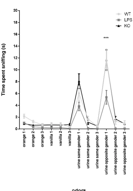
Olfactory habituation/dishabituation test has been performed as previously described [288, 318]. Briefly, 20 WT, 20 KO and 20 WT-LPS juvenile mice (6 weeks old) were placed in an empty cage and exposed to different odors, classified as non-social (vanilla and orange) and social odors (urine from same-gender or opposite-gender mice), in three consecutive trials of 2 min duration each. Time of olfactory investigation (considered as sniffing activity up to 2 cm distance from applicator) of each mouse was evaluated.
7.11. Adult mice behavior
7.11.1. Open field test
Open field test was performed as previously described [287]. Briefly, each mouse was acclimated in the procedure room for 10 min, then placed in the center of the arena (40 × 40 cm Plexiglas box) and allowed to freely explore for 5 min, during which time the mouse was recorded by a camera placed on top of the arena. The Plexiglas box was cleaned after each test session to delete any odor deposited by previous mouse. Mouse locomotor activity, total distance travelled, and average speed were automatically analyzed with Ethovision XT software (version 13, Noldus, Wageningen, The Netherlands).
7.11.2. Reciprocal social interaction test
Reciprocal social interaction test was performed following the Silverman protocol [319]. Briefly, mice were individually housed for 48 h preceding behavioral testing. For the test, after a period of acclimation of 10 min in the procedure room, each mouse was exposed to an age, sex and strain-matched unfamiliar subject (intruder subject) for 10 min. The test was video recorded and analyzed with Observer XT (version 14.1, Noldus, Wageningen, Netherlands). Specific social (nose- to-nose sniffing, anogenital sniffing, body sniffing, following, wrestling, mounting, body contact) and non-social (cage exploring, self-grooming) behaviors were evaluated.
7.11.3. Home cage observation test
In the home cage observation test mice were individually placed in a clean cage, with water and food, and observed for 1 h. The test was video recorded by a digital camera (HD, GigE monochrome) (Noldus, Wageningen, Netherlands) and analyzed by two independent observers. Time spent on self-grooming activity was measured.
7.11.4. Marble burying test
For this test, a protocol modified from Kedia [320] was applied. In detail, a rat cage (37 length × 18 width × 21 cm height) was filled with a 5-cm-deep sawdust layer. In the habituation phase, each mouse was placed in the cage and allowed to explore freely for 10 min after which it was removed. Twelve dark glass marbles were spaced evenly in a 3 × 4 grid on the surface of the sawdust. The mouse was then placed in the cage and was allowed to explore and interact with the marbles for 5 min. At the end of the test, the mouse was removed, and the number of the marbles buried were counted (a marble was considered buried if at least two-thirds of it was covered with sawdust). The test was recorded with digital camera (Noldus, Wageningen, Netherlands) and analyzed by the operator.
7.12. Statistical analysis
Statistical analysis was performed by Prism 6 software (GraphPad, San Diego, CA, USA). The data were analysed by one- or two-way analysis of variance (ANOVA) followed by post hoc tests. For mother-pup interaction, ultrasonic vocalizations pup’s analysis and the olfactory habituation/dishabituation tests, the repeated measurements (RM) two-way ANOVA, followed by Tukey’s multiple comparison test was used. For homing test and body weight the RM two-way ANOVA, followed by Sidak’s multiple comparisons test was performed. For serum analysis the one- way ANOVA followed by the Dunnett multiple comparison test was used. For analysis of minicolumns, the counts of GFAP and Iba1 cells, open field testing, as well as social, stereotyped and repetitive tests, the one-way ANOVA followed by a Tukey’s multiple comparison test was used. Data were presented as the means ± standard error mean (S.E.M.), with the statistical significance level set at p < 0.05.
8. Results
8.1. Both adult p50-/- mice and WT-LPS mice showed a long-lasting activation of peripheral and
central inflammation
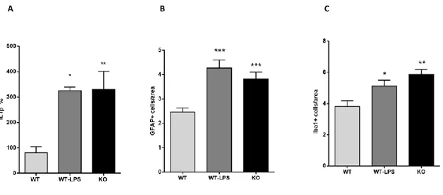
With this study we wanted to verify whether p50-/- mice can be considered a valid model of NDDs also comparing the offspring of KO dams with the offspring of a validated NDDs model, the LPS- induced model. Initially, we measured the inflammatory markers in both genetic (KO mice) and induced (LPS mice) adult offspring. In particular, we measured in adult mice offspring: a) IL-1β levels in the serum as a marker of peripheral inflammation by ELISA; b) Iba1- and GFAP-positive cells in the cortex by IHC as markers of central inflammation. As reported in Figure 1A, KO mice
Figure 1. Peripheral and central inflammation on adult KO and WT-LPS offspring. (A) Interleukin-1β (IL-
1β) serum levels in adult offspring (F (2, 12) = 7.268, p = 0.0086). *p = 0.017 for WT-LPS mice vs. WT mice, **p < 0.008 for KO mice vs WT mice. Data were analysed by one-way ANOVA followed by the Dunnett’s post-test analysis (B) GFAP-positive cells as marker of astrocytes in the cortex of adult offspring (F (2, 241) = 15.69, p < 0.0001). * p < 0.0001 for WT-LPS and KO mice vs. WT mice. (C) ionized calcium-binding adapter molecule 1 (Iba1) positive cells as marker of microglia in the cortex of adult offspring (F (2, 157) = 9.049, p = 0.0002). *** p = 0.0001 for KO mice vs. WT mice, *p = 0.02 for WT- LPS mice vs WT mice. Data were analysed by one-way ANOVA followed by Tukey’s post-test analysis.
and WT-LPS mice showed increased IL-1β levels compared with WT mice (% mean ± S.E.M.: WT 81.00 ± 23.75, WT-LPS325.3 ± 14.03, KO 329.6 ± 71.68). This indicates that KO mice show a long- lasting peripheral inflammation in the offspring similar to WT-LPS mice. Looking at the brain, both KO mice and WT-LPS mice reported a significant increase in both Iba1- and GFAP-positive cells in the cortex compared with WT mice (GFAP+ cells/area mean ± S.E.M.: WT 2.46 ± 0.16, WT-LPS 4.27 ± 0.32, KO 3.82 ± 0.27; Iba1+ cells/area mean ± S.E.M.: WT 3.81 ± 0.36, WT-LPS 5.13 ± 0.36, KO 5.88 ± 0.30) (Figure 1B-C). GFAP is a marker of astrocytes, whereas Iba1 is a microglial marker. Taken together, these results suggest that both KO and WT-LPS mice have a permanent alteration in both peripheral and central inflammation that can be compared with a MIA model of NDDs.
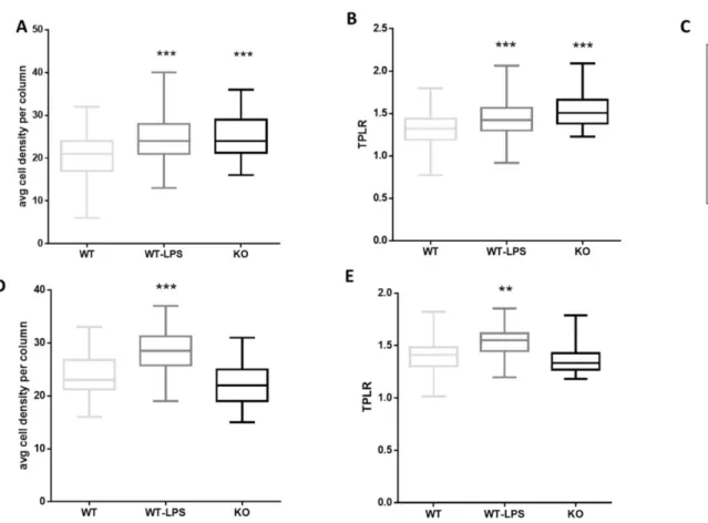
8.2. Abnormal organization in the cortex of adult p50-/- mice and WT-LPS mice
To examine the cytoarchitecture of adult KO mice and the effect of a maternal LPS treatment on the cytoarchitecture of offspring, columnar organization in the somatosensory (SS) cortex and anterior cingulate (AC) cortex of three mice per group was analyzed. These two areas were chosen for their importance in the contest of neurodevelopmental disorders [58,321-323]. Disruption of columnar organization can be detected by different parameters, such as an altered number of cells and cell positioning in the column [324]. Only pyramidal neurons were included in the analysis. KO mice showed an increase in cell density within a defined column in the SS cortex compared with WT mice, but not in the AC cortex (Figure 2A-D). Whereas, WT-LPS mice showed an increase in cell density within a defined column in both the SS and AC cortex compared with WT mice (cell density per column mean ± S.E.M.: somatosensory cortex, WT 20.70 ± 0.35, WT-LPS 24.27 ± 0.28, KO 24.82 ± 0.65; anterior cingulate cortex, WT 23.63 ± 0.48, WT-LPS 28.23 ± 1.05, KO 21.98 ± 0.56) (Figure 2A–D). Furthermore, the path length ratio (TPLR), a measurement of linearity of the cell column, was increased in both KO mice and WT-LPS mice SS cortex compared with WT mice (TPLR in the somatosensory cortex, mean ± S.E.M.: WT 1.31 ± 0.013, WT-LPS 1.44 ± 0.011, KO 1.54 ± 0.025). Only WT-LPS mice showed a significant difference in TPLR in the AC cortex compared to WT mice (TPLR in anterior cingulate cortex, mean ± S.E.M.: WT 1.40 ± 0.020, WT-LPS 1.53 ± 0.035, KO 1.36 ± 0.019) (Figure 2B-E). These data demonstrate that both the genetic (p50 KO mice) and induced (LPS) models, show specific cortical structure alterations.
Figure 2. Abnormal columnar organization in the cortex of adult KO and WT-LPS mice. Graphic
representation of data obtained from the minicolumn analysis in somatosensory (SS) cortex (A, B) and anterior cingulate (AC) cortex (D, E) of adult WT, WT-LPS and KO mice. (A) Average cell density per column in the SS cortex (F (2, 529) = 35.41, p<0.0001). *** p < 0.0001 for WT-LPS and KO mice vs. WT mice. (B) Path length ratio (TPLR) in the SS cortex (F (2, 529) = 41.27, p=0.0001). *** p < 0.0001 for WT-LPS and KO mice vs. WT mice. (D) Average cell density per column in AC (F (2, 119) = 18.65, p < 0.0001). ***p < 0.0001 for WT-LPS mice vs. WT mice. (E) TPLR in the AC cortex (F (2, 117) = 10.28, p < 0.0001). **p < 0.002 for WT-LPS vs WT mice. Data were analysed by one-way ANOVA followed by Tukey’s post-test analysis. (C) Representative images of minicolumns in WT, WT- LPS and KO mice SS cortex.
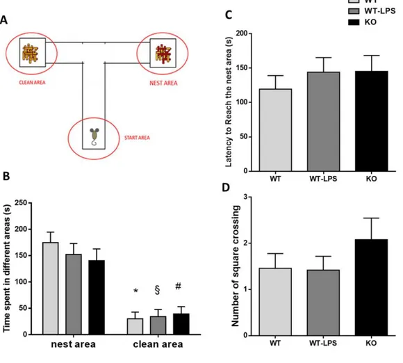
8.3. Reduced maternal care in p50-/- mice
Maternal care, such as nursing postures, non-nursing postures, pups grooming and nest building, were analyzed during the first postnatal week (PND1-7) of WT, WT-LPS and KO pups in order to examine whether there were any alterations in maternal behavior of each group (Figure 3). Maternal behavior has been analyzed because several studies have shown that alteration in maternal behaviors can affect pup’s development and have long-lasting effect on neuroendocrine and behavioral responses [325- 327] in particular on social behavior [328-330]. KO pups received significantly lower nursing
![Table 1. Main MIA animal models: rodent and primate models [70]](https://thumb-eu.123doks.com/thumbv2/123dokorg/5500628.63234/15.893.55.855.86.1086/table-main-mia-animal-models-rodent-primate-models.webp)
![Table 1 Main MIA animal models: rodent and primate models [70]](https://thumb-eu.123doks.com/thumbv2/123dokorg/5500628.63234/16.893.59.832.74.620/table-main-mia-animal-models-rodent-primate-models.webp)
![Table 2. A Sample of long-term behavioral and cognitive dysfunctions as identified in developmental immune activation models in rats and mice [11]](https://thumb-eu.123doks.com/thumbv2/123dokorg/5500628.63234/21.893.108.791.132.553/table-sample-behavioral-cognitive-dysfunctions-identified-developmental-activation.webp)