1.
General introduction ... 2
1.1.
Tumor: what it is? ... 1
1.2.
Defects of the mechanisms of cell replication and death. ... 3
1.3.
Cancer chemotherapy ... 4
1.4.
Microtubules ... 5
1.4.1.
How microtubules work throughout the cell division cycle ... 7
1.4.2.
Tubulin binding site ... 9
1.4.2.1 Colchicine binding site ... 10
1.4.2.2 The vinca domain ... 11
1.4.2.3. Taxol
binding site ... 12
1.4.3. Multi drug resistance ... 13
2.
Microtubules depolymerization inducing agents ... 17
2.1.
Hemiasterlins ... 17
2.2.
From Hemiasterlin to HTI-286 ... 20
2.2.1
SAR study on natural hemiasterlin ... 21
3.
Scopes and aims ... 26
3.1 Development of synthetic routes for the synthesis of A-fragment ... 26
3.1.1 Vedej’s synthetic approach to fragment A. ... 28
3.1.2. Durst’s synthetic approach ... 29
3.2. Toward the synthesis new analogues of HTI-286 ... 30
4. Results and discussion ... 34
4.1 Potentiality of silver oxidein nucleophile substitution reaction ... 34
4.2 Synthesis of the fragment BC ... 36
4.3 Synthesis of racemic mixture of bromoderivatives ... 37
4.4 Nucleophilic substitution reaction catalyzed by silver oxide ... 39
4.4.1 Synthesis of tert-Leucine derivatives ... 40
4.4.2 Synthesis of Valine derivatives ... 41
4.4.3 Synthesis of Phenylalanine derivatives at N-terminus ... 43
5. Biological evaluation ... 46
6. Conclusions and perspectives ... 52
6.1 Toward the pro-drug with stilbene 5c ... 54
6.1.1 Synergistic effect between (R,S)(S)(S)-76a, (R,S)(S)(S)-81a
6.1.2 Project for a reciprocal prodrug between stilbene 5c and (S)(S)(S) 76a ... 55
7.
Experimental section ... 58
7.1.
General methods ... 58
7.2.
Experimental procedures and analytical data. ... 59
7.2.1.
Synthesis of fragment BC ... 59
8.
Hemiasterlin: references ... 97
9.
Microtubules depolymerization inducing agents ... 100
9.1. Epothilones ... 100
9.2.
Structure and SAR of epothilones ... 102
9.2.1.
C12-C13 modifications ... 103
9.2.2.
Side chain modifications ... 104
9.2.3 C1-C5 and C6-C11 fragment modifications ... 105
9.2.4. Epothilones in clinical trials ... 106
10.
Scopes and aims ... 108
10.1. Project aims ... 108
10.2.
Retrosynthetic analysis ... 110
11.
Results and discussion ... 115
11.1. Synthesis of heterocyclic core ... 115
11.1.1
First synthetic approach ... 115
11.1.2 Second synthetic approach ... 116
11.1.3 Third synthetic approach ... 117
11.1.4 Fourth synthetic approach ... 118
11.1.5 Fifth synthetic approach ... 119
11.1.6 Choice of the best protecting group ... 120
11.1.6.1. Benzyl side chain protection ... 120
11.2. Synthesis of the homoallylic alcohol. ... 122
11.2.1 Diastereoselective aldol reaction ... 124
11.2.1.1. Evans aldol reaction ... 125
11.3. Toward the synthesis of the cyclopropane key intermediate 15 ... 127
11.4 Conclusions ... 129
12.
Experimental procedures ... 131
12.1. General Informations ... 131
Chapter 1
General introduction
1.
General introduction
By definition, the word natural refers to something that is present in or produced by nature and not artificial or man-made. Natural products may be extracted from tissue of plants, marine organisms or microorganism fermentation broths.
Structurally, natural products include different classes of compounds as terpenoids, polyketides, aminoacids, peptides, proteins, carbohydrates, lipids, nucleic acid bases, ribonucleic acid (RNA), deoxyribonucleic acid (DNA) and so forth, usually provided with biological and pharmacological activity.
They are considered the most productive source of leads for the development of drugs.
Indeed from 2005 to 2007 thirteen natural product-related drugs were approved, particularly as anti-infectives and anticancer agents.1,2
1.1. Tumor: what it is?
Cancer is a disease characterized by uncontrolled and diffuse multiplication of abnormal shape cells in the organism.3
Biologically, cancer is composed of more than one distinct disease, each with its own etiology and pathology, which is most closely related to the tissue of origin.
The treatment of these diseases has involved the development of many strategies, together with the discovery of mechanisms that allow disorderly cell growth, and many naturally-derived molecules have been found and employed to improve therapies. Moreover, natural compounds have often
been the starting point of a drug discovery process that was used to better understand targets and pathways of tumor onset.
For this reason, improvements in the area of cancer therapy are essential, considering that is forecasted to be major cause of death in the 21st century, in particular in industrialized countries.4
1.2. Defects of the mechanisms of cell replication and death.
During eukaryotic cell division, the mother cell must replicate its chromosomes exactly once in the synthetic phase (S phase of cell cycle), and then must separate the replicated chromosomes at the end of the mitotic phase to the two daughter cells provided with the same genetic heritage of the mother cell.
Defects in the coordination of chromosomes replication and segregation can have severe consequences leading to genetic instability and aneuploidy, and eventually fostering tumor malignancy.
To ensure correct transmission of genetic material during cell division, cells have evolved cellular regulatory mechanisms termed “cell cycle checkpoints”.
This control system prevents or delays cell cycle progression if certain cellular processes or proteins are disrupted, to gain time to repair the damage before cell division occurs.
When the damage is irreparable, the cell undergoes apoptosis through the triggering of specific biochemical pathways.
However, cancer cells often elude the control systems allowing uncontrolled cell proliferation, even when cell division does not occur properly.
About the 50% of human malignant tumor are induced by mutation of p53 gene, that codifies for the namesake protein involved in the reparation mechanism of DNA.3
1.3. Cancer chemotherapy
The aim of conventional antitumor therapy to date is to slow and hopefully halt the growth and spread of a cancer cells. There are three goals associated with the use of the most common anticancer agents:
• To damage the DNA of cancer cells.
• To stop cells replication by inhibition of the synthesis of new DNA strands.
• Block of mitosis, or the actual splitting of the original cell into two new daughter cells during replication.
In general, chemotherapy agents can be divided into three main categories based on their mechanism of action.
• Stop of the synthesis of pre-DNA molecule building blocks.
DNA building blocks are folic acid, heterocyclic bases, and nucleotides, which are made naturally in the cells.
All of these agents work to block some steps in the formation of nucleotides or deoxyribonucleotides (necessary for making DNA) and without these the cells can’t replicate.
Examples of drugs in this class include methotrexate (Abitrexate®) and fluorouracil (Adrucil®).
• Direct damage to the DNA in the cell nucleus.
These agents chemically damage DNA and RNA, either blocking totally replication or causing the formation of nonsense DNA or
Examples of drugs in this class include cisplatin (Platinol®) and antibiotics such as doxorubicin (Adriamycin®).
• Effect the synthesis or breakdown of the mitotic spindles.
Mitotic spindles serve as molecular railroads during cell replication, since they help to split the new copied DNA so that every copy goes to each of the two new cells during cell division.
These drugs paclitaxel (Taxol®) and vincristine (Oncovin®) disrupt the formation of the spindles and therefore interrupt cell division. Mitotic spindle structure is made up of dynamic substructures named microtubules.5
1.4. Microtubules
Microtubules are major dynamic structural components in cells since they are involved in very important cellular functions, such as the development and maintenance of cell shape, cell reproduction and division, cell signalling and movement.
For this reason, microtubules are the target of structurally different groups of anticancer drugs, most of which derive from natural products.
The mitotic inhibitors represent the single best cancer target identified to date.6
Microtubules are highly dynamic polymers of heterodimers of two closely related 55 KDa proteins termed α and β tubulin, arranged parallel to a cylindrical axis to form tubes of 25 nm diameter that may be many µ m long. The proteins are encoded by separate genes or small gene families, whose sequences are highly conserved throughout the eukaryotic kingdom. Another member of the tubulin family, γ-tubulin, is involved in the
It is found primarily in centrosomes and spindle bodies, since there are areas of most abundant microtubules nucleation.
Polymerization of microtubules occurs by a nucleation-elongation mechanism in which the formation of a short microtubules “nucleus” is followed by elongation phase characterized by reversible, noncovalent addition of tubulin dimers to the end of microtubule itself.
They exhibit complex polymerization dynamics that use energy provided by the hydrolysis of GTP.
Tubulin binds GTP in solution with high affinity, and little by little tubulin-GTP is added to the end of a growing microtubule, the tubulin-GTP is gradually hydrolyzed to GDP and Pi.
Ultimately, the Pi dissociates from the microtubules, leaving a microtubule core consisting of tubulin with stoichiometrically-bound GDP. The nucleotide remains non-dissociable and non-exchangeable until the tubulin subunit dissociates from the microtubule.
Microtubules dynamic is regulated by two phenomena, from one side “treadmilling” is the net growth at one microtubule end, on the other side “dynamic instability” is a process in which the individual microtubules ends switch between phases of relatively slow sustained growth and rapid shortening.
The transition between growth and shortening appears to be regulated by the presence or absence of the region of GTP-containing-tubulin at the microtubule end.
A microtubule can grow as long as maintains a stabilizing cap of tubulin-GTP or tubulin-GDP-Pi at its end. The loss of the cap induces depolymerization of microtubules.
Hydrolysis of tubulin bound GTP and the subsequent release of Pi establish conformational changes in the tubulin molecule, that destabilize the microtubule polymer, resulting in “catastrophe” and shortening of the microtubules.
The two ends of microtubules are not equivalent; one end, termed “plus end” is kinetically more dynamic than the “minus end” and although both ends can either grow or shorten, the changes in length at the first one end are much larger than the changes in length at the other one.7,8
Figure I-1. (a) Schematization of a microtubule made up of dimers of α and β
tubulin. (b) Representation of main microtubule dynamics.
1.4.1. How microtubules work throughout the cell division cycle
In a typical cell during the interphase of the cell cycle, the microtubules radiate from a central site near the nucleus, called “microtubules-organizing
centre” (MTOC). In animal cells, the MTOC consists of a centrosome made up of a lattice of MAPs, γ-tubulin and a pair of centrioles.
While the minus end of the microtubules lies in or near the centrosome, the plus end extends out toward the cell periphery. Before of the cell division, the cytosol is permeated by a fixed net of microtubules organized in a spindle-shape “mitotic spindle” that is the real director of the chromosomes segregation at anaphase.9
Mitotic spindle consists of three kinds of microtubules:
• Astral microtubules are shorter and stabler because they link the two poles of mitotic spindle to the cell ends.
• Polar microtubules are longer, rather stable and they look overlaid each other because of their structural role during mitosis.
• Kinetochore microtubules are unstable and directly linked to the chromosomes on the equatorial plate. They are the object of dynamic instability phenomenon given that the rapid removal of tubulin subunit determine the separation of the chromosomes.10
Figure I-2. Representation of microtubules network during the separation of
The interphase microtubules network disassembles at the onset of mitosis and is replaced by a new population of spindle microtubules that are 4 to 100 times more dynamic than microtubules in the interphase cytoskeleton. Mitotic spindle exchange its tubulin with that in the soluble pool with half-times in the order of 10-30 seconds. Recent studies have indicated that several antimitotic anticancer drugs appear to inhibit mitosis at the methaphase/anaphase transition by suppressing spindle tubules dynamics. In the presence of low, but effective drugs concentrations, spindle forms and mitosis can progress as far as the methaphase/anaphase transition. However, the spindles are completely unable to pass the mitotic cell cycle checkpoint and to initiate anaphase movements, or do so only after a long period of mitotic blockage, apparently due to their suppressed dynamics. Mitotically-blocked cells eventually die by apoptosis.6
1.4.2. Tubulin binding site
Tubulin was identified for the first time as the “colchicines-binding protein” by Borisy and Taylor in 1967, and the ability of colchicine to block cells in prometaphase/metaphase step of mitosis played a key role in the development of antimitotic drugs.
Antitubulin agents are divided into two categories based on their ability to bind to tubulin and change the ratio between assembled microtubules and dimeric tubulin.
In vitro, the equilibrium between the dimeric and polymeric forms of
tubulin can be altered by different effecters, such as DMSO, cofactors [ Mg2+, guanosine-5’-triphosphate (GTP), guanosine-5’-diphospahte (GDP)], or small molecule, which alter the stability of tubulin dimers or the
Three major classes of tubulin-binding agents have been identified: the colchicine-site binding agents, the vinca domain inhibitors, which block microtubules growth, taxanes and epothilones binding site inhibitors which stabilize microtubules.
Both the vinca alkaloids and the taxane drugs bind to the tubulin, but at different location on the protein: the first group binds to β-tubulin between amino acids 175 and 213, while paclitaxel (Taxol®) binds both to N-terminal unit on β-tubulin and to the region bounded by amino acids 217-231.
Colchicine that is not a clinically used drug for cancer binds to the β-subunit at the interface with α-monomer of the some tubulin molecule.9
1.4.2.1. Colchicine binding site
Colchicine (1), was isolated from the autumn crocus Colchicum autumnale. Although colchicine has significant in vitro antitumor effects, its medical uses, as well as use of its derivatives, has been limited because of its high toxicity, low bioavailability and poor water solubility (fig. I-3).11
The effect of colchicine on microtubule dynamics depends on drugs concentration. Thus, relatively high colchicine concentrations inhibit microtubule polymerization and depolymerize preformed microtubules. Colchicine binds to soluble tubulin and forms a poorly reversible tubulin-colchicine (TC) complex, which then is incorporate at the microtubule ends.6
A group of tubulin inhibitors that bind at this site include CA-4 (2) and ZD6126, both of which are currently in clinical development.
Figure I-3. Structures of colchicine and CA-4.
1.4.2.2. The vinca domain
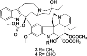
The antimitotic vinca alkaloids vinblastine (3), vincristine (4), vindesine and vinorelbine are widely used both as single agent and in combination with other antitumor drugs in cancer chemotherapy, particularly in a variety of hematologic and solid tumor (fig. I-4).
The two complex indole alkaloids, vinblastine and vincristine were originally isolated from the plant Catharantus Rosea, while vindensine and vinorelbine are semisinthetic compounds.
The binding site of vinblastine on tubulin also binds other compounds (hemiasterlins, crytophycins and halicondrins) and this kind of binding induces a conformationally change in tubulin that becomes more akin to vinblastine with the formation of vinblastine-tubulin spiral oligomers.9
1.4.2.3. Taxolbinding site
Taxol® (paclitaxel, 5) was isolated in 1971 by Well and Wani from the pacific yew Taxus Brevifolia and is effective used in the treatment of the breast, ovarian, and lung carcinomas.
Taxol appears to arrest cell in mitosis by stabilizing spindle microtubules, in particular this alkaloid induces the formation of morfologically-altered tubulin polymers, that organize themselves in 12 protofilaments rather than 13, both in vitro with pure tubulin and in vivo.6
Like vinblastine and colchicine, taxol slows and blocks mitosis at the metaphase/anaphase transition in a number of cell types, inducing accumulation in a metaphase-like state and ultimately apoptosis.12
The success of paclitaxel has spurred enormous interest in finding better pharmacokinetic profile analogs.
To date the only one approved for clinical use in the U.S. is docetaxel (6) that has a semi-synthetic origin.
Figure I-5. Structure of paclitaxel and docetaxel.
Both paclitaxel and docetaxel occupy the same binding site in the subunit of tubulin with a 1:1 stoichiometry.
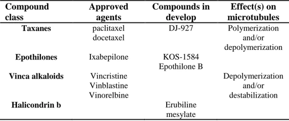
In the following tab (Table I-1) are reported the class, at the moment, more
O O O R OH O OH O H O O O 5 R= OCOCH3, R1= NHCOPh 6 R= OH; R1= OtBu R1 OH
However, there is an unmet need of new drugs since current therapies suffer from limited extension of survival time due to inherent or acquired resistance which is often associated with expression of the P-glycoprotein drug transporter. Compound class Approved agents Compounds in develop Effect(s) on microtubules Taxanes paclitaxel docetaxel DJ-927 Polymerization and/or depolymerization
Epothilones Ixabepilone KOS-1584 Epothilone B
Vinca alkaloids Vincristine Vinblastine Vinorelbine Depolymerization and/or destabilization Halicondrin b Erubiline mesylate
Table I-1. Classes of MTIs
1.4.3. Multi drug resistance
Multidrug resistance, the principal mechanism by which many cancers develop resistance to chemotherapy drugs, is a major factor in the failure of many forms of chemotherapy.
It affects patients with a variety of blood cancers and solid tumors, including breast, ovarian, lung, and lower gastrointestinal tract cancers. Tumors usually consist of mixed populations of malignant cells, some of which are drug-sensitive while others are drug-resistant. Chemotherapy kills drug-sensitive cells, but leaves behind a higher proportion of drug-resistant cells.
As the tumor begins to grow again, chemotherapy may fail because the remaining tumor cells are now resistant.
Resistance to therapy has been correlated to the presence of at least two molecular "pumps" in tumor-cell membranes that actively expel chemotherapy drugs from the interior. This allows tumor cells to avoid the toxic effects of the drug or molecular processes within the nucleus or the cytoplasm.
The two pumps commonly found to confer chemoresistance in cancer are P-glycoprotein and the so-called multidrug resistance–associated protein (MRP).
Drug resistance is a multifactor phenomenon in which different mechanisms are involved as failure of physiologic apoptosis, modified drug activation or degradation and modified transport of the drugs through the membrane because of a change its permeability.
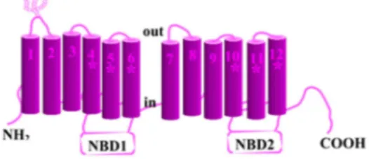
Pgp (Fig. I-6) is a trans-membrane protein, product of gene mdr-1, over-expressed in tumor cells, in particular those targets of chemotherapy. The primary structure of this protein is made up of 1280 amino acids, organized into two repeated units, everyone of 610 amino acids, linked by a bridge. Its trans-membrane domain (TMD) is directly involved in drugs binding, but is independent from ATP activity.13
Figure I-6. Scheme of P-glycoprotein and its functional domain. Domains directly
In this Ph.D. thesis I focused on two modified analogs of natural tubulin inhibitors, with the same target but different action mechanism that are respectively hemiasterlin and epothilone B.
These compounds are the result of the drug discovery process starting from natural template, aimed at finding new drugs that elude multi-drug resistance phenomenon.
Chapter 2
Hemiasterlins
2.
Microtubules depolymerization inducing agents
2.1. Hemiasterlins
Hemiasterlin (7), hemiasterlins A (8), B (9), and C (10) are members of a small family of cytotoxic tri-peptides that have been isolated from a South Africa sea sponge Hemiasterella minor (fig. II-1).
Structurally, hemiasterlins are characterized by the presence of tri- or tetramethylated tryptophan, tert-leucine, and N-methylvinylogous valine residues.
Figure II-1. Structures of hemiasterlins and relative numeration.
These natural substances show potent in vitro cytotoxicity against murine leukemia P388 and human breast, ovarian, colon, and lung cancer cell lines. Against human breast cancer MCF7 cells, compounds (7) and (8) are more cytotoxic and more potent mitotic blockers than vincristine, paclitaxel and nocodazole, while hemiasterlin C is the least potent derivative.14
The potency of hemiasterlin was also confirmed by the lowest IC50 values (about 2 pM) obtained in the human tumor cell lines OVCAR-3 and NCI-H460.
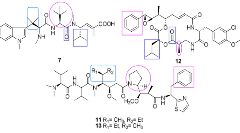
values are 0.59, 0.98, and 1.1 µM for dolastatin 10, hemiasterlin, and cryptophycin 1, respectively).
Figure II-2. Structure of hemiasterlin, cryptophicin 1, dolastatin. Boxed region
represents areas of common overlap identified through docking studies.
Several experimental studies probing the binding of this class of mitotic inhibitors to tubulin have appeared in literature.15
However, the complexity and diversity of tubulin-ligand binding compounded with the lack of corroborating structural evidence have served as limitation to an understanding of the detailed molecular interaction present in the system under study.
Rai and Wolff have localized the binding region of the microtubule destabilizing agent vinblastine on β-subunit, while the three peptides, probably, bind in a site distinct from that at which the vinca alkaloids bind, since they all noncompetitively inhibit the binding of radiolabeled vinca alkaloids to tubulin.
In contrast, hemiasterlin as well as cryptophycin 1 and chiral isomer of dolastatin 10 (13) competitively inhibit each other for binding of [3H]dolastatin 10 to tubulin (Table II-1).
Table II-1. Inhibition by hemiasterlin and other vinca domain drugs of the binding
of [3H]vinblastine and [3H]dolastatin 10 to Tubulina
Drug added % inhibition of [3H]vinblastine binding % inhibition of [3H]dolastatin 10 binding Hemiasterlin Dolastatin 10 Cryptophicin 1 Vinblastine 27 44 36 43 42 2 a
The 0.4 ml reaction mixtures contained 10 µM tubulin, 0.5% DMSO, the indicated potential inhibitor at 0.5 µM. Incubation was fro 30 min, and centrifugal gel filtrations of duplicated 0.19 ml aliquots were at room temperature. Avarages from two independent experiments are presented in the table. Stoichiometry of binding in the control reaction mixtures: 0.57 mol vinblastine and 0.59 mol of dolastatine 10 per mole of tubulin.
Dolastatin 10 is the most active, but phase I/II clinical trials revealed bone marrow toxicity, neuropathy together with a poor therapeutic index. Despite their structurally diversity, these antimitotic agents bind at the same active site, that is adjacent to the exchangeable GTP site on β-tubulin and is composed primarily of residues Ser171, Lys174, Val175, Asp177, Asn204, Glu205, Tyr208, Asp209, Phe212, Pro220, and Tyr222.16
Each of these antimitotic agents destabilizes and depolymerizes microtubules, resulting in the formation of aberrant non-microtubules rings and oligomers, in this case hemiasterlin-tubulin rings are 45 nm in diameter, and both contain 14 tubulin dimers.
Thanks to molecular dynamics simulations and molecular docking studies it was possible to understand which functional groups of the hemiasterlin
were important for the interaction with the binding site, comparing the three hydrophobic peptides.
Starting from the N-terminus of the hemiasterlin, the Trp residue provided with the two methyl group could be overlaid with the aliphatic side chain, while the Val residue probably overlaps the pyrrolidine ring of the dolastatin 10 (fig.II-2).
An overlap can be exist between the carbonyl of Val moieties and the aliphatic side chain of the Val, moreover Dil/Leu residues may show structural overlap. These results indicate that the Val and Ile/Leu residues may be the common elements forming the pharmacophore.15
2.2. From Hemiasterlin to HTI-286
Nieman and co-workers synthetized a number of analogues in order to define what portions of the structure were required for cytotoxicity and antimitotic activity and to prepare more potent analogs easier to synthesize.17
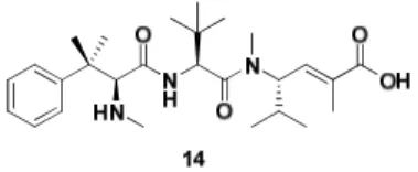
A great result of this study was the identification of a Synthetic Peptide Analogue 110 (SPA 110 or HTI-286, 14), obtained changing indole of hemiasterlin with phenyl ring.
At the moment, HTI-286 is in clinical trial after demonstration of biological activity in preclinical cancer models.
The library of products was prepared considering that the geminal methyl couldn’t be removed because they were thought to protect the tripeptide from proteolysis and/or to establish a preferred conformation that may be critical to biological activity.
Hemiasterlin has been the object of a series of single-point changes and corresponding new compounds have been tested for both antimitotic activity in MCF-7 cells expressing a dominant-negative mutant p-53 tumor suppressor gene and in vitro cytotoxicity.
It was found that there was a linear relation between antimitotic and cytotoxicity activity over a wide range of structural variations; this correlation suggest that, in these cells, cytotoxicity is solely due to inhibition of tubulin function at mitosis, and that hemiasterlins are pure antimitotic agents.
2.2.1. SAR study on natural hemiasterlin
C-10 trimethylammonium ion and the substitution of the isopropylic chain with a hydrogen atom caused a loss of activity, proving that the two functionality are crucial to metabolic stability and/or tubulin binding. The same outcome had the hydrogenation of the double bond to give a mixture of epimers that reduce the potency by roughly an order of magnitude.
The aromatic function is essential at N-terminus as demonstrated by the loss of potency due to the replacement of the N-methylindole ring with a methyl or hydrogen atom to give respectively tert-LEU and Val-hemiasterlins. Other modifications are well tolerated resulting in no significant loss in potency; these include the conversion of the B residue tert-Leu to Val,
formation of the methyl ester at and the replacement of the N-5 methyl substituent with a proton.
Only one change, the replacement of N-methylindole by aromatic ring made HTI-286 3-fold more potent than the natural product.
Figure II-4. Structural requirement for optimal cytotoxicity (IC50˂ 1 nM) nd
antimitotic activity (IC50 ˂ 1 NM)
The result of this study has highlighted four regions of hemiasterlins where single structural changes are possible without seriously compromising antimitotic activity:
• replacing N-methylindole with phenyl and methyl groups • replacing the tert-Leu residue with Valine
• removing the N-5 methyl substituent • making the C-terminal residue methyl ester
C-11 methyl substituents, the C-10 methylamino group, and the C-4 isopropyl groups are extremely important structural elements for potent antimitotic activity.18
With HTI-286 in hand, Rush and co-workers from Wyeth have undertaken a docking study to better understand the interaction of HTI-286 with the
tubulin and according their predictions, there are several important factors contributing to the HTI-286/tubulin interaction.
These include different hydrophobic interactions, hydrogen bonding, and electrostatic complementarity.
It is hypothesized the U-shaped curvature of the backbone of HTI-286, in particular the tertiary butyl group is found to occupy a spacious cavity in β-tubulin, proximal to the location of the guanosine 5’-diphosphate (GDP) (fig II.5).
The binding model finds the C-1 position of HTI-286 to be oriented in a region of β-tubulin that is flanked by two ASN residues (residues 186 and 101 of the β-tubulin subunit) providing hydrogen bonding opportunities with the carbonyl or hydroxyl group of the ligand.
The heteroatoms of the amide backbone of the HTI-286 are seen forming β-sheet-like interactions with the backbone atoms of nearby amino acids residues.
The interaction between the basic NH group of HTI-286 and the Asp179 residue of the β-tubulin subunit are quite significant in fact deleting them, the enthalpic and desolvatation contributions to binding might be perturbated.
The binding model indicates an association between Ser 174 and Ser 178 of the β-tubulin and the C-9, C-6 and N-8 position of the HTI-286 scaffold (Fig. II-4).19
Figure II-5. Surface view of HTI-286 docked to the β-tubulin portion of the
interdimer interface, emphasizing the binding pocket. The α-tubulin subunit is not shown for clarity. The tubulin surface is colored by lipophilic potential (brown, hydrophobic; green, neutral; and blue, hydrophilic). Hydrogens are removed for simplicity. The yellow-colored carbon atoms are poised for intramolecular interactions, and the tert-butyl carbons are colored purple.
Chapter 3
Scopes and aims
3.
Scopes and aims
3.1. Development of synthetic routes for the synthesis of A-fragment
In literature there are different total synthesis of hemisterlin, but the first one was completed by Andersen and co-workers in 1997 and it is the result of a sequence of coupling reactions between three modified amino acids A, B and C. Every amino acid apart the commercially available N-Boc-tert-leucine is synthesized singularly.
Figure III-1. Retrosynthetic analysis of hemiasterlin.
Structural assignment based on NMR and degradation studies are supported by X-ray diffraction analysis of hemiasterlin methyl ester, and it was found that the members of this family are known to have L-configuration (Fig. III-1).
The first approach was based on Evans’ oxazolidinone chemistry employing the electrophylic nitrogen source triisopropylphenylsulfonyl azide. The reduction of azide (23), followed by Boc-protection of amine (24) gave the desired functionalized tryptophan (28) in a good enantiomeric excess (> 98%).14
Scheme III-2. Reagents and conditions: (i): KN(SiCH3)2, THF, -78°C to 0°C, 3 h;
CH3I, -78°C to 0°C, 2 h; (ii): KN(SiCH3)2 (1.5 eq), then as in (i); (iii): i-Bu2AlH,
Et2O; (iv): TPAP, NMO, CH2Cl2, 4Å sieves; (v): Ph3P=CHOMe, THF, r.t.; (vi):
p-TsOH, H2O, dioxane, 60° C, 16 h; (vii): NaClO2, NaH2PO4, 2-methylbu2-ene,
t-BuOH, H2O, 0°C; (viii):CH3CCOCl, TEA, THF, -78°C; (ix): KN(SiMe3)2, THF,
-78°C; 2,4,6-triisopropylbenzenesulfonyl azide, THF, -78°C, 1 min; AcOH, 30-40°C, 1h; (x): SnCl2, dioxane, H2O, r.t., 36 h; (xi): (CH3CO2C)2O, dioxane, H2O, r.t., 16 h;
The reported synthesis of the tryptophan moiety starting from methyl ester of 3-indoylacetic acid (18) was 16 steps long and suffered from low yields of coupling reactions.
Several research groups tried to find a successful convergent approach that could give analogues more readily.
This would open the possibility of generating not only significant quantities of hemiasterlin itself but also analogues for screening and further biological assays.
3.1.1. Vedej’s synthetic approach to fragment A.
An improved enantiocontrolled route to the tetramethyltryptophan subunit (36) was developed by Vedejs’ group using an asymmetric Strecker synthesis (5 steps, 50% yield from 29).
Scheme III-3. Reagents and conditions: (i) Sc(OTf)3, (R)-2-phenylglycinol,
CH2Cl2, r.t.; Bu3SnCN, 0°C to r.t.; (ii) H2O2, K2CO3, DMSO, MeOH, 45°C; (iii) H2,
Pd(OH)2/C, MeOH, r.t.; (iv) BtsCl, Na2CO3, CH2Cl2-H2O, 0°C to r.t.; (v) CH3I,
This method exploits the high reactivity of a Bts-protected amino acid chloride in the difficult peptide coupling of sterically hindered amino acids residue like BC dipeptide.20
3.1.2. Durst’s synthetic approach to fragment A.
The same principle was followed by Durst’s group to get A-piece starting from N-methylindole (38), that was reacted with tin tetrachloride mediated ring opening cyanoepoxide.
The treating of the cyanohydrin (39) with NaOH gave the corresponding aldehyde (29) that was used in asymmetric Strecker methodology. This sequence led to the formation of an 85:15 diastereomeric mixture of α-cyano amines (41) direct precursor of Andersen intermediate (42).21
Scheme III-4. Reagents and conditions: (i) SnCl4, CH2Cl2, -78 °C, 70%; (ii) NaOH, EtOH 95%; (iii) (R)-phenylglycinol, CH2Cl2, TMSCN; (iv) Pb(OAc)4, MeOH/CH2Cl2; (v) 3 N HCl, Et2O, 55% over two steps; (vi) concentrated HCl reflux; (vii) (Boc)2O, Na2CO3, THF/H2O; (viii) NaH, MeI, DMF, 65% over three steps.
A-piece, whatever its origin, was coupled with the segment BC though amide bond formation.
To date, the first proposed synthetic route was extensively followed for SAR studies, but it is resulted poorly versatile, because every structurally different A amino acid has to prepared on purpose.
Moreover, this strategy is hardly scalable to obtain considerable amount of product.
3.2. Toward the synthesis new analogues of HTI-286
The aim of my Ph.D. project is the possibility to easily access to a series of synthetic analogues of HTI-286, opportunely modified at N-terminus. Molecular modeling studies, not reported in this context, have highlighted the ability of the binding site on the tubulin to host bulky derivatives at N-terminus.
For this reason, we considered the use of a common precursor from which a series of derivatives with modified A-portion could be obtained with one or very few steps.
Following previous works in the field, we have exploited the synthetic potentiality of 2-bromoacyl-peptides as versatile synthetic intermediates in reactions in which the pool of natural amino acids is used as starting material.
The modifications were directed mainly N-terminus of hemiastelin structure, but also the C-terminus, was involved in SAR studies.
At first we thought to invert the α,α-dimethylbenzyl group with the α-N-methyl group in order to get derivatives of the alanine (43) that was incorporated in the backbone of the hemiasterlin thanks to two following
Figure III-2. Inversion of methyl group with dimethylbenzyl radical at N-terminus.
From one side, a key role was played by silver oxide as promoter of substitution reactions with controlled chemistry, employed to introduce particular functional groups at N-terminus.23
Then, in order to further explore the size of the binding site, bulkier groups were introduced at N-terminus, employing bromoacyl peptides (44) derived from phenylalanine instead of alanine as starting materials.
Figure III-3. General structure of the synthesized products related to HTI-286.
Besides that, we also tried to obtain the direct analogue of natural hemiasterlin, bearing 2-(1-methyl-1H-indol-3-yl)propan-2-amine at C-10 of the tripeptidic backbone as derivative of alanine, but so far we haven’t obtained it.
Thus, we have thought to take advantage of this principle, planning to prepare a conjugate between the most active candidate and stilbene 5c, known to inhibit tubulin assembly (IC50 values from 2 to 8 nM) by binding to a site different from hemiasterlin, thanks to a linkage easily hydrolysable
in situ. The idea of a pro-drug came up also from the possible improvement
of the water-solubility of stilbene 5c.
By definition two or more agents or substances act in synergy if they produce an effect greater than the sum of their individual effects.24,25 In this context, this CA-4 analog was termed exactly as in a paper published by prof. Simoni and al.24,25
Chapter 4
Results and discussion
4. Results and discussion
4.1. Potentiality of silver oxide in nucleophile substitution
reaction
The research group, in which I carried out this Ph.D. project, has experience in the knowledge of the silver oxide chemistry as promoter of nucleophile substitution on substrates as peptides, depsi-peptides and pseudo-peptides.26 These applications are indicative of the great versatility of 2-bromoamides, and of the potential involvement of natural aminoacids.
Thus, the novelty of our synthetic approach is based on the consideration that (S)-α-amino-acid (45) can be diazotization and halogenated to give the corresponding bromo-acid (47) in turn derivatized to (S)-α-bromoacylpeptide or (S)-α-bromoacylamide (48) depending on the nature of R, maintaining the same configuration of the starting material.27
Scheme IV-1. Stereospecific synthesis of bromoacylpeptide from
(S)-α-aminoacid
This is the result of a double inversion of configuration in the sequence
In literature there are some examples that highlight the bromoacylamide (48) reactivity towards the substitution of the bromine with a nucleophile, according to different factors:
• nature of the nucleophile • solvent characteristics • presence of a silver promoter • nature of alkylic group in α position
Thus, the reaction of (S)-48 with an amine gives substitution compounds, whose N-terminus bears the side chain of (S)-45.
In absence or in presence of a soft Lewis acid Ag+, starting from (S)- bromoacylpeptide 49 the product with inverted configuration is obtained
(R)-51.
This reaction is quite slow, it takes hours to go to an end, and is performed with 1°, 2° amines and enolate that are good nucleophiles.
The combination of Ag+/hindered amine induced the amine to behave both as base and as nucleophile, thanks to a shift from a mechanism of electrophilic assistance to an alternative one, where Ag+ is responsible of an acidity-enhancement mechanism.
The addition of covalent silver oxide increases the rate of the reaction, in fact it was observed that with whatever amine, the product has the same configuration (S)-51 of the parent 2-bromoamide 49 in high ee was obtained.
Stereoselectivity depends on a favorable ratio between the rates of two competitive mechanisms that can operate.
By using an insufficient amount of Ag2O, in fact, the optical activity of the produced aminoamide decreas, because the rate of promoted and
Interaction between Ag2O and the solution-species would make the mechanism a complex one characterized by a labile aziridinone (α-lactam,
(R)-50).
Scheme IV-2. Reagents and conditions: (i) Ag2O; (ii) Nu:; (iii) Nu:, Ag + CF3SO3
4.2. Synthesis of the fragment BC
Scheme IV-3. Reagents and conditions: (i) CH3I, NaH 60% min. oil, THF; (ii)
CH3NHOCH3 ·HCl, 1-HOBT, WSC, NMM; (iii) LiAlH4, THF; (iv) PPh3, toluene;
(v) NaOH; (vi) 57, CH2Cl2; (vii) TFA, CHCl3
(S)(S)-Boc-N-methyl-(S)-valinale 52 obtained from reduction with LiAlH4 of the Weinreb amide (S)-54, was reacted with a stabilized Wittig reagent (57), previously prepared from 2-bromoester (56) to get γ-aminoester α,β-unsaturated (scheme 6).
The olefination was performed in CH2Cl2 affording stereoselectively the E-2-alkenoate (59) (scheme IV-3).
After deprotection under acidic conditions, the intermediate (S)-59 was coupled with (S)-Boc-Valine 60 or (S)-Boc-Tert-Leucine 61 employing TMAC to activate the amino acid (scheme IV-4).
Scheme IV-4. Reagents and conditions: (i) TMAC, DIPEA, THF, -78°C, 2h; (ii)
TFA, CHCl3, r.t.
4.3. Synthesis of racemic mixtures of bromoderivatives
From this known intermediates, we started our own project by reacting
2-bromoacyl-peptides (R,S)(S)(S)-65a,b as diasteromeric mixtures (scheme IV-5).
Scheme IV-5. Reagents and conditions: (i) TEA, DCM, 0°C, to r.t. overnight.
At first, the products were synthesized as mixture of diastereoisomers, in order to achieve easily both compounds, whose activity might be quickly tested and compared to each other. The most promising compounds were then prepared as single diasteroisomers in order to establish the identity of which one was responsible of the activity.
In this perspective, silver oxide was employed as coupling agent granting good yields and the correct stereochemistry of the product.
In this case, bromo-derivatives (S)(S)(S)-65a,b are the result of the reaction between (S)(S)-63a,b with (S)-2-propanoic acid 67, in turn obtained by diazotization-bromination of amino acid (L)-alanine 66.
Similarly, the (R)(S)(S)-65a,b series display both valine and tert-leucine were prepared using (D)-alanine as starting material (scheme IV-6).27
Scheme IV-6. Reagents and conditions: (i) NaNO2/H +
, KBr; (ii) TMAC, DIPEA, THF, -78°C
4.4. Nucleophilic substitution reaction catalyzed by silver oxide
The prepared bromoacyl intermediates 65a,b were subsequently reacted with different nucleophyles, mainly amines. Some amine were not commercial and were synthesized.
Indeed, most of amines 71a,d-e were prepared treating tertiary alcohol with sodium azide and TFA at 0°C, followed by catalytic hydrogenation with palladium on charcoal, while non commercially available alcohols 69d-e were obtained adding the correspondent ketone 68d-e on to freshly prepared Grignard’s reagent.29
With the aim to understand the importance of the two methyl groups R'' and R''', also amines as 71b,c, and N-methyl-indol-3-yl-methanamine 71f, yielded by reduction of the corresponding nitrile 73, were employed (scheme IV-7).
Scheme IV-7. Reagents and conditions: (i) CH3I, Mg, Et2O; (ii)NaN3, TFA, CHCl3;
(iii) H2, Pd/C; (iv) H2, Pd/C, EtOAc
In the same perspective, cyclohexylamine was used even though its structure is different from what is represented in the general scheme IV-7.
4.4.1. Synthesis of tert-Leucine derivatives
As regarding the reactivity, only the reaction with benzylamine 71c could run without Ag2O, while the other amines have required this promoter as they were quite bulky.
Also water was used as nucleophile even though hydroxy derivatives
(R,S)(S)(S)-78g and (R,S)(S)(S)-83g were reaction by-products more or less
present if the environment of the reaction was not strictly dry (scheme IV-8).22
Scheme IV-8. Reagents and conditions: (i) Ag2O, 70a-f, toluene, reflux; (iii) Ag2O,
Nu: (g: H2O or h:Cyclohexylamine), toluene; (ii) LiOH/MeOH/water, than TFA.
4.4.2. Synthesis of Valine derivatives
The following scheme shows the synthesis of some valine derivatives
(R,S)(S)(S)-81a,c in which only the amine 71a,c were used (scheme IV-9).
The mixtures (R,S)(S)(S)-75a,c, (R,S)(S)(S)-76a,c and (R,S)(S)(S)-80a,c,
(R,S)(S)(S)-81a,c weren’t separable either by flash chromatography or
HPLC, therefore the single diasteroisomers were stereoselectively synthetized starting from both (R) and (S)(S)(S)-65a,b thanks to the potentiality of Ag2O.
The de of the products is 95% and was measured by HPLC, 1H NMR and comparing the results with those of diasteromeric mixtures previously obtained.
Scheme IV-9. Reagents and conditions: (i) Ag2O, 71a,c, toluene, reflux; (iii) Ag2O,
H2O, toluene; (ii) LiOH/MeOH/water, than TFA.
Apart the hydroxy-derivatives (R,S)(S)(S)-78g, 83g, the final products
(R,S)(S)(S)-76a-f, (R,S)(S)(S)-79h, (R,S)(S)(S)-81a,c were all tested as
trifluoroacetate salts, after basic hydrolysis of ethyl ester and HPLC purification.
Moreover, we wanted expand our investigation on the versatility of synthetic procedure, together with the need of further SAR studies on a series of products modified at carboxyl-end position.
It was, therefore, easily synthetized a compound (R,S)(S)-88 whose fragment C was a rigid aromatic derivative of γ-aminoester α,β-unsaturated
(S)(S)-63b, condensing Boc-(L)-tert-leucine (S)-61 and 3-aminobenzoic
ethyl ester 84, in order to investigate if a modification at C-terminus might afford a series of products modified at this position (scheme IV-10).
Scheme IV-10. Reagents and conditions: (i) (1). DIPEA, TMAC, THF, -78°C, (2) TFA; (ii) DIPEA, TMAC, (R,S)-2-bromo-propionic acid (iii) Ag2O, toluene, reflux;
(iv) LiOH, MeOH, H2O, then TFA.
4.4.3. Synthesis of Phenylalanine derivatives at N-terminus
Computational studies suggested that the portion of the binding site that host the aromatic ring of HTI-286 was enough large to accommodate bulkier groups.
Moreover the most interesting derivatives were those bearing the dimethylbenzyl-amino group.
Thus, a bivalent ligand was prepared, including a further benzylic group at N-terminus.
The two diasteroisomers were synthetized separately following the same synthetic route shown in the scheme 12, but in place of alanine, (D) and (L)-phenylalanine 89 were used, as starting material for the corresponding bromo-acids (S)-90 and (R)-90 (scheme IV-11).
In the scheme is represented only the synthesis of diasteroisomer
(S)(S)(S)-93, but also the other (R)(S)(S)-93 were obtained.
Scheme IV-11. Reagents and conditions: (i) NaNO2/H+, KBr; (ii) TMAC, DIPEA,
Chapter 5
Biological evaluation
5. Biological evaluation
Small groups of compounds structurally different to each other were synthetized, tested and the biologic results have led us in our SAR study.22 The first synthetized compounds as racemic mixtures, bearing phenyl group at N-terminus apart series g that are hydroxyl-derivative, (R,S)(S)(S)-76a,c,
(R,S)(S)(S)-81a,c, (R,S)(S)(S)-78g, 83g were tested on lung cancer cells
A549.
While (R,S)(S)(S)-81c, (R,S)(S)(S)-78g and (R,S)(S)(S)-83g were inactive at the highest concentration used (3 µM), (R,S)(S)(S)-81a at the same concentration has shown weak activity.
On the other hand, tert-leucine derivatives 76a and
(R,S)(S)(S)-81a have a very interesting biologic profile, as they inhibit tumor cell
growth at nanomolar level.
It was also demonstrate that such concentrations are necessary to completely disrupt microtubules network and to arrest cell cycle at G2/M phase.
On purpose, immunofluorescent staining of A549 cells treated with
(R,S)(S)(S)-76a and (R,S)(S)(S)-81a was performed and it was found that
when these cells were treated with 100 nM of (R,S)(S)(S)-83g (inactive compound), there are no difference from the control cells with fully intactive microtubule network and normal mitotic spindles.
Cells treated with (R,S)(S)(S)-76a were blocked at mitotic phase as evidence of condensed chromosomes in DAPI (4’-6’-diammidine-2-phenylindole)
staining. The same effect was due to cells treatment with (R,S)(S)(S)-81a that exhibited a different pattern, in fact the microtubule network is not fully
In the mitotic cells identified by DAPI staining, the mitotic spindle shows a radial pattern characteristic for monastral spindle.
Because the structures of (R,S)(S)(S)-76a and (R,S)(S)(S)-81a are very similar except an additional methyl group in the B-piece, we thought the mechanism of these two compounds should be similar, and the different pattern seen in the immunofluorescent staining might be due to the potency. The two most active compounds were tested on several cellular panels after 48 hours incubation and it was observed IC50 values around 10 nM for both of them, but the ability of tumor cells growth inhibition of (R,S)(S)(S)-76a is higher than (R,S)(S)(S)-81a.
Figure V-1. Effect on cell growth of A-549 and H-1299 (cancer lung cells),
SNU-423 (hepatocellular cancer cells), MDA-MB (breast cancer cells), UCI-101 (ovarian cancer cells), HCT-116 (colon cancer cells) of compounds (R,S)(S)(S)-76a and
As (R,S)(S)(S)-76a and (R,S)(S)(S)-81a have proved to have the best pharmacologic profile, they were synthetized as single diastereoisomers and the cytotoxicty was examined in UCI-101 human ovarian cancer cells. Their activity is represented in figure V-2 together with racemic mixtures of new derivatives (R,S)(S)-88, (R,S)(S)(S)-76d and (R,S)(S)(S)-79h.
The two most active compounds are (R)(S)(S)-76a, 81a with IC50 of 20 nM, whereas (R,S)(S)(S)-79h and (R,S)(S)-88 have no cytotoxic activity even at 1 µM. Other two compounds (S)(S)(S)-76a and (R,S)(S)(S)-76d have IC50 at 200 nM. The fact that (R)(S)(S)-76a and 81a are more potent than corresponding stereoisomers (S)(S)(S)-76a and 81a highlights the essential role of the (R) configuration at N-terminus. This is not in line with taltobulin derivatives stereochemistry, in which (S)(S)(S) configurations are reported to have potent activity.
However, the bulky dimethyl benzyl group in the most active diastereomers occupies the same place in both series. It seems, therefore, that the correct placement of dimethyl benzyl group is more important than the secondary amine to give functional interaction with binding site.
The poor activity found with the aromatic 2-naphthyl-2-propyl group of
76d, suggests the presence of a large pocket that could be occupied by an
aromatic group, similarly to the indole ring in natural parent compound hemiasterlin.
Lack of the aryl portion also seems to be incompatible with growth inhibition, which is consistent with the results described in taltobulin series. Both other non aromatic substituents at nitrogen of fragment A as cyclohexyl and the rigid modified at fragment C analogue (R,S)(S)-88 led to loss of activity.
Figure V-2. (A) UCI-101 cells were treated with various concentrations of compounds for two days. The tumor growth suppression was determined by Alamar blue staining and plotted against concentrations of the treated drugs. Each concentrations was repeated in triplicates.
The lack of activity of (R,S)(S)(S)-79 h was unexpected as molecular modeling studies suggested that the pocket was enough large to host bulky groups. This aspect led us to synthetized other compounds aromatic like indole (R,S)(S)(S)-76f derivative just to confirm the data previously obtained.
It was found to be completely inactive in E2S cell viability test after 48 hours incubation.
Two diastereoisomers bearing p-bromine on the aromatic ring of dimethylbenzylamine were tested as they added and modified steric bulkiness at N-terminus.
They were found inactive with IC50 of 600 nM for (R)(S)(S)-76e and 100 nM for (S)(S)(S)-76e in a MTT cell vitality assay on MCF-7 breast cancer cells, and in this case there is a discrepancy referring the data collected so far, since diasteroisomer (R)(S)(S) was expected to be more active than the other one.
asymmetry centre was introduced at the most investigated region of the tripeptide that was tested as racemic mixture (R)(R,S)(S)(S)-76b, but it was found to be inactive showing IC50 >100 nM.
Phenylalanine was employed as source of bromoacyl intermediates for other derivatives were examined on MCF-7 with the aim to understand if the addition of a further bulky benzylic moiety in place of the simple methyl group of the alanine at N-terminus might increase the activity.
Thus, a couple of diastereoisomers (S)(S)(S)-93 and (R)(S)(S)-93 was synthetized and their respective activity compared with (R,S)(S)(S)-76a; unfortunately, the preliminary biologic data were not encouraging showing IC50 of 367 nM for (R)(S)(S)-93 and 240 nM for the other one in E2S cell viability test (figure V-3). Interestingly, in this case the activity of diasteroisomers showed that (S) is more active than (R), as for the couple bearing bromine, but with less difference.
In light of the results data, it seems that the presence at N-terminus, of bulkier aromatic groups decreases the activity, in fact (R,S)(S)(S)-76f and
(R,S)(S)(S)-76d are inactive.
Figure V-3.Cytotoxicity in ES-2 cells (duplicates, IC50 was calculated using Sigma
Chapter 6
Conclusions and perspectives
6. Conclusions and perspectives
This Ph.D. thesis reports the synthesis of a small numbers of compounds inhibitors of tubulin polymerization, involved in cells replication.
These product are isosters of patented tripeptide, HTI-286, and have been obtained with a modified synthetic route developed in our research group regarding mainly the N-terminus end.
Generally, the modification that we have done, are known in literature, but some of them have put forward again, with the aim to obtain compounds possibly more active than the parental, considering our modification at N-terminus.
Our synthetic approach has been studied to be more versatile thanks to the possibility to access to a variety of final products starting from a common building block, the bromoacylderivative characterized by a desidered stereochemistry.
The substitution of the bromine with a nucleophile, generally an amine, catalyzed by silver oxide, is the key step as it allowed us to obtain the derivative both as racemic mixture, both as single diasteroisomers.
Several compounds were examined in different biologic tests and IC50 values compared to establish some structural-activity relationships among them.
Our biological result are in line with the SAR data both of Nieman’ s group and Zask’s laboratory.17,18
• Compounds in which the B-piece is a valine aminoacid are less active than the corresponding with the tert-leucine.
• Both in Valine and in tert-leucine series, the presence of the geminal dimethyl group at A-fragment is essential for the activity, as demonstrate literature information.
• Among derivatives with the aromatic ring at N-terminus, the single diasteroisomers (R)(S)(S) are more active than the other series with
(S)(S)(S)stereochemistry, that is also the natural stereochemistry.
Inverting the groups bound on nitrogen and at C-α, only the first series might probably expose the radical in a correct spatial conformation, similar to that of HTI-286.
In the parental compounds, Zask’s group has demonstrate the stereochemistry (S)(S)(S) is critical for the activity of HTI-286 derivatives, in fact (S)(R)(S) and (S)(S)(R) isomers were completely inactive in inhibition polymerization tubulin assay. • Bulkier groups were introduce on the nitrogen at N-terminus as
suggested by molecular modeling informations.
We expected more potent compounds, but unfortunately they revealed a weak activity.
We have supposed, on the basis of the results, that the activity could be better related to the two methyl radicals near the bulky groups.
• Modifications at C-terminus that conferred rigidity to the backbone of the tripeptide were not tolerated.
• The most active compound is the (R)(S)(S)-76a with IC50 20 nM, value of the same order of magnitude of the referential compound, HTI-286.
6.1. Toward the pro-drug with stilbene 5c
On the basis of the reported results, a further progress in this project regarding antitubulin compounds in our research group, has been envisaged in the possible development of a pro-drug linking (R)(S)(S)-76a and stilbene 5c, whose activity is 10 nM in some solid tumor.
This compound suffers from low water solubility, and in the past our aim was to increase its in vivo biologic profile, by a linkage with a water soluble carrier.
As first information, we needed to know of any possible synergic activity between stilbene 5c and the most active hemiasterlin derivative.29
6.1.1. Synergistic effect between 76a, (R,S)(S)(S)-81a and stilbene 5c.
When the structure model of tubulin bound with colchicine and vinblastine was solved, the mechanism of interaction and how colchicine and vinblastine affect tubulin polymerization were unveiled. The tubulin-colchicine complex was kept in a curve conformation and unable to change into a straight conformation for polymerization, so that tubulin cannot be assembled.
The formation of a curve conformation in tubulin with colchicine site inhibitors also enhances the binding of vincristine by stabilization of the vincristine binding site in tubulin.
This structure information thus suggests a synergistic effect between colchicine site inhibitors and vincristine site inhibitors.
Since stilbene 5c is a colchicine-site tubulin inhibitor and (R,S)(S)(S)-76a and (R,S)(S)(S)-81a are inhibitors with non-colchicine related mechanism, synergistic effect between the two compounds and stilbene 5c was tested.
At first, it was carried out in vitro tubulin polymerization study by incubation of 1 µM stilbene 5c and (R,S)(S)(S)-76a individually and in combination. This concentration is barely effective in suppressing in vitro tubulin polymerization as shown in Figure VI-1.
However, the combination of both stilbene 5c and (R,S)(S)(S)-76a shows a much more robust inhibition of in vitro tubulin polymerization, suggesting a synergistic effect between them.
Figure VI-1. In vitro tubulin polymerization study. Purified tubulin was incubated in a 96-well plate with polymerization buffer and 1 µM stilbene 5c, (R,S)(S)(S)-76a
individually or in combination. OD340 was measured every min for 30 min in a plate reader at 37°C.
6.1.2. Project for a reciprocal prodrug between stilbene 5c and
(S)(S)(S) 76a
In order to further exploit the synergic effects of stilbene and hemiasterlin derivatives, as above reported, the synthesis of a mutual prodrug could be thought in order to possibly improve farmacokinetic of both components. The most important requirement is to find a linkage that is an in vivo hydrolysable function.
We would exploit the amine group of stilbene 5c and, on the other side, we have planned to react the bromo-acylpeptide with an amine conveniently bearing a group that is useful to be reacted with a linker (OH, NH2) and, in the same time maintaning a high biologic activity.
Figure VI-2. Model of the pro-drug between stilbene 5c and the compound with the
Chapter 7
Experimental section
7.
Experimental section
7.1. General methods
1
H and 13C NMR spectra were determined with a Mercury Place Varian spectroscope at 400 MHz. Chemical shifts (d) are reported in parts per million relative to residual chloroform (7.26 ppm) or dimethyl sulfoxide (2.49 ppm), TMS (0 ppm) as an internal reference. Coupling constants (J) are reported in Hertz (Hz). The peak shapes are denoted as follows: s, singlet; d, doublet; dd, double doublet; t, triplet; q, quartet; m, multiplet, br, broad. Electrospray (ES) mass spectra were recorded on a Micromass ZMD 2000 and MALDI mass on a Bruker Omniflex. IR spectra were recorded with an FT-IR Perkin Elmer Paragon 1000 using cm-1 as units. Optical rotation were determined in a polarimeter with a 10 cm cell, operating at 589 nm (sodium D line) at 20°C. The concentration was 1-2% in CHCl3. Chromatographic purifications were performed by flash chromatography using Merck 0.040-0.063 mm silica gel. Thin-layer chromatography (TLC) was performed on Merck silica gel precoated plates (Merck F254) using the indicated solvent systems. Analytical HPLC was performed with a Beckman System Gold with a 166 detector (visualization at 254 nm) and was run on a TSK Gel Super ODS (C18) column (4.6 mm x 10 cm, 2 mm) using a gradient solvent system (solvent A: 0.1% TFA/H2O, solvent B: 0.1% TFA/CH3CN). Unless otherwise noted, the gradient was: t = 0 min: 0% B, t = 25 min: 100% B. Retention times (tR) are reported. Preparative HPLC was performed with a Waters Delta Prep 4000 and was run on a XTerra (C18) column (30 x 50 mm, 5 mm) using a gradient solvent system (solvent A: 0:1% TFA/H2O, solvent B: 0:1% TFA/40% H2O/60% CH3CN). Unless otherwise noted, the gradient was: t = 0 min: 0% B, t = 25 min: 50%
B, t = 35 min: 100% B. All reactions mixtures were analyzed by HPLC and 1
H NMR to check the diastereomeric distribution before and after column chromatography, to avoid wrong conclusions, due to diastereomeric enrichment. Reagents and promoters are purchased from Alfa Aesar.
7.2. Experimental procedures and analytical data. 7.2.1. Synthesis of fragment BC
(S)-2-(tert-buthoxycarbonyl(methyl)amino)-3-methylbutanoic acid,
[(S)-53]
To a solution of Boc-L-Valine (14.7 g, 67.7 mmol) in dry THF (350 ml), methyl iodide (42.2 ml, 667 mol) was added dropwise.
At 0°C, NaH 60% min. oil (32.5 g, 667 mol) was added portion wise. The mixture was left stirring overnight at r.t.
After quench with water, the reaction solvent was concentrated in vacuo. The crude was suspended 150 ml of water and washed with 50 ml of EtOAc. The water phase was acidified with citric acid until pH 3.5, extracted with AcOEt (3x100 ml).
The organic phase, was washed with brine (3x50 ml) and dried over Na2SO4 to afford (15 g, 60 mmol, 96%) of yellow oil, that was used without any purification for the next reaction.
1H NMR (CDCl 3, 200 MHz) : δ 0.90 (d, 3H, J = 6.6 Hz), 1.01 (d, 3H, J = 6.6 Hz), 1.44 (s, 9H), 2.14 (m, 1H), 2.86 (s, 3H), 4.16 (m, 1H) 13C NMR (CDCl 3, 400 MHz) : δ 19.0, 19.9, 27.7, 28.4, 31.9, 65.6, 80.9, 157.1, 175.7