PhD Course in
“Clinical And Experimental Medicine”
XXVIII cicle
MOLECULAR ANALYSIS AND
GENOTYPE-PHENOTYPE CORRELATION IN
PATIENTS AFFECTED BY
HYPERPHENYLALANINEMIAS IN SOUTHERN
ITALY
Tutor PhD Student
Prof. Rosa Santacroce DrRoberta Trunzo
Supervisor
Prof. Maurizio Margaglione
_________________________________________________________________ Final Exam, Academic Year 2015-2016
A thesis submitted to the School of Life Science
of The University of Foggia
for the degree of
DOCTOR OF PHILOSOPHY
Department of Clinical and Experimental Medicine
University of Foggia
To my angel and my family,
for love and patience
during these years
There is no difficulty in holding current views, and we are all
clever in thinking the fashionable thoughts. But to create a new
idea, and foresee the development before the time is ripe, that is
insight.
To see further than the obvious, and to put things into a wider
context, is insight.
I
Sommario
L’Iperfenilalaninemia (HPA), una malattia genetica autosomica recessiva, è il risultato di un difetto dell’attività dell’enzima Fenilalanina Idrossilasi (PAH), con conseguente accumulo plasmatico di Fenilalanina. Nella maggior parte dei casi (circa il 98%) l’espressione fenotipica della patologia è il risultato della presenza di mutazioni a carico del gene PAH, codificante per l’enzima fenilalanina idrossilasi, con locus 12q24.1 sul cromosoma 12. La patologia ha una frequenza nella popolazione caucasica di circa 1 ogni 10.000, corrispondente ad una frequenza di portatori di circa 1 a 50; la maggior parte degli individui affetti sono identificati nel periodo neonatale mediante programmi di screening (obbligatori in diversi Paesi, quali l'Italia) sulla popolazione generale.
La HPA è espressa con una significativa variabilità di espressione fenotipica e con differenti gradi di gravità. La terapia alimentare, basata su una ristretta assunzione di fenilalanina, previene i danni neurologici.
I fenotipi dell’Iperfenilalaninemia sono definiti in base ai livelli di fenilalanina nel sangue alla nascita: Fenilchetonuria classica (PKU), con valori di fenilalanina nel plasma >1200 µmol/L (>20mg/dl); forma lieve di PKU con livelli di fenilalaninemia compresi tra 600 and 1200 µmol/L (10-20 mg/dl); Iperfenilalaninemia non-PKU (HPA- non PKU o MHP) quando i valori di fenilalaninemia sono al di sotto di 600 µmol/L (<10 mg/dl). Ad oggi, tramite l’analisi molecolare del gene PAH, sono state individuate circa 600 mutazioni (la maggior parte di tipo missenso) e oltre il 50% in condizione di eterozigosi composita. Il numero considerevole di mutazioni individuate rappresenta una consistente evidenza di eterogeneità allelica alla base della patologia che ne giustifica l’espressività variabile (anche nell’ambito di una stessa famiglia). Il progetto ha avuto, innanzitutto, l’obiettivo di incrementare le conoscenze sui meccanismi molecolari e biochimici, ancora oggi non del tutto noti, che sono alla base dell’eziopatogenesi ed espressività variabile dell’HPA basandoci su
II
un'attenta analisi delle mutazioni responsabili della patologia in pazienti provenienti dal Sud Italia.
La caratterizzazione genotipica dei pazienti con elevati livelli di Phe individuati con lo screening neonatale viene spesso eseguita per completare la diagnosi. Da questi dati, abbiamo anche correlato i genotipi con le attività enzimatiche residue (PRA) ottenute in esperimenti di espressione in vitro ed elencate nel database PAHdb (http://www.pahdb.mcgill.ca/), eseguendo una correlazione genotipo-fenotipo.
Nella sezione finale della tesi, si è puntato l’attenzione sulle implicazioni della PKU a livello metabolico, con particolare attenzione alla risposta al BH4: la
quantificazione dell’attività dell’enzima PAH, espresso in cellule in coltura, è stata eseguita tramite la tecnica della spettrometria di massa tandem presso la sezione di malattie metaboliche della clinica universitaria di Heidelberg (Germania). La spettrometria di massa consente l'uso di isotopi stabili per la quantificazione della fenilalanina e della tirosina e la misura dell'attività dell’enzima PAH delle mutazioni PKU, consentendo di testare e predire la loro risposta al BH4 in un sistema cellulare di mammifero.
In sintesi, i dati ottenuti in questo studio sulla frequenza e la distribuzione delle mutazioni del gene PAH rafforzano l’idea della notevole eterogeneità delle mutazioni nei pazienti HPA, con particolare riferimento al Sud Italia. Questo lavoro ha portato anche alla conclusione che il genotipo è il principale determinante del fenotipo biochimico nella maggior parte dei pazienti con deficit di PAH ed ha un grande valore nel determinare la risposta al cofattore. Inoltre, il calcolo dell’attività residua enzimatica proveniente dalle informazioni ottenute dai nostri esperimenti in vitro e da quelle disponibili nel database potrebbe essere utile per la previsione e/o l'esclusione di potenziali candidati per la terapia con BH4.
I risultati qui presentati forniscono quindi una delucidazione sui genotipi PKU, sui fenotipi, e sulla risposta al BH4 come riferimento per i medici, operatori
III
dei pazienti. Un numero significativo di pazienti affetti da PKU potrebbero trarre beneficio dalla terapia col BH4 che, combinata con una dieta meno rigida,
o usata in casi particolari come monoterapia, potrebbe ridurre al minimo le carenze nutrizionali e le disfunzioni neurologiche e psicologiche, contribuendo ad una migliore qualità di vita di questi pazienti.
Parole chiavi: fenilalanina idrossilasi umana, fenilchetonuria, mutazioni, espressione in vitro.
IV
Abstract
The hyperphenylalaninemia (HPA), an autosomal recessive genetic disorder, is the result of a defect of enzyme phenylalanine hydroxylase (PAH), resulting in the accumulation of phenylalanine (Phe) in the blood. In most cases (about 98%), the phenotypic expression of disease is the result of the presence of mutations in the PAH gene, coding for the enzyme PAH, with 12q24.1 locus on chromosome 12. The disease has a frequency in the Caucasian population of about 1 in 10,000 live births, corresponding to a carrier frequency of about 1 to 50; the majority of affected individuals are identified in the neonatal period by screening programs (mandatory in several countries, such as Italy) on the general population.
HPA is expressed with a significant variability of phenotypic expression and with different degrees of severity. Food therapy, based on a limited intake of phenylalanine, prevents neurological damage. Phenotypes of hyperphenylalaninemia are defined based on the levels of phenylalanine in the blood at birth: classical phenylketonuria (PKU), with values of Phe in the plasma >1200 µmol/L (>20 mg/dl); mild form of PKU with phenylalaninemia levels comprised between 600 and 1200 µmol/L (10-20 mg/dl); Non-PKU hyperphenylalaninemia (HPA-non PKU or MHP) when the values of phenylalaninemia are below 600 µmol/L (<10 mg/dl). To date, through the molecular analysis of PAH gene, about 600 mutations have been identified (the majority are missense) and over 50% of patients are composite heterozygous. The considerable number of identified mutations is a substantial evidence of allelic heterogeneity of the underlying pathology that justifies the variable expressivity (even within the same family).
The project aims first to increase the knowledge about the molecular and biochemical mechanisms of the disease, still not fully known, which are the basis of the etiology and variable expressivity of HPA, relying on the analysis
V
of gene mutations responsible of the disease in patients from Southern Italy. Genotyping of patients with elevated Phe levels detected in newborn screening is often performed to complete diagnosis. From these data, we also correlated genotypes with predicted residual activities (PRA) from in vitro expression experiments tabulated in PAHdb (http://www.pahdb.mcgill.ca/), performing a genotype–phenotype correlation.
The molecular bases of PKU and their implications at the metabolic level with focus on BH4 responsiveness were addressed in the final section of thesis. The
quantification of PAH activity expressed in cultured cells was performed by a tandem mass spectrometry assay at the section of Dietmar-Hopp-Metabolic Center of Universitatsklinikum of Heidelberg (Germany). Mass spectrometry allows the use of stable isotopes for Phe and Tyr quantification and PAH activity measurement of PKU mutations.
In summary, the data obtained in this study on the frequency and distribution of mutations in the PAH gene reinforce the idea of considerable heterogeneity of mutations in patients HPA, with particular reference to Southern Italy. This work has also led to the conclusion that the genotype is the main determinant of the biochemical phenotype in most patients with PAH deficiency and has greater value in estimation of BH4-responsiveness. In addition, calculating the
residual PAH activity from the information obtained from our in vitro experiments and those available in the database may be useful for predicting and/or exclusion of potential candidates for BH4 therapy.
The results presented herein provide then a clarification on PKU genotypes, on phenotypes, and response to BH4 as a reference available for clinicians, health
care professionals and researchers for diagnosis and establishment of tailored treatment of patients. A significant number of PKU patients is likely to benefit from BH4 treatment which, combined with a less strict diet, or in some cases as
VI
psychological dysfunctions, contributing to a better quality of life of these patients.
Keywords: human phenylalanine hydroxylase, phenylketonuria, mutations, in
VIII
Contents
S
OMMARIO... I
A
BSTRACT...
IVC
ONTRIBUTIONS TO THE PUBLICATIONS...
XIA
BBREVIATIONS...
XIICHAPTER 1 – INTRODUCTION
1.1
P
HENYLKETONURIA ANDH
YPERPHENYLALANINEMIA...
31.1.1 DIAGNOSIS AND CLASSIFICATION OF HYPERPHENYLALANINEMIAS5 1.1.2 PATHOPHYSIOLOGY OF PHENYLKETONURIA AND OUTCOME ... 6
1.1.3 PKU MATERNAL SYNDROME ... 11
1.1.4 PKU ACTUAL TREATMENT: THE LOW PHE DIET ... 12
1.1.5 ALTERNATIVES APPROACHES TO TREAT PKU ... 13
1.2
T
HEP
HENYLALANINE HYDROXYLASE SYSTEM...
181.2.1 STRUCTURAL BASIS AND REGULATION OF PHENYLALANINE HYDROXYLASE ... 18
1.2.2 CATABOLIC PATHWAY OF PHENYLALANINE ... 20
1.3
T
HEC
OFACTORT
ETRAHYDROBIOPTERIN(BH
4) ...
221.3.1 COFACTOR BIOSYNTHESIS AND FUNCTIONS... 22
1.3.2 ROLE OF BH4 AS A MOLECULAR CHAPERONE ... 24
1.3.3 INBORN ERRORS IN TETRAHYDROBIOPTERIN METABOLISM ... 26
1.4
M
OLECULARG
ENETICS ... 271.4.1 THE PAHGENE ... 27
1.4.2 CAUSATIVE MUTATIONS ... 29
1.4.3 GENOTYPE-PHENOTYPE CORRELATIONS ... 29
1.4.4 RESPONSIVENESS TO BH4 ... 32
CHAPTER 2 – AIM OF THE THESIS
2.
A
IM OF THE THESIS ... 38IX
CHAPTER 3 – MATERIAL AND METHODS
3.1
M
UTATION ANALYSIS OFPAH
GENE ... 443.1.1 PATIENTS ... 44
3.1.2 DNA EXTRACTION AND QUANTIFICATION ... 44
3.1.3 PCR AND DIRECT SEQUENCING ... 45
3.1.4 MLPA ANALYSIS ... 46
3.2
IN VITRO
E
XPRESSION OFPAH
MUTATIONS ... 473.2.1 MUTAGENESIS AND EXPRESSION IN E.COLI OF WILD TYPE AND MUTANT FORMS OF PAH ... 47
3.2.2 TRANSFECTION EXPERIMENTS ... 49
3.2.3 SAMPLE PREPARATION AND PAH ACTIVITY ASSAY ... 50
3.2.4 PHE AND TYR DETERMINATION BY LC-ESI-MSMS ... 51
3.2.5 WESTERN BLOT ... 51
CHAPTER 4 – RESULTS
4.1
M
OLECULAR DIAGNOSIS OFPAH
MUTATIONS ... 564.1.1 PAH MUTATION SPECTRUM IN MHP PATIENTS ... 56
4.1.2 GENOTYPE-PHENOTYPE CORRELATION IN MHP PATIENTS ... 61
4.1.3 PAH MUTATION SPECTRUM IN PKU PATIENTS ... 62
4.1.4 GENOTYPE-PHENOTYPE CORRELATION AND PREDICTION OF BH4 RESPONSIVENESS IN PKU PATIENTS ... 64
4.2
IN VITRO RESIDUAL PHENYLALANINE HYDROXYLASE
ACTIVITY ... 674.2.1 ENZYMATIC ACTIVITY ANALYSIS OF WILD TYPE AND MUTANT FORMS OF PAH ... 67
4.2.2 WESTERN BLOT ... 72
CHAPTER 5 – DISCUSSION
5.
D
ISCUSSION ... 76X
5.2 IN VITRO ANALYSIS ... 79CHAPTER 6 – CONCLUSION
6.
C
ONCLUSION ... 85R
EFERENCES...
87A
CKNOWLEDGMENTS...
94XI
Contributions to the publications
This thesis is based on the following papers.
I. Mutation analysis in Hyperphenylalaninemia patients from South Italy.
Trunzo R, Santacroce R, D'Andrea G, Longo V, De Girolamo G, Dimatteo C, Leccese A, Lillo V, Papadia F, Margaglione M.
Clin Biochem. 2013 Dec;46(18):1896-8.
II. Intra-familiar discordant PKU phenotype explained by mutation analysis in three pedigrees.
Trunzo R, Santacroce R, D'Andrea G, Longo V, De Girolamo G, Dimatteo C, Leccese A, Lillo V, Papadia F, Margaglione M.
Clin Biochem. 2014 Feb;47(3):233-5
III. Phenylalanine hydroxylase deficiency in South Italy: Genotype– phenotype correlations, identification of a novel mutant PAH allele and prediction of BH4 responsiveness.
Trunzo R, Santacroce R, D'Andrea G, Longo V, De Girolamo G, Dimatteo C, Leccese A, Bafunno V, Lillo V, Papadia F, Margaglione M.
XII
Abbreviations
BBB BH2 BH4 bp CBR cDNA CNS CSF DBS DHFR DHPR DNA GTPCH HMG-CoA Reductase HPA HPLC LC-ESI-MSMS Blood-Brain Barrier 7,8-Dihydrobiopterin (6R)-L-erythro-5,6,7,8-tetrahydrobiopterin ((6R)-BH4) Base pairCofactor Binding Region
Complementary Deoxyribonucleic Acid Central Nervous System
Cerebrospinal Fluid Dried Blood Spots Dihydrofolate Reductase Dihydropteridine Reductase Deoxyribonucleic Acid
Guanosine Triphosphate Cyclohydrolase 3-hydroxy-3-methyl-glutaryl-CoA Reductase Hyperphenylalaninemia
High-performance liquid chromatography Liquid Chromatography Electrospray Ionization Tandem Mass Spectrometry
XIII
LNAA MHP mRNA NADPH PAH PAHdb PAL PCR Phe PKU PTP PTPS qBH2 RNA SR TH TMS TPHLarge Neutral Amino Acids Mild Hyperphenylalaninemia Messenger Ribonucleic Acid
Nicotinamide Adenine Dinucleotide Phosphate Phenylalanine Hydroxylase
Phenylalanine Hydroxylase Locus Knowledgebase
Phenylalanine Ammonia Lyase Polymerase Chain Reaction L-Phenylalanine Phenylketonuria 6-pyruvoyl-5,6,7,8-tetrahydropterin 6-pyruvoyl-tetrahydropterin Synthase Quinoid-dihydrobiopterin Ribonucleic Acid Sepiapterin Reductase Tyrosine Hydroxylase Tandem Mass Spectrometry Tryptophan Hydroxylase
XIV
Trp Tyr
L-Tryptophan L-Tyrosine
1.1 P
HENYLKETONURIA ANDH
YPERPHENYLALANINEMIA1.1.1 HISTORY OF PHENYLKETONURIA (PKU)
PKU was first described by Asbjørn Følling, one of the first Norwegian physicians to apply chemical methods to the study of medicine. In 1934, the mother of two intellectually impaired children approached Følling to ascertain whether the strange musty odour of her children’s urine might be related to their intellectual impairment. The abnormally excreted substance in urine of these children was identified as phenylpyruvic acid, a transamination product from Phenylalanine (Phe). In this way, in 1934, Følling described a biochemical disorder associated with intellectual disability, the first such description. Further investigations lead to the conclusion that the defect is due to elevated concentrations of Phe in the blood. The name, “phenylketonuria”, was given to this disorder by Lionel Penrose, the first to recognize the importance of Følling’s discovery and to identify the autosomal recessive nature of inheritance of PKU.
Phe is an essential amino acid for humans taken up with nutrition. The evidence soon emerged that Phe-low diet could prevent the metabolic phenotype and thus mental retardation. The knowledge that PKU can be treated successfully greatly improved patients’ and families’ lives. This was also the urge of the bacteriologist Robert Guthrie, successfully trying to develop a more accurate test for PKU and promoting the need to test all newborns. He developed the first practical screening test for PKU in the early 1960s. The Guthrie test is a bacterial inhibition assay using a drop of blood from a heel prick spotted on filter paper. The filter paper is applied to an agar gel containing Bacillus subtilis, which requires Phe for growth. Bacterial growth only appears in samples with elevated Phe levels and the size of the bacterial colony gives a rough estimate of the Phe concentration in the sample. This test was cheap, reliable and robust. Today, neonatal screening and the use of Phe-restricted
diets is accepted as clinically effective and cost effective, and is universally applied across most of the world.
PKU emerged as the prototype for treatable human inherited genetic diseases by early diagnosis and showed that a simple Mendelian phenotype can also be a complex disorder. The understanding of the links between gene, mutations, enzyme function, metabolism and clinical phenotype provide opportunities to better understand the pathophysiology of disease.
1.1.2 EPIDEMIOLOGY OF PKU
Today, almost all cases of PKU are identified through national neonatal screening programs. The prevalence of phenylketonuria varies widely around the world. It is highest in Caucasian or east Asian subjects, at about 1 case per 10 000-15 000 live births [1,2].
Persistent hyperphenylalaninemia is detected in about one in every 4 000 births in Turkey because of high consanguinity within the population, and in Northern Ireland. Finland has the lowest prevalence in Europe with one case per 100 000. In the USA the prevalence is one case per 15 000. In Latin America it varies from about 1 per 50 000 to 1 per 25 000 births, with a generally lower prevalence in the north. Estimates of prevalence rates in Asia vary from about 1 per 15 000 to 1 per 100 500 births in regions of China [3,4], less than one per 200 000 in Thailand [5], and about one per 70 000 in Japan. Spain differs from other European countries, having a relatively high incidence of relatively mild elevations of blood Phe arising from partial activation of PAH [6].
The prevalence of PKU in subjects of African or South Asian descent may be lower than the prevalence in Caucasian populations.
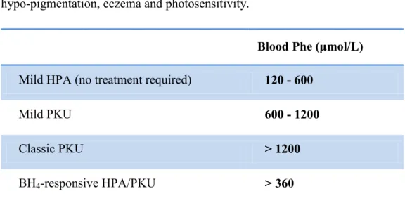
1.1.3 DIAGNOSIS AND CLASSIFICATION OF
HYPERPHENYLALANINEMIAS
Hyperphenylalaninemia (HPA) and phenylketonuria (PKU; OMIM #261600) are a heterogeneous group of autosomal recessive disorders [7] with an incidence of 1/10 000 in Caucasians and a variable frequency in other populations. It is caused by a deficiency of phenylalanine hydroxylase (PAH, EC 1.14.16.1), a hepatic enzyme which catalyzes the conversion of phenylalanine (Phe) to tyrosine, using tetrahydrobiopterin (BH4) as its
coenzyme. The deficiency of PAH enzyme, which determines an accumulation of L-Phe in body fluids and the conversion to other metabolites toxic for the central nervous system, is caused by mutations in the PAH gene (GenBank accession no. U49897.1). Based on blood phenylalanine (Phe) levels, we can distinguish between mild hyperphenylalaninemia (MHP) with blood Phe levels less than 600 µmol/L (< 10mg/dl), mild PKU showing Phe levels between 600 and 1200 µmol/L (10-20 mg/dl), and classical PKU with Phe levels above 1200 µmol/L (> 20mg/dl) (Table 1). All infants are screened for PKU between the ages of 2 and 7 days, in order to allow timely dietary intervention to protect children with PKU from neurological damage. The newborn screening test involves the measurement of blood L-Phe levels by Guthrie test, a bacterial inhibition assay or by Tandem Mass Spectrometry (TMS). Normally, blood L-Phe levels are below 120µmol/L (2mg/dl). Some infants, particularly those born prematurely, may demonstrate immaturity of enzyme systems involved in amino acid metabolism, resulting in a transient elevation of blood Phe to a level sufficient to test positive in a PKU screening test. A second test is required for confirmation of HPA and to eliminate the possibility of transient HPA. The results of early PKU screening should also be interpreted with caution in sick neonates, due to interference with a Guthrie test by antibiotics [8]. If not diagnosed early, untreated or late-treated, classical PKU patients can develop severe intellectual disability, seizures, ataxia, motor deficits, and behavioural
problems, and in many cases, features of autism. Furthermore, this biochemical defect can result in a variety of cutaneous abnormalities, including diffuse hypo-pigmentation, eczema and photosensitivity.
Blood Phe (μmol/L) Mild HPA (no treatment required) 120 - 600
Mild PKU 600 - 1200
Classic PKU > 1200
BH4-responsive HPA/PKU > 360
Table 1: Classification of hyperphenylalaninemias according to blood Phe levels. Normal blood Phe levels are below 120 μmol/L.
PAH requires BH4 as a cofactor. About 1–2% of cases of
hyperphenylalaninemia are due to mutations in genes coding for enzymes involved in BH4 biosynthesis or regeneration pathways lead to disturbed Phe
metabolism. The differentiation of a PAH defect and a defect in BH4
metabolism is done by analysis of urinary or blood pterins and determination of dihydropteridine reductase (DHPR).
1.1.4 PATHOPHYSIOLOGY OF PHENYLKETONURIA AND OUTCOME The major effect of hyperphenylalaninemia (HPA) in PKU patients is on the brain function and development. Symptoms of untreated or late-diagnosed PKU patients are generally summarized as mental retardation, but the clinical effects of HPA/PKU are highly variable (several intellectual disability, seizures, ataxia, motor deficits, behavioural problems, and, in many cases, features of autism). In
adults, anxiety disorders and depression have been reported [9,10,11]. Microcephaly and impaired growth were revealed in adult non-treated PKU patients. A characteristic of classic PKU already known from 1930s is a mousy odor that results from the excretion of phenylketone bodies in the urine. Hypopigmentation in PKU patients is a result of deficient melanin synthesis inhibited by the elevated Phe levels [12]. Many of these symptoms are only rarely observed nowadays because of the introduction of neonatal screening. Children with a late diagnosis and start of treatment can still markedly benefit from dietary treatment. Cognitive performances may improve in children as well as intellectual functioning, behavior and improved quality of life but they depend on the age at which the treatment is started. Nevertheless, intellectual disabilities remain and chronic neurotoxic consequences from high Phe levels are irreversible.
All these symptoms led to the conclusion that a PAH deficiency mainly affects the central nervous system and leads to impaired brain development and function. The pathogenesis of brain dysfunction is not completely known. However, there are several hypotheses addressing possible causes of the neurotoxicity. It is highly likely that the neurocognitive damage in PKU is caused by different processes, all occurring together, discussed briefly below:
Impared LNAA uptake into brain and dopamine and serotonin synthesis The most important neurotoxic pathophysiological mechanisms are thought to be the direct effects of high Phe levels in combination with reduced levels of brain large neutral amino acids (LNAAs). Phe’s entry into the brain is mediated by the large neutral aminoacid carrier L-aminoacid transporter 1 (LAT1). The other LNAA, valine, leucine, isoleucine, methionine, threonine, histidine, tryptophan and tyrosine, are also using this route in a competitive manner [13]. In addition, for each LNAA taken into the brain, the LAT1 transporter excretes one LNAA. Moreover, the LAT1 transporter shows highest affinities for Phe among all the LNAA’s. High concentrations of Phe in the blood can inhibit LAT1 and other large neutral amino acids, including tyrosine (Tyr) and tryptophan (Trp), from
entering the brain, increasing the potential for neurotransmitter dysfunction and their availability for protein synthesis. The low levels of Tyr and Trp might contribute to the depletion of the neurotransmitters dopamine and serotonin. The dopamine depletion in patients with PKU, either due to a deficiency of its precursor Tyr or secondary to hypomyelinisation, might play a prominent role in the development of the neurocognitive impairments. Since the prefrontal cortex is highly susceptible for changes in Tyr levels, the deficits established in PKU patients are frequently specific to the cognitive functioning of the frontal lobes of the brain, which is rich in nerve endings from dopamine-containing neurons, although not all studies support this hypothesis. In addition, cerebral serotonin deficiency may explain the increased occurrence of anxiety and depression disorders in PKU patients. Synthesis of serotonin occurs by hydroxylation of tryptophan by tryptophan hydroxylase. Like dopamine, tryptophan competes with Phe across the BBB. At elevated plasma Phe concentrations, brain tryptophan concentrations, and consequently brain serotonin levels, might be reduced. Although dopamine and serotonin are involved in postnatal brain development and maturation, these findings do not explain the severe mental retardation of PKU patients, but they are likely to explain certain cognitive deficiencies.
Altered myelin metabolism (white matter abnormalities)
Myelin is a cerebral protein often found abnormal in PKU and associated with white matter abnormalities detected in PKU patients. High concentrations of Phe and/or reduced availability of other LNAA may inhibit the development of myelin formation in untreated patients, and to oedema within myelin in early-treated patients with PKU. These lesions may be reversible over a period of months following the achievement of improved Phe control. White matter damage on magnetic resonance imaging has been correlated with cognitive impairment in some patients with PKU. However, such structural impairments usually do not correlate with functional neuropsychological status, and impaired
myelin function may not completely account for the principal pathophysiological defect in PKU.
Other mechanisms
Other possible mechanisms for hyperphenylalaninaemia-induced damage to the brain include reduced activity of pyruvate kinase, disturbed glutamatergic neurotransmission, reduced activity of the enzyme 3-hydroxy-3-methylglutaryl coenzyme A reductase (HMG-CoA reductase), and the function of monoamine oxidase B as a modifying gene (Figure 1). HMG-CoA reductase is a rate-limiting enzyme in the metabolic synthesis pathway of cholesterol.
As protein and cholesterol are essential parts of myelin, this finding is in line with observations of hypomyelination and gliosis in brain cells of PAHenu2 and wild type mice and in brains of deceased PKU patients [14].
It is however not clear, whether impaired cholesterol synthesis leads to reduced myelination in PKU or whether synthesis of HMG-CoA reductase is reduced, suggesting that reduced cerebral protein synthesis may affect enzymes in myelin formation. Finally, DNA damage and oxidative stress may also play a role on PKU pathogenesis [15,16]. The accumulation of toxic metabolites may lead to the induction of free radical production, increasing the risk of oxidative stress of tissue damage in PKU.
Figure 1. Potential mechanisms of neurocognitive impairment by hyperphenylalaninemia (HPA). (Figure adapted from Feillet et al. 2010.).
Patients with non-phenylketonuric hyperphenylalaninaemia have a lower risk of neuropsychological dysfunction than do those with phenylketonuria, although compared with healthy controls, some might have decreased executive functioning. Despite most development of the brain occurring in the early years of life, it seems that discontinuation of dietary management of phenylketonuria during adolescence leads to subtle but measurable deficits in neuropsychological functioning during adult life. The universal experience of those caring for individuals with phenylketonuria is that the dietary treatment results in a pronounced improvement in cognitive outcome but imposes a social burden.
1.1.5
PKU
MATERNAL SYNDROMEAnother important health concern related to PKU pathogenesis is the relation between prenatal L-Phe exposure and the offspring outcome. During pregnancy, Phe crosses the placenta by active transport, resulting in 70% to 80% increased fetal concentration of Phe compared with maternal concentration. An elevated Phe concentration is toxic and teratogenic to a developing fetus.
Untreated maternal phenylketonuria or hyperphenylalaninemia during pregnancy may lead to maternal phenylketonuria syndrome in the neonate. This syndrome consists of low birth weight, microcephaly, congenital heart disease, intellectual or developmental disability and facial dysmorphisms. In this respect, women of childbearing age with all forms of phenylketonuria, including mild variants such as mild hyperphenylalaninemia, are advised to use a strict diet that starts before conception to prevent teratogenic effects in the fetus. Normal pregnancy and neonatal outcome have been achieved in women with PKU who have blood Phe concentrations between 120 and 360 µmol/L before conception or by 8 weeks of gestation at the latest. Achieving this degree of control requires a major commitment by the woman and support by her treating professionals, continually throughout pregnancy, is important, as cognitive outcome in these offspring is better than in children whose mothers began or resumed dietary phenylalanine restriction after conception. Offspring born to women who are fed with a normal diet, unless, as in rare cases, also have phenylketonuria. Mothers with maternal phenylketonuria can breastfeed their non-phenylketonuria infants without restriction. These infants carry a mutant gene for phenylketonuria but their residual PAH activity is sufficient to adequately metabolize phenylalanine, even the additional amount they receive from their mother´s breast milk [7].
1.1.6
PKU
ACTUAL TREATMENT:
THE LOWP
HE DIETThe aim of PKU treatment is the reduction of blood Phe to a level allowing normal brain development. An individual’s blood Phe depends upon dietary intake of Phe and the residual activity of Phe hydroxylase. As stated in 1.1.3 and 1.1.4 sections, PKU related symptoms are severe, therefore it is necessary to treat this disease since birth. At present, PKU is treated with a lifelong dietary protein restriction, in which many common foods, such as milk and dairy products, meat, eggs, wheat, beans, corn, peanuts, lentils, and other grains, are prohibited to the patients (see also Figure 2).
Figure 2. Low Phe diet: foods that are allowed are in the inner part of the diagram.
A semi-synthetic diet is used which comprises:
foods of low phe content in unlimited amounts such as many fruits and vegetables;
weighed amounts of foods containing medium amounts of Phe (e.g. broccoli, potato). The amount of Phe ingested is often calculated using an exchange system. In the UK system 1 ‘exchange’= 50 mg Phe which is approximately 1 g protein;
Phe-free amino acid mixtures to provide normal total protein intake; vitamins, minerals and trace elements.
The diet should be strictly followed with these food groups evenly distributed throughout the day. Aspartame should be avoided as it contains large amounts of Phe. Infant formulae feeds which are Phe-free are available; many contain added essential fatty acids. These are used in conjunction with a small amount of standard infant formulae. It is possible to continue breast feeding even in severe PKU by giving a measured amount of Phe-free formula prior to a breast feed. All PKU diets should be administered with the advice of a specialist dietician [17].
1.1.7
A
LTERNATIVES APPROACHES TO TREATPKU
As mentioned in 1.1.6 section, PKU related symptoms can be prevented by a strict dietary restriction from early infancy. Although it is recognized that dietary treatment initiated early in life is successful in avoiding the severe mental retardation of PKU, this treatment presents many limitations, since PKU patients must adhere to an unpalatable, expensive and lifelong diet that is frequently stopped prematurely, leading to an unsatisfactory clinical outcome. As long-term dietary compliance is difficult, there is a need for alternative modes of treatments:
Treatment with large neutral amino acids (LNAAs)
Because phenylalanine competes with other LNAAs for transport (LAT 1) across the blood–brain barrier, supplementation with these aminoacids other than Phe, could provide a potential treatment approach.
In fact, LNAA administration was shown to reduce toxic Phe levels in the brain of patients despite constantly elevated serum concentrations [18,19,20].
In this way, other amino acids with high affinity for the BBB transport system keep high plasma Phe concentrations from entering the brain and it seems that this treatment has a beneficial effect on executive functioning [7]. It may not replace Phe-low diet, at least in childhood or in pregnant women with PKU, but may help to relax dietary restriction of treated individuals or help in management of untreated adults with classic PKU. However, further clinical trials and data are required to examine safety and efficacy of LNAA therapy still in development.
Glycomacropeptide
Another strategy is the supplementation of glycomacropeptide (GMP). Glycomacropeptide is a natural protein from cheese whey, that is rich in specific essential amino acids but contains no Phe, Tyr, or tryptophan (Trp). It is being investigated as a useful adjunct to dietary treatment for PKU by allowing a greater intake of ‘natural’ protein in the diet, thus, allowing a decrease in artificial amino acid mixture. It has been shown in the mouse model to improve bone density [17] and some studies have given promising results also in terms of palatability, safety and improved compliance, but further evidence is still required [21,22,23].
Enzyme substitution therapy
Enzyme therapy can be addressed in two different ways, either by replacing PAH or by substitution with a foreign protein capable of metabolizing Phe. Replacement of PAH is highly challenging, as the whole, intact multi-enzyme complex for PAH catalysis including BH4-cofactor is required. The enzyme of
choice for substituting PAH is phenylalanine ammonia lyase (PAL, EC 4.3.1.5), derived from plants and compared to PAH, sufficiently more stable as oral formulation. PAL targets the intestinal system and is also involved in Phe metabolism, capable of lowering blood Phe in humans. This exogenous enzyme converts Phe to transcinnaminic acid and negligible amounts of ammonia without the need of a cofactor, making it catalytically less complex than PAH. In the mouse model of phenylketonuria, blood and brain concentrations of Phe were reduced during 90 days of treatment with injections of PAL. The PEGylation (attachment of polyethylene glycol polymers to lysine side chains) diminished immune-mediated detection and elimination of the injected enzyme [24]. Clinical trials have been initiated in PKU patients and preliminary results showed a significant decrease in Phe blood levels after a single injection. A phase II clinical trial is in progress in addition to development of an orally administrable PAL form [25]. PAL would probably be used in addition to a less stringent Phe diet.
BH4 therapy
Some mutations are associated with a BH4-sensitive phenotype of
phenylketonuria, in which giving pharmacological doses of exogenous BH4
results in an increase in the activity of PAH that is sufficient to reduce circulating Phe with a therapeutically relevant effect. These mutations usually present with substantial residual activity when expressed in eukaryotic cell systems and are located in all regions of PAH. However, the relation between
genotype and BH4-responsiveness is complex. Thus, although genotyping is
useful in excluding non-responders (classic phenylketonuria), it is insufficient for a reliable prediction of those who are BH4 responsive. Mechanisms of BH4
-responsiveness are multifactorial, but the main mechanism seems to be stabilisation of the PAH tetramer by preventing misfolding, subunit aggregation, proteolytic degradation, and thermal inactivation [7].
Since 2008, sapropterin dihydrochloride (Kuvan® or Biopten®, BioMarin Pharmaceutical Inc), the synthetic analogue of BH4, has been market approved
for treatment and is commercially available in the US, Japan and Europe. It acts as a pharmacological chaperone by stabilizing PAH. In BH4-responsive
patients, BH4 decreases the blood Phe concentration and/or increases the dietary
Phe tolerance. Correct and efficient identification of BH4-responsive patients is
important, both to improve the fast assessment, as well as to avoid false expectations and unnecessary costs. Unfortunately, there is still no golden standard on how to assess BH4 responsiveness most efficiently. Three methods
have been proposed for the prediction of BH4 responsiveness: the 7–28 days
BH4 challenge, the 48-hour BH4 loading test and, the START (Sapropterin
Therapy Actual Response Test) BH4 challenge and genotyping protocol.
Genotype was frequently reported to be useful in predicting or excluding BH4
responsiveness [26]. The proportion of BH4-sensitive patients increases as the
severity of the phenotype decreases, which is due to the mechanistic action of administered BH4, requiring sufficient active hepatic PAH protein (residual
enzyme activity). The online BIOPKU database (www.biopku.org) tabulates available data on almost 800 genotypes, phenotypes and BH4-response in
patients previously tested and is used as a reference tool in consulting whether to challenge a patient based on previous similar genotype results.
Pharmacological Chaperons
The use of pharmacological chaperones to stabilize or promote correct folding of mutant proteins represents a promising new approach in the treatment of many genetic diseases causing protein misfolding. Proteins and small molecules in addition to tetrahydrobiopterin may act as chaperones to assist in the folding of PAH [27].
Gene therapy
Gene therapy for the treatment of PKU has been the focus of multiple research groups over the last two decades. In a mouse model of PKU, important progress has been made by the use of an adenovirus related gene directed into the liver [28].
However, the vector’s genome is gradually eliminated as it is not integrated into the hepatocyte’s DNA and re-injection was not effective due to immunological responses. Studies of PKU mouse models have also shown that gene therapy can be successfully delivered to non-hepatic tissues such as the muscle. It is easily accessible and does not undergo cell division. In fact, the insertion into the muscle cells of vectors containing the necessary genes for PAH and tetrahydrobiopterin synthesis results in a system that could convert phenylalanine into tyrosine, mimicking the role of hepatic phenylalanine metabolism [29].
Liver Transplantation
Hepatocyte transplantation has been performed in preclinical studies using various animal models, as well in humans with metabolic disorders, such as urea cycle defects or glycogen storage disorders. This cellular approach could be possible for the permanent treatment of PKU if a selective growth advantage
could be achieved for donor hepatocytes. This treatment has been reported to be successful in an animal model with a selective advantage for the donor cells. However, cell based therapies using stem cells or more differentiated progenitor cells may represent the future of cell transplantation for treatment of metabolic liver diseases such as PKU [27].
1.2 T
HEP
HENYLALANINE HYDROXYLASE SYSTEM1.2.1 STRUCTURAL BASIS AND REGULATION OF PHENYLALANINE
HYDROXYLASE
PAH is an hepatic monooxygenase that catalyzes the conversion of Phe to Tyrosine (Figure 3), using 6R-lerythro-5,6,7,8-tetrahydrobiopterin (BH4) as a
coenzyme. Molecular oxygen and iron are essential for the hydroxylation of Phe to Tyrosine.
Figure 3. Overview of the reaction catalyzed by phenylalanine hydroxylase.
The structure of PAH is similar to the other principal aromatic amino acid hydroxylases, tyrosine hydroxylase (TH) and tryptophan hydroxylases (TPH) 1 and 2. The size of an individual subunit of PAH is about 50kDa, and the active enzyme exists as dimer or (mainly) tetramer of these subunits, with these forms
existing in equilibrium, according to cytosolic pH. Each subunit of PAH consists of three structural and functional domains:
N-terminal regulatory domain (residues 1-142);
Large catalytic domain (residues 143-410), which contains the active site with the iron center and the binding sites for Phe and BH4;
Small C-terminal tetramerization domain (residues 411-452), which mediates the association of subunits into tetramers (Figure 4.).
Figure 4. 3D crystal structure of the human PAH. A: Interactive site, the iron atom is
shown in red. The N-terminus starting over the active site, as well as the rest of the regulatory domain is highlighted in blue; catalytic domain in yellow; and tetramerization domain is in green. B: The native tetramer form of the enzyme. C: Structure of a monomer of human Phenylalanine Hydroxylase full-length composite model. In figure are shown the regulatory domain (residues 19–142), the catalytic domain (residues 143–410) and the tetramerization domain.
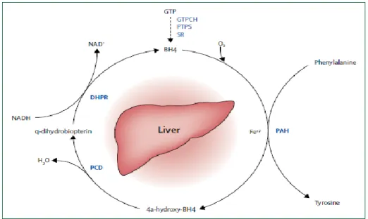
1.2.2 CATABOLIC PATHWAY OF PHENYLALANINE
The L-Phe amino acid is hydroxylated by the enzyme PAH to form Tyr. PAH is an iron-containing enzyme depending on 6(R)-L-erythro-tetrahydrobiopterin (BH4) as cofactor and molecular oxygen for efficient
catalysis. During the reaction, BH4 is oxidized to 4a-hydroxypterin and needs to
be regenerated for continuous catalysis. The regenerating enzymes pterin-4a-carbinolamine (PCD) and dihydropteridine reductase (DHPR) complete the phenylalanine hydroxylating system (Figure 5).
Figure 5. Phenylalanine hydroxylating system: during the hydroxylation of
phenylalanine by phenylalanine hydroxylase (PAH), and when molecular oxygen (O2)
and iron (Fe+2) are present, tetrahydrobiopterin (BH4) is oxidised to a
4a-hydroxy-BH4 intermediate, which is subsequently regenerated back to BH4 via quinonoid (q)
dihydrobiopterin by the enzymes carbinolamie-4adehydratase (PCD) and by the NADH-dependent dihydropteridine reductase (DHPR). BH4 is synthesised from
guanosine triphosphate (GTP) by three additional enzymes GTP cyclohydrolase I (GTPCH), 6-pyruvoyl-tetrahydropterin synthase (PTPS), and sepiapterin reductase (SR). Mutations in genes coding for PCD, DHPR, GTPCH, PTPS, and SR result in BH4 deficiency.
catecholamines (dopamine, adrenaline and noradrenaline), melanin and thyroid hormones.
Therefore, the biosynthesis of the neurotransmitter monoamines dopamine, norepinephrine and serotonin is dependent upon the availability of the precursor amino acids Tyr and Trp within the brain and the presence of a normal Phe concentration. A defect in PAH system, leading to excess Phe and a relative lack of Tyr and Trp in the brain, has consequences for the subsequent enzymatic reactions.
To maintain Phe homeostasis in vivo, PAH is highly sensitive to changes in substrate concentrations and its activity is tightly regulated and requires the binding of all three of its substrates- Phe, molecular oxygen and the cofactor BH4– prior to any reaction occurs. The first 30 residues of PAH act as an
autoregulatory sequence that includes the Ser16, a substrate for cAMP-dependent protein kinase and is termed autoregulatory, as it sterically limits substrate access unless Phe binding to the regulatory domain activates the enzyme.
The activation by the Phe substrate results in a significant increase in the initial rate of Tyr formation [30]. Activation is probably related to a conformational change induced in the PAH protein upon binding which is transmitted throughout the enzyme, displacing the autoregulatory sequence and leading to the propagation of the activating process to the adjacent subunit in the dimer and finally to the other dimer through the oligomerization domain. While Phe activates the enzyme, at the same time BH4 acts as an inhibitor in addition to its
role as a cofactor, keeping the enzyme in a low activity state, and blocking the substrate-activating conformational change [31]. BH4 interacts with the
N-terminal autoregulatory sequence and leads to a dead-end PAH–BH4 complex,
closing the entrance to the active site and leaving the enzyme in a latent, low-activity state [32,33,34]. Phe binds with lower affinity to PAH than the cofactor BH4. The high affinity binding of BH4 and the inhibitory regulatory effect in
chain with residues from the catalytic and regulatory domain. The iron atom is also essential for catalytic activity. It is coordinated to two histidine residues (H285 and H290), as well as to one oxygen from E330. The rest of the coordination sites of iron are occupied by water molecules, which are all displaced upon substrate and cofactor binding. This leaves an open coordination site for the reaction with O2 and generates an activated intermediate in the
hydroxylation of the Phe and BH4.
Therefore, the composite model of full-length PAH provided an important basis for analysis of the numerous mutations resulting in deficient PAH activity.
1.3 T
HEC
OFACTORT
ETRAHYDROBIOPTERIN(BH
4)
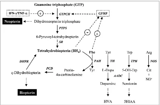
1.3.1 COFACTOR BIOSYNTHESIS AND FUNCTIONS
BH4 is the short name of its correct chemical name
2-amino-4-hydroxyl-6-[L-erythro-1’,2’-dihydroxypropyl]-tetrahydrobiopterin. BH4 is essential for
many diverse processes and ubiquitously present in all tissues of higher organisms. Consequently, BH4 plays a key role in a number of biological
processes and pathological states associated with monoamine neurotransmitter formation, cardiovascular and endothelial dysfunction, the immune response and pain sensitivity. BH4 is formed de novo from GTP via a sequence of three
enzymatic steps carried out by GTP cyclohydrolase I (GTPCH), 6- pyruvoyltetrahydropterin synthase (PTPS) and sepiapterin reductase (SR). Detailed reviews of the biosynthesis of BH4 have been provided in Figure 6.
Figure 6. Overview of the biosynthesis of tetrahydrobiopterin (BH4) and a summary of
its actions as a cofactor in enzymatic reactions.
In the last two steps of the novo biosynthetic pathway, 6-pyruvoyl-tetrahydropterin is thought to be converted to 6-pyruvoyl-tetrahydropterin derivatives with a different succession of side chain reductions involving SR but potentially also alternative carbonyl and aldose reductases.
BH4 itself is hydroxylated during the aromatic amino acid reactions of PAH,
TH and TPH. It is therefore essential that the cofactor be regenerated to ensure a continuous supply of reduced cofactor and to prevent accumulation of harmful metabolites. BH4 is oxidized to BH4-4a-carbinolamine and via two
further catalytic reactions and quinoid dihydrobiopterin (qBH2) intermediate
reduced to BH4. The enzymes involved are PCD and DHPR.
BH4 acts not only as a cofactor of the aromatic amino acid monooxygenases as
described above. Additional functions on the cellular level were found upon the discovery that BH4 is essential for all three isoforms of nitric oxide synthase
L-citrulline in two-step reactions [36]. The role of BH4 in these reactions is
different from aromatic amino acid monooxygenases. BH4 is not involved in
oxygen activation, donates only one electron, and is regenerated without the need for external enzymes. Werner E. et al. recently compared the mechanisms involving BH4 in hydroxylases and NOSs [37]. The synthases are ubiquitously
involved in vascular and cardiac functions, establishing a role for BH4 in
diseases like hypertension, diabetes, atherosclerosis, cardiac hypertrophy and failure, but also Parkinson’s and Alzheimer’s disease.
1.3.2 ROLE OF BH4 AS A MOLECULAR CHAPERONE
BH4 supplementation is known to result in increased enzyme activity.
Besides this non-chaperon stimulatory effect of BH4 supplementation, the
binding of BH4 to PAH may also increase the stability of protein and
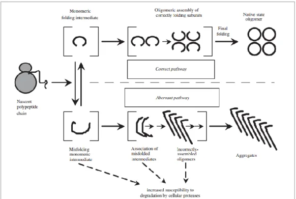
prevent its degradation, a phenomenon described as a “chaperone” effect. The term molecular chaperones identifies the large molecular machines that in an energy-dependent manner ensure the correct folding of intracellular proteins. Together with the ubiquitin-proteasome and the selective autophagy systems, molecular chaperones constitute the quality control system acting at different subcellular localizations, working in concert to reduce the accumulation of misfolded proteins by either refolding or destroying them (Figure 7).
Figure 7. Model of competing pathways for correct vs. aberrant protein folding and
assembly (Figure based on ref. [38]).
Many of the point mutations on the PAH sequence leading to missense in translation were found to affect protein folding and assembly leading to secondary effects on enzyme function. They can lead to increased aggregation and destabilized native conformation, causing loss-of-function pathologies (or eventually gain-of-function). The aggregates or nonfunctional conformations are no longer removed by the cells quality control systems like proteases. The protein architecture of PAH is very sensitive to single point mutations throughout the protein sequence. It results in a greater proportion of protein entering the aberrant pathway, where it is more susceptible to proteolytic degradation in human cells than protein proceeding through the pathway of correct folding and assembly.
However, BH4 was shown to favour the formation of tetrameric PAH where
tetramers, and thus increased the stability of the PAH protein and slowed its inactivation in vitro [39,40].
These data suggest that molecular chaperones, like BH4, can be an efficient
treatment option for PKU, although further development regarding mechanism and action of clinical effects is required before application in patients is possible.
1.3.3 INBORN ERRORS IN TETRAHYDROBIOPTERIN METABOLISM
About 2% all Phe level elevations detected by the newborn screening (higher in some countries where frequent consanguinity tends to maintain the presence of genetic disorders within families, e.g. Turkey or Saudi Arabia) are due to disorders in BH4 metabolism, highlighting the importance of always
considering the differential diagnosis for every even slightly elevated blood Phe level. BH4 deficiencies are more severe than PKU with regard to their response
to therapy and treatment is substantially different. Low-Phe diet is not effective and early substitution with dopamine and serotonin precursors, as well as with the synthetic BH4 is crucial for a good outcome. Analysis of dried blood spot
(DBS) or urine for neopterin and biopterin and measurement of DHPR activity in the DBS is essential for the exact diagnosis and should be performed as early as possible. A BH4 loading test and measurement of neurotransmitter
metabolites, pterins, and folates in cerebrospinal fluid add further important information about the severity of the disease. Some patients with DHPR deficiency show a normal blood or urinary neopterin and biopterin profile. Therefore, DHPR activity measurement is essential in all patients with HPA, regardless of pterin measurements [41].
BH4 deficiencies arise from mutations in the genes coding for the enzymes in
BH4 biosynthesis and regeneration pathways. Due to the secondary disturbance
nervous system neurotransmitter biosynthesis and generally lead to severe and heterogeneous phenotypes. BH4 deficiencies are treatable diseases although
with variable outcomes. The clinical signs and symptoms of a BH4 deficiency
largely arise from neurotransmitter depletion and compromised nitric oxide synthesis [42]. The few patients that present with a mild phenotype generally show normal brain neurotransmitter metabolism and require only BH4 monotherapy. Symptoms of BH4 deficiency may develop only after a few
weeks or months of life. Therefore, most patients are initially maintained on a low-Phe diet until final diagnosis. All BH4 deficiencies associated with elevated
Phe levels are inherited in an autosomal recessive manner and often share common clinical symptoms. Clinical features manifested with GTPCH, PTPS and DHPR deficiencies are abnormal movements together with impaired tone and posture, convulsions, seizures, mental retardation, as well as light pigmentation and microcephaly.
1.4 M
OLECULARG
ENETICS1.4.1 THE PAHGENE
The human phenylalanine hydroxylase gene (PAH) is located on chromosome region 12q22 - 12q24.2 containing the nucleotide sequence coding for the hepatic enzyme phenylalanine hydroxylase (PAH, phenylalanine 4-monooxygenase, EC 1.14.16.1). The PAH gene is 90 kb long and consists of 13 exons and their required introns, adding up to 171 kb including the flanking regions, encoding a 51.9 kDa polypeptide sequence with 452 amino acids. The exonic sequences in PAH take up less than 1% of the genomic sequence. The complete sequences have been catalogued in GenBank under NM_000277 (mRNA, 2680 bp), U49897.1 (cDNA, 1359 bp), AF404777 (gDNA, 171 kb)
and NP_000268.1 (protein, 51.9 kDa) or in Ensembl under the reference number ENSG00000171759.
PKU arises from mutations on this gene and is inherited as an autosomal recessive disorder. Different possibilities of a child to develop PKU, according to the PAH status of its parents is depicted in Figure 8.
Figure 8. Overview of the mode of inheritance of PKU (Figure adapted from website
http://www.pku.com/en/What_is_PKU/Who_gets_PKU/who_gets_pku.html).
Thus, if only one parent has a defective copy of the PAH gene, offsprings have a 50% chance of being a carrier themselves, with no possibility of inheriting clinical PKU (Figure 8. panel a). All children of a parent with PKU will have at least one defective copy of PAH, but at risk of PKU only if the other parent carries a PAH mutation that affects its activity (Figure 8. panels b and c). An offspring of parents who each have one defective copy of the PAH gene has a 25% chance of having PKU, a 50% chance of being a carrier of a PAH mutation, a 25% chance of inheriting two fully functional copies of the PAH gene (Figure 8. panel d). Finally, all children of two parents with PKU will inherit the disease.
1.4.2 CAUSATIVE MUTATIONS
The PAH gene is the only gene associated with PAH deficiency, which results from mutations in both alleles disturbing enzyme function. Isolation and sequencing of PAH cDNA opened new fields for intense exploration of the molecular causes of PKU. The Human PAH Mutation Knowledgebase (hPAHdb), a database of naturally occurring mutations in the human PAH gene, was summarised in 2009 and includes a total of 567 separate mutations. Since the latest updates of this database roughly 61% of all mutations listed are missense mutations, followed by small deletions (13%), splice mutations (11%), silent and nonsense mutations (5 - 6%), and small insertions (2%). The position and nature of the mutation dictates its effect on the activity of the PAH enzyme, which determines the hyperphenylalaninemic phenotype of the patient. Little or no enzyme activity (for example p.R408W, the most common mutation) results in the classic phenylketonuria phenotype. Other mutation, such as p.E390G, p.Y414C and p.A300S only partly inhibit enzyme activity so that dietary Phe tolerance is higher, giving rise to mild phenylketonuria or mild hyperphenylalaninemia. The majority of mutations are scattered over the entire gene length but with a different frequency in distinct populations and geographic areas and a number of them have been analyzed and characterized. Thanks to different studies, it is now known that most of PAH missense mutations result in a misfolding of the protein which increases its turnover both in vitro and probably in vivo, pointing to a decreased conformational stability as the major molecular mechanism for the loss of PAH function in PKU [43].
1.4.3 GENOTYPE-PHENOTYPE CORRELATIONS
The compilation of all information about PAH mutations and establishment of genotype-phenotype correlations is highly useful in predicting the course of disease for a patient’s diagnosis. The ability to predict the
phenotype already in a newborn with PAH deficiency not only enables the design and early implementation of an optimal dietary regimen, it also greatly improves counseling of the patient’s family.
The BIOPKU database (www.biopku.org) was created at the University of Zürich and it is based on the genotype and phenotype data of over 9700 PAH-deficient patients from all over the world. More than 900 different PAH gene variants are tabulated in the database and these result in almost 2000 different genotypes. More than 75% of all PKU patients are compound heterozygotes bearing two different variants. Some of the mutation combinations of patients listed are not always associated with the same phenotype. In general, genotype-phenotype correlation is possible, but there are always exceptions. Mutations for which inconsistencies regarding genotype/phenotype correlation are reported in this database as well as in the literature, are the R261Q mutation in the homozygous state or in combination with the R158Q mutation, the L48S mutation in the homozygous state or in combination with the R158Q mutation, and the Y414C mutation in combination with the R408W mutation [41].
As already mentioned, a substantial number of PKU patients are compound heterozygotes expressing two different PAH alleles. Thus, the genotype is determined by the type of PAH gene variation, which may result in a totally inactive enzyme or a protein with substantial residual activity (up to 100% of the wild-type PAH). However, the interallelic complementation (IC) phenomena may occur, leading to a milder (positive IC) or a more severe phenotype (negative IC) than expected in the homozygous constellation [44]. Interallelic complementation effects arise from the combination of PAH monomer variants yielding a heterotetrameric PAH protein with functional and/or structural properties different from the wild-type homotetrameric protein. The interallelic complementation of variant protein monomers and effect of possible protein modifiers may complicate the interpretation and lead to inconsistencies between the same genotype and different phenotypes as demonstrated by Trefz et al. [45]. Lichter-Konecki et al. [46] correlated
genotypes and residual in vitro enzyme activity with the phenotypes of PKU patients. As expected, patients with classic PKU harbored more severe mutations with less or no residual PAH activity [47].
General truths about genotype/phenotype correlations in PKU that have emerged from the data cataloged in the databases, especially in the BIOPKU data base, and in literature are:
1. Mutation combinations that allow for <15% in vitro enzyme activity cause classic PKU and do not respond to BH4. Mutation combinations
that allow for >20% residual activity responds to BH4. Responders have
moderate to mild phenotypes.
2. Splice site mutations may cause classic or mild PKU depending on ‘read through’ (i.e. normal splicing may sometimes occur despite the mutation), and the fact that they have different phenotype associations is listed in the available databases.
3. Specific mild mutation/classic mutation combinations with identical predicted residual enzyme activity may have different phenotype associations (due to negative intra-allelic complementation) but the phenotype associations of different mutation combinations can be found in the available data bases making prediction unnecessary.
4. The BH4 responsiveness of many mutation combinations (complete
genotypes) has been well established several times. Patients that have those genotypes may not need to undergo BH4 responsiveness testing
[41].
Although many factors affect phenotypes, the specific PAH genotype is the main determinant of metabolic phenotype in most cases.
1.4.4 RESPONSIVENESS TO BH4
Some mutations have been associated with a BH4- sensitive PKU
phenotype, where the administration of pharmacological doses of exogenous BH4 results in an increase in the activity of PAH sufficient to reduce circulating
phenylalanine levels to a clinically significant extent and daily tolerance for phenylalanine.
These mutations usually give rise to milder forms of PKU, with substantial residual PAH activity. Some analyses have identified mutations which facilitate stabilization of the structure of the PAH protein by BH4, which is consistent
with the BH4 responsive phenotype [48,49].
It was emphasized from these studies that a significant residual PAH activity is a prerequisite for BH4 responsiveness. This was further characterized on
biochemical, molecular and physiological levels and several possible mechanisms for BH4 responsiveness were proposed based upon results of in vitro expression studies and structural implications of the mutations [50].
Many mutations and genotypes were found associated with responsiveness and are listed in the BIOPKU database (www.biopku.org).
These mutations with substantial residual activity are located in all regions of PAH (Figure 9).
Figure 9.: Three-dimensional crystal structure of the human phenylalanine hydroxylase monomer with most common BH4-responsive mutations The most mutations (70%) were
positioned in the catalytic domain (blue), 16% of mutations are in the regulatory domain (red), and 14% are in the tetramerization domain (lilac). (source BIOPKU database; http://www.bh4.org/biopku.html)
About 70% of all mutations are located in the catalytic domain of PAH, 16% in the regulatory domain, 14% are located in the tetramerization domain.
Based on the presently detected genotypes of BH4 -responsive patients, it would
appear that the allelic PAH mutation-combination is the most important indicator of BH4-responsiveness. Two severe mutations found on the two
alleles for PAH will very likely result in severe PKU, and in turn little or no PAH enzymatic activity, and very likely no BH4-responsive. It would be very
difficult to propose a possible mechanism for BH4-responsiveness in patients
with homozygosity for severe null mutations. Nevertheless, a few severe/classical PKU patients (with blood Phe levels >1200 µmol/L) have been
found to be BH4-responsive, all of them with at least one partially active allele
(as determined by enzymatic activity assays performed on expressed protein in
vitro). Similarly, patients with two mild mutations that show relatively high
residual activity (i.e., for example >30% activity as compared to wild-type PAH) will likely display HPA or mild PKU, and possibly be BH4-responsive.
The combination of one mild mutation with one severe mutation will questionably be BH4-responsive, and based on the currently known genotypes
that are BH4-responsive, this will depend upon the combination of the
mutations present in the genotype. Thus, most of the genotypes found currently to be BH4-responsive consist of one mild mutation and one severe mutation, or
two relatively mild mutations, and they also display high residual enzymatic activity [51]. However, the relation between genotype and BH4-responsiveness
is complex. A study in Croatian patients with phenylketonuria showed that the prevalence of BH4-responsive PKU was lower than would have been expected
from genotyping alone [52]. This study also clearly documented that only the full genotype may predict the phenotype and BH4–responsiveness. Overall,
genotyping alone is currently insufficient for prediction of the BH4 –sensitive
PKU phenotype, but it is helpful in excluding non-responders to BH4 therapy