European Journal of Endocrinology
180:3 177–187
L Zhang and others FNA cytology in thyroid lymphoma
Fine-needle aspiration to diagnose primary
thyroid lymphomas: a systematic review and
meta-analysis
Lu Zhang1, Marco Castellana2, Camilla Virili3, Anna Crescenzi4, Francesco Giorgino2, Emanuele Zucca5,6,7,
Luca Ceriani8, Franco Cavalli7, Luca Giovanella8 and Pierpaolo Trimboli8
1Institute of Hematology, Union Hospital, Tongji Medical College, Huazhong University of Science and Technology, Wuhan, Hubei,
China, 2Section of Internal Medicine, Endocrinology, Andrology and Metabolic Diseases, Department of Emergency
and Organ Transplantation, University of Bari Aldo Moro, Bari, Italy, 3Department of Medico-Surgical Sciences and
Biotechnologies, Sapienza University of Rome, Latina, Italy, 4Pathology Unit, University Hospital Campus Bio-Medico,
Rome, Italy, 5Division of Medical Oncology, Oncology Institute of Southern Switzerland, Bellinzona, Switzerland, 6Medical Oncology, University of Bern, Bern, Switzerland, 7Institute of Oncology Research, and 8Department of
Nuclear Medicine and Thyroid Centre, Oncology Institute of Southern Switzerland, Bellinzona, Switzerland
Abstract
Background: Primary thyroid lymphoma (PTL) is a rare malignancy, and its prognosis depends significantly on its early diagnosis. While fine-needle aspiration (FNA) represents the gold standard to identify differentiated thyroid carcinoma, its reliability for the detection of PTL is still unclear. Here, we conducted a systematic review and meta-analysis to evaluate the diagnostic performance of FNA in PTL.
Research design and methods: A comprehensive literature search of PubMed/MEDLINE and Scopus databases was conducted to retrieve papers reporting histologically proven PTL undergone FNA. The last search was performed in February 2018 without language and time restrictions.
Results: Thirty-two studies describing 593 PTL were included and the pooled FNA sensitivity was 0.48 (95% CI = 0.38– 0.58). FNA sensitivity was 0.51 in 20 studies published before 2010 and 0.39 in those published later, 0.50 in six articles with at least 20 cases and 0.44 in nine series enrolled after 2000. This performance was similar in 12 articles including diffuse large B-cell lymphoma (0.54) and those six on marginal zone lymphoma (0.56). Remarkably, FNA sensitivity increased to 0.72 when considering also FNA reports suspicious for PTL reported in 14 articles. Heterogeneity among the series was found. Publication bias was not always detected.
Conclusions: The present meta-analysis demonstrated that FNA has low sensitivity in diagnosing PTL. However, this rate increased when considering also FNA reports suspicious for PTL, which is relevant from a clinical standpoint. This result could support indirectly the use of additional imaging and/or core biopsy when PTL is suspected.
Introduction
Primary thyroid lymphoma (PTL) is a rare disease, accounting for 1–5% of thyroid malignancies and up to 2.5% of all extranodal lymphomas, with women more commonly affected than men (1, 2, 3). The majority of lymphomas arising in the thyroid gland are non-Hodgkin’s lymphomas of B-cell origin (3, 4). Most commonly, they are diffuse large B-cell lymphomas (DLBCL), which
represents about half of the cases or marginal zone lymphomas of the mucosa-associated lymphoid tissue type (MALT lymphoma), accounting for up to a quarter of cases. Composite cases with both MALT lymphoma and DLBCL can occasionally be seen and may represent an ongoing histologic transformation process. Follicular lymphomas may also be found in the thyroid gland,
Correspondence should be addressed to L Giovanella Email [email protected] European Journal of Endocrinology (2019) 180, 177–187
Clinical Study
Published by Bioscientifica Ltd. Printed in Great Britain© 2019 European Society of Endocrinology
https://eje.bioscientifica.com
https://doi.org/10.1530/EJE-18-0672
Downloaded from Bioscientifica.com at 01/28/2021 10:45:13AM via free access
European Journal of Endocrinology
but are less common (approximately 10% PTL). Other histologic subtypes are rare at this site and comprise small lymphocytic lymphomas (3%), together with Burkitt’s, mantle cell and lymphoblastic lymphomas, each accounting for <1% of cases. T-cells lymphomas and Hodgkin lymphomas are extremely rare (3, 4, 5, 6, 7, 8). PTL serves (along with salivary gland lymphomas associated with myoepithelial sialadenitis) as the paradigm of the link of autoimmunity with lymphomas. The normal thyroid gland does not contain native lymphoid tissue and PTL typically arises from a background of chronic lymphocytic thyroiditis (9, 10). Indeed, patients with Hashimoto’s thyroiditis (HT) are at greater risk for developing PTL, with a relative risk of 67 compared to those without thyroiditis (9). In the context of chronic antigenic stimulation, abnormal B-cell clones acquiring successive genetic abnormalities can progressively replace the normal B-cell population of the inflammatory tissue, giving rise to the lymphoma.
The driving mechanisms might be, however, distinct in each autoimmune disease (11) and impaired immune-surveillance may also contribute to the lymphomagenesis process (12, 13).
Albeit the thyroid is not a mucosal organ, the lymphoid tissue occurring in HT shares many features with MALT (14). The differential diagnosis between a thyroid MALT lymphoma and its benign reactive precursor, HT, is not always straightforward (10, 14) and clonal B-cells can be present in (a minority of) HT (15). Detection of PTL is difficult and its early diagnosis remains challenging even in our era of emerging technologies. A PTL should be suspected in the presence of a rapidly enlarging neck mass, particularly in women with HT. Certain ultrasound features, such as enhanced posterior echoes, may also suggest the diagnosis. Fine-needle aspiration (FNA) represents the gold standard to identify differentiated thyroid carcinoma, and ultrasonography (US) and ultrasound-guided FNA are the most reliable and most commonly used first-line diagnostic procedures for risk stratification of thyroid nodules. However, the real accuracy of FNA for the detection of PTL is still unclear. FNA may lead to the diagnosis of DLBCL, while due to the continuum spectrum between HT and MALT lymphoma, it is considered less reliable for the diagnosis of MALT lymphoma. A combination of the cytological examination with flow cytometry analysis of FNA has been proposed (16), but it requires the availability of experienced specialists in hematology, flow cytometry and thyroid cytology. Therefore, biopsy is ultimately needed for a complete diagnostic work-up. Less aggressive
than surgical biopsies, core needle biopsy (CNB) may provide enough tissue for both the accurate histological diagnosis and additional ancillary assays (17, 18, 19, 20), and it is currently recommended when PTL is suspected (1, 20). Nevertheless, despite its expected limitations for the diagnosis of PTL, US-guided FNA remains the most frequently used tool for the evaluation of thyroid nodules.
The actual reliability of FNA in detecting PTL has not yet been definitely assessed. Here, we report the results of a systematic review and meta-analysis on the sensitivity of FNA in the identification of PTL.
Methods
Registration of review
The present systematic review was registered in PROSPERO (n = CRD42018091734).
Search strategy
A five-step search strategy was planned. Firstly, we searched sentinel studies in PubMed. Secondly, keywords and MeSH terms were identified in PubMed. Thirdly, the terms ‘primary thyroid lymphoma’, ‘cytology’, ‘FNA’, ‘FNAB’, ‘FNAC’, ‘fine-needle’, ‘fine needle’ and ‘biopsy’ were searched in PubMed, in order to test the strategy. Fourthly, PubMed/MEDLINE and Scopus were screened. Finally, the references of included studies were screened for additional papers. The last search was performed on February 3, 2018. No language or time restriction was adopted.
Studies reporting the detection rate by FNA in histology-confirmed PTL were eligible for inclusion. The exclusion criteria were (a) articles not within the field of interest of this review; (b) review articles, editorials, letters or comments; (c) articles that did not provide clear study characteristics or reports that had overlapping patient data; (d) case reports and (e) case series reporting less than five PTL undergone FNA. Three investigators (P T, C V and M C) independently searched papers, screened titles and abstracts of the retrieved articles, and reviewed full-texts and selected articles for their inclusion. Incongruities were resolved in a consensus meeting involving all authors of the paper.
Data extraction
For each study that was included in the research, the following information was extracted independently
European Journal of Endocrinology
by three investigators (P T, C V and M C) in a piloted form: (1) general information on the study (author, year of publication, journal, country and type of study); (2) number of patients diagnosed with PTL and who underwent FNA with year of diagnosis; (3) overall FNA results and (4) lymphoma subtype-specific FNA results. If more than one FNA was performed, the first one was considered. The main paper and Supplementary data (see section on supplementary data given at the end of this article) were searched. Since the analysis of the sentinel studies revealed a high heterogeneity in FNA results reporting, data were cross-checked and any discrepancy was discussed.
Study quality assessment
The risk of bias of included studies was assessed independently by two reviewers (P T and M C) through the Quality Assessment of Diagnostic Accuracy Studies (QUADAS-2) tool for the following aspects: patient selection, index test, reference standard, flow and timing. Risk of bias and concerns about applicability were rated as low, high and unclear risk (21). Data presentation was arranged using the Review Manager computer program (RevMan version 5.3. Copenhagen: The Nordic Cochrane Centre, The Cochrane Collaboration, 2014).
Statistical analysis
The sensitivity of FNA in PTL was calculated from each article on a per-lesion-based analysis. ‘Highly suspicious’ and ‘highly suggestive’ results were input as positive.
A random effect model was then used statistically to pool the data. Pooled data were presented with 95% CI. I2 index was used to test for heterogeneity among the
studies (significant heterogeneity was defined as having an I2 value >50%). Egger’s test was used to evaluate
publication bias. Statistical analyses were performed by using StatsDirect statistical software (StatsDirect Ltd; Altrincham, UK).
Results
The search yielded 843 potentially relevant articles, of which 632 on PubMed and 211 on Scopus and other seven records were retrieved by other sources. Duplicates were excluded by using a specific software, and titles and abstracts of 819 references were screened leading to the exclusion of 655 articles. The remaining 164 were evaluated in full-text, and 32 (20, 22, 23, 24, 25, 26, 27, 28, 29, 30, 31, 32, 33, 34, 35, 36, 37, 38, 39, 40, 41, 42, 43, 44, 45, 46, 47, 48, 49, 50, 51, 52) were finally included for the present review (Fig. 1).
Study quality assessment
The risk of bias of the included studies was shown in Fig. 2 (see also Supplementary data). Overall, a low risk of bias was found: all consecutive patients diagnosed with PTL in a specific period were included. FNA was conducted and interpreted before histology. Reference standard bias was rated as high since histology is commonly performed once the results of the index test
Figure 1
Flow diagram of the search to retrieve eligible studies.
European Journal of Endocrinology
are known. Flow and timing biases were rated as low since PTL is a chronic condition. Finally, all studies met the predefined criteria for applicability concerns. The only exceptions to the statements above include the studies by Adhikari et al., in which 13 specimens had a prior diagnosis of lymphoma before FNA (22), those by Dustin et al., in which cases with insufficient cytological material for interpretation were not included (28) and by Matsuda et al., in which no information regarding patient selection was found (37).
Qualitative analysis
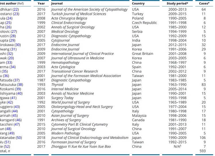
The main characteristics of the included articles were summarized in Table 1. The studies were published between 1987 and 2018 and reported cohorts ranging from 5 to 106 patients. Twenty-seven articles were single-center, three two-center, one three-center and one four-center. Seventeen studies were performed in Asia, eight in Europe and seven in North America. Overall, a number of 593 histologically proven PTL diagnosed in
Figure 2
Risk of bias and applicability concerns graph: review authors’ judgments about each domain presented as percentages across included studies.
Table 1 Characteristics of the 32 articles included in the meta-analysis.
First author (Ref) Year Journal Country Study period* Cases#
Adhikari (22) 2016 Journal of the American Society of Cytopathology USA 2000–2013 64
Bostanci (23) 2017 Turkish Journal of Medical Sciences Turkey 2009–2015 11
Bula (24) 2008 Acta Chirurgica Belgica Poland 1990–2005 8
Cap (25) 1999 Clinical Endocrinology Czech Republic 1991–1998 6
Cha (26) 2002 Annals of Surgical Oncology USA 1985–2000 12
Colovic (27) 2007 Medical Oncology Serbia 1994–1999 5
Dustin (28) 2012 Diagnostic Cytopathology USA 1992–2009 15
Gupta (29) 2005 CytoJournal India 1998–2004 10
Hirokawa (30) 2017 Endocrine Journal Japan 2012–2015 32
Hwang (31) 2009 Endocrine Journal Korea 1991–2006 29
Joshi (32) 2009 International Journal of Clinical Practice Great Britain 2001–2008 9
Kwak (20) 2007 Journal of Ultrasound in Medicine Korea 2003–2005 6
Lam (33) 1999 Hematopathology China 1968–1997 9
Lerma (34) 2003 Acta Cytologica Spain 1992–2001 6
Li (35) 2017 Transational Cancer Research China 2002–2012 9
Lu (36) 2001 Journal of the Formosan Medical Association Taiwan 1981–2000 11
Matsuda (37) 1987 Diagnostic Cytopathology Japan 1983–1985 5
Matsuzuka (38) 1993 Thyroid Japan 1963–1990 83
Mizokami (39) 2016 Internal Medicine Japan 2005–2014 9
Nishiyama (40) 2003 Annals of Nuclear Medicine Japan 1990–2001 15
Ogawa (41) 2001 Surgery Today Japan 1993–1998 5
Pyke (42) 1992 World Journal of Surgery USA 1965–1989 20
Ruggiero (43) 2005 Otolaryngology-Head and Neck Surgery USA 1977–2004 15
Sangalli (44) 2001 Cytopathology Italy 1980–1998 17
Sarinah (45) 2010 Asian Journal of Surgery Malaysia 1998–2006 15
Skarsgard (46) 1991 Archives of Surgery Canada 1981–1990 18
Stacchini (47) 2015 Cytometry Part B: Clinical Cytometry Italy 2001–2013 11
Sun (48) 2010 Journal of Surgical Oncology China 1991–2007 11
Wang (49) 2005 Modern Pathology USA 1990–2005 5
Watanabe (50) 2018 Journal of Clinical Endocrinology and Metabolism Japan 1990–2009 106
Wu (51) 2016 Formosan Journal of Surgery Taiwan 1992–2015 9
Xie (52) 2017 Zhongguo Yi Xue Ke Xue Yuan Xue Bao China N/A§ 7
Total, n 593
European Journal of Endocrinology
the period from 1963 to 2015 and submitted to FNA were described. Other examinations were described alongside to FNA cytology. Among cytological ancillary testing, flow cytometry was the most widely used (22, 26, 30, 43, 47); other techniques included PCR, immunocytochemistry and G-banding chromosome examination (22, 29, 44, 46, 47). In other studies, CNB/tru-cut or surgical biopsy were reported (20, 23, 26, 31, 32, 35, 36, 38, 39, 42, 43, 44, 45, 46, 47, 48, 50, 51); in one study, a lymph node biopsy was performed (51). Among histological ancillary testing, immunohistochemistry was the most widely used (26, 30, 33, 34, 35, 49, 51); only a few reports included immunoglobulin heavy-chain gene analysis and flow cytometry studies (30, 43).
Quantitative analysis
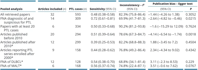
In order to calculate the pooled sensitivity of FNA in diagnosing PTL, we initially included all 32 papers (20, 22, 23, 24, 25, 26, 27, 28, 29, 30, 31, 32, 33, 34, 35, 36, 37, 38, 39, 40, 41, 42, 43, 44, 45, 46, 47, 48, 49, 50, 51, 52), leading to a sensitivity of FNA of 0.48 with high heterogeneity and no publication bias. In the attempt to reduce the heterogeneity, we then calculated FNA sensitivity in some subgroups. There were 14 articles describing the number of FNA reports as diagnostic of and suspicious for PTL separately (22, 23, 28, 32, 34, 35, 41, 42, 43, 44, 47, 48, 50, 51, 52); when we considered these two FNA reports as a whole, a sensitivity of 0.72 was found, with high heterogeneity and publication bias. Also, by analyzing FNA sensitivity combining only data from the six papers with at least 20 PTL cases (22, 30, 31, 38, 42, 50), we found a sensitivity of 0.50 with significant heterogeneity but no publication bias. Then, we evaluated the impact of timing of publication. Firstly, in the 12 articles published more recently (i.e., we arbitrary selected papers published after 2010) (22, 23, 28, 30, 35, 39, 45, 47, 48, 50, 51, 52), we found a sensitivity of 0.39 with heterogeneity and no publication bias (Fig. 3). Secondly, in the nine papers reporting PTL series enrolled after 2000 (20, 22, 23, 30, 32, 35, 39, 47, 52), the pooled FNA sensitivity was 0.44 with heterogeneity, but there was no publication bias (Fig. 4). Lastly, we analyzed the pooled FNA sensitivity in the specific histologic PTL types. The most prevalent PTL variants in the 32 retrieved articles were DLBCL and MALT. The pooled FNA sensitivity in DLBCL from 12 articles (20, 22, 28, 30, 31, 34, 36, 43, 44, 47, 48, 51) was 0.54 with mild heterogeneity and no publication bias (Fig. 5). The pooled FNA sensitivity in MALT from six articles (30, 31, 39, 44, 47, 50) was 0.56
with mild heterogeneity (Fig. 6). All the above results are detailed in Table 2 (view also Supplementary data).
Discussion
The aim of the current study was to evaluate the available evidence on the reliability of FNA cytology in diagnosing PTL. Although PTLs include some of the most aggressive thyroid malignancies, they are very rare; consequently, evidence on the reliability of diagnostic tools is limited.
Figure 3
Forest plot of detection rate (i.e. diagnostic sensitivity) of FNAC in PTL (random effect), including 95% confidence intervals, in papers published since 2010.
Figure 4
Forest plot of detection rate (i.e. diagnostic sensitivity) of FNAC in PTL (random effect), including 95% confidence intervals, in articles reporting series of enrolled after 2000.
European Journal of Endocrinology
Here, we focused on the FNA performance –this being the gold standard test to identify thyroid cancer. By applying a specific search strategy, we found 32 articles reporting PTL cases subjected to FNA and published over a very large period (1987–2018) by Asian, European and American authors. The most significant finding was that the FNA sensitivity in diagnosing PTL was quite low (48%) when we considered the overall series of papers and remained unchanged even after selecting various subgroups of articles or focusing on different histological PTL types. However, sensitivity increased to 72% when FNA reports ‘suspicious for’ and ‘diagnostic of’ PTL were considered together. From the clinical standpoint, this high
diagnostic sensitivity is a most interesting observation because the same diagnostic approach is indicated in both FNA instances; in fact, since FNA is inadequate for initial lymphoma diagnosis, a biopsy is recommended to provide adequate tissue for the precise identification/confirmation of the histological lymphoma subtype (53). While FNA is the pivotal diagnostic tool for the assessment of thyroid nodules, it is usually performed in a clinical setting that may lack specific expertise on lymphomas. Furthermore, PTL being a rare condition, its precise identification at FNA may depend on the specific expertise of the cytologist who often is a thyroid specialist. Nevertheless, FNA performance for the lymphoma diagnosis remains lower than that observed in differentiated thyroid carcinomas, in which FNA sensitivity is reported >85% (1, 54, 55, 56). The present data also indicate that the recognized accuracy of FNA in detecting thyroid cancers must be referred only to papillary carcinoma and not to other types of malignancies (1, 56, 57, 58). Surprisingly, when we analyzed the subgroup of articles published more recently or enrolling patients in the last two decades, we found even lower FNA sensitivity. We cannot explain these findings with certainty, but it could be speculated that PTL diagnosed in earlier decades (i.e. in 80s or 90s) could have been detected in a late or more advanced stage of the disease. This condition has to be taken into account as a potential factor of risk of bias. Due to its aggressive nature, the presence of DLBCL might have been clinically suspected leading to FNA performed as a diagnostic proof rather than question (or accompanied by a core biopsy) with blinded cytopathologists.
The cytological presentation of PTL is quite variable and may be sometimes challenging due to the variability of the cytological picture and presence of thyrocytes included in the sample. The cytological features of PTL are similar to those reported for lymph nodes: the main characteristic is the presence of monomorphism of the neoplastic population in contrast with the mixture of small and large lymphocytes that are present in varying proportions in reactive lymph nodes. While large cell lymphomas are readily recognized as malignant tumors, small cell lymphomas usually require immunophenotyping of the neoplastic population to be correctly identified. This approach requires the availability of a cell block for immunohistochemistry or a cytological preparation for flow cytometry (59). In this setting, the clinical history is a crucial point to achieve the correct diagnosis: if a lymphoproliferative disorder is suspected before FNA, appropriate ancillary studies can be planned and performed. From the therapeutic point of view,
Figure 5
Forest plot of detection rate (i.e. diagnostic sensitivity) of FNAC in DLBCL (random effect), including 95% confidence intervals.
Figure 6
Forest plot of detection rate (i.e. diagnostic sensitivity) of FNAC in MALT (random effect), including 95% confidence intervals.
European Journal of Endocrinology
non-Hodgkin lymphomas are distinct clinicopathological entities defined through a comprehensive evaluation of morphological, immunophenotypic and genetic features and clinical data, and their complete assessment is the base for the classification and the planning of specific treatments (60). Some authors found that flow cytometry applied to FNA might enhance the accuracy of cytological diagnosis in lymphoproliferative disorders and allow further subclassification in more than half of the cases (61). This implies that the remaining patients will need adjunctive analysis. This is also true for those patients who receive a cytological diagnosis of PTL (either suspicious or definitive) based on morphological assessment only. In such cases, FNA may serve as a screening test, which should be followed by an excisional (or core) biopsy for the appropriate histological assessment.
While ultrasound combined with FNA is pivotal in the initial assessment of thyroid nodule due to its high sensitivity and specificity to rule out primary and metastatic solid tumors, its accuracy drops significantly in the assessment of PTL. Indeed, PTL frequently presents itself as a large mass and not as a defined nodule. In addition, some of the ancillary ultrasonographic features typical for thyroid malignancy (such as microcalcifications, irregular margins, marked hypoechogenicity, taller-than-wide shape) are often absent (1). Alternative imaging tools may be taken into account. 18F-FDG PET-CT is
a functional imaging modality increasingly used for the staging and response assessment in patients with lymphomas and provides high accuracy in the detection of nodal and extranodal disease (53, 62). Nevertheless, in the PTL context also 18F-FDG PET-CT has important
limitations. In fact, inflammatory cells, in particular
leukocytes, show an increased glucose metabolism after antigenic activation. This phenomenon explains the high tracer uptake detected by PET in HT and may affect the specificity of the PET imaging (63, 64). In contrast with differentiated thyroid cancer, which usually depicts focal FDG uptake, a diffuse (either homogeneous or irregular) increase of thyroid uptake is the most characteristic PET feature in PTL (63, 65). However, this pattern cannot be considered pathognomonic since the same is frequently found in patients with HT or Graves’ disease (65, 66, 67) and, more rarely, in cases of metastatic cancer involvement of the thyroid (68, 69). On the other hand, focal uptake may be occasionally found also in patients with a secondary lymphomatous involvement of the thyroid gland (70). Additionally, HT is considered as the ‘etiologic background’ of PTL (9, 71), and in some cases, it is very difficult to discriminate between the two coexisting processes (72). Some authors demonstrated higher SUVmax
values in PTL compared to HT or Graves’ disease patients (73); nevertheless, a wide range of SUVmax values (7.4– 39.6) has been reported in cases of PTL (63, 74, 75, 76). The FDG uptake values of malignant lymphomas differ according to the histological subtypes and aggressive PTL such as DLBCL show higher values than indolent lymphomas (i.e. MALT) (77); this makes it difficult to use the SUVmax to discriminate between HT/Graves’ disease and PTL. Therefore, the PET imaging may only suggest or support the clinical suspicion of PLT but cannot be used as surrogate of the pathologic diagnosis. Some authors also reported that patients with PTL exhibit decreased CT density of the thyroid gland as compared to healthy subjects and patients with autoimmune thyroiditis (73, 78, 79). Nevertheless, different CT patterns have
Table 2 Results of the meta-analysis on the overall series of articles and in specific subgroups.
Pooled analysis Articles included (n) PTL cases (n) Sensitivity (95% CI) Inconsistency – I
2
(95% CI)
Publication bias – Egger test
(95% CI) P
All retrieved papers 32 593 0.48 (0.38–0.58) 82.3% (75.8–86.4) −1.44 (−4.26 to 1.38) 0.3052
FNA diagnostic of and
suspicious for PTL 14 309 0.72 (0.61–0.81) 69.9% (41.7–81.3) −2.64 (−4.82 to −0.46) 0.0215
Papers with at least 20
PTL cases 6 334 0.50 (0.33–0.68) 90.2% (81.2–93.8) −1.6 (−15.29 to 12.09) 0.7624
Articles published
before 2010 20 294 0.51 (0.39–0.64) 78.6% (67.3–84.7) −4.14 (−6.54 to −1.74) 0.0018
Articles published after
2010* 12 299 0.39 (0.25–0.53) 82.2% (68.8–88.3) 1.88 (−3.45 to 7.2) 0.4504
Articles reporting PTL series enrolled after 2000*
9 158 0.44 (0.28–0.62) 76.8% (49.2–86.4) 2.34 (−4.34 to 9.02) 0.4342
FNA of DLBCL* 12 128 0.54 (0.38–0.70) 68.8% (34.1–81.4) 3.11 (−2.3 to 8.53) 0.229
FNA of MALT* 6 168 0.56 (0.37–0.74) 74.8% (22.4–87.1) 3.51 (−0.6 to 7.62) 0.0767
Sensitivity ranged from 0 to 1.0. Absence of heterogeneity was set at I2 <50%. Egger test significantly identified the presence of publication bias.
European Journal of Endocrinology
been associated with PTL (solitary nodule surrounded by normal thyroid tissue, multiple nodules in the thyroid and both homogeneously enlarged thyroid lobes with a reduced attenuation, with or without peripheral thin hyperattenuating thyroid tissue), such that the use of the decreased attenuation as a diagnostic parameter is problematic in clinical practice (80, 81). Hence, biopsy confirmation is always needed for the diagnosis of PTL. CT and MRI may contribute to the local stage of the disease by defining the location and extension of the primary lesion and by assessing the possible invasion of the adjacent structures in areas that are poorly assessed by ultrasonography. Since MRI has a high contrast resolution for soft tissues, it may better detect pseudocapsules and define the uninvolved thyroid tissues (82, 83). Conversely,
18FDG PET-CT plays a pivotal role in the initial staging
of PTL, providing a reliable map of regional and distant disease dissemination (84). At the time of presentation, 56% of PTLs are Ann Arbor stage IE, 32% stage IIE, 13% stage III–IVE, with 5-year disease-specific survival of 86, 81, and 64%, respectively (8, 85). Stage and histological subtype are predictors of outcome and therefore also affect the therapeutic approach to PTLs. Patients with localized and indolent disease (e.g., MALT lymphoma stage IE) usually receive loco-regional treatment such as radiotherapy alone or surgery (86, 87, 88), while those with disseminated disease as well as those with aggressive histological subtypes, such as DLBCL, should be treated with chemotherapy and consolidation radiotherapy (89, 90, 91, 92). Changes of FDG uptake represent a sensitive and reliable tool for monitoring the response of the disease to treatment and for detecting early relapses with higher accuracy than CT imaging (68, 84, 93).
In view of the potential limitations of FNA and imaging tools in the detection of PTL, current guidelines suggest using other biopsy techniques; firstly, CNB and surgical excision (1). This approach is not really supported by evidence-based data, probably due to the rareness of PTL (14). However, the present study underlines FNA and ultrasound limits and indirectly corroborates the use of CNB and/or other imaging tools when PTL is clinically suspected. The results of the present meta-analysis should not be affected by these limitations. We feel that the very large number of included papers and the wide period during which they were published can reduce the publication bias. The results (and heterogeneity) were unchanged when we evaluated several subgroups of articles, also if arranged by time periods. Specifically, FNA sensitivity further decreased when we meta-analyzed the subgroup of recent studies; this can avoid the bias frequently present
in meta-analyses on studies reporting preliminary and recent results, because positive findings are more likely to be published. Therefore, we are confident to have excluded significant search and statistical weakness. On the other hand, we could not evaluate FNA sensitivity in some specific contexts, such as limited versus advanced stage or with ancillary cytological examinations available; data on these issues were not reported in the retrieved articles. As a main inclusion criterion, here we selected studies reporting the detection rate by FNA in histologically proven PTL. On one hand, this study design allowed us to have a strong standard of reference (i.e. histology) for FNA assessment. On the other hand, this approach could introduce another potential bias of our data. In fact, multidisciplinary teams from many centers may rely only on FNA results, and later stage and treat their patients without core or open biopsy. Then, these studies were not found in our search due to our major inclusion criterion (i.e. histologically proven PTL).
The present meta-analysis showed that FNA cytology has a low sensitivity in diagnosing PTL, ranging between 39 and 56% of cases in a large series of articles reporting on PTL subjected to this procedure. Moreover, this percentage increased to 72% when we included also FNA reports suspicious for PTL, and this can be a useful information for clinical practice. These data indirectly suggest that the high reliability recognized for ultrasound-guided FNA in thyroid malignancy should be specifically referred to papillary carcinomas. Furthermore, these results indirectly support the use of other imaging tools and the need of core or excisional biopsy when PTL is suspected.
Supplementary data
This is linked to the online version of the paper at https://doi.org/10.1530/ EJE-18-0672.
Declaration of interest
The authors declare that there is no conflict of interest that could be perceived as prejudicing the impartiality of this study.
Funding
This research did not receive any specific grant from any funding agency in the public, commercial or not-for-profit sector.
References
1 Haugen BR, Alexander EK, Bible KC, Doherty GM, Mandel SJ, Nikiforov YE, Pacini F, Randolph GW, Sawka AM, Schlumberger M et al. 2015 American Thyroid Association Management guidelines for adult patients with thyroid nodules and differentiated thyroid cancer: the American Thyroid Association guidelines task force on thyroid nodules and differentiated thyroid cancer. Thyroid 2016 26 1–133. (https://doi.org/10.1089/thy.2015.0020)
European Journal of Endocrinology
2 Stein SA & Wartofsky L. Primary thyroid lymphoma: a clinical review. Journal of Clinical Endocrinology and Metabolism 2013 98 3131–3138. (https://doi.org/10.1210/jc.2013-1428)
3 Pavlidis ET & Pavlidis TE. A review of primary thyroid lymphoma: molecular factors, diagnosis and management. Journal of Investigative Surgery 2017 Epub. (https://doi.org/10.1080/08941939.2017.1383536) 4 Graff-Baker A, Roman SA, Thomas DC, Udelsman R & Sosa JA.
Prognosis of primary thyroid lymphoma: demographic, clinical, and pathologic predictors of survival in 1408 cases. Surgery 2009 146 1105–1115. (https://doi.org/10.1016/j.surg.2009.09.020) 5 Thieblemont C, Mayer A, Dumontet C, Barbier Y,
Callet-Bauchu E, Felman P, Berger F, Ducottet X, Martin C et al. Primary thyroid lymphoma is a heterogeneous disease. Journal of Clinical Endocrinology and Metabolism 2002 87 105–111. (https://doi. org/10.1210/jcem.87.1.8156)
6 Pedersen RK & Pedersen NT. Primary non-Hodgkin’s lymphoma of the thyroid gland: a population based study. Histopathology 1996 28 25–32. (https://doi.org/10.1046/j.1365-2559.1996.268311.x) 7 Alzouebi M, Goepel JR, Horsman JM & Hancock BW. Primary thyroid
lymphoma: the 40 year experience of a UK lymphoma treatment centre. International Journal of Oncology 2012 40 2075–2080. (https:// doi.org/10.3892/ijo.2012.1387)
8 Onal C, Li YX, Miller RC, Poortmans P, Constantinou N, Weber DC, Atasoy BM, Igdem S, Ozsahin M & Ozyar E. Treatment results and prognostic factors in primary thyroid lymphoma patients: a rare cancer network study. Annals of Oncology 2011 22 156–164. (https:// doi.org/10.1093/annonc/mdq310)
9 Holm LE, Blomgren H & Lowhagen T. Cancer risks in patients with chronic lymphocytic thyroiditis. New England Journal of Medicine 1985 312 601–604. (https://doi.org/10.1056/NEJM198503073121001) 10 Isaacson PG. Lymphoma of the thyroid gland. Current Topics in
Pathology 1997 91 1–14.
11 Zucca E & Bertoni F. The spectrum of MALT lymphoma at different sites: biological and therapeutic relevance. Blood 2016 127 2082–2092. (https://doi.org/10.1182/blood-2015-12-624304) 12 Drugarin D, Negru S, Koreck A, Zosin I & Cristea C. The pattern of a
T(H)1 cytokine in autoimmune thyroiditis. Immunology Letters 2000 71 73–77. (https://doi.org/10.1016/S0165-2478(99)00156-X) 13 Nanba T, Watanabe M, Inoue N & Iwatani Y. Increases of the Th1/
Th2 cell ratio in severe Hashimoto’s disease and in the proportion of Th17 cells in intractable Graves’ disease. Thyroid 2009 19 495–501. (https://doi.org/10.1089/thy.2008.0423)
14 Hyjek E & Isaacson PG. Primary B cell lymphoma of the thyroid and its relationship to Hashimoto’s thyroiditis. Human Pathology 1988 19 1315–1326. (https://doi.org/10.1016/S0046-8177(88)80287-9) 15 Saxena A, Alport EC, Moshynska O, Kanthan R & Boctor MA. Clonal
B cell populations in a minority of patients with Hashimoto’s thyroiditis. Journal of Clinical Pathology 2004 57 1258–1263. (https:// doi.org/10.1136/jcp.2004.018416)
16 Cibas ES & Ali SZ. The 2017 Bethesda system for reporting thyroid cytopathology. Thyroid 2017 27 1341–1346. (https://doi.org/10.1089/ thy.2017.0500)
17 Trimboli P & Crescenzi A. Thyroid core needle biopsy: taking stock of the situation. Endocrine 2015 48 779–785. (https://doi.org/10.1007/ s12020-014-0382-z)
18 Buxey K & Serpell J. Importance of core biopsy in the diagnosis of thyroid lymphoma. ANZ Journal of Surgery 2012 82 90. (https://doi. org/10.1111/j.1445-2197.2011.05935.x)
19 Nam M, Shin JH, Han BK, Ko EY, Ko ES, Hahn SY, Chung JH & Oh YL. Thyroid lymphoma: correlation of radiologic and pathologic features. Journal of Ultrasound in Medicine 2012 31 589–594. (https:// doi.org/10.7863/jum.2012.31.4.589)
20 Kwak JY, Kim EK, Ko KH, Yang WI, Kim MJ, Son EJ, Oh KK & Kim KW. Primary thyroid lymphoma: role of ultrasound-guided needle biopsy. Journal of Ultrasound in Medicine 2007 26 1761–1765. (https://doi.org/10.7863/jum.2007.26.12.1761)
21 Whiting PF, Rutjes AW, Westwood ME, Mallett S, Deeks JJ, Reitsma JB, Leeflang MM, Sterne JA & Bossuyt PM. QUADAS-2: a revised tool for the quality assessment of diagnostic accuracy studies. Annals of Internal Medicine 2011 155 529–536. (https://doi.org/10.7326/0003-4819-155-8-201110180-00009)
22 Adhikari LJ, Reynolds JP & Wakely PE Jr. Multi-institutional study of fine-needle aspiration for thyroid lymphoma. Journal of the American Society of Cytopathology 2016 5 170–176. (https://doi.org/10.1016/j. jasc.2015.11.002)
23 Bostanci H, Dikmen K, Akyurek N, Buyukkasap AC, Yavuz A, Yalcin MM & Akin M. Eleven patients with primary thyroid lymphoma: a single center experience. Turkish Journal of Medical Sciences 2017 47 1322–1327. (https://doi.org/10.3906/sag-1611-91) 24 Bula G, Waler J, Niemiec A, Trompeta J, Steplewska K &
Gawrychowski J. Unusual malignant thyroid tumours – a clinical study of 20 cases. Acta Chirurgica Belgica 2008 108 702–707. (https:// doi.org/10.1080/00015458.2008.11680320)
25 Cap J, Ryska A, Rehorkova P, Hovorkova E, Kerekes Z & Pohnetalova D. Sensitivity and specificity of the fine needle aspiration biopsy of the thyroid: clinical point of view. Clinical Endocrinology 1999 51 509–515. (https://doi.org/10.1046/j.1365-2265.1999.00847.x)
26 Cha C, Chen H, Westra WH & Udelsman R. Primary thyroid lymphoma: can the diagnosis be made solely by fine-needle aspiration? Annals of Surgical Oncology 2002 9 298–302. (https://doi. org/10.1007/BF02573069)
27 Colovic M, Matic S, Kryeziu E, Tomin D, Colovic N & Atkinson HD. Outcomes of primary thyroid non-Hodgkin’s lymphoma: a series of nine consecutive cases. Medical Oncology 2007 24 203–208. (https:// doi.org/10.1007/BF02698041)
28 Dustin SM, Jo VY, Hanley KZ & Stelow EB. High sensitivity and positive predictive value of fine-needle aspiration for uncommon thyroid malignancies. Diagnostic Cytopathology 2012 40 416–421. (https://doi.org/10.1002/dc.21802)
29 Gupta N, Nijhawan R, Srinivasan R, Rajwanshi A, Dutta P,
Bhansaliy A & Sharma SC. Fine needle aspiration cytology of primary thyroid lymphoma: a report of ten cases. Cytojournal 2005 2 21. (https://doi.org/10.1186/1742-6413-2-21)
30 Hirokawa M, Kudo T, Ota H, Suzuki A, Kobayashi K & Miyauchi A. Preoperative diagnostic algorithm of primary thyroid lymphoma using ultrasound, aspiration cytology, and flow cytometry. Endocrine Journal 2017 64 859–865. (https://doi.org/10.1507/endocrj.EJ17-0111)
31 Hwang YC, Kim TY, Kim WB, Shong YK, Yi KH, Shong M, Jo YS, Kim WS & Chung JH. Clinical characteristics of primary thyroid lymphoma in Koreans. Endocrine Journal 2009 56 399–405. (https:// doi.org/10.1507/endocrj.K08E-355)
32 Joshi A, Chan J, Bruch G, Jeannon JP, Mikhaeel NG, Fields PA & Simo R. Thyroid lymphoma and airway obstruction – is there a rationale for surgical management? International Journal of Clinical Practice 2009 63 1647–1652. (https://doi.org/10.1111/j.1742-1241.2009.02050.x)
33 Lam KY, Lo CY, Kwong DL, Lee J & Srivastava G. Malignant lymphoma of the thyroid. A 30-year clinicopathologic experience and an evaluation of the presence of Epstein-Barr virus. American Journal of Clinical Pathology 1999 112 263–270. (https://doi. org/10.1093/ajcp/112.2.263)
34 Lerma E, Arguelles R, Rigla M, Otal C, Cubero JM, Bague S, Carreras AM, Eulalia E, Gonzalez-Campora R, Galera H et al. Comparative findings of lymphocytic thyroiditis and thyroid lymphoma. Acta Cytologica 2003 47 575–580. (https://doi. org/10.1159/000326571)
35 Li L, Wáng YXJ, Shi L, Yu C & Feng X-L. Primary thyroid lymphoma: CT findings of a rare malignant tumor with pathologic correlations. Translational Cancer Research 2017 6 578–587. (https://doi. org/10.21037/tcr.2017.03.84)
European Journal of Endocrinology
36 Lu JY, Lin CW, Chang TC & Chen YC. Diagnostic pitfalls of fine-needle aspiration cytology and prognostic impact of chemotherapy in thyroid lymphoma. Journal of the Formosan Medical Association 2001 100 519–525.
37 Matsuda M, Sone H, Koyama H & Ishiguro S. Fine-needle aspiration cytology of malignant lymphoma of the thyroid. Diagnostic
Cytopathology 1987 3 244–249. (https://doi.org/10.1002/dc.2840030314) 38 Matsuzuka F, Miyauchi A, Katayama S, Narabayashi I, Ikeda H,
Kuma K & Sugawara M. Clinical aspects of primary thyroid lymphoma: diagnosis and treatment based on our experience of 119 cases. Thyroid 1993 3 93–99. (https://doi.org/10.1089/thy.1993.3.93) 39 Mizokami T, Hamada K, Maruta T, Higashi K, Yamashita H,
Noguchi Y, Noguchi H & Tajiri J. Development of primary thyroid lymphoma during an ultrasonographic follow-up of Hashimoto’s thyroiditis: a report of 9 cases. Internal Medicine 2016 55 943–948. (https://doi.org/10.2169/internalmedicine.55.5428)
40 Nishiyama Y, Yamamoto Y, Yokoe K, Satoh K & Ohkawa M. Diagnosis of thyroid lymphoma and follow-up evaluation using Ga-67 scintigraphy. Annals of Nuclear Medicine 2003 17 351–357. (https:// doi.org/10.1007/BF03006600)
41 Ogawa Y, Kato Y, Ikeda K, Aya M, Ogisawa K, Kitani K, Onoda N, Ishikawa T, Haba T, Wakasa K et al. The value of ultrasound-guided fine-needle aspiration cytology for thyroid nodules: an assessment of its diagnostic potential and pitfalls. Surgery Today 2001 31 97–101. (https://doi.org/10.1007/s005950170190)
42 Pyke CM, Grant CS, Habermann TM, Kurtin PJ, van Heerden JA, Bergstralh EJ, Kunselman A & Hay ID. Non-Hodgkin’s lymphoma of the thyroid: is more than biopsy necessary? World Journal of Surgery 1992 16 604–609; discussion 609–610. (https://doi.org/10.1007/ BF02067333)
43 Ruggiero FP, Frauenhoffer E & Stack BC Jr. Thyroid lymphoma: a single institution’s experience. Otolaryngology-Head and Neck Surgery 2005 133 888–896. (https://doi.org/10.1016/j.otohns.2005.07.040) 44 Sangalli G, Serio G, Zampatti C, Lomuscio G & Colombo L. Fine
needle aspiration cytology of primary lymphoma of the thyroid: a report of 17 cases. Cytopathology 2001 12 257–263. (https://doi. org/10.1046/j.1365-2303.2001.00338.x)
45 Sarinah B & Hisham AN. Primary lymphoma of the thyroid: diagnostic and therapeutic considerations. Asian Journal of Surgery 2010 33 20–24. (https://doi.org/10.1016/S1015-9584(10)60004-8) 46 Skarsgard ED, Connors JM & Robins RE. A current analysis of
primary lymphoma of the thyroid. Archives of Surgery 1991 126 1199–1203; discussion 1203–1194. (https://doi.org/10.1001/ archsurg.1991.01410340037006)
47 Stacchini A, Pacchioni D, Demurtas A, Aliberti S, Cassenti A, Isolato G, Gazzera C, Veltri A, Sapino A, Papotti M et al. Utilility of flow cytometry as ancillary study to improve the cytologic diagnosis of thyroid lymphomas. Cytometry Part B: Clinical Cytometry 2015 88 320–329. (https://doi.org/10.1002/cyto.b.21204)
48 Sun TQ, Zhu XL, Wang ZY, Wang CF, Zhou XY, Ji QH & Wu Y. Characteristics and prognosis of primary thyroid non-Hodgkin’s lymphoma in Chinese patients. Journal of Surgical Oncology 2010 101 545–550. (https://doi.org/10.1002/jso.21543)
49 Wang SA, Rahemtullah A, Faquin WC, Roepke J, Harris NL & Hasserjian RP. Hodgkin’s lymphoma of the thyroid: a clinicopathologic study of five cases and review of the literature. Modern Pathology 2005 18 1577–1584. (https://doi.org/10.1038/ modpathol.3800501)
50 Watanabe N, Narimatsu H, Noh JY, Iwaku K, Kunii Y, Suzuki N, Ohye H, Suzuki M, Matsumoto M, Yoshihara A et al. Long-term outcomes of 107 cases of primary thyroid mucosa-associated lymphoid tissue lymphoma at a Single Medical Institution in Japan. Journal of Clinical Endocrinology and Metabolism 2018 103 732–739. (https://doi.org/10.1210/jc.2017-01478)
51 Wu S-Y, Chu C-H, Duh Q-Y, Hsieh C-B, Yu J-C & Shih M-L. Management for primary thyroid lymphoma: experience from a
single tertiary care centre in Taiwan. Formosan Journal of Surgery 2016 49 201–207. (https://doi.org/10.1016/j.fjs.2016.07.001)
52 Xie Y, Liu W, Liu Y, Wang W, Wang M, Liu H, Li X & Gao W. Diagnosis and clinical analysis of primary thyroid lymphoma. Zhongguo Yi Xue Ke Xue Yuan Xue Bao 2017 39 377–382.
53 Cheson BD, Fisher RI, Barrington SF, Cavalli F, Schwartz LH, Zucca E & Lister TA. Recommendations for initial evaluation, staging, and response assessment of Hodgkin and non-Hodgkin lymphoma: the Lugano classification. Journal of Clinical Oncology 2014 32 3059–3068. (https://doi.org/10.1200/JCO.2013.54.8800)
54 Chung SR, Suh CH, Baek JH, Choi YJ & Lee JH. The role of core needle biopsy in the diagnosis of initially detected thyroid nodules: a systematic review and meta-analysis. European Radiology 2018 28 4909–4918. (https://doi.org/10.1007/s00330-018-5494-z) 55 Misiakos EP, Margari N, Meristoudis C, Machairas N, Schizas D,
Petropoulos K, Spathis A, Karakitsos P & Machairas A. Cytopathologic diagnosis of fine needle aspiration biopsies of thyroid nodules. World Journal of Clinical Cases 2016 4 38–48. (https://doi.org/10.12998/ wjcc.v4.i2.38)
56 Bongiovanni M, Trimboli P, Rossi ED, Fadda G, Nobile A & Giovanella L. DIAGNOSIS OF ENDOCRINE DISEASE: High-yield thyroid fine-needle aspiration cytology: an update focused on ancillary techniques improving its accuracy. European Journal of Endocrinology 2016 174 R53–R63. (https://doi.org/10.1530/EJE-15-0817)
57 Trimboli P, Guidobaldi L, Bongiovanni M, Crescenzi A, Alevizaki M & Giovanella L. Use of fine-needle aspirate calcitonin to detect medullary thyroid carcinoma: a systematic review. Diagnostic Cytopathology 2016 44 45–51. (https://doi.org/10.1002/dc.23375) 58 Trimboli P, Treglia G, Guidobaldi L, Romanelli F, Nigri G, Valabrega S,
Sadeghi R, Crescenzi A, Faquin WC, Bongiovanni M et al. Detection rate of FNA cytology in medullary thyroid carcinoma: a meta-analysis. Clinical Endocrinology 2015 82 280–285. (https://doi. org/10.1111/cen.12563)
59 Kalina T, Flores-Montero J, van der Velden VH, Martin-Ayuso M, Bottcher S, Ritgen M, Almeida J, Lhermitte L, Asnafi V, Mendonca A et al. EuroFlow standardization of flow cytometer instrument settings and immunophenotyping protocols. Leukemia 2012 26 1986–2010. (https://doi.org/10.1038/leu.2012.122)
60 Cozzolino I, Rocco M, Villani G & Picardi M. Lymph node fine-needle cytology of non-Hodgkin lymphoma: diagnosis and classification by flow cytometry. Acta Cytologica 2016 60 302–314. (https://doi.org/10.1159/000448389)
61 Zeppa P, Marino G, Troncone G, Fulciniti F, De Renzo A, Picardi M, Benincasa G, Rotoli B, Vetrani A & Palombini L. Fine-needle cytology and flow cytometry immunophenotyping and subclassification of non-Hodgkin lymphoma: a critical review of 307 cases with technical suggestions. Cancer 2004 102 55–65. (https://doi.org/10.1002/ cncr.11903)
62 Barrington SF, Mikhaeel NG, Kostakoglu L, Meignan M,
Hutchings M, Mueller SP, Schwartz LH, Zucca E, Fisher RI, Trotman J et al. Role of imaging in the staging and response assessment of lymphoma: consensus of the International Conference on Malignant Lymphomas Imaging Working Group. Journal of Clinical Oncology 2014 32 3048–3058. (https://doi.org/10.1200/JCO.2013.53.5229) 63 Basu S, Chryssikos T, Moghadam-Kia S, Zhuang H, Torigian DA
& Alavi A. Positron emission tomography as a diagnostic tool in infection: present role and future possibilities. Seminars in Nuclear Medicine 2009 39 36–51. (https://doi.org/10.1053/j. semnuclmed.2008.08.004)
64 Gotthardt M, Bleeker-Rovers CP, Boerman OC & Oyen WJ. Imaging of inflammation by PET, conventional scintigraphy, and other imaging techniques. Journal of Nuclear Medicine 2010 51 1937–1949. (https://doi.org/10.2967/jnumed.110.076232)
65 Karantanis D, Bogsrud TV, Wiseman GA, Mullan BP, Subramaniam RM, Nathan MA, Peller PJ, Bahn RS & Lowe VJ. Clinical significance of diffusely increased 18F-FDG uptake in the
European Journal of Endocrinology
thyroid gland. Journal of Nuclear Medicine 2007 48 896–901. (https:// doi.org/10.2967/jnumed.106.039024)
66 Yasuda S, Shohtsu A, Ide M, Takagi S, Takahashi W, Suzuki Y & Horiuchi M. Chronic thyroiditis: diffuse uptake of FDG at PET. Radiology 1998 207 775–778. (https://doi.org/10.1148/ radiology.207.3.9609903)
67 Chen W, Parsons M, Torigian DA, Zhuang H & Alavi A. Evaluation of thyroid FDG uptake incidentally identified on FDG-PET/CT imaging. Nuclear Medicine Communications 2009 30 240–244. (https://doi. org/10.1097/MNM.0b013e328324b431)
68 Win AZ & Aparici CM. Bilateral diffuse fluorodeoxyglucose uptake in thyroid gland diagnosed by fluorodeoxyglucose-positron emission tomography/computed tomography. World Journal of Nuclear Medicine 2014 13 138–141. (https://doi.org/10.4103/1450-1147.139147) 69 Are C, Hsu JF, Schoder H, Shah JP, Larson SM & Shaha AR. FDG-PET
detected thyroid incidentalomas: need for further investigation? Annals of Surgical Oncology 2007 14 239–247. (https://doi. org/10.1245/s10434-006-9181-y)
70 Lin M, Wong C, Lin P, Shon IH, Cuganesan R & Som S. The prevalence and clinical significance of (18) F-2-fluoro-2-deoxy-D-glucose (FDG) uptake in the thyroid gland on PET or PET-CT in patients with lymphoma. Hematological Oncology 2011 29 67–74. (https://doi.org/10.1002/hon.955)
71 Kato I, Tajima K, Suchi T, Aozasa K, Matsuzuka F, Kuma K & Tominaga S. Chronic thyroiditis as a risk factor of B-cell lymphoma in the thyroid gland. Japanese Journal of Cancer Research 1985 76 1085–1090.
72 Arabi M, Dvorak R, Smith LB, Kujawski L & Gross MD. Fluorodeoxyglucose positron emission tomography in primary thyroid lymphoma with coexisting lymphocytic thyroiditis. Thyroid 2011 21 1153–1156. (https://doi.org/10.1089/thy.2011.0064) 73 Nakadate M, Yoshida K, Ishii A, Koizumi M, Tochigi N, Suzuki Y,
Ryu Y, Nakagawa T, Umehara I & Shibuya H. Is 18F-FDG PET/CT useful for distinguishing between primary thyroid lymphoma and chronic thyroiditis? Clinical Nuclear Medicine 2013 38 709–714. (https://doi.org/10.1097/RLU.0b013e31829b2686)
74 Lee CJ, Hsu CH, Tai CJ & Lin SE. FDG-PET for a thyroid MALT lymphoma. Acta Oncologica 2008 47 1165–1167. (https://doi. org/10.1080/02841860701732923)
75 Chander S, Zingas AP, Bloom DA, Zak IT, Joyrich RN & Getzen TM. Positron emission tomography in primary thyroid lymphoma. Clinical Nuclear Medicine 2004 29 572–573. (https://doi. org/10.1097/01.rlu.0000134987.48416.05)
76 Mehta A & Muthukrishnan A. Stage IE non-Hodgkin’s thyroid lymphoma on (18)F-FDG-PET/CT. Hellenic Journal of Nuclear Medicine 2011 14 186–187.
77 Schoder H, Noy A, Gonen M, Weng L, Green D, Erdi YE, Larson SM & Yeung HW. Intensity of 18fluorodeoxyglucose uptake in positron emission tomography distinguishes between indolent and aggressive non-Hodgkin’s lymphoma. Journal of Clinical Oncology 2005 23 4643–4651. (https://doi.org/10.1200/JCO.2005.12.072) 78 Kamijo K. Clinical studies on thyroid CT number in chronic
thyroiditis. Endocrine Journal 1994 41 19–23. (https://doi.org/10.1507/ endocrj.41.19)
79 Derringer GA, Thompson LD, Frommelt RA, Bijwaard KE, Heffess CS & Abbondanzo SL. Malignant lymphoma of the thyroid gland: a clinicopathologic study of 108 cases. American Journal of Surgical Pathology 2000 24 623–639. (https://doi.org/10.1097/00000478-200005000-00001)
80 Kim HC, Han MH, Kim KH, Jae HJ, Lee SH, Kim SS, Kim KH & Chang KH. Primary thyroid lymphoma: CT findings. European Journal
of Radiology 2003 46 233–239. (https://doi.org/10.1016/S0720-048X(02)00134-1)
81 Li XB & Ye ZX. Primary thyroid lymphoma: multi-slice computed tomography findings. Asian Pacific Journal of Cancer Prevention 2015 16 1135–1138. (https://doi.org/10.7314/APJCP.2015.16.3.1135) 82 Shibata T, Noma S, Nakano Y & Konishi J. Primary thyroid lymphoma:
MR appearance. Journal of Computer Assisted Tomography 1991 15 629–633. (https://doi.org/10.1097/00004728-199107000-00019) 83 Wang JH, Chen L & Ren K. Identification of primary thyroid
lymphoma with medical imaging: a case report and review of the literature. Oncology Letters 2014 8 2505–2508. (https://doi. org/10.3892/ol.2014.2542)
84 Basu S, Li G, Bural G & Alavi A. Fluorodeoxyglucose positron emission tomography (FDG-PET) and PET/computed tomography imaging characteristics of thyroid lymphoma and their potential clinical utility. Acta Radiologica 2009 50 201–204. (https://doi. org/10.1080/02841850802620689)
85 Freeman C, Berg JW & Cutler SJ. Occurrence and prognosis of extranodal lymphomas. Cancer 1972 29 252–260. (https://doi.org/10.1002/1097-0142(197201)29:1<252::AID-CNCR2820290138>3.0.CO;2-#)
86 Tsang RW, Gospodarowicz MK, Pintilie M, Wells W, Hodgson DC, Sun A, Crump M & Patterson BJ. Localized mucosa-associated lymphoid tissue lymphoma treated with radiation therapy has excellent clinical outcome. Journal of Clinical Oncology 2003 21 4157–4164. (https://doi.org/10.1200/JCO.2003.06.085) 87 Harrington KJ, Michalaki VJ, Vini L, Nutting CM, Syrigos KN,
A’Hern R & Harmer CL. Management of non-Hodgkin’s lymphoma of the thyroid: the Royal Marsden Hospital experience. British Journal of Radiology 2005 78 405–410. (https://doi.org/10.1259/ bjr/31803121)
88 Sakorafas GH. What’s the role of surgery in the management of primary thyroid lymphoma? Acta Oncologica 2011 50 319–320. (https://doi.org/10.3109/0284186X.2010.500301)
89 Watanabe N, Narimatsu H, Noh JY, Kunii Y, Mukasa K, Matsumoto M, Suzuki M, Sekiya K, Ohye H, Yoshihara A et al. Rituximab-including combined modality treatment for primary thyroid lymphoma: an effective regimen for elderly patients. Thyroid 2014 24 994–999. (https://doi.org/10.1089/ thy.2013.0523)
90 Karlin L & Coiffier B. Improving survival and preventing recurrence of diffuse large B-cell lymphoma in younger patients: current strategies and future directions. OncoTargets and Therapy 2013 6 289–296. (https://doi.org/10.2147/OTT.S42574)
91 Miller TP, Dahlberg S, Cassady JR, Adelstein DJ, Spier CM,
Grogan TM, LeBlanc M, Carlin S, Chase E & Fisher RI. Chemotherapy alone compared with chemotherapy plus radiotherapy for localized intermediate- and high-grade non-Hodgkin’s lymphoma. New England Journal of Medicine 1998 339 21–26. (https://doi.org/10.1056/ NEJM199807023390104)
92 Watanabe N, Noh JY, Narimatsu H, Takeuchi K, Yamaguchi T, Kameyama K, Kobayashi K, Kami M, Kubo A, Kunii Y et al. Clinicopathological features of 171 cases of primary thyroid lymphoma: a long-term study involving 24553 patients with Hashimoto’s disease. British Journal of Haematology 2011 153 236–243. (https://doi.org/10.1111/j.1365-2141.2011.08606.x) 93 Naswa N, Sharma P, Nazar AH, Mohapatra TK, Bal C & Kumar R. (18)
F-FDG PET/CT for initial assessment and response monitoring in a case of high grade primary lymphoma of the thyroid gland: a case report and review of literature. Indian Journal of Nuclear Medicine 2014 29 94–96. (https://doi.org/10.4103/0972-3919.130291)
Received 10 August 2018
Revised version received 16 November 2018 Accepted 17 December 2018