Human Periodontal Stem Cells Release Specialized
Proresolving Mediators and Carry
Immunomodulatory and Prohealing Properties
Regulated by Lipoxins
ELEONORACIANCI,a,b,cANTONIORECCHIUTI,cORIANATRUBIANI,b,cFRANCESCADIOMEDE,b,c MARCOMARCHISIO,a,bSEBASTIANOMISCIA,a,bROMAINA. COLAS,dJESMONDDALLI,d CHARLESN. SERHAN,dMARIOROMANOb,c
Key Words. Periodontal ligament stem cellsx Lipoxin A4x Immunomodulation x Lipid mediators x
Regeneration
ABSTRACT
Unresolved inflammation and tissue destruction are underlying mechanisms of periodontitis, which is
linked to dysregulated polymorphonuclear neutrophil (PMN) functions. Lipoxin A4(LXA4) is a
special-ized proresolving lipid mediator (SPM) that dampens excessive inflammation, promotes resolution, and protects from leukocyte-mediated tissue damage. Human periodontal ligament stem cells (hPDLSCs) represent key players during tissue regeneration and may contribute to resolution of in-flammation; thus, they may represent a promising tool in regenerative dentistry. In the present study, we investigated the actions of hPDLSCs on PMN apoptosis and antimicrobial functions, and
deter-mined the impact of LXA4on hPDLSCs. hPDLSCs significantly reduced apoptosis and stimulated
micro-bicidal activity of human PMNs, via both cell-cell interactions and paracrine mechanisms. Lipid mediator metabololipidomics analysis demonstrated that hPDLSCs biosynthesize SPMs, including
resolvin D1, D2, D5, and D6; protectin D1; maresins; and LXB4; as well as prostaglandins D2, E2,
and F2a. LXA4significantly enhanced proliferation, migration, and wound healing capacity of hPDLSCs
through the activation of its cognate receptor ALX/FPR2, expressed on hPDLSCs. Together, these re-sults demonstrate that hPDLSCs modulate PMN functions, and provide the first evidence that stem
cells generate SPM and that the LXA4-ALX/FPR2 axis regulates regenerative functions of hPDLSCs by a
novel receptor-mediated mechanism. STEMCELLSTRANSLATIONALMEDICINE2016;5:20–32
SIGNIFICANCE
These findings uncovered unappreciated features of stem cells from the periodontal ligament, sup-porting the notion that these cells may act as master regulators of pathophysiological events through the release of mediators that promote the resolution of inflammation and bacterial killing. The study also demonstrated that it is possible to modulate important functions of periodontal stem cells using
lipoxin A4, a potent endogenous stop signal of inflammation. Thus, this study revealed an
unappre-ciated anti-inflammatory proregenerative circuit that may be exploited to combat periodontal pathologies using resident stem cells. Moreover, the data may represent a more general template to explain the immunomodulatory functions of stem cells.
INTRODUCTION
Acute inflammation is a protective host response, which, if timely and self-limited, leads to elimina-tion of pathogens and to homeostasis restoraelimina-tion [1, 2]. In oral tissues, excessive or unresolved pathogen-induced inflammation causes tissue in-jury and periodontitis (PD) [3, 4]. Hence, while PD etiology is bacterial, a failure to resolve in-flammation is a pathophysiological determinant in PD progression [5]. PD is associated with
cardiovascular disease [6]; therefore, the need for efficacious treatment of periodontal inflam-mation is urgent.
Resolution of acute inflammation is an active and highly coordinated response, involving en-dogenous proresolving mediators, biosynthe-sized locally, that control leukocyte trafficking and restore homeostasis [7, 8]. Among them, specialized proresolving lipid mediators (SPMs), derived from polyunsaturated fatty acids (PUFAs), including lipoxins (LXs), resolvins (Rvs), protectins,
a
Department of Medicine and
Aging Science,bStemTeCh
Group, andcDepartment of
Medical, Oral and Biotechnological Sciences, “G. D’Annunzio” University of Chieti-Pescara, Chieti, Italy
d
Center for Experimental Therapeutics and
Reperfusion Injury, Harvard Institutes of Medicine, Brigham and Women’s Hospital and Harvard Medical School, Boston,
Massachusetts, USA Correspondence: Mario Romano, M.D., Department of Medical, Oral and Biotechnological Sciences, “G. D’Annunzio” University of Chieti-Pescara, via Luigi Polacchi, 13, 66100 Chieti, Italy. Telephone: 39 0871-541475; E-Mail: [email protected]
Received July 20, 2015; accepted for publication October 7, 2015; published Online First on November 25, 2015.
©AlphaMed Press 1066-5099/2015/$20.00/0
http://dx.doi.org/ 10.5966/sctm.2015-0163
T
ISSUE
-S
PECIFIC
P
ROGENITOR AND
S
TEM
C
ELLS
by guest on January 10, 2016
http://stemcellstm.alphamedpress.org/
and maresins, perform multipronged actions that improve the outcome of inflammation-related pathologies in experimental
models and clinical trials [8–11]. LXA4and its isomer 15-epi-LXA4
are biosynthesized from arachidonic acid (AA) by transcellular mechanisms during inflammation resolution [12, 13]. They control polymorphonuclear neutrophil (PMN) infiltration into inflamed tissues and attract monocytes/macrophages, enhancing efferocytosis and bacterial phagocytosis, hallmarks of resolution [14]. These actions are mediated by the activation of a specific G-protein-coupled receptor (GPCR) termed ALX/FPR2 [15, 16].
LXA4is found in the gingival crevicular fluid and in peripheral blood
of patients with PD [17, 18]. Moreover, SPMs combat excessive in-flammation and tissue destruction in experimental PD [4]. Along
these lines, LXA4, 15-epi-LXA4, and related synthetic stable analogs
limit PMN infiltration and activation triggered by microorganisms in vitro [17, 19] and in an animal model of ligature plus
infection-induced PD, improving the clinical outcome [20]. In addition, LXA4
significantly reduces PD-related bone loss [20], supporting the re-lationship between nonresolving inflammation and tissue destruc-tion in PD, and providing a radestruc-tional approach to stimulate wound healing and tissue regeneration. Nevertheless, it is unclear whether the protective actions of LXs in PD include, in addition to anti-PMN-driven periodontal damage, the stimulation of proregenerative ac-tivities of cells in the periodontium.
Periodontal ligament (PDL), the soft connective tissue in-volved in teeth stability, homoeostasis, and repair [21, 22], con-tains a niche of multipotent stem cells [23, 24] that retain tissue regenerative capacity in adulthood [21, 22]. Human peri-odontal ligament stem cells (hPDLSCs) possess self-renewal and multilineage (osteo-, chondro-, odonto-, adipo-, and neurogenic) differentiation capability [24, 25]. Like bone marrow mesen-chymal stem cells (BM-MSCs), hPDLSCs express mesenmesen-chymal antigens as well as cementoblastic/osteoblastic markers [23]. Because of these properties, hPDLSCs have been used in preclinical studies to assess their periodontal healing capability [26, 27]. Along these lines, the potential therapeutic applications of MSCs in regenerative medicine have been broadened by their unique immunomodulatory properties [28, 29] via cell-cell contact and/or paracrine mechanisms [30, 31]. Similarly to BM-MSCs, hPDLSCs, expanded ex vivo, possess paracrine immunosuppres-sive capability by inhibiting peripheral blood mononuclear cell (PBMC) proliferation [32]. Whether they also form and release proresolution mediators remains to be determined.
In the present study, we provide evidence that hPDLSCs reg-ulate PMN functions related to inflammation resolution and bac-terial killing, and demonstrate that they biosynthesize SPMs.
Moreover, we show that LXA4 stimulates receptor-dependent
functions of hPDLSCs relevant to tissue healing. Together, these results contribute to the elucidation of novel pathways that can be exploited for stem cell-based treatment of PD.
MATERIALS ANDMETHODS Ethics Statement
The protocol and informed consent to use human periodontal lig-ament biopsy specimens were approved by the medical ethics
committee at the Medical School,“G. D’Annunzio” University
of Chieti-Pescara, Italy (no. 266/17.04.14). PDL biopsy specimens
were obtained from healthy subjects (ages 20–35 years) without
systemic and oral diseases who signed an informed consent form before specimen collection.
hPDLSC Isolation, Culture, and Characterization
hPDLSCs were isolated according to Trubiani et al. [33]. PDL tis-sues were incubated with MSC basal medium supplemented with a specific bullet kit (Lonza Group, Walkersville, MD, https://www.
lonza.com) at 37°C in 5% carbon dioxide (CO2) for 20 days, during
which cells spontaneously migrated from the explants. Stemness surface molecules and pluripotency-associated markers were evaluated, as previously described [34]. The hPDLSC pheno-type was determined by flow cytometry using the following antibodies: fluorescein isothiocyanate (FITC)-conjugated CD13, CD14/FITC, phycoerythrin (PE)-conjugated CD29, CD31/FITC, peridinin chlorophyll protein (PerCP)-cyanine (Cy) 5.5-conjugated CD34, CD44/FITC, CD45/FITC, CD73/PE, CD90/FITC, CD105/PE, CD106/FITC, allophycocyanin (APC)-conjugated CD117, CD133 /PE, CD144/FITC, CD146/PE, CD166/FITC, CD326/PerCP-Cy5.5, Alexa488-conjugated human leukocyte antigen (HLA)-ABC, HLA DR/PE (BD Bioscience, Franklin Lakes, NJ, https://www.bdbiosciences.com). Pluripotent embryonic markers NANOG/PE, octamer-binding transcription factor 4 (Oct4)/PE, stage-specific embryonic antigen (SSEA4)/FITC, and (sex determining region Y)-box 2 (Sox2)/FITC were purchased from BD Bioscience. Cells were ana-lyzed with a FACSCalibur flow cytometer (BD Bioscience), using the Cellquest (BD Bioscience) and FlowJo (Ashland, OR, http:// www.flowjo.com) software. Unstained cells were the negative control.
Colony-Forming Unit-Fibroblast Assay
To assess their colony-forming unit (CFU) capability, hPDLSCs were
seeded at 13 103cells per 10 ml onto 100-mm culture dishes in
growth medium (Lonza Group) kept at 37°C in 5% CO2. After 14
days, cells were fixed with cold methanol, stained with 0.5% crystal violet, washed with distilled water, and dried. Aggregates of 50 or more cells were scored as 1 colony forming unit-fibroblast. Osteogenic, Chondrogenic, and
Adipogenic Differentiation
The formation of a mineralized bone matrix was monitored for 21 days using human MSC (hMSC) osteogenic differentiation me-dium (Lonza Group), as previously described [35]. Osteogenic dif-ferentiation was determined by staining mineralized deposits with Alizarin Red S (Sigma-Aldrich, Milan, Italy, https://www. sigmaaldrich.com) and by assessing the expression of osteogenic markers (i.e., alkaline phosphatase [ALP], runt-related transcrip-tion factor 2 [RUNX2], and osteopontin [OPN]) by real-time poly-merase chain reaction (PCR).
To drive hPDLSC chondrogenic differentiation, cells were grown in three-dimensional (3D) cultures incubated for 21 days with chondro-inductive medium (CIM) made of high-glucose
Dulbecco’s modified Eagle’s medium (DMEM) containing 1 mM
dexamethasone, 1mM ascorbate-2-phosphate (Sigma-Aldrich), 1%
sodium pyruvate (Sigma-Aldrich), 10% insulin-transferrin-selenium 1003 concentration (Thermo Fisher Scientific, Waltham, MA, https://www.thermofisher.com), and 10 ng/ml transforming
growth factor (TGF)-b3(PeproTech, London, U.K., https://www.
peprotech.com). After 3 weeks, cells were fixed, cut into 3-mm sections and stained with 1% (wt/vol) Alcian blue 8GX (Sigma-Aldrich) for histological examination. In parallel, hPDLSC
mono-layers were cultured in 6-well plates for 14–21 days with CIM to
assess chondrogenic-related gene expression (see below) and chondrogenic matrix deposition using Alcian blue. Adipogenesis
by guest on January 10, 2016
http://stemcellstm.alphamedpress.org/
was induced using the hMSC adipogenic differentiation medium (Lonza Group) for 4 weeks. Lipid-drop accumulation and triglycer-ide content were respectively evaluated by Red Oil staining and the AdipoRed assay (Lonza Group) [35]. All experiments were
car-ried out with hPDLSCs at passages 3–7.
PMN Isolation and Coculture With hPDLSCs
PMNs were isolated from healthy volunteers (25–40 years old),
who denied taking medication at least for 5 days, using dextran-histopaque sedimentation [37]. Isolated PMNs were, on average, 93% viable, as determined by flow cytometric analysis of annexin V (annV) and propidium iodide (PI) staining (annexin V-FITC Ap-optosis Detection Kit; eBioscience, San Diego, CA, https://www. ebioscience.com). For coculture experiments, PMNs were incubated with hPDLSCs at different ratios (1:1, 5:1, 10:1) for 18 hours at 37°C
and 5% CO2. In some experiments, PMNs were cultured for 18 hours
with hPDLSC-conditioned medium (CdM). In others, PMNs were ex-posed to human interleukin (IL)-8 (100 ng/ml) (PeproTech) for 30 minutes at room temperature (RT) before coculture with hPDLSCs. PMNs maintained in complete medium were used as controls.
To obtain CdM, hPDLSCs (passages 3–6) were seeded in 6-well
plates at 33 105cells per well in 2 ml of complete DMEM. Cells
were cultured for 24, 48, and 72 hours at 37°C and 5% CO2. At each
time point, CdM was collected, centrifuged to remove cellular
de-bris, and snap frozen at280°C until use.
PMN Apoptosis
Apoptosis of PMNs with or without hPDLSCs or hPDLSC-CdM was evaluated by flow cytometry using annexinV and PI (eBioscience)
double staining. Approximately 104cellular events were acquired
for each sample. Bacterial Killing
The reference laboratory Pseudomonas aeruginosa strain PAO1 (ATCC 15692, ATCC, Manassas, VA, https://www.atcc.org) was grown (at 37°C overnight) on Muller-Hinton (MH) agar plates. At 24 hours before experiments were conducted, a single colony was picked up from the agar plate, inoculated in 5 ml of MH broth, and grown overnight under continuous agitation (300 rpm, 37°C). The following day, confluent PA01 was diluted (1:10) with fresh
MH medium and grown for ∼2 hours in a shaking incubator
(300 rpm, 37°C) to midlog phase (optical density of 550 nm
[OD550] = 0.456 0.05), corresponding to ∼2 3 108CFU/ml).
Bac-teria (2 ml) were opsonized (30 minutes, 37°C) with 10% AB
hu-man serum (Sigma-Aldrich), adjusted to an OD550of∼0.45 with
Dulbecco’s phosphate buffered saline (PBS), and further diluted
(1:5) with Roswell Park Memorial Institute (RPMI) medium con-taining 20% AB human serum. PMNs cocultured with hPDLSCs (10:1) for 18 hours were counted, washed twice with PBS, and
sus-pended in antibiotic-free RPMI at a density of 106cells per
milli-liter. Opsonized PA01 suspension (∼3 3 107
CFU, 500ml) was
added to 500ml of human PMNs (∼6 3 105) and the mixtures
were maintained in constant motion on a rotating wheel at 37°C. PA01 kept alone served as a control of maximal bacterial growth. After 90 minutes, aliquots of each mixture were re-moved, serially diluted with sterile PBS, and plated on MH agar to enumerate the viable CFUs. The PMN killing index was
calcu-lated as follows: [(CFU in the absence of PMN2 CFU in the
pres-ence of PMN)/CFU in the abspres-ence of PMN3 100].
Multiplex Cytokines/Chemokines Analysis
The concentration of epidermal growth factor (EGF), 10, 6, IL-8, and vascular endothelial growth factor (VEGF) in 24-hour cul-tured hPDLSC-CdM was determined using a Milliplex human cytokines/chemokines kit (EMD Millipore, Billerica, MA, https://
www.emdmillipore.com) according to the manufacturer’s
instruc-tions. Briefly, 25ml of medium was added to 25 ml of assay buffer,
followed by 25ml of antibody-coated magnetic beads incubated
for 2 hours at RT during shaking. Plates were washed twice with
buffer and incubated (1 hour, RT) with 25ml of biotinylated
anti-bodies followed by streptavidin-PE (30 minutes, RT). Plates were analyzed using a Luminex 100/200 platform (Luminex, Austin, TX, https://www.luminexcorp.com) equipped with the xPONENT 3.1 software. Standard curves for each analyte were generated using reference proteins. Analyte concentrations were determined by a five-parameter logistic curve. Analyses were carried out in dupli-cate and all incubation steps were performed in the dark. Lipid Mediator Metabololipidomics
hPDLSCs were cultured in 150-mm plates with 9 ml of phenol-red free medium until 80% confluence. After treatment, medium and cells were harvested, using a scraper in 15-ml tubes, snap frozen
in an acetone dry ice bath, and rapidly stored at280°C. Two
vol-umes of ice-cold methanol containing 500 pg of deuterium
(d)-labeled d4-LTB4, d5-LXA4, d5-RvD2, and d4-PGE2(Cayman
Chemical, Ann Arbor, MI, https://www.caymanchem.com) were then added and samples stored at 4°C for 45 minutes to allow for protein precipitation. Supernatants were collected after centrifu-gation (3,000 rpm, 10 minutes, 4°C) and brought to less that 10% of methanol content using nitrogen. All samples for liquid chromatography-tandem mass spectrometry (LC-MS-MS)
analy-sis were extracted using C18solid-phase extraction (SPE) columns
(Waters, Milford, MA, https://www.waters.com), equilibrated
with 6 ml of methanol and 12 ml of H2O. Sample supernatants
were acidified (pH∼3.5) and immediately loaded onto the SPE
column. Samples were washed with 12 ml of hexane, eluted with 12 ml of methyl formate, taken to dryness, and suspended in phase (1:1 methanol to water) for MS-MS analysis. The LC-MS-MS system (QTrap6500; ABSciex, Framingham, MA, https:// sciex.com) equipped with an Agilent HP1100 binary pump (Agilent Technologies, Santa Clara, CA, https://www.chem.agilent.com) and an automatic injector was setup and operated, as described by Colas et al. [10]. To monitor and quantify the levels of the various lipid mediators (LMs), a multiple reaction monitoring method was developed with signature ion fragments for each molecule, also as described by Colas et al. [10].
Analysis of ALX/FPR2 Expression
Flow Cytometry
Surface and intracellular expression of ALX/FPR2 was evaluated in hPDLSCs from different donors using a monoclonal anti-ALX/FPR2 primary antibody (Aldevron, Fargo, ND, https://www.aldevron.
com). Briefly, 53 105cells per sample were fixed with 4%
parafor-maldehyde (PFA), washed, and incubated with buffer containing pri-mary antibody for 60 minutes at 4°C. After removing the excess of primary antibody, cells were incubated with an anti-mouse FITC-conjugated secondary antibody. For intracellular staining, cells were permeabilized with FACS lysing and Perm2 solutions (BD Bioscience) before staining. Secondary antibody-matched controls were used to assess unspecific fluorescence.
by guest on January 10, 2016
http://stemcellstm.alphamedpress.org/
Immunofluorescence and Confocal Microscopy
hPDLSCs grown on glass coverslips were fixed with 4% PFA, washed, and processed for ALX/FPR2 immunofluorescence. Cells were permeabilized with Triton X-100, neutralized with 5% skimmed milk in PBS for 30 minutes. Incubation with primary mouse anti-ALX/FPR2 antibody (Aldevron) (1:100) was followed by exposure to Alexa Fluor 488 green fluorescence conjugated goat anti-mouse (Molecular Probes; Thermo Fisher Scientific, Eugene, OR, https://www.thermofisher.com). For actin cytoskeleton and nuclei counter-staining, cells were incubated with AlexaFluor 594 conjugated phalloidin (Thermo Fisher Scientific), and TO-PRO (Thermo Fisher Scientific). Samples were analyzed using a Zeiss LSM510 META confocal system (Zeiss, Jena, Germany, www.zeiss. com) connected to an inverted Zeiss Axiovert 200 microscope equip-ped with a Plan Neofluor oil-immersion objective (403/1.3 NA). Images were collected using an argon laser beam with excita-tion lines at 488 nm and a helium-neon source (543 nm and 665 nm).
RNA Isolation and Quantitative Real-Time PCR
hPDLSC total RNA was isolated with the RNeasy Plus Universal Mini Kit (Qiagen, Valencia, CA, https://www.qiagen.com) accord-ing to the manufacturer’s instructions. Quantitative PCR for oste-ogenic markers and ALX/FPR2 was performed with an ABI PRISM 7900 HT Sequence Detection System (Applied Biosystems, Foster City, CA, https://www.appliedbiosystems.com). TaqMan gene ex-pression assays (ALP, Hs01029144_m1, RUNX2, Hs00231692_m1, OPN, Hs00959010_m1, fatty acid binding protein 4 [FABP4],
Hs01086177_m1, peroxisome proliferator-activated receptorg
[PPARg], Hs01115513_m1, and ALX/FPR2 Hs00265954_m1 from Applied Biosystems; and ACAN, Hs.PT.56a.742783 and COMP, Hs. PT.58.2694031 from Integrated DNA Technologies, San Jose, CA, https://www.idtdna.com) were used according to standard pro-tocols. Beta-2 microglobulin (B2M, Hs99999907_m1; Applied Bio-systems) was used for template normalization. Duplicates were set up for each sample and the mRNA expression was analyzed
by the comparative 2-ΔΔCtrelative quantification method [38].
Proliferation Assay
hPDLSCs (23 103cells per well) were seeded in 96-well
micro-plates with complete DMEM overnight at 37°C and 5% CO2. The
following day, cells were starved overnight with DMEM plus 1% fetal bovine serum (FBS), gently washed, and exposed to 0.001
to 1.0 nM LXA4 (Calbiochem, San Diego, CA, https://www.
merckmillipore.com/) or vehicle (0.01% ethanol maximum) diluted with 1% FBS-containing DMEM. Cells were enumerated after 24, 48, and 72 hours using trypan blue exclusion and data were expressed as proliferation index calculated as ratio between
LXA4-and vehicle-treated hPDLSCs. In some experiments, hPDLSCs
were exposed to the ALX/FPR2 antagonist WRW4
(Calbio-chem) (10mM, 30 minutes) before LXA4, and the cellular
prolifer-ation index was calculated as ratio between LXA4- and WRW4-LXA4
treated cells. Each condition was tested at least in triplicate and ex-periments were repeated three times using hPDLSCs from different donors.
In Vitro Wound Healing Assay
hPDLSCs (53 104cells per well) were grown in 6-well plates in
complete medium until 80% to 90% confluence and then starved
overnight with DMEM plus 1% FBS. Wounds (∼580 mm wide) were created by scraping the monolayers with a 200-ml pipette tip. Cell debris was removed by vigorous rinsing with PBS to avoid random reattachment. Thereafter, serum-free medium
contain-ing increascontain-ing concentrations of LXA4(0.01 to 1 nM) or vehicle
(0.01% ethanol, maximum) were added to each well. hPDLSCs maintained in DMEM plus 10% FBS served as a control of maximal healing capacity. Wounds were monitored at 0, 3, 6, 24, and 48 hours using an optical microscope equipped with a digital camera (Canon, Tokyo, Japan, https://www.canon.com). Each well was photographed at three locations along the original wound boundary and wound width was quantified in three fields using ImageJ software (U.S. National Institutes of Health, Bethesda, MD, https://imagej.nih.gov/ij). To examine the involvement of
the ALX/FPR2 receptor in LXA4-regulated migration, wounded
cells were exposed to the ALX/FPR2 antagonist WRW4 (10mM,
30 minutes) before LXA4, and wound closure was monitored at
0, 3, 6, 24, and 48 hours. Duplicates for each condition were examined.
Statistical Analysis
Data were expressed as mean 6 SEM. Differences between
groups were determined using the Student’s t test. Differences
were considered significant when p, .05.
RESULTS
Isolation and Characterization of hPDLSCs
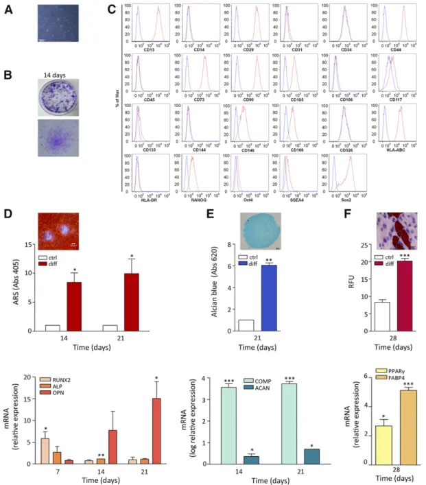
hPDLSCs from PDL biopsy specimens consisted of cells with a ho-mogeneous fibroblast-like morphology (Fig. 1A), reminiscent of other MSCs (e.g., bone marrow) [23] and capable of generating fibroblast colonies from single cells after 14 days of culture (Fig. 1B). hPDLSCs expressed specific MSC antigens, such as CD13, CD29, CD44, CD73, CD90, CD105, CD117, CD146, and CD166. Furthermore, these cells expressed the histocompatibility antigen HLA-ABC as well as markers of pluripotency (NANOG, Oct4, SSEA4, and Sox2). They stained negative for the hematopoi-etic markers CD14, CD31, CD34, CD45, and HLA-DR as well as for surface vascular endothelial-cadherin (CD144) and epithelial cell adhesion molecule (CD326) (Fig. 1C). Thus, hPDLSCs met the min-imal criteria for being classified as MSCs [39].
To evaluate hPDLSC differentiation, we induced osteogenic; chondrogenic, and adipogenic commitment. Under osteo-inductive conditions for 3 weeks, hPDLSCs produced mineral-ized extracellular matrix that was stained with Alizarin Red S and quantified (Fig. 1D, upper panel). Consistent with this, ALP and RUNX2 mRNA accumulated at early differentiation stages (7–14 days) to progressively decrease at 21 days when OPN was upregulated (Fig. 1D, lower panel).
After 21 days in chondrogenic conditions, the deposition of chondrogenic-like matrix was revealed by Alcian blue staining (Fig. 1E, upper panel), whereas real-time PCR showed a higher ex-pression of chondrogenic-related genes as aggrecan (ACAN) and cartilage oligomeric matrix protein (COMP) compared with
undif-ferentiated hPDLSCs (p, .01) (Fig. 1E, lower panel).
When hPDLSCs were subjected to adipogenic differentiation for 4 weeks, Oil Red O-positive lipid-droplet accumulation within cells was observed accompanied by an increase in
triglycer-ide content compared with control cells (p, .0001) (Fig. 1F,
upper panel). Consistent with this, hPDLSCs under adipogenic
by guest on January 10, 2016
http://stemcellstm.alphamedpress.org/
Figure 1. Human periodontal ligament stem cells (hPDLSCs) immunophenotype and differentiation. (A): Light microscopy image of passage-2 hPDLSCs. Original magnification:310. (B): Representative images of hPDLSC colony-forming units at 14 days. (C): Flow cytometric analysis of mesenchymal stem antigens in hPDLSCs. Blue histograms represent unstained cells; red histograms are stained for specific antibodies. Results are representative of five preparations. (D): Qualitative and quantitative analysis of hPDLSC osteogenic differentiation. Upper panel: bars rep-resent spectrophotometric quantification of extracted ARS in control and differentiated hPDLSCs after 14 and 21 days (n = 3;p, p , .05). ARS-positive calcified nodules are shown in the inset (original magnification:310). Lower panel: mRNA evaluated by real-time polymerase chain reaction of the osteorelated genes ALP, RUNX2, and OPN at 7, 14, and 21 days of culture (n = 3;p, p , .05; pp, p , .01). (E): Qualitative and quantitative analysis of hPDLSC chondrogenic differentiation. Upper panel: bars represent Alcian blue content in adherent hPDLSCs in normal or chondrogenic conditions at days 14 and 21 (n = 3;pp, p , .01). A representative image of Alcian blue staining of chondrogenic nodules obtained from hPDLSCs after 21 days of 3-dimensional culturing is shown (inset, original magnification:310). Lower panel: mRNA expression of the chondrogenic-related genes ACAN and COMP at days 14 and 21 (n = 3;p, p , .05; ppp, p , .001). (F): Qualitative and quantitative analysis of hPDLSC adipogenic differentiation. Upper panel: bars depict the triglyceride content in hPDLSCs after 4 weeks of normal or adipogenic cul-turing (n = 3;ppp, p , .001). A representative image is shown of Oil Red O-positive lipid vacuoles (H&E staining for nuclei and cytoplasm). Lower panel: mRNA expression of adipogenic-related genes, such as FABP4 and PPARg after 4 weeks of differentiation (mean 6 SEM of triplicates; p, p , .05; ppp, p , .001). Abbreviations: abs, absorbance; ARS, Alizarin Red S; ctrl, control; diff, differentiated cells; HLA, human leukocyte antigen; OCT, octamer-binding transcription factor; RFU, relative fluorescence unit; Sox2, (sex determining region Y)-box 2; SSEA, stage-specific embryonic antigen.
by guest on January 10, 2016
http://stemcellstm.alphamedpress.org/
culturing conditions expressed high levels of adipogenic genes as FABP4 and PPARg compared with control cells (Fig. 1F, lower panel).
hPDLSCs Inhibit Apoptosis and Enhance Bactericidal Activity of PMNs
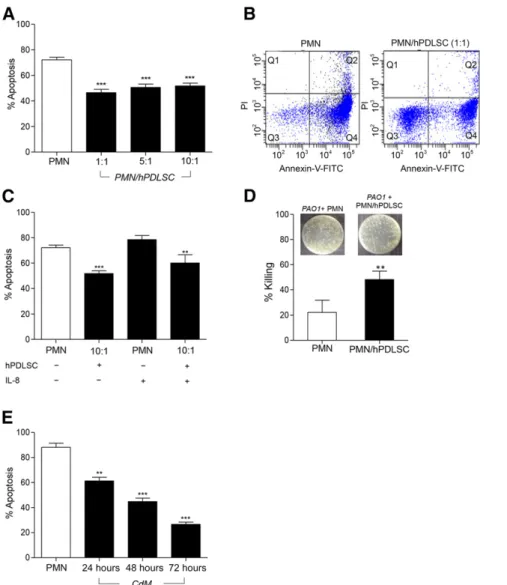
To evaluate the potential pathophysiological relevance of hPDLSCs in PD, we examined their impact on selected func-tions of PMNs, key regulators of the periodontal homeostasis [2, 40, 41]. To this end, PMNs were cocultured with hPDLSCs at different ratios for 18 hours and their survival and bactericidal activity were evaluated. hPDLSCs protected PMNs from
spon-taneous apoptosis (p, .0001), with the 1:1 ratio being most
efficient (∼36% inhibition) (Fig. 2A). Also, the percentage of
early apoptotic PMNs (annV+/PI2) was markedly reduced
(from approxiately∼90% to 50%; p = .0002) by hPDLSCs, while
the percentage of viable cells (annV2/PI2) increased (from
∼7% to 37%; p = .0007).
Since IL-8 drives PMN recruitment at inflammatory sites [40], we ran hPDLSC/PMN coincubations in the presence of IL-8 to mimic an inflammatory environment. Figure 2C shows that hPDLSCs also protected IL-8-activated PMNs from apoptosis, compared with PMNs treated with IL-8 alone.
To investigate whether hPDLSCs, in addition to sustaining PMN viability, maintained their bactericidal activity, we exposed PMNs from cocultures with hPDLSCs to the P. aeruginosa strain PAO1. PMNs derived from hPDLSC cocultures (1:10) showed higher
Figure 2. Effects of hPDLSCs on PMN apoptosis and bacterial killing. (A): PMNs were cultured with or without hPDLSCs for 18 hours. Apoptosis was evaluated by annexin V (annV)-FITC and PI staining. Results are given as the mean6 SEM of6 independent experiments (ppp,p , .0001). (B): Representative flow cytometry dot-plot of PMNs cultured with or without hPDLSCs (1:1) for 18 hours. Lower left quadrants: viable PMNs (annV2/PI2); lower right quadrants: early apoptotic cells (annV+); upper left quadrants: necrotic cells (PI+); upper right quadrants: late apoptotic PMNs (annV+/PI+). (C): Percentage of apoptotic IL-8-activated PMNs cultured with or without hPDLSCs (1:10). Results are given as the mean6 SEM of 4 separate ex-periments (pp, p = .04; ppp, p , .0001 ). Apoptosis is expressed as percentage of annV+cells. (D): PMNs cocultured with hPDLSCs for 18 hours were incubated with Pseudomonas aeruginosa strain PAO1 (1:50) and plated on agar. Insets show the PAO1 colony-forming units grown overnight. Results are given as the mean6SEMof3experiments(pp,p=.0197).(E):PMNswereincubatedwith24-,48-and72-hourCdMfromhPDLSCs.Thepercentage of apoptotic cells was evaluated by annV-FITC/PI staining. Results are given as the mean6 SEM of 4 independent experiments (pp, p = .0003; ppp, p,.0001).Abbreviations:CdM,conditionedmedium;FITC,fluoresceinisothiocyanate;hPDLSC,humanperiodontalligamentstemcell;IL,interleukin; PI, propidium iodide; PMN, polymorphonuclear neutrophil.
by guest on January 10, 2016
http://stemcellstm.alphamedpress.org/
bactericidal activity compared with PMNs kept alone (p = .0197) (Fig. 2D), as indicated by the lower CFU number in the presence
of PMNs isolated from cocultures (7.36 log106 0.16 vs. 7.53
log106 0.16 CFU/ml; p = .0197). Collectively, these results
pro-vide the first epro-vidence that hPDLSCs promote survival and anti-bacterial activities of PMNs.
To assess whether hPDLSCs antagonized PMN apoptosis by re-leasing survival factors, we incubated PMNs (18 hours, 37°C) with CdM collected from hPDLSCs cultured for 24, 48, and 72 hours. As shown in Figure 2E, hPDLSC-CdM from all 3 time points significantly
inhibited PMN apoptosis (p = .0003; p, .0001; p , .0001,
respec-tively). Notably, 72-hour hPDLSC-CdM exhibited greater protective
effect (26.616 1.77 annV+/PI2PMNs) compared with 48 hours
(44.79 6 2.71 annV+/PI2 PMNs) or 24 hours (61.33 6 2.85
annV+/PI2PMNs) CdM (Fig. 2E). Remarkably, the antiapoptotic
effect of 72-hour CdM was more potent than that observed
with hPDLSCs (46.506 2.64 annV+/PI2PMNs, 1:1 ratio) (Fig. 2A).
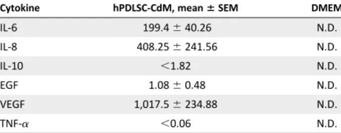
To gain insights into this finding, we carried out multiplex cyto-kine analysis of hPDLSC-CdM and consistently observed that hPDLSC supernatants contain IL-6 and IL-8, which are known to inhibit PMN death [41, 42], as well as IL-10, VEGF, and EGF (Table 1). These results indicate that hPDLSCs can regulate PMN functions in a paracrine manner, producing a protective milieu that can influence resolution of inflammation and bacterial clearance.
hPDLSCs Produce SPMs
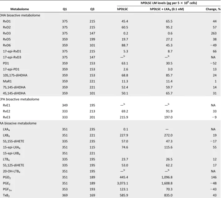
We asked whether SPM biosynthesis was a component of the im-munomodulatory properties of hPDLSCs, because SPMs promote inflammation resolution and are protective in experimental PD [4]. To address this point, we performed LC-MS-MS-based LM metabololipidomics. hPDLSCs generated PUFA-derived LM from both lipoxygenase (LOX) and cyclooxygenase (COX) pathways (Fig. 3A, 3B; Table 2). In these incubations, we identified media-tors from the docosahexaenoic acid (DHA)-derived bioac-tive metabolome including the D-series resolvins (RvD1, RvD2, RvD5, and RvD6), protectin D1 (PD1), and maresins (MaR1 and 7S,14S-diHDHA). These mediators were identified using char-acteristic retention times and MS-MS fragmentation in accor-dance with published criteria [10], as illustrated for MaR1,
RvD1, and LXB4(Fig. 3B). In these hPDLSC incubations, we
also identified mediators from the AA bioactive metabolome,
including the LOX products LXB4and 15-epi-LXA4, as well as
the COX products PGD2, PGE2, and PGF2a. Moreover, we
iden-tified RvE2 and RvE3 from the eicosapentaenoic acid (EPA) metabolome (Fig. 3A, 3B; Table 2). These findings demon-strate that explanted hPDLSCs produce SPMs, suggesting that they may contribute to the resolution of periodontal inflamma-tion in vivo.
LXA4Modulates the Metabolipidomic Profile of hPDLSCs Next, we sought to determine whether proresolving mediators and receptors influenced hPDLSC activities. We focused on
the ALX/FPR2-LXA4axis, which is known to improve the
out-come of experimental PD [20]. hPDLSCs express ALX/FPR2 at the mRNA and protein levels, as confirmed by real-time PCR, and flow cytometry and confocal microscopy, respec-tively (supplemental online Fig. 1). Furthermore, ALX/FPR2 expression was not altered by cryopreservation (data not shown).
Once ALX/FPR2 expression was demonstrated in these cells,
we examined LXA4bioactions. To this end, we determined whether
exposure to LXA4for 24 hours influenced the lipid mediator
pro-file of hPDLSCs. LXA4increased D series Rvs as well as PGD2,
while it decreased PD1 and PGE2 (Table 2). Although these
changes did not reach statistical significance, because of inter-donor variability in these primary hPDLSC cultures, these results
suggest that LXA4 may stimulate the production of SPMs by
hPDLSCs, likely triggering a proresolution feed-forward loop in the periodontium.
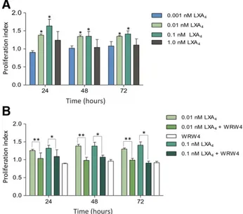
LXA4Stimulates ALX/FPR2-Dependent hPDLSC Proliferation and Migration
Because hPDLSCs modulate PMN activities and release media-tors of inflammation and resolution, their self-renewal and migration capabilities are fundamental to achieve a patho-physiological impact. Therefore, we determined whether these
functions could be modulated by LXA4. hPDLSCs exposed to
increasing concentrations of LXA4(0.001–1 nM) for 24, 48, and
72 hours displayed a concentration- and time-dependent incre-ment in proliferation, with a maximum at 24 hours (Fig. 4A). This effect was suppressed by WRW4, an ALX/FPR2 antagonist peptide
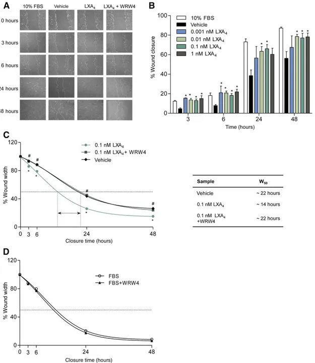
[44] (Fig. 4B). Likewise, LXA4concentrations as low as 0.01–1 nM
significantly enhanced hPDLSC directional migration in vitro, accel-erating wound closure (Fig. 5A, 5B). At 3 and 6 hours after
wounding, the percentage of filled wound in LXA4-treated cells
was 3.5-fold higher compared with vehicle-exposed cells, and comparable to that observed in cells with 10% FBS, our control
for maximal wound-healing capacity (Fig. 5A, 5B). LXA4
short-ened the time needed to reduce wound width by 50% (W50) from
22 to 14 hours (∼50%) (Fig. 5C). This effect was blunted by
WRW4 (Fig. 5A–5C), which, instead, did not influence W50in
cells exposed to FBS (Fig. 5D).
DISCUSSION
PD is characterized by persistent inflammation and it is asso-ciated with local and systemic morbidities [3, 6] Therefore,
Table 1. Cytokine quantification in hPDLSC-CdMa
Cytokine hPDLSC-CdM, mean6 SEM DMEM
IL-6 199.46 40.26 N.D. IL-8 408.256 241.56 N.D. IL-10 ,1.82 N.D. EGF 1.086 0.48 N.D. VEGF 1,017.56 234.88 N.D. TNF-a ,0.06 N.D. a
Aliquots of hPDLSC-CdM were incubated with magnetic beads coated with antibodies specific for each cytokine for 2 hours at RT under shaking. Biotinylated detector antibodies followed by streptavidin–PE (30 min, RT) were added to the samples. Plates were analyzed using a Luminex 100/200 platform equipped with the xPONENT 3.1 software. Standard curves for each analyte were generated by using reference proteins. Analyte concentration, expressed as pg/ml, was determined with a five-parameter logistic curve. Results are given as mean6 SEM from n = 3.
Abbreviations: CdM, conditioned medium; DMEM, Dulbecco’s modified Eagle’s medium; EGF, epidermal growth factor; hPDLSC, human periodontal ligament stem cell; IL, interleukin; N.D., not detectable; TNF, transforming growth factor; VEGF, vascular endothelial cell growth factor.
by guest on January 10, 2016
http://stemcellstm.alphamedpress.org/
knowledge of mechanisms and therapeutic approaches for this pathology is relevant for human health. In this study, we focused on stem cells isolated from the periodontal ligament. These cells have been added recently to the growing list of stem cells [23] and are likely to represent a promising tool to combat PD and related systemic consequences. hPDLSCs exhibit fibroblast-like morphology and colony-forming efficiency, and express em-bryonic and mesenchymal, but not hematopoietic or endothelial, markers (Fig. 1). They also express SSEA4, an embryonic marker, and the transcription factors NANOG, Oct4, and Sox2, associated with pluripotency (Fig. 1). With appropriate media, hPDLSCs differ-entiated into osteogenic, chondrogenic, and adipogenic lineages (Fig. 1). They can also produce cementum and PDL Sharpe’s fibers [23, 45]. These properties, combined with their easy accessibility, make hPDLSCs excellent candidates for periodontal regenerative medicine.
It is widely accepted that stem cells exert immunoregulatory func-tions, which, in some clinical settings, represent a primary therapeutic
mechanism [46]. In PD, the regulation of the host immune response is paramount for resolution [47]; PMNs are key players because they contribute to the removal of oral pathogens and tissue rebuilding [48]. Consistent with this scenario, PMN key activities, for instance chemotaxis and bacterial phagocytosis, are impaired in patients with periodontitis, thus contributing to tissue damage [40, 41].
Here, we show for the first time that hPDLSCs sustain PMN survival and bactericidal activity (Fig. 2). Cell-cell contact was not strictly necessary for these effects to take place, indicat-ing paracrine immunoregulatory functions of hPDLSCs. Indeed, they release cytokines and growth factors (i.e., IL-8, VEGF, and IL-6) that promote PMN recruitment and survival [49, 50] (Table 1). They also release IL-10 (Table 1), which limits the proin-flammatory activities of PMNs [51] and improve wound repair during PD [52]. This is consistent with a proresolutive, antimicrobial profile of the hPDLSC secretoma and prompted us to evaluate the capability of these cells to produce SPMs, pivotal regulators of in-flammation resolution [8].
Figure 3. Bioactive lipid mediators formed by hPDLSCs. hPDLSCs were incubated for 24 hours and lipid mediators (LMs) were analyzed by LM metabololipidomics. (A, B): Representative multiple reaction monitoring traces, representative of seven cell preparations, for the identified LMs along with (B) accompanying tandem mass spectrometry spectra used for identification of MaR1, RvD1, and LXB4.
by guest on January 10, 2016
http://stemcellstm.alphamedpress.org/
Using an established LM metabololipidomic approach [53], we obtained the first evidence that hPDLSCs produce and re-lease a broad spectrum of proresolutive mediators, including DHA-derived D-series resolvins, protectin D1, and maresin 1;
EPA-derived resolvin E2 and E3; and AA-derived LXB4 (Fig.
3). These mediators carry immunoregulatory/proresolutive bioactions by regulating granulocyte trafficking, stimulat-ing efferocytosis and microbial clearance [8, 54]. In particu-lar, D- and E-series resolvins regulate PMN chemotaxis and increase their capability to phagocytose and kill bacteria through specific cell-surface receptors, namely ALX/FPR2 and chemerin receptor 23 (ChemR23), widely expressed on PMNs [37, 54, 55]. Furthermore, these SPMs prevent bone re-sorption in experimental periodontitis [20]. hPDLSCs also
pro-duce large amounts of PGE2, which regulates bone formation
and resorption [56]. Hence, hPDLSCs generate LMs that or-chestrate inflammation resolution, immunomodulation, and bone metabolism.
Although it will be interesting to evaluate SPM production by stem cells from other sources, the present results provide new el-ements for the understanding of stem cell biology. It has been re-ported that stem cells exert a number of paracrine functions related to immunomodulation and control of the inflammation [28, 29]. Thus, the timely release of lipid mediators of inflammation and resolution may be regarded as a pivotal mechanism underlying these functions. Evidence that supports this hypothesis is accumu-lating. For instance, MSCs from umbilical cords reduce mast cell
de-granulation and alleviate murine atopic dermatitis through PGE2
release [57]. PGE2is also key for the attenuation of
graft-versus-host disease by BM-MSCs [58] and for the anti-inflammatory
Table 2. Lipid mediators produced by hPDLSCs in culture: Impact of LXA4 a (n = 7 cell incubations) Metabolome Q1 Q3 hPDLSC LM levels (pg per 53 106 cells) hPDLSC hPDLSC + LXA4(0.1 nM) Change, %
DHA bioactive metabolome
RvD1 375 215 45.4 65.5 44 RvD2 375 215 60.5 95.2 57 RvD3 375 147 0.2 0.6 263 RvD5 359 199 19.7 27.2 38 RvD6 359 101 88.7 45.3 249 17-epi-RvD1 375 215 5.3 8.7 66 17-epi-RvD3 375 147 —b —b NA PD1 359 153 63.1 30.5 252 17-epi-PD1 359 153 2.6 3.0 13 10S,17S-diHDHA 359 153 68.8 85.7 24 MaR1 359 221 11.3 11.4 1 7S,14S-diHDHA 359 221 52.4 59.7 14 4S,14S-diHDHA 359 101 50.1 65.7 31
EPA bioactive metabolome
RvE1 349 195 —b —b NA RvE2 333 213 69.2 91.9 33 RvE3 333 201 215.9 197.0 29 AA bioacive metabolome LXA4 351 235 0.1 — NA LXB4 351 221 227.9 272.0 19 5S,15S-diHETE 335 235 57.0 47.3 217 15-epi-LXA4 351 115 74.6 115.6 55 15-epi-LXB4 351 221 LTB4 335 195 23.7 26.5 12 5S,12S-diHETE 335 195 53.0 62.2 17 20-OH-LTB4 351 195 —b —b NA PGD2 351 189 445.4 1,096.8 146 PGE2 351 189 3,073.1 1,608.8 248 PGF2a 353 193 123.1 70.3 243 TxB2 369 169 585.9 835.0 43
aHuman periodontal ligament stem cells (hPDLSC) were incubated with or without 0.1 nM LXA
4and LMs were assessed using LM metabololipidomics
(details are available in Materials and Methods).
bBelow the limit of detection (the detection limit was approximately 0.1 pg).
Abbreviations:—, not quantified; AA, arachidonic acid; DHA, docosahexaenoic acid; EPA,eicosapentaenoicacid; LM, lipidmediator;LXA4, lipoxin A4; Q1,
M-H (parent ion); NA, not applicable; Q3, diagnostic ion in the tandem mass spectrometry (daughter ion).
by guest on January 10, 2016
http://stemcellstm.alphamedpress.org/
effects of adipose-derived MSCs on chondrocytes and synoviocytes
from osteoarthritis patients [59]. Although PGE2was the most
abundant eicosanoid generated by hPDLSCs under routine cul-ture conditions (Table 1), the observation, never reported be-fore, to our knowledge, that these cells can generate SPM from DHA and EPA, which act at subnanomolar to nanomolar concentrations, suggest that: (a) the relative abundance of bioactive lipid mediators generated by stem cells could be mod-ulated by the dietary intake of precursor fatty acids and might change in pathological conditions, and (b) the overall impact of stem cells on immuno-inflammatory responses may be dictated by a network of lipid mediators, which act in concert with peptides (Table 1).
On the other hand, the features of hPDLSCs uncovered in the present study propose these cells as key players within the con-text of PD, as their recruitment, proliferation, and release of pro-resolutive mediators can influence the outcome of PD. Here, we provide the first evidence that these processes can be
modu-lated by an LXA4-driven, receptor-mediated mechanism as
hPDLSCs express ALX/FPR2 (supplemental online Fig. 1). In
ad-dition to LXA4, this receptor recognizes RvD1 and annexin-A1,
both carrying anti-inflammatory, proresolution functions [37]. Consistent with earlier reports showing expression of this GPCR in human BM-MSCs and its role in stem cell adhesion, migration, and homing for tissue repair [60, 61], the activation of ALX/FPR2
by LXA4stimulated hPDLSC proliferation and migration in vitro
(Figs. 4, 5). The effect of LXA4on wound healing was significant
al-ready at 3 hours, ruling out the involvement of cell proliferation, at least at the earlier time points. These effects are of considerable rel-evance as hPDLSCs can differentiate into osteoblasts and regenerate PDL [62]; therefore, their recruitment and expansion within a dis-eased periodontium may accelerate tissue healing. Along these
lines, LXA4levels are reduced in crevicular fluids and plasma
from patients with PD [17, 18], thus potentially limiting endoge-nous hPDLSC-dependent tissue-repair mechanisms. Whether
LXA4can trigger differentiation pathways in hPDLSCs, similar
to what occurs with embryonic stem cells exposed to PD1 [63], remains to be determined.
LXA4also changed the LM profile of hPDLSCs (Table 2), although
it did not have appreciable impact on PMN survival. Cells exposed to
LXA4, showed an increase in D-series Rvs, which control leukocyte
trafficking and have antimicrobial properties in vivo [64]. LXA4also
changed the AA metabolome: It increased PGD2while decreasing
PGE2formation (Table 2). This is consistent with a proresolution
pro-file [65]. Therefore, it appears that LXA4can sustain a
receptor-mediated proresolution feed-forward loop that involves hPDLSCs in the periodontium.
CONCLUSION
The main finding of this study is that stem cells isolated from the periodontal ligament can release SPM and other mediators that carry relevant immunomodulatory, proresolution, and prohealing properties. This hPDLSC characteristic can be harnessed for therapeutic purposes in PD, although it is likely to represent a more general mechanism by which stem cells from differ-ent sources can influence the outcome of inflammatory disor-ders. We also demonstrate that the ALX/FPR2 receptor is crucial to promote the recruitment and expansion of hPDLSCs, thus representing a novel molecular target to enhance stem cell-mediated proresolving circuits. Collectively, these results
highlight the involvement of SPM and their receptors in stem cell biology, providing useful knowledge for innovative stem cell therapeutics.
ACKNOWLEDGMENTS
We thank the dentists of the Department of Medical Science, Oral and Biotechnology of the University G. d’Annunzio, Chieti, Italy, and all the people who donated the periodontal ligament and blood. We also thank Ilaria Merciaro and Alessia Lamolinara for technical support. This work was partially supported by a grant from the Cari-Chieti Foundation (Italy) to the StemTeCh Group and grants from the Italian Ministry of Education, University and Research to M.R. (PRIN 2010YK7Z5K_002), O.T. (PRIN 20102ZLNJ5), and S.M. (FIRB RBAP1047J_006). J.D., R.C., and C.N.S. were supported by NIH GM-095467 to C.N.S.
AUTHORCONTRIBUTIONS
E.C. and A.R.: conception and design, collection and assembly of data, data analysis and interpretation, manuscript writing, final approval of manuscript; O.T.: financial support, collection and assembly of data, data analysis and interpretation, final approval of manuscript; F.D.: provision of patients, collection and assembly of data, data analysis and interpretation, final approval of manuscript; M.M.: collection and assembly of data, data analysis and interpretation, final ap-proval of manuscript; S.M.: financial support, data analysis and interpretation, final approval of manuscript; R.A.C. and J.D.: collection and assembly of data, data analysis and interpretation, manuscript writing, final approval of manuscript; C.N.S.: data
Figure 4. ALX-FPR2-dependent stimulation of human periodontal ligament stem cell (hPDLSC) proliferation by LXA4. (A): hPDLSCs were
treated with LXA4(0.01–1 nM) or vehicle for 24, 48, and 72 hours.
Pro-liferation was evaluated by cell count and trypan blue exclusion. Data are expressed as proliferation index (cell number with LXA4per cell
number with vehicle) and are given as mean6 SEM from 4 indepen-dent experiments with triplicates (p, p , .05 vs. vehicle). (B): hPDLSCs were treated with WRW4 (10mM) before LXA4(0.01–0.1 nM) (pp, p ,
.01 WRW4 + 0.01 nM LXA4vs. 0.01 nM LXA4;p p , .05 WRW4 + 0.1 nM
LXA4vs. 0.1 nM LXA4). Results are given as the mean6 SEM of five
experiments with triplicates. LXA4, lipoxin A4.
by guest on January 10, 2016
http://stemcellstm.alphamedpress.org/
analysis and interpretation, manuscript writing, final approval of manuscript; M.R.: conception and design, financial support, man-uscript writing, final approval of manman-uscript.
DISCLOSURE OFPOTENTIALCONFLICTS OFINTEREST
C.N.S. is a compensated Corbus scientific advisory board member; has compensated research funding from the NIH;
and is an uncompensated inventor on patents assigned to
Brigham and Women’s Hospital and Partners HealthCare
on the composition of matter, uses, and clinical development of anti-inflammatory and pro-resolving lipid mediators. These in-clude lipoxins and resolvins and related compounds that are licensed for clinical development. The resolvins are licensed to Resolvyx Pharmaceutical. The other authors indicated no potential conflicts of interest.
Figure 5. ALX/FPR2-dependent stimulation of human periodontal ligament stem cell (hPDLSC) migration by LXA4. (A, B): Analysis
of hPDLSC migration after exposure to LXA4(0.01–1 nM) or vehicle. Representative images of wound closure at 0, 3, 6, 24, and 48 hour
(magnification:310) (A). Wound closure was quantified at 3, 6, 24, and 48 hours postwounding, by measuring the area not filled by cells, using ImageJ software. Results are given as the mean6 SEM of percentage wound closure of 5 experiments (p, p , .05 vs. vehicle) (B). (C): Wounded hPDLSCs were treated with WRW4 before LXA4(0.1 nM). The arrow shows the difference (∼10 hours) between time needed to
reduce wound width by 50% (W50) of LXA4-treated hPDLSCs incubated with or without WRW4 (p, p , .05 LXA4vs. vehicle; #, p# .01 LXA4+
WRW4 vs. LXA4). The table displays W50values. (D): Effects of WRW4 treatment on FBS-induced migration. Results are given as the
mean6 SEM of five experiments. FBS, fetal bovine serum; LXA4, lipoxin A4.
by guest on January 10, 2016
http://stemcellstm.alphamedpress.org/
REFERENCES
1 Kumar V, Abbas AK, Fausto N et al. Robbins and Cotran Pathologic Basis of Disease, Professional Edition: Expert Con-sult-Online. St. Louis, MO: Elsevier Health Sciences, 2009.
2 Scapini P, Cassatella MA. Social network-ing of human neutrophils within the immune system. Blood 2014;124:710–719.
3 Pihlstrom BL, Michalowicz BS, Johnson NW. Periodontal diseases. Lancet 2005;366: 1809–1820.
4 Russell CD, Schwarze J. The role of pro-resolution lipid mediators in infectious disease. Immunology 2014;141:166–173.
5 Van Dyke TE, Serhan CN. Resolution of in-flammation: A new paradigm for the pathogen-esis of periodontal diseases. J Dent Res 2003;82: 82–90.
6 Holmlund A, Holm G, Lind L. Severity of periodontal disease and number of remaining teeth are related to the prevalence of myocar-dial infarction and hypertension in a study based on 4,254 subjects. J Periodontol 2006; 77:1173–1178.
7 Ortega-G ´omez A, Perretti M, Soehnlein O. Resolution of inflammation: An integrated view. EMBO Mol Med 2013;5:661–674.
8 Serhan CN. Resolution phase of inflamma-tion: Novel endogenous anti-inflammatory and proresolving lipid mediators and pathways. Annu Rev Immunol 2007;25:101–137.
9 Stables MJ, Gilroy DW. Old and new generation lipid mediators in acute inflam-mation and resolution. Prog Lipid Res 2011; 50:35–51.
10 Colas RA, Shinohara M, Dalli J et al. Iden-tification and signature profiles for pro-resolving and inflammatory lipid mediators in human tissue. Am J Physiol Cell Physiol 2014;307: C39–C54.
11 Wu SH, Chen XQ, Liu B et al. Efficacy and safety of 15(R/S)-methyl-lipoxin A(4) in topical treatment of infantile eczema. Br J Dermatol 2013;168:172–178.
12 Serhan CN, Hamberg M, Samuelsson B. Lipoxins: Novel series of biologically active com-pounds formed from arachidonic acid in human leukocytes. Proc Natl Acad Sci USA 1984;81: 5335–5339.
13 Cl `aria J, Serhan CN. Aspirin triggers previously undescribed bioactive eicosanoids by human endothelial cell-leukocyte inter-actions. Proc Natl Acad Sci USA 1995;92: 9475–9479.
14 Maderna P, Godson C. Lipoxins: Resolu-tionary road. Br J Pharmacol 2009;158:947–959. 15 Fiore S, Maddox JF, Perez HD et al. Iden-tification of a human cDNA encoding a func-tional high affinity lipoxin A4 receptor. J Exp Med 1994;180:253–260.
16 Romano M, Recchia I, Recchiuti A. Lipoxin receptors. ScientificWorldJournal 2007;7:1393– 1412.
17 Pouliot M, Clish CB, Petasis NA et al. Lipoxin A(4) analogues inhibit leukocyte re-cruitment to Porphyromonas gingivalis: A role for cyclooxygenase-2 and lipoxins in periodontal disease. Biochemistry 2000;39: 4761–4768.
18 Fredman G, Oh SF, Ayilavarapu S et al. Im-paired phagocytosis in localized aggressive
periodontitis: rescue by Resolvin E1. PLoS One 2011;6:e24422.
19 B¨orgeson E, L¨onn J, Bergstr¨om I et al. Lipo-xin A₄ inhibits Porphyromonas gingivalis-induced aggregation and reactive oxygen species pro-duction by modulating neutrophil-platelet in-teraction and CD11b expression. Infect Immun 2011;79:1489–1497.
20 Serhan CN, Jain A, Marleau S et al. Re-duced inflammation and tissue damage in transgenic rabbits overexpressing 15-lipoxy-genase and endogenous anti-inflammatory lipid mediators. J Immunol 2003;171:6856– 6865.
21 Bartold PM, McCulloch CA, Narayanan AS et al. Tissue engineering: a new paradigm for periodontal regeneration based on molecular and cell biology. Periodontology 2000 2000; 24:253–269.
22 Shimono M, Ishikawa T, Ishikawa H et al. Regulatory mechanisms of periodontal regen-eration. Microsc Res Tech 2003;60:491–502.
23 Seo BM, Miura M, Gronthos S et al. Inves-tigation of multipotent postnatal stem cells from human periodontal ligament. Lancet 2004; 364:149–155.
24 Lindroos B, M¨aenp¨a¨a K, Ylikomi T et al. Characterisation of human dental stem cells and buccal mucosa fibroblasts. Biochem Bio-phys Res Commun 2008;368:329–335.
25 Huang GT, Gronthos S, Shi S. Mesenchy-mal stem cells derived from dental tissues vs. those from other sources: Their biology and role in regenerative medicine. J Dent Res 2009;88: 792–806.
26 Sonoyama W, Liu Y, Fang D et al. Mesen-chymal stem cell-mediated functional tooth re-generation in swine. PLoS One 2006;1:e79.
27 Park JY, Jeon SH, Choung PH. Efficacy of periodontal stem cell transplantation in the treatment of advanced periodontitis. Cell Transplant 2011;20:271–285.
28 Spaggiari GM, Abdelrazik H, Becchetti F et al. MSCs inhibit monocyte-derived DC matu-ration and function by selectively interfering with the generation of immature DCs: Central role of MSC-derived prostaglandin E2. Blood 2009;113:6576–6583.
29 Di Nicola M, Carlo-Stella C, Magni M et al. Human bone marrow stromal cells suppress T-lymphocyte proliferation induced by cellular or nonspecific mitogenic stimuli. Blood 2002; 99:3838–3843.
30 Jiang XX, Zhang Y, Liu B et al. Human mes-enchymal stem cells inhibit differentiation and function of monocyte-derived dendritic cells. Blood 2005;105:4120–4126.
31 Krampera M, Glennie S, Dyson J et al. Bone marrow mesenchymal stem cells inhibit the response of naive and memory antigen-specific T cells to their cognate peptide. Blood 2003;101:3722–3729.
32 Wada N, Menicanin D, Shi S et al. Immu-nomodulatory properties of human periodontal ligament stem cells. J Cell Physiol 2009;219: 667–676.
33 Trubiani O, Di Primio R, Traini T et al. Morphological and cytofluorimetric analysis of adult mesenchymal stem cells expanded ex vivo from periodontal ligament. Int J Immunopathol Pharmacol 2005;18:213–221.
34 Eleuterio E, Trubiani O, Sulpizio M et al. Proteome of human stem cells from periodontal
ligament and dental pulp. PLoS One 2013;8: e71101.
35 Trubiani O, Scarano A, Orsini G et al. The performance of human periodontal ligament mesenchymal stem cells on xenogenic biomate-rials. Int J Immunopathol Pharmacol 2007;20 (suppl 1):87–91.
36 Trubiani O, Piattelli A, Gatta V et al. As-sessment of an efficient xeno-free culture system of human periodontal ligament stem cells. Tissue Eng Part C Methods 2015;21: 52–64.
37 Krishnamoorthy S, Recchiuti A, Chiang N et al. Resolvin D1 binds human phago-cytes with evidence for proresolving recep-tors. Proc Natl Acad Sci USA 2010;107: 1660–1665.
38 Livak KJ, Schmittgen TD. Analysis of relative gene expression data using real-time quantitative PCR and the 2(-Delta Delta C(T)) method. Methods 2001;25: 402–408.
39 Dominici M, Le Blanc K, Mueller I et al. Minimal criteria for defining multipotent mes-enchymal stromal cells. The International Soci-ety for Cellular Therapy position statement. Cytotherapy 2006;8:315–317.
40 Van Dyke TE, Horoszewicz HU, Cianciola LJ et al. Neutrophil chemotaxis dysfunction in human periodontitis. Infect Immun 1980;27: 124–132.
41 MacFarlane GD, Herzberg MC, Wolff LF et al. Refractory periodontitis associated with abnormal polymorphonuclear leukocyte phago-cytosis and cigarette smoking. J Periodontol 1992;63:908–913.
42 Asensi V, Valle E, Meana A et al. In vivo interleukin-6 protects neutrophils from apopto-sis in osteomyelitis. Infect Immun 2004;72: 3823–3828.
43 Kettritz R, Gaido ML, Haller H et al. Interleukin-8 delays spontaneous and tumor necrosis factor-alpha-mediated apoptosis of human neutrophils. Kidney Int 1998;53: 84–91.
44 Shin EH, Lee HY, Kim SD et al. Trp-Arg-Trp-Trp-Trp-Trp antagonizes formyl peptide receptor like 2-mediated signaling. Biochem Biophys Res Commun 2006;341:1317–1322.
45 Trubiani O, Orsini G, Zini N et al. Regener-ative potential of human periodontal ligament derived stem cells on three-dimensional bioma-terials: A morphological report. J Biomed Mater Res A 2008;87:986–993.
46 Weng JY, Du X, Geng SX et al. Mesenchy-mal stem cell as salvage treatment for refrac-tory chronic GVHD. Bone Marrow Transplant 2010;45:1732–1740.
47 Ohlrich EJ, Cullinan MP, Seymour GJ. The immunopathogenesis of periodontal disease. Aust Dent J 2009;54(suppl 1):S2–S10.
48 Hasturk H, Kantarci A, Van Dyke TE. Oral inflammatory diseases and systemic inflammation: Role of the macrophage. Front Immunol 2012; 3:118.
49 Bickel M. The role of interleukin-8 in inflammation and mechanisms of regu-lation. J Periodontol 1993;64:5(suppl):456– 460.
50 Sinnathamby T, Yun J, Clavet-Lanthier ME et al. VEGF and angiopoietins promote in-flammatory cell recruitment and mature blood vessel formation in murine sponge/
by guest on January 10, 2016
http://stemcellstm.alphamedpress.org/
Matrigel model. J Cell Biochem 2015;116: 45–57.
51 Bazzoni F, Tamassia N, Rossato M et al. Understanding the molecular mechanisms of the multifaceted IL-10-mediated anti-inflammatory response: lessons from neutrophils. Eur J Immunol 2010;40:2360–2368.
52 Garlet GP. Destructive and protective roles of cytokines in periodontitis: A re-appraisal from host defense and tissue de-struction viewpoints. J Dent Res 2010;89: 1349–1363.
53 Lee SH, Williams MV, DuBois RN et al. Targeted lipidomics using electron capture at-mospheric pressure chemical ionization mass spectrometry. Rapid Commun Mass Spectrom 2003;17:2168–2176.
54 Oh SF, Dona M, Fredman G et al. Resolvin E2 formation and impact in inflammation reso-lution. J Immunol 2012;188:4527–4534.
55 Herrera BS, Hasturk H, Kantarci A et al. Impact of resolvin E1 on murine neutrophil phagocytosis in type 2 diabetes. Infect Immun 2015;83:792–801.
56 Raisz LG. Physiologic and pathologic roles of prostaglandins and other eicosanoids in bone metabolism. J Nutr 1995;125:7(suppl): 2024S–2027S.
57 Kim HS, Yun JW, Shin TH et al. Human um-bilical cord blood mesenchymal stem cell-derived PGE2 and TGF-b1 alleviate atopic dermatitis by reducing mast cell degranulation. STEM CELLS 2015;33:1254–1266.
58 Auletta JJ, Eid SK, Wuttisarnwattana P et al. Human mesenchymal stromal cells attenuate graft-versus-host disease and maintain graft-versus-leukemia activity fol-lowing experimental allogeneic bone mar-row transplantation. STEM CELLS 2015;33: 601–614.
59 Manferdini C, Maumus M, Gabusi E et al. Adipose-derived mesenchymal stem cells ex-ert antiinflammatory effects on chondrocytes and synoviocytes from osteoarthritis patients through prostaglandin E2. Arthritis Rheum 2013;65:1271–1281.
60 Kim MK, Min S, Park YJ et al. Expression and functional role of formyl peptide receptor
in human bone marrow-derived mesenchy-mal stem cells. FEBS Lett 2007;581:1917– 1922.
61 Viswanathan A, Painter RG, Lanson NA Jr et al. Functional expression of N-formyl peptide receptors in human bone marrow-derived mes-enchymal stem cells. STEM CELLS 2007;25: 1263–1269.
62 Hoang AM, Oates TW, Cochran DL. In vitro wound healing responses to enamel ma-trix derivative. J Periodontol 2000;71:1270– 1277.
63 Yanes O, Clark J, Wong DM et al. Meta-bolic oxidation regulates embryonic stem cell differentiation. Nat Chem Biol 2010;6: 411–417.
64 Spite M, Norling LV, Summers L et al. Resolvin D2 is a potent regulator of leukocytes and controls microbial sepsis. Nature 2009;461: 1287–1291.
65 Gilroy DW, Colville-Nash PR, Willis D et al. Inducible cyclooxygenase may have anti-inflammatory properties. Nat Med 1999;5: 698–701.
See www.StemCellsTM.com for supporting information available online.
by guest on January 10, 2016
http://stemcellstm.alphamedpress.org/