Porphyromonas gingivalis
lipopolysaccharide stimulation
in human periodontal ligament
stem cells: Role of epigenetic
modifications
to the inflammation
Francesca Diomede,1Soundara Rajan Thangavelu,2 Ilaria Merciaro,1Monica D’Orazio,3 Placido Bramanti,2Emanuela Mazzon,2 Oriana Trubiani1
1Stem Cells and Regenerative Medicine Laboratory, Department of Medical, Oral and Biotechnological Sciences, University “G. d’Annunzio” Chieti-Pescara, Chieti, Italy
2IRCCS Centro Neurolesi “Bonino Pulejo”, Messina, Italy
3Department of Rheumatology and Immunology, Medical University of Graz, Austria
Abstract
Periodontitis is a chronic oral inflam-matory disease produced by bacteria. Gingival retraction and bone and connec-tive tissues resorption are the hallmarks of this disease. Chronic periodontitis may con-tribute to the risk of onset or progression of neuroinflammatory pathological conditions, such as Alzheimer’s disease. The main goal of the present study was to investigate if the role of epigenetic modulations is involved in periodontitis using human periodontal ligament stem cells (hPDLSCs) as an in
vitro model system. hPDLSCs were treated
with lipopolysaccharide of Porphyromonas
gingivalis and the expression of proteins
associated with DNA methylation and his-tone acetylation, such as DNMT1 and p300, respectively, and inflammatory transcrip-tion factor NF-kB, were examined. Immunofluorescence, Western blot and next generation sequencing results demonstrated that P. gingivalis lipopolysaccharide signif-icantly reduced DNA methylase DNMT1, while it markedly upregulated the level of histone acetyltransferase p300 and NF-kB in hPDLSCs. Our results showed that P.
gingivalis lipopolysaccharide markedly
reg-ulate the genes involved in epigenetic mechanism, which may result in inflamma-tion inducinflamma-tion. We propose that P. gingivalis lipopolysaccharide-treated hPDLSCs could be a potential in vitro model system to study epigenetics modulations associated with
periodontitis, which might be helpful to identify novel biomarkers linked to this oral inflammatory disease.
Introduction
Periodontal disease is a chronic inflam-matory disease caused by sub gingival bac-terial biofilm.1This biofilm formation
caus-es gingival retraction and bone and connec-tive tissue resorption.2 Moreover, it has
been demonstrated that in chronic condition inflammatory modulators associated with periodontitis may cause systemic inflamma-tion, participate in the onset or in the pro-gression of neuroinflammatory conditions, including Alzheimer’s disease (AD) and produce cognitive decline.3-5Although the
real mechanism underlying the destruction of tooth supporting structures is not com-pletely understood, bacterial interaction with the cells present in the oral cavity could be considered as a key pathogenetic step followed by biofilm synthesis. Moreover, mesenchymal stem cells (MSCs) present in the oral cavity are affected by bacteria and bacterial derivatives.6Different
types of dental MSCs have been reported: dental pulp stem cells,7exfoliated
decidu-ous teeth stem cells,8periodontal ligament
stem cells,9apical papilla stem cells,10
den-tal follicle stem cells,11 and gingiva stem
cells.12,13Human periodontal ligament stem
cells (hPDLSCs) displayed substantial pro-liferation capacity ex vivo14,15 and
mesen-genic differentiation capacity.16
Various bacteria and their pathogen associated molecules including lipopolysac-charides (LPS) activate a cascade of chronic inflammatory events and modulate the host tissue response. Many types of gram-nega-tive anaerobic bacteria are involved in peri-odontitis. Porphyromonas gingivalis is the major periodontitis pathogen, that triggers initiation and progression of periodontal diseases.17,18In addition to bacteria, genetics
and environmental factors also play a cru-cial role in the etiology of periodontitis via regulating epigenetic modifications.19
Bacteria and their products can produce alterations in DNA methylation, which modifies the regulation of inflammatory genes followed by disease progression.20-22
DNA methylation and histone acetylation are the major epigenetic modifications induced by diseases and environmental fac-tors.23,24 DNA (cytosine-5)
methyltrans-ferase 1 (DNMT1) and histone deacetylases (HDACs) are the key controllers, which regulate DNA methylation and histone acetylation, respectively.25
In periodontal disease condition, his-tone acetylation promotes the transcription of inflammatory genes such as p300/CBP histone acetyltransferase, NF-kB and other proinflammatory cytokines.26However, the
impact of histone modifications during the progression of periodontitis remains unclear. NF-kB signaling pathway could be involved in sustained histone modifications which further augments the disease progres-sion.27 Nuclear transcription factor NF-kB
has a key role to activate innate immunity which causes osteoclast differentiation and to induce bone resorption.28DNA
methyla-tion is regulated by two different types of DNA methyltransferases (DNMTs): de
novo methyltransferases (DNMT3a and
DNMT3b), which are active during early development29 and maintenance
methyl-transferase (DNMT1), which regulates methylated and unmethylated CpG sites in the cells.30-32
In the present study, we have investigated the epigenetic modifications elicited by
Porphyromonas gingivalis LPS (LPS-G) using
hPDLSCs as a model system to study novel
Correspondence: Oriana Trubiani, Department of Medical, Oral and Biotechnological Sciences, University “G. d’Annunzio” Chieti-Pescara, Via dei Vestini 31, 66100 Chieti, Italy.
Fax: +39.0871.3554033. E-mail: [email protected]
Key words: Periodontitis; inflammation; human periodontal mesenchymal stem cells; epigenetic regulation; DNMT1; p300; Alzheimer’s disease.
Acknowledgments: this work has been sup-ported with OT ex 60% funds, University “G. d’Annunzio”.
Contributions: FD, SRT, IM, experiments per-forming; MDO, samples collection; FD, SRT, MDO, data analysis, manuscript revision; EM, OT manuscript drafting; PB, EM, OT, study design. All authors have edited, read and approved the final manuscript.
Conflict of interest: the authors declare no conflict of interest.
Received for publication: 9 June 2017. Accepted for publication: 2 August 2017. This work is licensed under a Creative Commons Attribution-NonCommercial 4.0 International License (CC BY-NC 4.0).
©Copyright F. Diomede et al., 2017 Licensee PAGEPress, Italy
European Journal of Histochemistry 2017; 61:2826 doi:10.4081/ejh.2017.2826
Non
commercial
biomarkers linked to this oral inflammatory disease. To this end, we have examined the expression of DNMT1, p300 and NF-kB fol-lowed by LPS-G treatment in hPDLSCs.
Materials and Methods
Ethic statement
The present study was approved by the Medical Ethics Committee at the Medical School, “G. d’Annunzio” University, Chieti, Italy (n. 266/17.04.14). All healthy volunteers enrolled in this study have signed the informative consent form. The Department of Medical, Oral and Biotechnological Sciences and the Laboratory of Stem Cells and Regenerative Medicine are certified according to the quality standard ISO 9001:2008 (certificate n. 32031/15/S).
Cell culture
Periodontal ligament biopsies were col-lected from premolar teeth of healthy vol-unteers. All patients provided written informed consent to participate in the study. Before the biopsy collection, each patient was pre-treated for one week with profes-sional dental hygiene and chlorhexidine. Explants were obtained from alveolar crest and horizontal fibers of the periodontal lig-ament by scraping the roots using a Gracey’s curette.33 Periodontal tissue
frag-ments were cut, washed with PBS (Lonza, Basel, Switzerland) and placed in a TheraPEAK™MSCGM-CD™ Bullet Kit serum free, chemically defined (MSCGM-CD) medium (Lonza) at 37°C for the growth of human MSCs. Cells sponta-neously migrated from the explants after reaching about 80% of confluence were trypsinized (LiStar Fish, Milan, Italy), and subsequently subcultured until passage 2 (P2). Cells utilized for the experimental assays were at P2.
LPS-G treatment
hPDLSCs were divided in two groups: group 1, untreated control (hPDLSCs); and group 2, cells treated with 5 μg/mL LPS-G (InvivoGen, San Diego, CA, USA) (hPDLSCs/LPS-G) for 24 h.
Morphological evaluation
After 24 h, hPDLSCs and hPDLSCs treated with LPS-G were fixed with 2.5% glutaraldehyde in 0.1 M cacodylate buffer pH 7.4 for 2 h, stained with toluidine blue solution and observed by inverted optical microscope Leica DMIL (Leica Microsystems, Milan, Italy).
MTT assay
Cell viability was evaluated by 3-(4,5- dimethyl-2-thiazolyl)-2,5-diphenyl-2-H-tetrazolium bromide (MTT) test. 1.5×10⁴
cells of each group were plated in 96-well plates and were incubated with 200 µl cul-ture medium. After incubation, 20 µL MTT solution was added to each well and incu-bated for 3 h.34The absorbance was
meas-ured on an automated microplate reader (Sinergy HT, Biotek Instruments, Bad Friedrichshall, Germany) at 570 nm.
Immunofluorescence analysis
hPDLSCs and hPDLSCs/LPS-G were processed as previously reported by Trubiani et al.35 The following primary
monoclonal antibodies were used: anti-human NF-kB (1:250, rabbit) (OriGene Technologies, Inc., Rockville, MD, USA), anti-DNMT1 (1:250, rabbit) (OriGene), anti-p300 (1:250, rabbit) (OriGene). Then, cells were incubated for 1 h at 37°C with Alexa Fluor 568 red fluorescence conjugat-ed goat anti-rabbit secondary antibodies (1:200) (Molecular Probes). Subsequently, cells were incubated with Alexa Fluor 488 phalloidin green fluorescence conjugate (1:200, Molecular Probes) to mark cytoskeleton actin. Cell nuclei were stained with TOPRO (1:200, Molecular Probes) for 1 h at 37°C. Glass coverslips were placed upside down on glass slides and mounted with Prolong antifade (Molecular Probes).36
Samples were observed with Zeiss LSM510META confocal system (Zeiss, Jena, Germany) connected to an inverted Zeiss Axiovert 200 microscope equipped with a Plan Neofluar oil-immersion objec-tive (63x). Images were collected using an argon laser beam with excitation lines at 488 nm and a helium-neon source at 543 and 665 nm.
Nuclei isolation
Treated and untreated hPDLSCs (5×10⁶) were washed with PBS and
resus-pended in 500 μL of a hypotonic/detergent buffer [10 mM Tris-Cl pH 7.8, 0.125% Nonidet P-40, 1 mM β-mercaptoethanol, 1 mM phenylmethylsulfonyl fluoride, 10 μL/mL cocktail inhibitor (Protease Inhibitor Cocktail, Sigma-Aldrich, St. Louis, MO, USA), 0.1 μg/mL soybean trypsin inhibitor]. Cells were sheared by 5 passages through a 22-gauge syringe needle.37Nuclei
were recovered by low speed centrifugation (800×g for 10 min) and washed two times in 10 mM Tris-Cl pH 7.4, 2 mM MgCl2 plus protease inhibitors as mentioned above. The pellet, representing the intact nuclei fraction, was used for biochemical experiments.
Western blot analysis
Thirty micrograms of proteins from hPDLSCs and hPDLSCs/LPS-G and from extracted nuclei were separated on SDS-PAGE and subsequently transferred to nitrocellulose sheets using a semidry blot-ting apparatus. Sheets were saturated for 120 min at room temperature in blocking buffer (1xTBS, 5% milk, 0.1% Tween-20), then incubated overnight at 4°C in blocking buffer containing primary antibodies to NF-kB (1:2000, rabbit) (OriGene), DNMT1 (1:1000, rabbit) (OriGene), p300 (1:750, rabbit) (OriGene) and β-actin (1:750, mouse) (Santa Cruz Biotechnology, Santa Cruz, CA, USA). After four washes in TBS containing 0.1% Tween-20, samples were incubated for 60 min at room temperature with peroxidase-conjugated secondary anti-body diluted 1:1000 in 1x TBS with 2.5% dry milk and 0.1% Tween-20.38Bands were
visualized by ECL method and quantified by Alliance 2.7 (UVItec Ltd., Cambridge, UK).
Next generation sequencing (NGS)
Total RNA was isolated using a Reliaprep RNA Cell Miniprep System (Promega, USA). TruSeq RNA Access library kit (Illumina, Inc., San Diego, CA, USA) was applied to make RNA sequenc-ing libraries. Fifty ng of total RNA was fragmented at 94°C for 8 min on a thermal cycler. First strand cDNA synthesis was produced with random hexameres and SuperScript II Reverse Transcriptase (Invitrogen) using the following tempera-ture profile: 25°C for 10 min, 42°C for 15 min and 70°C for 15 min. Then, RNA tem-plates were removed and double stranded cDNA was produced using dUTP. Subsequently, the 3`ends of the cDNA were adenylated and ligated with indexing adap-tor. The following PCR temperature profile was used for the selective enrichment of adaptor-ligated DNA fragments: 98°C for 10 s, 60°C for 30 s and 72°C for 30 s (15 cycles). Then, the libraries were validated and were hybridized with the selective probes to capture exome. Probes with enriched libraries were captured using streptavidin coated magnetic beads, eluted from the beads, cleaned up with AMPure XP beads and processed for the second PCR amplification using following temperature profile: 98°C for 10 s, 60°C for 30 s and 72°C for 30 s (10 cycles). Later, the libraries were quantified with KAPA Library Quantification Kit-Illumina/ABI Prism® (Kapa Biosystems, Inc., Wilmington, MA, USA) and validated using Agilent High Sensitivity DNA Kit on a Bioanalyzer. DNA fragments were
meas-Non
commercial
ured in the range of 200-650 bp. Peak was observed around 250 bp. After normaliza-tion to 12 pM, DNA libraries were processed for cluster and single read sequencing (150 cycles) using MiSeq tool (Illumina, Inc.). The libraries were loaded on a MiSeq Flow Cell v3 to make clustering and were sequenced. Later, the reads were grouped with specific indexes using demul-tiplexing software tool CASAVA (ver. 1.8.2, Illumina, Inc.) and were mapped against “Homo sapiens UCSC hg19” reference sequences using RNA-Seq Alignment ver. 1.0.0. TopHat 2 (Bowtie 1) was applied for the Read mapping. Fragments per kilobase of gene model per million mapped reads (FPKM) values were calculated as described by Rajan et al.12
Statistical analysis
All experiments were performed in tripli-cate. The data are presented as means ± stan-dard error and were analyzed using SIGMA-STAT (SPSS Inc., Chicago, Ill., USA). One-way repeated-measurement analysis of vari-ance (ANOVA), followed by the post-hoc Holm-Sidak test (when appropriate) and the Student’s paired t-test were used for statistical analysis. A P-value less than 0.05 was consid-ered statistically significant. In NGS experi-ment, the statistical analysis on the read counts to measure the proportion of differen-tially expressed genes between two samples was performed by The Cufflinks Assembly & DE package ver. 2.0.0. False discovery rate (FDR; Q) value less than 0.05 was considered statistically significant.
Results
Morphological and viability evaluation
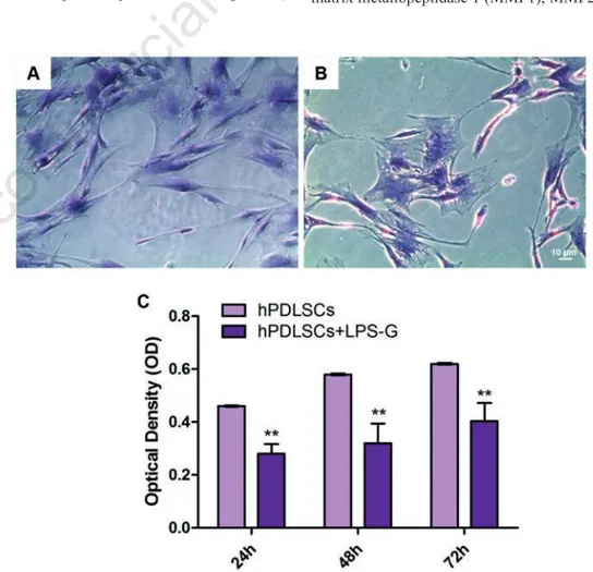
First, we have examined the morpho-logical changes and viability in hPDLSCs treated with LPS-G. hPDLSCs (Figure 1A) and LPS-G treated hPDLSCs (Figure 1B) showed a fibroblastic-like shape. However, LPS-G treated cells showed long cytoplas-mic processes and a low cell density when compared to the control cells. The prolifer-ation rate of hPDLSCs was assessed by means of MTT assay. Obtained data showed a significant exponential cell growth during all endpoint in hPDLSCs, while LPS-G induces a significant reduction in cell via-bility (P<0.01) (Figure 1C).
LPS-G induces epigenetic
modula-tions in hPDLSCs
Then, we have investigated the modula-tions in epigenetic regulation caused by
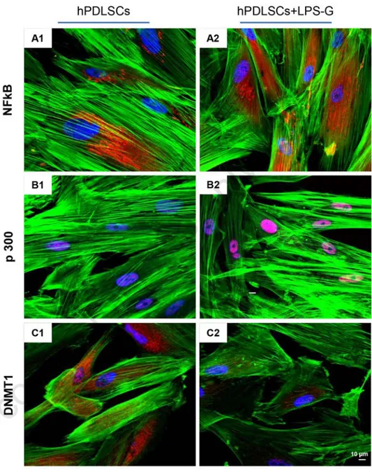
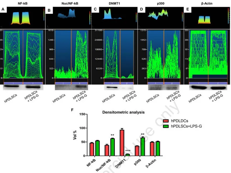
LPS-G treatment. Expression and subcellu-lar localization of NF-kB, DNMT1 and p300 were observed by immunofluores-cence with confocal microscopy. Increased nuclear translocation of NF-kB was observed in LPS-G-treated hPDLSCs (Figure 2 A2), while its predominant cyto-plasmic expression of NF-kB was noticed in control untreated cells (Figure 2 A1). Nuclear histone acetyl transferase p300 was enhanced in hPDLSCs treated with LPS-G (Figure 2 B2), while basal expression was observed in untreated cells (Figure 2 B1). On the contrary, DNMT1 expression was significantly suppressed in LPS-G- treated cells (Figure 2 C2), when compared to untreated cells (Figure 2 C1). These results were further confirmed with western blot analysis. Total expression of NF-kB was unaltered (Figure 3A), while nuclear expression of NF-kB was markedly increased in LPS-G-treated hPDLSCs, but not in the untreated cells (Figure 3B). Similarly, p300 level was increased in the cells treated with LPS-G (Figure 3D). As expected, DNMT1 was markedly reduced followed by LPS-G treatment (Figure 3C). Densitonetric analysis of specific band pro-vided a protein quantification (Figure 3F).
These results revealed that LPS-G signifi-cantly suppressed DNMT1, while upregu-lated the expression of p300 and NF-kB, which may trigger the transcription of inflammatory genes in hPDLSCs. NGS data supported these results. DNMT1 was down-regulated and p300 was updown-regulated in LPS-G-treated hPDLSCs (Figure 4) (Q<0.05). Histone deacetylase HDAC1 was reduced while HDAC2 was increased after LPS-G treatment (Figure 4) (Q<0.05). Moreover, proinflammatory cytokine tran-scripts, tumor necrosis factor alpha induced protein 1 (TNFAIP1) and interleukin 6 sig-nal transducer (IL6ST) were significantly enhanced in LPS-G-treated hPDLSCs (Figure 3) (Q<0.05).
LPS-G modulates genes associated
with AD in hPDLSCs
Considering the connection between periodontitis and AD, we have examined the expression of genes linked with AD in LPS-G treated hPDLSCs. We have noticed that AD-linked genes such as amyloid beta precursor protein (APP), amyloid beta pre-cursor protein binding protein 2 (APPBP2), interferon gamma receptor 1 (IFNGR1), matrix metallopeptidase 1 (MMP1), MMP2
Figure 1. Morphological evaluation. A) Isolation and culture of hPDLSCs from peri-odontal ligament tissues; cells showed a fibroblastic morphology. B) After 24 h of LPS-G treatment, cells showed a low density and morphological changes, such as long and numerous cytoplasmic processes. C) MTT showed a reduction in cell proliferation rate in all examined endpoint in LPS-G treated cells. Magnification: 10x; **P<0.01.
Non
commercial
and MMP16 were markedly increased in LPS-G-treated hPDLSCs than in control untreated hPDLSCs (Table 1; Q<0.0001). Relative log2 fold change expression of these genes in hPDLSCs and LPS-G-treated hPDLSCs are shown in Table 1. These results suggest that LPS-G-induced inflam-mation in hPDLSCs may activate the genes associated with AD.
Discussion
Periodontitis is a prevalent inflammato-ry disease arising from the gum plaques produced by oral pathogens including
Porphyromonas gingivalis. Advanced form
of periodontitis results in the retraction of gingiva, loss of periodontal ligament, and degeneration of surrounding alveolar bone. Chronic periodontitis may increase the risk of neuroinflammatory diseases such as AD.39,40 Complex molecular mechanisms
are involved in the stimulation and mainte-nance of inflammation, among which epige-netic pathways received special attention because of their upstream regulations. Epigenetic modifications include chemical alterations of DNA and associated proteins, leading to remodeling of the chromatin and activation or inactivation of gene transcrip-tion. These changes can contribute to the development and maintenance of cancer, autoimmune and inflammatory diseases, including periodontitis. Interestingly, some epigenetic modifications are reversible and can be stimulated by environmental fac-tors.41,42Knowledge on the modification of
epigenetic mechanisms may provide sub-stantial inputs regarding the mechanism of the key regulatory pathways of the genes involved in the maintenance of chronic inflammation. Indeed, the crucial role of DNA and histone modifications, the two major epigenetic regulations, have been described in periodontitis.26Several studies
have demonstrated that in periodontal dis-ease, gene expression can be influenced by DNA methylation;26 however, only few
studies have reported the histone
modifica-tions induced by LPS-G stimulus.43Earlier
studies have demonstrated that in B cells, LPS-G induced methylation and acetylation in H3 and H4 histone proteins.26Recently, it
has been demonstrated that periodontitis is
characterized by chronic inflammation that might be linked with aberrant DNA methy-lation in the gingival tissues.44,45 The
increased methylation in the gene promoter region is associated with a reduction in gene
Table 1. LPS-G modulates genes associated with Alzheimer’s disease. NGS data revealed that LPS-G treatment significantly increased the expression of AD-linked genes such as APP, APPBP2, IFNGR1, MMP1, MMP2 and MMP16 in hPDLSCs (Q<0.0001).
Gene hPDLSCs hPDLSCs+LPS-G log2 fold change Q-value
APP 405.63 1057.55 1.38 0.0001 APPBP2 4.48 6.94 0.63 0.0001 IFNGR1 14.82 34.78 1.23 0.0001 MMP1 13.84 161.31 3.54 0.0001 MMP2 975.87 1359.89 0.48 0.0001 MMP16 2.30 8.99 1.97 0.0001
Figure 2. Immunofluorescence. Cells grown on coverslips were fixed and stained with the indicated primary and secondary antibodies and DAPI, and subjected to confocal microscopy. NF-kB expression in hPDLSCs (A1) and in hPDLSCs treated with LPS-G (A2). p300 expression in hPDLSCs (B1) and in hPDLSCs treated with LPS-G (B2). DNMT1 expression in hPDLSCs (C1) and in hPDLSCs treated with LPS-G (C2). Data are representative of three independent experiments. Magnification: 63x.
Non
commercial
expression, while hypomethylation pattern is closely associated with transcriptional activation.46Moreover, it has been
demon-strated that LPS-G downregulated the expression of DNA methylases DNMT3a and DNMT1 in keratinocytes.44
In our recent study we reported that in hPDLSCs, LPS-G treatment activates the TLR4/MyD88 complex, induces NF-kB nuclear translocation and triggers the secre-tion of proinflammatory cytokines includ-ing TNF-α.47 Based on these findings, we
assume that hPDLSCs may represent an appropriate stem cell modelling through which we may understand the biological machinery of epigenetic system and inflam-mation in response to LPS-G in periodontal tissues. To this end, we have investigated the expression of DNMT1, p300 and NF-kB in hPDLSCs administered with LPS-G. Immunofluorescence and Western blot results demonstrated that LPS-G treatment significantly suppressed the expression of DNMT1 in hPDLSCs. This result is
corrob-Figure 3. Western blot analysis of NF-κB (A), NF-κB in nuclear extract (B), DNMT1 (C) and p300 (D) expression in hPDLSCs and in hPDLSCs treated with LPS-G for 24 h. β-actin was used as a housekeeping protein (E). Densitometric analysis of protein specific bands (F). **P<0.01; ***P<0.001.
Figure 4. Gene expression. Next generation sequencing demonstrated the modulation of genes expressed in untreated and LPS-G treated hPDLSC. P300, TNFAIP1, IL6ST and HDAC2 were expressed greater than 2-fold (Log2 fold change; Q<0.05). DNMT1 and HDAC1 genes were significantly downregulated in LPS-G stimulated hPDLSCs when compared with untreated cells (Q<0.05). Up-regulated transcripts are highlighted in red color; downregulated transcripts are highlighted in green color.
Non
commercial
orated with the findings from Yin and Chung,48 which showed that P. gingivalis
(whole bacteria) could cause a decrease in DNMT1 gene expression in oral epithelial cell and that P. gingivalis may modulate DNA methylation status of the genes involved in the pathogenesis of periodonti-tis. Moreover, LPS stimulation down regu-lated DNMT1 expression in human HaCaT keratinocytes and in oral ephitelial cells.44
LPS triggers many intracellular signal-ing cascades among which NF-kB pathway is a crucial one.49,50NF-kB represents a
fam-ily of transcription factors essential for the induction of the most important classes of inflammatory genes, including genes involved in periodontitis progression. Both recruitment of NF-kB to target promoters and NF-kB-induced transcriptional genes could be modulated through chromatin modification.50CBP/p300 are general
tran-scriptional co-activators that help NF-kB to bridge with the basal transcription machin-ery.51CBP/p300 interact with a large array
of transcription factors to integrate multiple cellular signaling pathways.52,53In addition,
CBP/p300 possess chromatin-remodeling capabilities owing to their histone- and tran-scription factor-acetylating properties. p300 is not only a transcriptional adaptor but also a histone acetyltransferase.54 In our
hPDLSCS-based stem cell model, we found that LPS-G increased the expression of NF-kB and its coactivator p300, indicating the critical regulatory effect of these molecules during the inflammation process triggered by LPS-G. Moreover, we noticed that his-tone deacetylase HDAC1 was decreased while HDAC2 was increased followed by LPS-G treatment. These results suggested the differential regulatory effect of these deacetylases over epigenetics regulation induced by LPS-G. In addition, NGS data revealed the upregulation of proinflamma-tory cytokine transcripts TNFAIP1 and IL6ST in LPS-G-treated hPDLSCs, which suggested the activation of inflammatory cytokines followed by LPS-G.
Emerging evidence suggests that poor oral health influences the initiation and/or progression of diseases such as atheroscle-rosis (with sequelae including myocardial infarction and stoke), diabetes mellitus and neurodegenerative diseases.55 In particular,
chronic periodontitis is associated with an increase in cognitive decline in Alzheimer’s disease.5 Considering the association
between periodontitis and AD, in the pres-ent study we have examined if LPS-G treat-ment may modulate the genes linked with AD in hPDLSCs. NGS data showed that genes involved in AD pathogenesis such as APP, APPBP2, IFNGR1, MMP1, MMP2
and MMP16 were significantly upregulated in LPS-G-treated hPDLSCs. These data support the notion that periodontitis may increase the risk of AD. We assume that LPS-G-treated hPDLSCs might be a poten-tial in vitro model system to explore the link between periodontitis and AD. The impor-tance of epigenetics in periodontal disease has been studied with growing interest in the last few years due to its strict correlation to the systemic diseases Recent advances in epigenomic approaches allow mapping of the methylation state in the genome, which may help to identify new biomarkers as well as to develop novel therapeutic strate-gies for preventing periodontal disease.
In conclusion, we demonstrated the activation of epigenetic modifications in LPS-G-treated hPDLSCs. Reduced DNMT1 and increased NF-kB and p300 were noticed in hPDLSCs treated with LPS-G. We proposed that hPDLSCs-based stem cell model could be a potential in vitro tool to evaluate epigenetic changes associated with periodontitis.
References
1. Demmer RT, Behle JH, Wolf DL, Handfield M, Kebschull M, Celenti R, et al. Transcriptomes in healthy and dis-eased gingival tissues. J Periodontol 2008;79:2112-24.
2. Kornman KS. Mapping the pathogenesis of periodontitis: a new look. J Periodontol 2008;79:1560-8.
3. Sochocka M, Sobczynski M, Sender-Janeczek A, Zwolinska K, Blachowicz O, Tomczyk T, et al. Association between periodontal health status and cognitive abilities. The role of cytokine profile and systemic inflammation. Curr Alzheimer Res 2017 doi: 10.2174 /1567205014666170316163340 [Epub ahead of print]
4. Lee YT, Lee HC, Hu CJ, Huang LK, Chao SP, Lin CP, et al. Periodontitis as a Modifiable risk factor for dementia: A nationwide population-based cohort study. J J Am Geriatr Soc 2017;65:301-5. 5. Ide M, Harris M, Stevens A, Sussams R,
Hopkins V, Culliford D, et al. Periodontitis and Cognitive Decline in Alzheimer’s Disease. PloS One 2016;11:e0151081.
6. Chatzivasileiou K, Kriebel K, Steinhoff G, Kreikemeyer B, Lang H. Do oral bac-teria alter the regenerative potential of stem cells? A concise review. J Cell Mol Med 2015;19:2067-74.
7. Diomede F, Caputi S, Merciaro I, Frisone S, D’Arcangelo C, Piattelli A, et
al. Pro-inflammatory cytokine release and cell growth inhibition in primary human oral cells after exposure to endodontic sealer. Int Endod J 2014;47: 864-72.
8. Miura M, Gronthos S, Zhao M, Lu B, Fisher LW, Robey PG, et al. SHED: stem cells from human exfoliated deciduous teeth. Proc Natl Acad Sci USA 2003;100:5807-12.
9. Rajan TS, Giacoppo S, Diomede F, Ballerini P, Paolantonio M, Marchisio M, et al. The secretome of periodontal ligament stem cells from MS patients protects against EAE. Sci Rep 2016;6: 38743.
10. Sonoyama W, Liu Y, Fang D, Yamaza T, Seo BM, Zhang C, et al. Mesenchymal stem cell-mediated functional tooth regeneration in swine. PloS One 2006; 1:e79.
11. Morsczeck C, Gotz W, Schierholz J, Zeilhofer F, Kuhn U, Mohl C, et al. Isolation of precursor cells (PCs) from human dental follicle of wisdom teeth. Matrix Biol 2005;24:155-65.
12. Rajan TS, Scionti D, Diomede F, Grassi G, Pollastro F, Piattelli A, et al. Gingival stromal cells as an in vitro model: Cannabidiol modulates genes linked with amyotrophic lateral sclerosis. J Cell Biochem 2017;118:819-28.
13. Trubiani O, Toniato E, Di Iorio D, Diomede F, Merciaro I, C DA, et al. Morphological analysis and interleukin release in human gingival fibroblasts seeded on different denture base acrylic resins. Int J Immunopathol Pharmacol 2012;25:637-43.
14. Rajan TS, Giacoppo S, Trubiani O, Diomede F, Piattelli A, Bramanti P, et al. Conditioned medium of periodontal liga-ment mesenchymal stem cells exert anti-inflammatory effects in lipopolysaccha-ride-activated mouse motoneurons. Exp Cell Res 2016;349:152-61.
15. Diomede F, Zini N, Gatta V, Fulle S, Merciaro I, D’Aurora M, et al. Human periodontal ligament stem cells cultured onto cortico-cancellous scaffold drive bone regenerative process. Eur Cell Mater 2016;32:181-201.
16. Libro R, Scionti D, Diomede F, Marchisio M, Grassi G, Pollastro F, et al. Cannabidiol modulates the immunophe-notype and inhibits the activation of the inflammasome in human gingival mes-enchymal stem cells. Front Physiol 2016;7:559.
17. Zhang P, Liu JZ, Xu QA, Harber G, Feng X, Michalek SM, et al. TLR2-dependent modulation of osteoclastogenesis by Porphyromonas gingivalis through
differ-Non
commercial
ential induction of NFATc1 and NF-kappa B. J Biol Chem 2011;286:24159-69. 18. Lamont RJ, Jenkinson HF. Life below
the gum line: Pathogenic mechanisms of Porphyromonas gingivalis. Microbiol Mol Biol Rev 1998;62:1244-63 19. Offenbacher S, Barros SP, Beck JD.
Rethinking periodontal inflammation. J Periodontol 2008;79(8 Suppl):1577-84. 20. Fraga MF, Ballestar E, Villar-Garea A,
Boix-Chornet M, Espada J, Schotta G, et al. Loss of acetylation at Lys16 and trimethylation at Lys20 of histone H4 is a common hallmark of human cancer. Nat Gene 2005;37:391-400.
21. Hur K, Niwa T, Toyoda T, Tsukamoto T, Tatematsu M, Yang HK, et al. Insufficient role of cell proliferation in aberrant DNA methylation induction and involvement of specific types of inflam-mation. Carcinogenesis 2011;32:35-41. 22. Bobetsis YA, Barros SP, Lin DM,
Weidman JR, Dolinoy DC, Jirtle RL, et al. Bacterial infection promotes DNA hypermethylation. J Dent Res 2007;86: 169-74.
23. Lod S, Johansson T, Abrahamsson KH, Larsson L. The influence of epigenetics in relation to oral health. Int J Dent Hyg 2014;12:48-54.
24. Sadakierska-Chudy A, Kostrzewa RM, Filip M. A comprehensive view of the epigenetic landscape Part I: DNA methy-lation, passive and active DNA demethy-lation pathways and histone variants. Neurotox Res 2015;27:84-97.
25. Kim Y, Kim K, Park D, Lee E, Lee H, Lee YS, et al. DNA methyl transferase I acts as a negative regulator of allergic skin inflammation. Mol Immunol 2013;53:1-14.
26. Martins MD, Jiao Y, Larsson L, Almeida LO, Garaicoa-Pazmino C, Le JM, et al. Epigenetic Modifications of Histones in Periodontal Disease. J Dent Res 2016; 95:215-22.
27. Dev A, Iyer S, Razani B, Cheng G. NF-kappaB and innate immunity. Curr Top Microbiol Immunol 2011;349:115-43. 28. Sanchez-Pernaute O, Ospelt C, Neidhart
M, Gay S. Epigenetic clues to rheumatoid arthritis. J Autoimmun 2008;30:12-20. 29. Reik W, Kelsey G, Walter J. Dissecting
de novo methylation. Nat Genet 1999;23:380-2.
30. Colot V, Rossignol JL. Eukaryotic DNA methylation as an evolutionary device. Bioessays1999;21:402-11.
31. Bird A. DNA methylation patterns and epigenetic memory. Genes Dev 2002; 16:6-21.
32. Vaissiere T, Sawan C, Herceg Z. Epigenetic interplay between histone modifications and DNA methylation in gene silencing. Mutat Res 2008;659:40-8. 33. Diomede F, Rajan TS, Gatta V, D’Aurora M, Merciaro I, Marchisio M, et al. Stemness maintenance properties in human oral stem cells after long-term passage. Stem Cells Int 2017;2017:5651287.
34. Soundara Rajan T, Giacoppo S, Scionti D, Diomede F, Grassi G, Pollastro F, et al. Cannabidiol activates neuronal pre-cursor genes in human gingival mes-enchymal stromal cells. J Cell Biochem 2017;118:1531-46.
35. Trubiani O, Guarnieri S, Diomede F, Mariggio MA, Merciaro I, Morabito C, et al. Nuclear translocation of PKCalpha isoenzyme is involved in neurogenic commitment of human neural crest-derived periodontal ligament stem cells. Cell Signal 2016;28:1631-41.
36. Carotti S, Perrone G, Amato M, Gentilucci UV, Righi D, Francesconi M, et al. Reelin expression in human liver of patients with chronic hepatitis C infec-tion. Eur J Histochem 2017;61:2745. 37. Orciani M, Trubiani O, Guarnieri S,
Ferrero E, Di Primio R. CD38 is consti-tutively expressed in the nucleus of human hematopoietic cells. J Cell Biochem 2008;105:905-12.
38. Verrucci M, Pancrazzi A, Aracil M, Martelli F, Guglielmelli P, Zingariello M, et al. CXCR4-independent rescue of the myeloproliferative defect of the Gata1low myelofibrosis mouse model by Aplidin. J Cell Physiol 2010;225:490-9. 39. Gaur S, Agnihotri R. Alzheimer’s
dis-ease and chronic periodontitis: Is there an association? Geriatr Gerontol Int 2015;15:391-404.
40. Ganesh P, Karthikeyan R, Muthukumaraswamy A, Anand J. A Potential role of periodontal inflamma-tion in Alzheimer’s Disease: A review. Oral Hlth Prev Dent 2017;15:7-12. 41. Wilson AG. Epigenetic regulation of
gene expression in the inflammatory response and relevance to common dis-eases. J Periodontol 2008;79(8 Suppl):1514-9.
42. Barros SP, Offenbacher S. Epigenetics: connecting environment and genotype to phenotype and disease. J Dent Res. 2009;88:400-8.
43. Lod S, Johansson T, Abrahamsson KH, Larsson L. The influence of epigenetics in relation to oral health. Int J Dent Hyg 2014;12:48-54.
44. de Camargo Pereira G, Guimaraes GN, Planello AC, Santamaria MP, de Souza AP, Line SR, et al. Porphyromonas gin-givalis LPS stimulation downregulates DNMT1, DNMT3a, and JMJD3 gene expression levels in human HaCaT ker-atinocytes. Clin Oral Investig 2013;17:1279-85.
45. Gomez RS, Dutra WO, Moreira PR. Epigenetics and periodontal disease: future perspectives. Inflamm Res 2009; 58:625-9.
46. Jones PA, Laird PW. Cancer epigenetics comes of age. Nat Gen 1999;21:163-7. 47. Diomede F, Zingariello M, Cavalcanti
M, Merciaro I, Pizzicannella J, De Isla N, et al. MyD88/ERK/NFkB pathways and pro-inflammatory cytokines release in periodontal ligament stem cells stimu-lated by Porphyromonas gingivalis. Eur J Histochem 2017;61:2791.
48. Yin L, Chung WO. Epigenetic regulation of human beta-defensin 2 and CC chemokine ligand 20 expression in gingi-val epithelial cells in response to oral bac-teria. Mucosal Immunol 2011;4:409-19. 49. Jain S, Darveau RP. Contribution of Porphyromonas gingivalis lipopolysac-charide to periodontitis. Periodontol 2000 2010;54:53-70.
50. Kraus D, Winter J, Jepsen S, Jager A, Meyer R, Deschner J. Interactions of adiponectin and lipopolysaccharide from Porphyromonas gingivalis on human oral epithelial cells. PloS One 2012;7: e30716.
51. Merika M, Williams AJ, Chen GY, Collins T, Thanos D. Recruitment of CBP/p300 by the IFN beta enhanceo-some is required for synergistic activa-tion of transcripactiva-tion. Mol Cell 1998; 1:277-87.
52. Blobel GA. CREB-binding protein and p300: molecular integrators of hematopoietic transcription. Blood 2000; 95:745-55.
53. Kamei Y, Xu L, Heinzel T, Torchia J, Kurokawa R, Gloss B, et al. A CBP inte-grator complex mediates transcriptional activation and AP-1 inhibition by nuclear receptors. Cell 1996;85:403-14. 54. Ogryzko VV, Schiltz RL, Russanova V,
Howard BH, Nakatani Y. The transcrip-tional coactivators p300 and CBP are histone acetyltransferases. Cell 1996;87: 953-9.
55. Scannapieco FA, Cantos A. Oral inflam-mation and infection, and chronic med-ical diseases: implications for the elderly. Periodontol 2000 2016;72:153-75.