43 ACTA oTorhinolAryngologiCA iTAliCA 2013;33:43-46
Sleep disorders
Efficacy and safety of maxillomandibular
advancement in treatment of obstructive sleep
apnoea syndrome
Risultati dell’avanzamento maxillo-mandibolare nel trattamento della sindrome
delle apnee ostruttive del sonno
M. Giarda1, M. Brucoli1, F. arcuri1, r. Benech1, a. BraGhiroli2, a. Benech1
1 Maxillofacial unit, Piemonte orientale university; novara, italy; 2 Sleep Medicine unit, Veruno, italy
SummAry
To assess the effectiveness of maxillomandibular adavancement for treatment of adults with obstructive sleep apnoea, we report the results obtained after maxillomandibular advancement. A group of 16 patients were studied before surgery, at 6 months after surgery and at follow-up. The analysis included: upper airway endoscopy during mueller’s manoeuvre, lateral cephalometry, polysomnography and Epworth Sleepiness Scale. The results of surgical treatment were divided into “surgical success” and “surgical cure”. The former was defined as an Ahi < 20 events/hour and a > 50% reduction in Ahi after surgical procedure, while the latter was defined as an Ahi < 5 events/hour after surgical procedure. At follow-up, all patients had Ahi < 20 events/hour with a surgical success rate of 100%. The surgical cure rate was 37.5%, with 6 patients having an Ahi < 5 events/hour. Surgical success and long term stability of outcomes confirm the efficacy and safety of mmA for treatment of obstructive sleep apnoea syndrome. however, a continuous follow-up of these patients is necessary to control their lifestyle and to detect possible relapse.
KEy WorDS: Maxillomandibular advancement • Obstructive sleep apnoea syndrome • Surgery treatment in OSAS
riASSunTo
Obbiettivo del nostro studio è stato valutare l’efficacia dell’avanzamento maxillo-mandibolare come trattamento dell’apnea ostruttiva del sonno negli adulti. Sedici pazienti sono stati studiati prima dell’intervento chirurgico, a sei mesi dopo l’intervento ed al follow-up. L’analisi includeva: l’endoscopia delle vie aeree superiori durante la manovra di Mueller, la cefalometria laterale, la polisonnografia e la scala della sonnolenza di Epworth. I risultati del trattamento chirurgico sono stati riportati in termini di successo chirurgico (riduzione superiore al 50% dell’ AHI con valori inferiori a 20 eventi / ora) e successo terapeutico (AHI <5 eventi / ora dopo l’intervento chirurgico) Al follow up (T2) tutti i pazienti avevano AHI <20 eventi / h con un “successo chirurgico” del 100%. Il tasso di “successo terapeutico” è stato del 37,5%, con 6 pazienti con un AHI <5 eventi / h. Il successo chirurgico e la stabilità a lungo termine dei risultati confermano l’efficacia e la sicurezza dell’avanzamento maxillo-mandibolare per il trattamento della OSAS. Tuttavia, un continuo follow-up di questi pazienti è necessario per controllare il loro stile di vita e di rilevare eventuali possibili recidive.
PArolE ChiAvE: Avanzamento maxillo-mandibolare • Sindrome delle apnee ostruttive del sonno • Trattamento chirurgico dell’OSAS
Acta Otorhinolaryngol Ital 2013;33:43-46
Introduction
Continuous positive airway pressure therapy (CPAP) is first line treatment for patients with obstructive sleep apnoea syndrome (oSAS). CPAP prevents upper airway collapse, relieves symptoms such daytime sleepiness and
decreases cardiovascular events 1 2. however, for various
reasons this treatment has poor compliance 3.
An alternative approach to the oSAS is upper airway surgery, with the goal of increasing the posterior airway space and decreasing resistance to airflow, removing the
site or sites of upper airway collapse 4.
Different surgical approaches have been proposed,
includ-ing tracheostomy, uvulopalatopharyngoplasty, velouvulo-pharyngeal lift, hyoid suspension, partial glossectomy, lingual suspension, tongue base resection, genioglossus
advancement and maxillomandibular advancement 5-8.
Tracheostomy is the surgical treatment for oSA with a success rate of 100% because of by-pass of the site of collapse; it is indicated as a treatment of last resort af-ter the failure of other surgical procedures. other surgical procedures have given results that are not always positive. The reported surgical success rate for soft tissue surgical
procedures is approximately 40-60% 9.
maxillo-mandibular advancement (mmA) enlarges the pharyngeal space by expanding the skeletal framework
M. Giarda et al.
44
so that the soft tissue pharyngeal structures and tongue
attach, resulting in reduced pharyngeal collapsibility 10.
mmA is currently the most effective surgical treatment
for management of oSA in adults 11 12. herein, we report
the results obtained after mmA in a group of adult pa-tients affected by oSAS.
Materials and methods
Between 2004 and 2009, 16 patients (13 males and 3 fe-males) with a mean age of 49.31 years (SD 8.22) diagnosed with oSAS underwent mmA. maxillary and mandibular advancements were obtained by le Fort i osteotomy and bilateral sagittal split osteotomy, respectively. Surgery was always performed by the same surgical team.
Concomitant to bimaxillary surgery, 5 patients underwent septoplasty/turbinoplasty; 1 was subjected to uvulopalat-opharyngoplasty and 3 patients underwent simultaneously septoplasty/turbinoplasty and uvulopalatopharyngoplasty. Additional procedures included (1) piriform rim recontour-ing, (2) anterior nasal spine modelling and (3) alar base cinch suture. no patients underwent genioplasty.
osteosynthesis was performed using plates and monocorti-cal/bicortical screws, as dictated by the magnitude of the advancements and the anatomic variations. To assess surgi-cal success and outcomes, we evaluated both objective and subjective parameters before surgery (T0), at 6 months af-ter surgery (T1) and at follow-up (T2). The average length of follow-up was 48.6 months (SD 25.1 months).
objective examinations were evaluated by upper airway fi-broscopy during mueller’s manoeuvre, lateral cephalometry and polysomnography. Subjective examinations were evalu-ated by the Epworth Sleepiness Scale (ESS) questionnaire. With upper airway endoscopic evaluation, performed us-ing a flexible fibre optic endoscope in the supine position during mueller’s manoeuvre, we assessed the localization of site/sites of collapse (n: nose, o: oropharynx, h: oy-popharynx), pattern of collapse (c: circular, t: transversal, ap: antero-posterior) and grade of collapse (grade 0, 1, 2,
3, 4) (noh classification) 13.
With lateral cephalometry, performed on latero-lateral tel-eradiography by the same operator, we evaluated skeletal relationship by angular measurements (SnA, SnB) as well as the posterior air space (PAS) between the base of
the tongue and the posterior wall of the pharynx 14.
using polysomnography, we evaluated the average number of apnoeas and hypopnoeas per hour during sleep (Ahi), the average number of oxyhaemoglobin desatu-ration per hour during sleep (oDi) and the average time spent with oxyhaemoglobin saturation below 90% during sleep (Sao2 < 90).
Data were analyzed using the paired Student’s t-test and AnovA post hoc analysis with Bonferroni’s correction considering p < 0.001 as statistically significant.
This research was approved by our institution.
Results
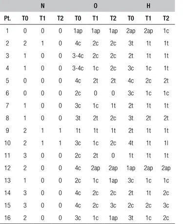
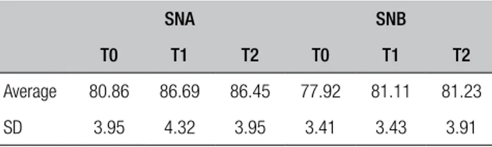
Presurgical upper airway fibroscopy with mueller’s ma-noeuvre identified oropharyngeal and hypopharyngeal collapse in all patients. The most frequent pattern of oro-pharyngeal collapse was the circular type (o: c), and was observed in 14 patients (87.5%), while the most frequent pattern of hypopharyngeal collapse was the transversal type (h: t) and was observed in 10 patients (62.5%). At T1, we observed a reduction of the grade of collapse in all patients. From T1 to T2, we observed a worsening of the grade of collapse in 4 patients, although the T2 grade of collapse was less than the preoperative grade (T0); these patients had significant improvement in clinical outcomes (Table i). We referred these patients to sleep endoscopy. For the cephalometric skeletal measurements (SnA and SnB), no statistically significant changes (p > 0.001) were observed from T1 to T2 (Table ii). These data con-firmed long-term skeletal stability.
The posterior airway space (PAS) was increased in length from T0 to T1, respectively, from 3.73 mm (SD 2.1 mm) to 9.7mm (SD 2.7 mm) (p < 0.001). From T1 to T2, the differences in the PAS were not statistically significant (p > 0.001) (Table iii).
in Table iv, polysomnographic data is shown; from T0 to T1, there was a statistically significant decrease of Ahi, respectively, from 47.1 events/h (SD 22.5 events/h) to
Table I. Upper airway endoscopic evaluation during Mueller’s manoeuvre. NOH (nose oropharynx hypopharynx) classification.
N O H
Pt. T0 T1 T2 T0 T1 T2 T0 T1 T2 1 0 0 0 1ap 1ap 1ap 2ap 2ap 1c 2 2 1 0 4c 2c 2c 3t 1t 1t 3 1 0 0 3-4c 2c 2c 2t 1t 1t 4 1 0 0 3-4c 1c 2c 3c 1c 1t 5 0 0 0 4c 2t 2t 4c 2c 2t 6 0 0 0 2c 0 0 3c 1c 1c 7 1 0 0 3c 1c 1t 2t 1t 1t 8 1 0 0 3t 2t 2c 3t 2t 2t 9 2 1 1 1t 1t 1t 2t 1t 1t 10 2 1 1 3c 1c 2c 4t 1t 1l 11 3 0 0 2c 2t 0 1t 1t 1t 12 2 0 0 4c 2ap 2ap 1ap 2ap 2ap 13 1 0 0 2c 1c 1ap 3c 1c 1c 14 3 0 0 4c 2c 2c 2t 1t 2c 15 3 0 0 4c 2c 3c 2c 2c 3c 16 2 0 0 3c 1c 1ap 3t 1c 2c
Efficacy and safety of maxillomandibular advancement in treatment of obstructive sleep apnoea syndrome
45
16.1 events/h (SD 17.5 events/h) (p < 0.001). moreover, from T1 to T2 we observed a decrease in Ahi, but this change was not statistically significant (p > 0.001). The postoperative result remained stable over time. The oDi
also maintained the same trends. Sao2 < 90% worsened
from T1 to T2, (10 ± 18% vs 19.3 ± 39.3%). Comparing the saturation during wake before and after surgery, this result can be explained as a worsening of the clinical pic-ture and not just as worsening of the sleep disorder. From T0 to T1, the ESS score had a statistically signifi-cant reduction (12.93 ± 1.69 vs. 2.56 ± 1.99) (p < 0.001), while from T1 to T2 the difference in ESS score was not statistically significant (2.56 ± 1.99 vs 4.12 ± 2.52) (p > 0.001) (Table v).
Discussion
mmA is currently considered to be the craniofacial surgery that is most effective in treatment of oSAS in
adults 11 12. The results of surgical treatment of oSAS are
divided into “surgical success” and “surgical cure”. Sur-gical success is defined as an Ahi < 20 events/hour and a > 50% reduction in Ahi 50% after surgical procedure.
Surgical cure is defined as an Ahi < 5 events/hour after surgical procedure 15.
holty and guilleminault performed a meta-analysis and systematic review to estimate the clinical efficacy of mmA in treating oSAS involving 627 adults with oSAS
undergoing mmA 15. The mean Ahi decreased from 63.9
events/hour to 9.5 events/hour following surgery. The surgical success and cure rates were 86.0 ± 30.9% and 43.2 ± 11.7% respectively. They also observed the main-tenance of surgical success at 44 months after surgery. in our study, at follow up (T2) all patients had Ahi < 20 events/hour with a “surgical success” rate of 100%. The surgical cure rate was 37.5%, with 6 patients having an Ahi < 5 events/hour. These results are in agreement with data reported in the above-mentioned meta-analysis and confirm the efficacy of mmA. improvements in Ahi were stable over time. There were no significant changes be-tween the data collected at 6 months (T1) and at follow-up (T2). At follow-up, Ahi sufferers had further a reduction in Ahi registered at T1; this trend, although not statisti-cally significant, could be explained by oedema of the soft tissue that persists for several months after surgery. We observed a significant improvement in daytime sleepi-ness, and all patients had subjective benefits of mmA (Ta-ble v).
The analysis of skeletal cephalometric values (SnA and SnB) at T1 and T2 did not show any statistically significant differences (p > 0.001), confirming the long-term stability of skeletal advancement. no patient had skeletal relapse. in agreement with literature reports, postoperative PAS (T1) showed a significant increase (3.73 ± 9.07 mm vs. 2.1 ± 2.7 mm). At T2, PAS maintained stable values; the differences between measurements performed at T1 and at T2 were not statistically significant (p > 0.001). Even the endoscopic data are encouraging; in fact, all pa-tients showed a reduction in the grade of collapse at T1. At T2, we observed a worsening of the grade of collapse in 4 patients, despite the resolution of symptoms and significant reduction in Ahi. however, in these patients the T2 grade of collapse was less than the T0 grade. one probable ex-planation lies in the extreme subjectivity of the assessment method, and we referred these patients to sleep endoscopy. Skeletal advancement was 9.1 mm ± 1.3 mm for the max-illa and 8.9 mm ± 1.8 mm for the mandible. lye et al. found a statistically significant correlation between the
degree of maxillary advancement and reduction in Ahi 12.
however, others have reported no association between the degree of maxillary advancement and improvement in Ahi
after mmA 16. in our study, mmA was personalized to the
fa-cial characteristics of each patient, and probably a mmA of 1 cm protects patients from relapse in both skeletal soft tissues. initially, mmA was practiced as a second stage of treat-ment, after failure of previous nasal and/or oropharyngeal
surgery 14, and as a first stage of treatment in patients with
substantial craniofacial deformities 5 17. our study
under-Table II. Cephalometric skeletal measurements.
SNA SNB
T0 T1 T2 T0 T1 T2
Average 80.86 86.69 86.45 77.92 81.11 81.23 SD 3.95 4.32 3.95 3.41 3.43 3.91
Data are the angle of measure (°)
Table III. Cephalometric PAS measurements. PAS
T0 T1 T2
Average 3.73 9.7 10.3
SD 2.1 2.7 2.3
Data are number (mm).
Table IV. Polysomnographic data.
T0 T1 T2 AHI 47.1 ± 22.5 16.1 ±17.5 10.4 ± 5.4 Supine AHI 41.1 ± 16.2 17.1 ± 24.2 12.6 ± 5.6 Lateral AHI 11.7 ± 14.0 5.2 ± 9.4 3.2 ± 4.7 ODI 20.4 ± 21.4 7.3 ± 10.1 6.6 ± 6.0 Sa < 90% 25.0 ± 7.0 10.0 ± 18.0 19.3 ± 39.3 Table V. ESS scores.
ESS
T0 T1 T2
Average 12.93 2.56 4.12
M. Giarda et al.
46
lines the possibility to extend mmA, as first stage treat-ment, even in patients without dento-facial anomalies. however, this choice should also be guided by the sever-ity of the disorder, seversever-ity of daytime symptoms, degree of pharyngeal collapse and restriction of PAS.
mmA is generally safe with a major surgical complication maxillary (ischaemic necrosis, cardiac complication) rate
of 1% 15, a minor complication (mandibular relapse, facial
paraesthesia, tempor-mandibular joint disease) 15 18 19 rate
of 3.1% and no reported deaths. immediately after sur-gery, facial paresthesia was observed in 4 patients. At fol-low-up however, no patients reported facial paraesthesia. To reduce the excessive protrusion of maxilla and upper lip, we performed piriform rim recontouring, anterior nasal spine modelling and alar base cinch suture. in our study, no patients reported worsening of their appearance; 13 patients showed a degree of rejuvenation after mmA and 3 reported no postoperative change.
Conclusions
oSAS is a chronic disease, and the goal of treatment is control of symptoms and control of oSAS-related risks by reducing the severity of the disorder. Surgical success and long-term stability of outcomes confirm the efficacy and safety of mmA for treatment of oSAS. however, continuous follow-up of these patients is necessary to control their lifestyle and detect possible relapse.
References
1 Engleman hm, Douglas nJ. Sleepiness, cognitive function and quality of life in obstructive sleep apnea/hypopnea syn-drome. Thorax 2004:59;618-22.
2 haentjeens P, van meerhaeghe A, moscariello A, et al. The impact of continuous positive airway pressre on blood pres-sure in patients with obstructive sleep apnea syndrome: evidence from a meta-analysis of placebo-controlled rand-omized trials. Arch intern med 2007;167:757-64.
3 mcArdle n, Devereux g, heidarnejad h, et al. Long term use of CPAP therapy for sleep apnea/hypopnea syndrome.
Am J respir Crit Care med 1999;159:1108-14.
4 Sher AE, Schechtman KB, Piccirillo JF. The efficacy of sur-gical modifications of the upper airway in adults with ob-structive sleep apnea syndrome. Sleep 1996;56:596-602.
5 hochban W. Surgical treatment of obstructive sleep apnea.
otorhinolaryngol nova 2000;10:149-56.
6 mantovani m, minetti A, Torretta S, et al. The velo-uvulo-pharyngeal lift or “roman blinds” technique for treatment
of snoring: a preliminary report. Acta otorhinolaryngol ital 2012;32:48-53.
7 vicini C, Frassineti S, la Pietra mg, et al. Tongue Base Reduction with Thyro-Hyoido-Pexy (TBRTHP) vs. Tongue Base Reduction with Hyo-Epiglottoplasty (TBRHE) in mild-severe OSAHS adult treatment. Preliminary findings from a prospective randomised trial. Acta otorhinolaryngol ital 2010;30:144-8.
8 Caples Sm, rwley JA, Prinsell Jr, et al. Surgical modifi-cations of the upper airway for obstructive sleep apnea
in adults: a systematic review and meta-analysis. Sleep
2010;33:1396-407.
9 lin hC, Friedman m, Chang hW, et al. The efficacy of multi-level surgery of upper airway in adults with obstructive sleep
apnea/hypopnea syndrome. laryngoscope 2008;118:902-8.
10 Fairburn SC, Waite PD, vilos g, et al. Three-dimensional changes in upper airways of patients with obstructive sleep
apnea following maxillomandibular advancement. J oral
maxillofac Surg 2007;65:6-12.
11 Fusetti m, Fioretti AB, valenti m, et al. Cardiovascular and metabolic comorbidities in patients with obstructive
sleep apnoea syndrome. Acta otorhinolaryngol italica
2012;32:320-5.
12 lye KW, Waite PD, meara D, et al. Quality of life evalua-tion of maxillo-mandibular advancement surgery for
treat-ment of obstructive sleep apnea. J oral maxillofac Surg
2008:66:968-72.
13 vicini C, mira E. NOH classification. A proposal. Atti del
Xlvii raduno gruppo Alta italia di otorinolaringoiatria. riccione 2001, p. 155-163.
14 riley rW, Powell nB, guilleminaul C. Obstructive sleep ap-nea syndrome: a surgical protocol for dynamic upper airway reconstruction. J oral maxillofacial Surg 1993;51.742-7.
15 holty JC, guilleminault C. Maxillomandibular advancement for treatment of obstructive sleep apnea: a systematic review and meta analysis. Sleep med rev 2010;14:287-97.
16 Smatt y, Ferri J. Retrospective study of 18 patients treated by maxillomandibular advancement with adjunctive proce-dures for obstructive sleep apnea syndrome. J Craniofac Surg 2005;16:770-7.
17 miles PJ, nimkarn y. Maxillomandibular advancement sur-gery in patients with obstructive sleep apnea: mandibular
morphology and stability. int J Adult orthod orthognath
Surg 1995;10:193-200.
18 hendler Bh, Costello BJ, Silverstein K, et al. A protocol for uvulopalatopharyngoplasty, mortised genioplasty, and maxillomandibular advancement in patients with obstructive sleep apnea: an analysis of 40 cases. J oral maxillofac Surg 2001;59:892-7.
19 van Sicklels JE, hatch JP, Dlce C, et al. Effects of age, amount of advancement, and genioplasty on neurosensory disturbance after bilateral sagittal split osteotomy. J oral maxillofac Surg 2002;60:1012-7.
Address for correspondence: mariangela giarda, S.C.D.u. di Chirurgia maxillo-Facciale, ospedale maggiore della Ca-rità, corso mazzini 18, 28100 novara, italy. Tel. +39 0321 3733893/3733783/3733895. Fax +39 0321 3733893. E-mail: [email protected]