SCUOLA NORMALE SUPERIORE
ANNO ACCADEMICO 2008-2009
Tesi di Perferzionamento in
Neurobiologia
Visual discrimination learning and
LTP-like changes in primary visual cortex
Relatori: Candidato:
Alessandro Sale Roberto De Pasquale
Lamberto Maffei
INTRODUCTION...4
THE CONCEPT OF LEARNING
...5
Declarative memory ...6
Non-declarative memory ...8
VISUAL DISCRIMINATION LEARNING
...10
Visual perceptual learning ...11
IN SEARCH OF A PHYSIOLOGICAL MODEL FOR VISUAL DISCRIMINATION
LEARNING...16
Cellular mechanisms underlying memory and learning... 17
The discovery of LTP ...18
LTP: A PHYSIOLOGICAL SUBSTRATE FOR MEMORY AND LEARNING...20
Pharmacological approaches ...21
Genetic approaches...24
Does learning produce LTP-like changes?...26
Does the induction of LTP influence subsequent learning? ...28
Does learning influence the induction of LTP? ...31
LTP: A CELLULAR POINT OF VIEW
...32
LTP in the primary visual cortex ...35
LTP IN PRIMARY VISUAL CORTEX AND VISUAL DISCRIMINATION LEARNING
..39
MATERIALS AND METHODS ...41
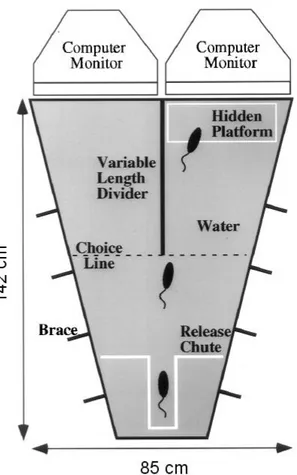
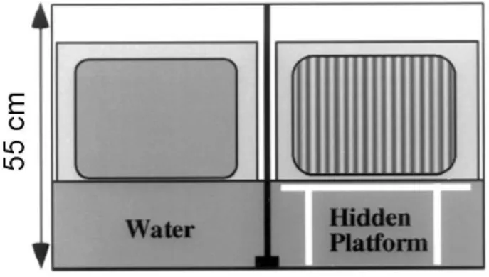
ANIMALS...41
EXPERIMENTAL SETTINGS
...41
Visual water task ...41
Electrophysiology...44
Drug administration ...44
E
XPERIMENTAL PROCEDURES...45
Visual discrimination improvement assessment ...45
Change of stimuli orientation...47
Mimicry and occlusion of LTP ...48
Pharmacological interference...49
RESULTS...50
VISUAL DISCRIMINATION IMPROVEMENT
...51
SELECTIVITY OF ORIENTATION
...58
VISUAL DISCRIMINATION LEARNING CAUSES LTP-LIKE CHANGES IN PRIMARY
VISUAL CORTEX...60SATURATION OF PRIMARY VISUAL CORTEX LTP BY VISUAL DISCRIMINATION
LEARNING...62
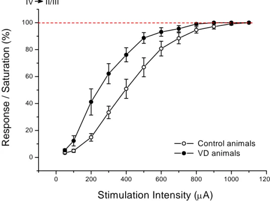
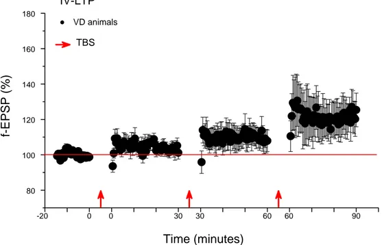
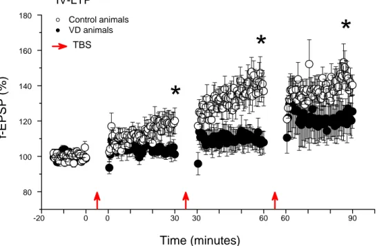
IV-LTP ...62
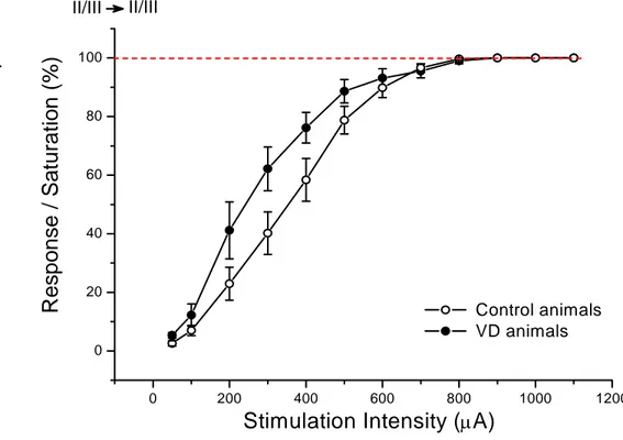
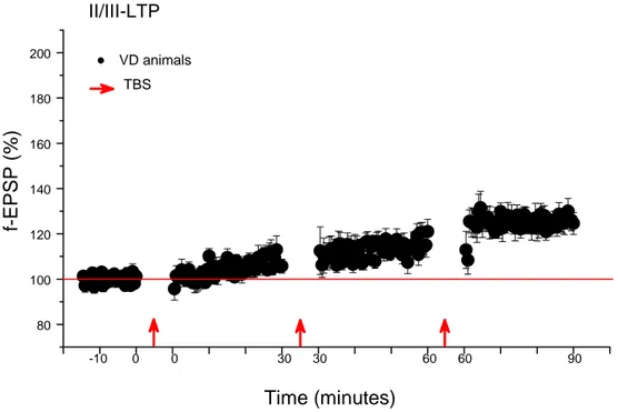
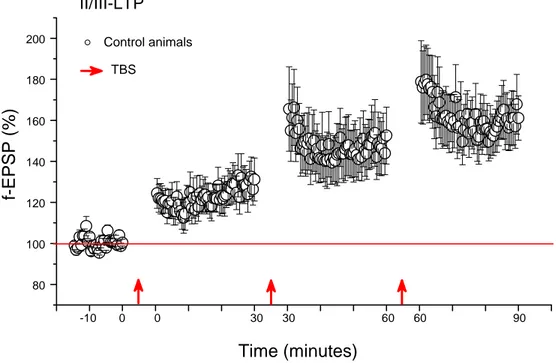
II/III-LTP ...65
PHARMACOLOGICAL BLOCKADE OF LTP IMPAIRS VISUAL DISCRIMINATION
LEARNING...68
DISCUSSION ...70
BIBLIOGRAPHY ...78
Introduction
Visual discrimination learning is a visual process that refers to the ability to differentiate one visual target from another. This ability is fundamental for an individual to interact with the environment. For example, the ability to learn to visually discriminate letters and words becomes essential in learning to read and deficits in visual discrimination are a common cause of reading problems. One must be able to discriminate visually in terms of colour, foreground-background, form, size, and position in space.
Visual discrimination learning is a property of visual perception that is supposed to rely upon modifications of synaptic strength in neurons of neural structures concerning visual perception. The study of the physiological and cellular mechanisms underlying this plasticity contributes to increase our knowledge about how the brain makes use of visual information from the external world. This knowledge is fundamental to better understand how to intervene in diseases related to visual discrimination skills.
The principal mechanisms involved in visual discrimination learning is probably visual perceptual learning, which is defined as the increase in visual abilities after training. Numerous studies tried to explain the link between visual perceptual learning and the sensorial plasticity underlying it. Results from these studies showed that cortical areas operate in this process as low as primary visual cortex (V1) at the first levels of perception (Vogels & Orban 1985, Shiu & Pashler 1992 and Schoups et al. 1995). In particular, V1 is actually known as the cortical field in which visual perceptual learning is more likely to take place involving simple visual stimuli such as gratings (Schoups et al. 2001, Furmansky et al. 2004, Maertens & Pollmann 2005, Frenkel et al. 2006, Pourtois et al. 2007 and Yotsumoto et al. 2008).
The aim of this study is to verify the relation between visual discrimination learning and Long Term Potentiation (LTP), which is one of the best characterized forms of synaptic plasticity underlying various kinds of learning (Rogan et al. 1997, Moser et al. 1998, Rioult-Pedotti et al. 2000 and Whitlock et al. 2006). However, before analysing this topic
in details, it is useful to spend some words about the general concepts of learning and memory.
The concept of learning
Learning is the acquisition of new knowledge or skills from the external world. The expression of learning is the emergence of new behavioural patterns or simply the modification of pre-existing ones. Since the concept of learning implies that information has to be conserved to be subsequently recalled or re-used, investigating learning implyes the study of memory. Memory is the faculty that allows information from the environment to be stored. A basic and generally accepted classification of memory is based on the duration of memory retention, and identifies two distinct types of memory, short term memory and long term memory (Mc Gaugh 1966). When sensorial information is transferred to short-term memory, this allows one to recall it from several seconds up to minutes. Short-term memory is supported by transient patterns of neuronal communication (Bauer & Fuster 1976 and Jonides et al. 1998). Storage of short-term memory generally has a strictly limited capacity and duration. Information is available for a certain period of time, but is not retained indefinitely. To be long lasting a memory trace has to be consolidated through processes involving long term memory storage (Kesner & Connor 1972). In biological terms, long-term memories are maintained by stable and permanent changes in neural connections widely spread throughout the brain (Ordy & Schjeide 1973 and Markowitsch 1985).
Another kind of classification divides memory in two distinct, independent and parallel systems. This idea about memory organization became a topic of experimental interest when evidence from normal subjects, amnesic patients, and experimental animals converged on the same view (Scoville & Milner 1957 and Squire 1992). A fundamental distinction can be made between declarative or explicit memory that is accessible to awareness and non-declarative or implicit memory that is not (Leritz et al. 2006 and Speekenbrink et al. 2008).
Declarative memory
Declarative or explicit memory refers to the capacity of acquiring or modifying knowledge about facts and events. It is the kind of memory that is impaired in amnesia and it can be divided into semantic memory concerning facts about the world and episodic memory concerning the capacity to re-experience an event in the context in which it originally occurred (Tulving 1983).
Focusing on visual declarative memory, it has been shown that when information is acquired through the visual pathways, visual stimuli are coded by declarative memory systems as explicit knowledge (Desimone 1996 and Wolfe 1998). Initially, information processing occurs in a group of anatomically linked cortical fields, the so-called object-analyzer system, often called the “ventral visual stream” (Murray et al. 2007). It comprises several visual areas including V1 and the inferior temporal cortex (Stotnick 2004). Subsequently, to persist as memories, visual features are to be consolidated by the temporary intervention of the medial temporal lobe (MTL).
Determination of specific systems involved in memory consolidation began with the finding that damage to the MTL produced severe amnesia (see Warrington & Weiskrantz 1969). MTL is a term of convenience for referring collectively to the hippocampus, dentate gyrus, subicular complex, amygdala, and perirhinal, entorhinal, and parahippocampal cortex. These structures make selective contributions to declarative memory. Hippocampus is involved in processing information about places and paths, while perirhinal cortex seems to be more involved in processing information about objects. During amnesia, while remote memories usually remain intact, recently acquired declarative memories do not. This happens because amnesic patients with MTL damage have great difficulty in forming new long term memories (Scoville & Milner 1957, Penfield & Milner 1958 and Corkin 1984). Another important observation is that when brain pathology includes damage to the neocortex, remote memory is often impaired (Graham & Hodges 1997, Squire et al. 2001 and Bayley et al. 2003).
These findings suggest that initial acquisition and retrieval of declarative memories require MTL, while subsequent storage of information in various neocortical areas occurs without a further significant MTL contribution (Squire 1992, McClelland et al. 1995,
Squire et al. 2004 and Nadel & Moscovitch 1997). Memories gradually become independent from MTL as they are consolidated in neocortical circuits that serve as remote memory storage (Alvarez & Squire 1994 and Squire & Alvarez 1995). Studies of hippocampus-dependent memory in animals have largely confirmed this idea (Zola-Morgan & Squire 1990, Kim & Fanselow 1992, Kim et al. 1995, Anagnostaras et al. 1999, Frankland et al. 2001, Sutherland et al. 2001 and Clark et al. 2002). Moreover, there is strong evidence suggesting that synaptic structural changes take place in the neocortex during consolidation (Maviel et al. 2004 and Frankland et al. 2004), probably in order to stabilize remote memories. Consolidation processes are likely to be distribuited in different regions of the neocortex, including visual areas (Roland & Gulyàs 1995 and Mc Gaugh 2000).
But why does declarative memory need two complementary systems? Gradual interleaving of memories into the neocortex is essential for discovery of generalities and the eventual formation of knowledge structures. Using connectionist models, it can be shown that the rapid incorporation of new information into an existing knowledge system would cause catastrophic interference (Marr 1970, Marr 1971 and McClelland et al. 1995).
Essentially, new information would dominate and erase previously acquired information. Probably this explains why cortical consolidation is a slow, extended process, and why the hippocampus is needed as a temporary link between distributed cortical memories. New memories need to be incorporated into existing knowledge structures in the cortex through a gradual, interleaving process to avoid the loss of old information. This might happen during periods of inactivity and sleep, when bursts of activity, called sharp-waves (SPWs), are generated in the hippocampus (Buzsaki 1989 and Hasselmo 1999). SPWs could provide the activation required to drive intercortical plasticity and to promote cortical consolidation. This periodic activity seems to operate by a mechanism called synaptic re-entry reinforcement (Shimizu et al. 2000, Cui et al. 2004 and Wittenberg & Tsien 2002). Recent observations show that experiences are replayed during sleep synchronously in the hippocampus and in the visual cortex (Mehta 2007). During slow-wave sleep in rats, multicell spiking patterns in the visual cortex and in the hippocampus are organized into frames, defined as periods of stepwise increase in neuronal population activity. The multicell firing sequences evoked by awake experience are replayed during these frames in
both regions and coordinated to reflect the same experience (Ji & Wilson 2007). This probably implies simultaneous reactivation of coherent memory traces in the visual cortex and hippocampus during sleep. This reactivation may contribute to or reflect the result of the memory consolidation process.
Non-declarative memory
Non-declarative or implicit memory refers to processes known to be dispositional, expressed through performance, that have the ability to gradually extract common elements from a series of separate events. Memories occur as modifications within specialized performance systems. They are categorized in groups based loosely on functional properties and sometimes more strongly on functional or anatomical dissociations. They are revealed through reactivation of the systems within which learning originally occurred (Schacter 1992, Ashby & Waldron 1999 and Smith 2008). Typical examples are non-associative learning (habituation, sensitization and dishabituation), associative learning, skill learning, priming, perceptual learning and emotional learning (Roediger 3rd 1990).
Habituation, sensitization and dishabituation are the simplest forms of learning giving rise to non-declarative memory (Carew 1989). During habituation, repetition of a non relevant stimulus leads to a decrease in reflexive response, while during sensitization a strong aversive stimulus leads to an increase in sensitivity of other aversive sensory/motor reflexes. Dishabituation is the case in which sensitization is formed to override the previous habituation. These forms of implicit learning are non-associative because habituation involves the modification of a single sensory channel and not the association of two different ones, while sensitization is not specific to any sensory channel.
Associative learning occurs when two or more sensory streams, motor rules or cognitive rules are associated. The best described forms are classical conditioning and operant conditioning. In classical conditioning a non relevant conditioned stimulus (CS) is coupled with a relevant unconditioned stimulus (US), so that the CS becomes subsequently relevant (Pavlov 1927). In operant conditioning the positive or the negative reinforcement
of a behavioural pattern leads to the modification of the subsequent use of the same specific behaviour (Skinner 1935).
Skill learning is the acquisition of new behavioural abilities with practice and is defined as facilitation on a range of abilities in a particular task (Squire 1992 and Squire & Zola 1996). It relies upon basal ganglia and cerebellum activity. The initial cognitive stage requires working memory capacity. This stage is the categorization of skills used to guide behavior. In the subsequent associative stage behavior becomes tuned and errors are eliminated, while in the subsequent autonomous stage there is a gradual continued improvement of skill with little reliance upon working memory.
Perceptual learning is the specific and relatively permanent modification of perception and behavior following sensory experience (Schacter 1990). It involves structural and functional changes in primary sensory cortices.
Priming is an improvement in a perceptual or conceptual task from a one trial learning perceptual exposure to the stimulus being used in the task (Squire 1992). Priming is thought to happen in primary sensory areas and results from an improvement in processing efficiency. Much of priming results in a decrease in response time or in an increase in probability of correct response.
Emotional learning concerns the unconscious learning by storage of information about the emotional significance of events (LeDoux 1993). The neural system underlying emotional learning critically involves the amygdala and structures with which it is connected. It includes all the emotional reactions that are built over time by simple exposure.
Visual discrimination learning
During visual discrimination learning, the process of perception becomes adapted to the environment. Experience increases the attention paid to features that are important, and decreases the attention to irrelevant ones. Attention can be selectively directed toward important stimulus aspects at several different stages in information processing. Researchers in animal learning and human categorization have described shifts toward the use of dimensions that are useful for tasks (Nosofsky 1986) or have previously been useful (Lawrence 1949). Thus, experience can lead to the separation of perceptual dimensions comprised in a single stimulus. Dimensions that are originally treated as fused often become segregated with development or training. The subject shifts from perceiving stimuli in terms of holistic, overall aspects to analytically decomposing objects into separate dimensions. This trend has received substantial support from developmental psychology.
As mentioned before, two memory systems can be distinguished in terms of the different kinds of information they process and the principles by which they operate. In visual discrimination learning both systems may be potentially utilized. The critical aspect is the strategy implemented during the discrimination learning, which reflects which memory system is principally engaged. Categorizing the objects that are to be discriminated requires attending to the object-based spatial frequency information collected by different spatial frequency channels of the visual system. This drives a visual perceptual learning process of the spatial frequencies that facilitate the particular categorization of the object (Sowden & Schyns 2006). However, sinse retinal information about object spatial frequencies varies in size with distance, the critical bands of diagnostic spatial frequencies are seen by different channels. Support is provided by knowledge whenever the ability to abstract and generalize is needed to optimize visual discrimination performance (Sowden & Schyns 2006). Thus, in a visual discrimination task, recognition may also be useful and an interaction may happen between the top-down conscious object-based indications and the bottom-up information coming from the spatial frequency channels organization of the visual system. The top-down and bottom-up contribution in discrimination may vary according to the complexity of the object features and to the categorizing ability of the discriminating organism (Sowden & Schyns 2006).
In visual pattern discrimination tasks, monkeys with large MTL lesions show no deficit concerning learning and retention of pattern discriminations (Squire & Zola-Morgan, 1983). Amnesic patients learn such tasks in a few trials, like normal individuals, but they later loose awareness of what they previously learned (Squire et al. 1988). The difference appears to lie in the fact that monkeys learn the pattern discrimination task gradually, during several hundred of trials in a manner reminiscent of visual perceptual learning (Iversen, 1976) while humans approach the task as a simple problem of conscious memorization. These findings show that it is possible to observe an experimental situation in which only one of the two systems is substantially working, but more generally, almost anytime visual discrimination is occurring, both systems might be utilized together with different respective contributions, according to different strategies usable to learn a task. It is often problematic to completely isolate the single contribution of one of the two different complexes engaged, especially working with animals which cannot suggest a verbal check of the conscious aspects of the information acquired.
Visual perceptual learning
Visual perceptual learning is maybe the principal mechanism operating in visual discrimination learning concerning simple visual stimuli such as gratings (Schoups et al. 2001, Furmansky et al. 2004, Maertens & Pollmann 2005, Frenkel et al. 2006, Pourtois et al. 2007 and Yotsumoto et al. 2008). It involves relatively long-lasting changes to an organism’s visual system that improves its ability to respond to the environment. A major consequence of visual perceptual learning is that perceptions become increasingly differentiated from each other. By differentiation, stimuli that were initially perceptually indistinguishable become separated. Laboratory studies have extensively studied training effects involving simple discriminations, noting that improvement comes after several training sessions (Magnussen & Greenlee 1999) or, in some cases, as the effect of a mere exposition to a stimulus (Magnussen 2000). In order to focus the investigation exclusively on the non-declarative aspects of learning coinciding with visual perceptual learning, the impact of verbal or categorical coding has to be minimized. In current research this impact
is reduced by studying the retention of single dimensions or attributes of the visual stimulus. The decay of memory is tracked in delayed discrimination tasks with variable time intervals interposed between the stimuli that are to be compared. Memory performance is indexed by the resultant discrimination thresholds or some equivalent measures (Kinchla & Smyzer 1967, Laming & Scheiwiller 1985, Regan 1985, Magnussen et al. 1990 and Magnussen & Greenlee 1999).
Visual perception is known to concern various proprieties of visual stimuli including orientation, direction of motion, texture, deepness, spatial position and spatial frequency (Shapley & Lennie 1985, Baker Jr & Mareschal 2001 and Derrington et al. 2004). In order to make visual perceptual learning strictly specific, during the training discrimination improvement is directed to one particular property of the stimuli (Gilbert 1994). This specificity has deep implications for the understanding of neural mechanisms underlying visual perceptual learning. For example, some features such as orientation, contrast and colour exhibit a slight decay in the short-term memory range of visual perceptual learning, whereas others, such as spatial frequency and motion, are stored with precision (Nilsson & Nelson 1981, Vogels & Orban 1986, Lee & Harris 1996, Blake et al. 1997 and Reinvang et al. 1998). Moreover, trained performance on a horizontal discrimination task frequently does not transfer to a vertical version of the same task (Fahle & Edelman 1993 and Poggio et al. 1992), nor does it transfer to new retinal locations (Fahle et al. 1995 and Shiu & Pashler 1992), and it does not completely transfer from the trained eye to the untrained eye (Fahle et al. 1995).
During the task of discriminating changes along a single property (for example spatial frequency) in a multiple property test, human observers are able to extract the relevant information from concurrent changes along other properties, for example contrast or orientation, as precisely as when the stimuli to be compared vary along a single property (Burbeck 1987 and Heeley et al. 1993). These observations suggest an interesting model for visual discrimination. A set of second-order neural representations might combine information from neural representations tuned to different properties of the visual stimulus (Magnussen et al. 1998 and Olzak & Thomas 1999). These second-order mechanisms might be organized in a modular way. Parallel mechanisms that are dedicated to the processing of one property (for example, spatial frequency) would abstract information
across other properties simultaneously (for example, orientation and contrast). Each property-dedicated mechanism would be organized in terms of an array of memory stores that would be linked in a lateral inhibitory network and each store would code a restricted range of values along the property. According to this model the operating system should be a neural structure where representation of the basilar proprieties of the stimulus is strictly organized.
Learning tasks concerning simple stimuli, with specificity for properties like spatial frequency or stimulus orientation, are likely to be mediated by mechanisms involving the first steps of cortical elaboration (Vogels & Orban, 1985; Shiu & Pashler, 1992 and Schoups et al. 1995), where receptive fields are smaller, visual topography is finely organized and there is a fine selectivity for orientation and spatial frequency. V1 is known to have neurons, called simple cells, with high selectivity for stimulus orientation (Hubel & Wiesel 1959), which is an important feature of the organization of V1 columnar architecture (Hubel & Wiesel 1977). Recent investigations utilizing Magnetic Functional Resonance (fMRI) and Transcranical Magnetic Stimulation (TMS) found direct evidence that visual discrimination improvements show changes at the first states of visual information cortical elaboration (Furmansky et al. 2004 and Maertens & Pollmann 2005). For example, one of these studies directly showed that improved visual perceptive performance was linked to increased V1 neural activity (Furmansky et al. 2004). Subjects were trained to recognize a low contrast grating, while fMRI recordings occurred before and after the training. Primary visual cortex response was increased after learning and this effect was specific for location and orientation of the training stimulus.
Important findings about V1 involvement in visual perceptual learning were also obtained by electroencephalogram (EEG) recording experiments. One study examined the period in which visual perceptual learning took place in subjects trained in a texture discrimination task (Pourtois et al. 2007). This approach had a temporal resolution which was able to define the latency of the effects observed after training. The target produced a change in the visual evoked potential in V1, which was the earliest component of the whole cortical response. This effect only occurred when target was present in a previously trained location and in corrispondence of the upper part of the visual field. Thus, this study showed that plasticity in V1 can underlie the consolidation of a recent perceptive ability. This
ability is acquired by modeling the initial charge of sensorial input which occurs at the first visual cortical area.
All these ideas deriving from imaging experiments, were also supported by findings of correlations between electrophysiological recordings and orientation learning in monkeys V1 (Schoups et al. 2001). Behavioral improvement in this type of learning has been linked to an improved neuronal performance of trained compared to naive neurons. Improved long-term neuronal performance resulted from changes in the characteristics of orientation tuning of individual neurons. More particularly, the slope of the orientation tuning curve, that was measured at the trained orientation, increased only for the subgroup of trained neurons, most likely to code the orientation identified by the monkey. No modifications of the tuning curve were observed in orientations for which the monkey had not been trained.
However, in another study (Ghose at al. 2002) the authors showed that in V1 perceptual learning consisted only in a little reduction of the response amplitude of single neurons tuned on the training orientation. There were no modifications in the receptive field proprieties. They argued the psychophysical change was more probably obtained by a decoding strategy specifically optimized for training, than by a better neural representation of orientation in the primary visual areas. However, differences in the specificity of training experimental design may influence the contribution of the brain areas involved. In particular, learning observed by Schoups and colleagues was eye and location specific (Schoups et al. 2001), which is consistent with neural changes in V1, while Ghose et al. found a transfer of learning improvement from one eye to another and between different retinotopic locations (Ghose at al. 2002).
This discrepancy between these two studies might be explained by a recent interesting fMRI study (Yotsumoto et al. 2008). Authors examined V1 changing activity during the occurrence of visual perceptual learning, in a texture discrimination task. During every training session subjects were asked to point to a letter while a target stimulus was constantly presented for a brief time in a peripheral location of their visual field. Subjects were asked to identify the letter and to define the orientation of the target stimulus. In separated imaging session, fMRI activity of subjects was measured while the level of the task performance was evaluated. Relation between the level of performance and V1 activity
deferred along the whole period of learning task. Visual plasticity could be distinguished in two phases. In the first one, an increasing of performance level corresponded to an increasing fMRI signal recorded on the visual cortex. Authors suggested that an increase in the number and strength of synapses might have occurred during this phase. These changes probably underlined both the fMRI signal increase, and the higher level of performance. In the second phase, instead, a stable saturation of improved performance occurred together with a decrease of the cortical fMRI signal. After saturation of performance level, the number and the strength of synapses involved, might have been reduced and only the ones that were essential to continue the task might have been kept activated. This experiment suggested a model in which the local network of visual cortex can be reorganized to acquire and consolidate information during learning, but once the task has been completed the level of performance can be kept without further reorganizations.
While in primates the neural substrate involved in perceptual learning may have a deep dependence on training specificity, in rodents the relation between learning and neural changes might be more simple. Repeated exposition to a stimulus of defined orientation, leads to a specific potentiation of the response in primary visual cortex of awake mice (Frenkel et al. 2006). This was recently demonstrated by the evidence in V1 of changes in amplitude of visual evocated potentials (VEPs), recorded during visual exposure tests. Repeated exposure to a specific oriented stimulus leads to an increase to its evoked response. Modifications underlying potentiation were resistant even after the subsequent exposure to an orthogonally oriented stimulus. The animals were in a condition of passive exposure to stimuli, so the cortical modification required in case of an active training might have a more consistent effect. It is anyway interesting to note that cortical changes observed in mice are very similar to fMRI signal increase caused by training in human V1 (Furmansky et al. 2004) and are also similar to the results obtained by recordings in V1 of monkeys (Schoups et al. 2001).
Perceptual learning may also be associated to changes in the contextual modulation of neurons response in V1 (Crist et al. 2001 and Li et al. 2004). After a training of bisection of three lines or after a Vernier training (little discrepancy of orientation discrimination) monkeys showed an improvement with no change in the receptive fields basal proprieties in V1. However, there was a change in the contextual modulation, a high order property of V1
cells: the modulation of the response due to the introduction, out of one cell receptive field, of further stimuli having spatial relation with the cell preferred stimulus.
When improvement requires a mechanism that takes count of the contest, top-down interactions between multiple brain areas control physiological changes by a combination of local circuitry and feed back connections from higher cortical levels (Grossberg 1999, Gilbert et al. 2000 and Gilbert & Sigman 2007). Indeed, the function of primary visual cortex is known to result from an interaction between feed forward and top-down information. Internal representations of the world, behavioral requirements, attention and expectation affect the brain strategy for analyzing the visual field. Complex information that is represented at higher stages may control perceptual learning by influencing simpler processes occurring at antecedent stages by top-down corticocortical connections.
In search of a physiological model for visual discrimination
learning
I discussed how visual discrimination learning requires processes of visual perceptual learning, which are principally related to changes occurring specifically in the primary visual cortex (V1). This issue makes V1 the visual area most likely to show plastic changes and the most reasonable to choose for an in-depth examination aimed at the investigation of physiological and cellular mechanisms operating in the network of neurons involved.
However, before analysing in details a suited model for visual discrimination learning, it is useful to review the most used approaches for the study of biological aspects of memory and learning.
Cellular mechanisms underlying memory and learning
The concept of neuronal networks is dated as early as 1884 (Exner 1884) and was further defined by describing algorithms that developed its fundamental principles (Hodges 1983). Since the birth of this concept, ensembles of neurons are thought to participate in maintaining a representation that serves as a memory trace. Such ensembles require dynamic interactions among neurons and the ability to modify these interactions. This implies use-dependent changes in the activity of the network, which is most easily altered by changes in synaptic function. Hebb formalized this idea in his postulate: “When an axon of cell A is near enough to excite cell B and repeatedly or persistently takes part in firing it, some growth process or metabolic change takes place in one or both cells such that A’s efficiency, as one of the cells firing B, is increased (Hebb 1949) ”. The use of the Hebb rule in a distributed memory system can lead to an efficient storage of a number of representations within the same neural network.
Since the first attempts on the research of the physiological and molecular modifications underlying memory and learning, neural networks resulted very complex. Thus, this pioneer approach to neuroscience began with the study of the simpler nervous system of invertebrates, in order to define the synaptic plasticity underlying simple forms of learning, like habituation, sensitization and classical conditioning. The experiments on Aplysia californica are the most famous example. Results showed that molecular mechanisms underlying classical conditioning in Aplysia Californica, are a modification of the simpler processes known to involve sensitization (Castellucci et al. 1970, Carew et al. 1971, Walters et al. 1979 and Kandell 2001). All results converged suggesting that complex kinds of learning might be constituted by an ensemble of mechanisms similar to those underlying simpler kinds (Kosower 1972). This observations moved a lot of researchers to pass to an appropriate model suitable for the more complex mammalian nervous systems.
Among mammals, rodents are ideally suited for such a research in neurobiology. Understanding their neural structures might be crucial for elucidating the fundamental structure and function of the mammalian brain, because rodents are the largest order of mammals, representing over 40% of mammalian species. They are among the smallest
mammals known with adult weights in the range of few hundreds grams and they have a short generation time, on the order of ten weeks from being born to giving birth. This make rodents relatively easy to house. Females breed prolifically in the lab with an average of 5-10 pups per litter, this number is big enough to allow statistically robust sample sizes. Moreover, most laboratory-bred strains are relatively docile and easy to handle.
As soon as the brain of rodents was recognized as a suited mammalian model for neurobiology, investigation about physiological mechanisms of learning and memory started principally with the study of hippocampal circuitry. In fact, since the occurrence of amnesia after hippocampal lesion was well known, hippocampus has always been acknowledged as one of the functional structure principally involved in declarative memory. Electrophysiological studies of hippocampal circuitry soon revealed a phenomenon of increase in synaptic transmission whose mechanisms might also underlie the occurrence of learning and memory. This phenomenon is called Long Term Potentiation (LTP) and is probably the most powerfull model to investigate physiological and cellular mechanisms operating in the network of neurons involved in synaptic plasticity.
The discovery of LTP
Bliss & Lømo first found the proof that hippocampal neurons show plastic modifications that might be the ones necessary for declarative memory (Bliss & Lømo 1973). They observed that tetanic stimulation of the perforant path in anesthetized rabbits increased the slope of the population excitatory post-synaptic potential (field EPSP) recorded extracellularly in the dentate gyrus of the hippocampus. They labelled that phenomenon Long-Term Potentiation (LTP).
LTP is the long-lasting improvement in synaptic transmission between two neurons, occurring after a high-frequency stimulus to the presynaptic fiber. LTP improves the ability of neurons to communicate with one another across synapses and it is recognized as one of the best known form of synaptic plasticity. LTP is ruled by multiple mechanisms that vary
according to different brain region, animal age and species. In the best known form of LTP, enhanced communication is predominantly reached by improving the postsynaptic neuron sensitivity to neurotransmitters.
Most types of LTP are known to be N-methyl-D-aspartate (NMDA) receptor dependents (Collingridge et al. 1983) and this dependence is directly correlated to two important proprieties: associativity and cooperativity. Indeed, in order for LTP to occur, depolarization of postsynaptic cells and contemporaneous presynaptic activity (as predicted by Hebb’s postulated) is necessary and the cooperation of more than one activated fibre is needed. Associativity and cooperativity depend on NMDA receptor which is well suited to be involved in hebbian plasticity mechanisms (Tsien 2000, Brown et al. 1988 and Collingridge & Bliss 1995).
NMDA is a voltage-dependent glutamate receptor subtype. For LTP induction, the NMDA receptor must be activated by the neurotransmitter glutamate while simultaneously there must be sufficient depolarization of the postsynaptic membrane to relieve a Mg2+ block in the NMDA-associated ion channel, which allows the entry of Ca2+ into the postsynaptic neuron. Ca2+ activate a number of Ca2+-sensitive second messenger pathways.
Because NMDA receptors are sensitive to both presynaptic transmitter release and postsynaptic depolarization, they act as hebbian coincidence detectors. This property can explain cooperativity and associativity through temporal and spatial summation. Thus, activated NMDA receptors at synapses that are proximal to active sites of depolarization, may be depolarized sufficiently to relieve the Mg2+ block and initiate the cascade of events
that leads to LTP induction. This cascade may occur even if the activity of a particular synapse alone is not sufficient to induce LTP. NMDA receptors can account for the association of two separate afferent projections to the same cell, one strongly and the other weakly active (Kelso & Brown 1986 and Levy & Steward 1979), and for the cooperative requirement by which a threshold number of fibers are active.
LTP: a physiological substrate for memory and learning
Since neurons communicate by synapses and memories are believed to be stored within synapses, it is not surprising that LTP is probably the most popular accepted cellular mechanisms of how memory traces could be stored in the neuronal networks. The reason for such popularity is probably the fact that those changes in the synaptic strength fitt quite well with the theoretical predictions of Hebb’s postulate. (Bliss & Lynch 1988, Morris 1989 and Montague & Sejnowski 1994). If memory is stored in networks of neurons and if network efficiency is mediated by persistent activity, then LTP induced by persistent stimulation of an afferent pathway appears as a likely mechanism by which the brain stores information.
Progress has been made in demonstrating that LTP possesses a number of features that would be expected for a computational device used to store information. Among these are the fact that LTP meets the durability requirement for longer lasting memories and the fact that repetition of LTP induction produces longer lasting LTP (Barnes 1979), just as practice improves behavioural retention. Moreover, there are three similarities between LTP and learning in support of the notion that LTP is a memory mechanism: LTP is specific to tetanized inputs, it is associative and it lasts for a long time.
Simple neural reflexes may be incorporated into conditioned reflexes that are known to involve specific neural pathways. Both pre- and postsynaptic specificity have been demonstrated under certain conditions also in LTP. LTP is specific in the way that only tetanized afferents show potentiation. However, the idea of specificity of tetanized afferents has become clouded by reports that LTP induction might involve molecules and retrograde messengers that diffuse into adjacent neurons (O’Dell et al. 1991, Schuman & Madison 1991, Bonhoeffer et al. 1989). This lack of specificity might however play a role, in the sense that diffuse alterations in different presynaptic elements may permit the storage of the temporal order of inputs (Montague & Sejnowski 1994).
Another interesting property of LTP, which led some researchers to suggest that it is a memory mechanism, is associativity. As already said, if weak non-LTP-inducing stimulation in one afferent is paired with strong LTP-inducing stimulation in another
afferent to the same cell population, then the weakly stimulated afferent will also exhibit LTP (Levy & Steward 1979, McNaughton et al. 1978). The property of associativity is reminiscent of classical conditioning, but the temporal constraints of associative LTP are dissimilar to those of classical conditioning. In addition, the necessary temporal ordering of CS and UCS are absent in associative LTP, and a mechanism as simple as associative LTP cannot account for the behavioral complexity observed in classical conditioning. Associative LTP does, instead, bear comparison to sensory preconditioning, another psychological example of learning (Mackintosh 1974).
Even if all these observations suggest that LTP is a substrate of memory, they are not sufficient to validate this hypothesis. In order to demonstrate such hypothesis, stronger evidence from more specific experimental approaches is needed.
Pharmacological approaches
Since the first evidence of LTP in hippocampal neurons, the pharmacological approach has been one of the most common attempts to verify the involvement of LTP-like mechanisms in learning. Unfortunately, administration of drugs is often far from being selective on networks directly involved in learning and memory. It might induce an effect on learning through a sensory, motor, motivational, attentional or other variable (Martinez et al. 1991). These concerns complicate the interpretation of studies using this strategy. Here the discussion is limited to pharmacological studies that used relatively localized, or at least intra-CNS administration of drugs, so that as far as possible the effects described can be interpreted of an action of the drug in a circumscribed area of the brain.
Subsequent to the demonstration of the important role of NMDA-type glutamate receptors in LTP induction, a number of behavioural researchers rushed to characterize the effects of NMDA-receptor antagonists on learning. One of the most comprehensive study examined intracerebroventricular (ICV) administration of AP5, the selective NMDA antagonist, on learning in a Morris water maze task (Morris et al. 1986). Prior research indicated that the hippocampus is important in the acquisition of this task, that is, when rats
have to learn the location of a hidden platform in a water pool, with respect to distal cues in the environment (Morris et al. 1982). Researchers first assured that NMDA antagonist caused no apparent sensorimotor impairments, then infused a group of animals with AP5 and showed a significant, but not striking, spatial learning impairment. Authors suggested that learning in the Morris water maze can involve non-spatial elements and that other hippocampus-independent strategies are employed in the initial stages of learning. In this view, spatial deficits should be most apparent at the point of asymptotic learning, and performance in the probe trials should be sensitive to spatial-learning deficits. Thus, for many researchers, the most convincing indication of memory deficits is observed in the probe trials. During this test the platform is removed, and the amount of time the animal spends in the quadrant where the platform was located is measured. Animals treated with AP5 showed no preference for the original location of the platform. By contrast, animals that received either saline or the inactive stereoisomer of AP5 showed a significant preference for the quadrant where the platform had been located, which indicates that the animals treated with NMDA antagonist had no spatial memory of the platform. The effect of AP5 on LTP induction was also assessed to compare the behaviour-impairing and LTP-induction-impairing action of AP5. LTP was induced by stimulation of the perforant-path dentate synapse. The drug had no effect on the low-frequency evoked responses, while it impaired acquisition of the water maze task and completely blocked LTP induction (Morris et al. 1982).
It is important to take consideration, however, that animals can also choose different strategies that do not include the expected learning strategy. Indeed Morris and his colleagues, using the Morris swim task (Bannerman et al. 1995), have provided evidence that, under some circumstances, LTP may not be necessary to learn the solution of a spatial problem. Normally, acquisition of this task is prevented by hippocampal NMDA receptor blockade; however, if rats are pretrained in the same apparatus in a different room, acquisition is essentially normal under NMDA receptor blockade but is nevertheless prevented by hippocampal lesions. It is assumed that rats solve the swim task by learning the relationships between the remote visual cues and the hidden platform. If this were true, one might conclude that hippocampal LTP is unnecessary for this form of learning. Two other possibilities are the uncertainty of whether NMDA receptor blockade is sufficiently
complete and the possibility that the rats do not always use a spatial strategy to solve the problem.
Metabotropic glutamate receptors seem also to be implicated in the induction of LTP. This evidence prompted the assessment of the role of these glutamate receptors in spatial learning. Perfusion of the metabotropic antagonist [RS]-α-methyl-4-carboxyphenylglycine (MCPG) did not produce deficits in animals during acquisition of a Morris water maze (Richter-Levin et al. 1994), although a significant deficit was observed in probe trials given 24 h after the last training trial. In these same animals, equivalent quantities of MCPG attenuated the magnitude but did not block the induction of perforant-path dentate LTP. Thus antagonism of metabotropic glutamate receptors produces some late deficits in LTP and spatial learning. In summary, localized receptor blockade does produce observable deficits. These deficits are similar to those observed with extensive hippocampal lesions.
More recent experiments of pharmacological intervention enlarged this kind of approach to different type of molecular interference. For example, both spatial memory and LTP were influenced in parallel, also by a GABA-B receptor antagonist (Stäubli et al. 1999), by an oxytocin antagonist (Tomizawa et al. 2003) and by an antisense oligodeoxynucleotide used to inhibit a protein known to be associated with cytoskeletal proteins in hippocampal neurons. Pharmacological approaches also seem to be easily effective in various brain structures and types of learning, like shown by blockade of NR2B subunit of the NMDA receptor, which impaired the induction of cingulate LTP and the formation of early contextual fear memory (Zhao et al. 2005).
These findings suggest not only that LTP may contribute to mechanisms underlying various forms of learning, but also that it may be a fundamental mechanism of information storage.
Genetic approaches
Long term changes of cell function occurring in long-term memory storage are known to be controlled by gene expression and resultant protein production. Many research groups investigated the chain of cellular events that underlie induction and maintenance of LTP (Grant et al. 1992, Silva et al. 1992a, Silva et al. 1992b) by recurring to genetic approaches. In these studies the mouse model was chosen, given that its genome is well characterized. Expression of specific genes was altered and the resultant effect was studied in whole transgenic mice for LTP and learning. When alteration leads to a complete blockage of gene expression the animals are called knock-out mice. The gene of interest, usually a well-characterized gene, is cloned and this altered DNA is introduced into embryonic stem cells derived from blastocysts. The gene combines with the DNA of the stem cells, and those cells in which the gene is inserted at appropriate regions of the DNA can be isolated and inserted into developing blastocysts. Subsequent cells arising from these altered cells have the modified gene. The resulting animal is a heterozygous chimera (combination of normal and mutant cells) that, with cross breeding, can generate progeny that are homozygous for the modified targeted gene.
In studies of genes related to LTP, an area of focus in the study of transgenes has been kinases. A group of researchers (Silva et al. 1992b) engineered knockout mice to be deficient in α-calcium-calmodulin-dependent kinase II (α-CaMKII). Although the probability of induction of LTP was greatly reduced in the mutants, LTP in some animals was virtually indistinguishable from LTP observed in wild-type controls. A subsequent study (Silva et al. 1992a) assessed the ability of α-CaMKII mutants to learn the Morris water maze. The α-CaMKII mutants were impaired in their ability to find the hidden platform on the first session of training. In the probe trial, the mutant mice took roughly twice as long to find the platform. An additional test employed a randomly located platform. Some trials were conducted with the hidden platform randomly located at other sites. Mutant mice took as long to find refuge at the random sites as to find refuge at the original location, whereas wild-type mice took less time to find the original location and longer times to find the random platforms, which indicates negative transfer. Thus, the evidence suggests that the α-CaMKII mutants had a deficit in the ability to learn the spatial
maze. What is not so clear is whether this spatial deficit is related to LTP. In the mutant mice only the probability of LTP induction was altered. LTP induction, however was not abolished.
Other groups targeted genes specific for subtypes of the glutamate receptor. One group (Sakimura et al. 1995) created mice with a mutation of the GluRe subunit of the NMDA-receptor channel. During training in the Morris water maze the mutants showed an initial latency deficit that disappeared by the end of training. The authors considered their findings positive evidence for the participation of the GluRe subunit of the NMDA receptor in both LTP and the acquisition of spatial learning. The gene mutation, however, did not abolished LTP nor spatial learning.
One group created a metabotropic glutamate receptor 1 (mGluR1) mutant to test involvement of mGluR1 in LTP and contextual-fear conditioning (Aiba et al. 1993). The mGluR1 mutants had a reduced LTP magnitude and were impaired in the hippocampus-dependent contextual-fear conditioning task. The authors concluded that the mGluR1 receptor modulates neural plasticity, apparently expressed as the magnitude of LTP. Another group (Conquet et al.1994) found that in Morris water maze, the mGluR1 mutants could not find the platform and evidenced no learning. They concluded that the observed deficit was due to an impairment of spatial ability mediated by mGluR1 receptors and probably in the mossy-fiber CA3 system, because LTP was greatly reduced only in the mossy fiber-CA3 system.
Knockout strategy provided a strong evidence that LTP is a substrate of learning. However despite of its specificity of elimination this strategy was weakened by the complexity of the mutant creature that had developed without a particular gene. Many questions concerned whether an animal’s motor and sensorial systems were competent to perform what was required. Fortunately, these problems has been in part overcome by the use of inducible and reversible form of transgenic mutants (Mansuy et al. 1998 and Malleret et al. 2001). Subsequent studies succeeded in confirming that mutant forms of specific molecular factors interfering in the mouse forebrain, cause changes in both memory storage and LTP induction efficacy (Miller et al. 2002, Morozov et al. 2003, Kelleher 3rd et al. 2004, Seeger et al. 2004, Moosmang et al. 2005, Costa-Mattioli et al. 2005 and Moretti et al. 2006).
Does learning produce LTP-like changes?
In order to gain further evidence of the LTP pertinence as a model for memory and learning, a reasonable check is the verification that changes found after the induction of LTP match the same modifications noticed after the learning task, when analysing the memory structure principally involved. For example, a group of researchers recorded responses in the mossy-fiber projections of the hippocampus, as animals learned a radial arm maze (Mitsuno et al. 1994). Incremental increases were observed in mossy-fiber field EPSPs over the course of learning. Changes in evoked responsiveness were evident three days after learning. In another study specific learning task was substituted with enriched environment, as rearing animals in complex environments produces changes that are thought to be a result of increase in learning opportunity (Bennett et al. 1964, Greenough et al. 1973, Rosenzweig et al. 1962). The field EPSP slopes of in vitro hippocampal slices taken from animals exposed to an enriched environment was larger in rats raised in a complex environment than in rats housed in standard laboratory conditions (Green & Greenough 1986).
LTP-like changes after learning are likely to happen also in other brain areas and structures beside hippocampus, suggesting that similar cellular mechanisms are involved wherever synaptic plasticity underlies formation of memory traces. For example, fear conditioning is known to induce associative LTP-like changes in the amygdala (Rogan et al. 1997). This has been seen by measuring CS evoked field potentials in lateral nucleus of amygdala (LA), before, during and after fear conditioning in freely behaving rats. The CS was an acoustic tone able to trigger the acquisition of an evoked waveform from the electrode in LA. Slope and amplitude of the waveform were unchanged by unpaired presentation of the CS and the aversive unconditioned stimulus US, but increased significantly when the CS was paired with the US.
There is also a study that shows evidence for LTP like modifications following learning involving the cerebral cortex: rats were trained to reach their food through a hole in a box with a single forpaw, in order to retrieve small food pellets using a grasping motion (Rioult-Pedotti et al. 1998). Field potentials evoked by stimulation of primary
motor area horizontal connections were increased after learning and practicing the skilled reaching task.
In summary, all these studies show changes in synaptic strength that may be due to LTP-like mechanisms. However, to reach such conclusion, there’s still an important matter to point out. Why should changes in evoked-response amplitude following a single learning episode be detectable? According to the view of distributed memory systems, changes underlying learning should occur in a very small fraction of the available synapses, and there is no reason to expect that such sparse changes would be evident in synaptic activation evoked by the stimulation of thousands of afferent fibers activated by a stimulating electrode. The amygdala and hippocampal memory systems could have a small capacity and utilize most synapses when storing information. In such a system an evoked response might reveal the existence of a stored memory. The information in these low-capacity systems would have to be erased or have to decay rapidly in order to store new information. Some researchers suggest that mossy-fiber projections to CA3 constitute a low-capacity store (Lynch & Granger 1986) because LTP in mossy fibers can decay quite rapidly (within hours) in vitro (Mitsuno et al. 1994). However, learning-induced changes in evoked mossy-fiber responses are observed three days after the end of training, bringing evidence against the neural changes representing a transient, low-capacity store.
The troubles regarding non measurable changes can be overcome by a suited strategy. Synaptic changes in responses mediated by a large number of afferents do not need to be observed. The evoked response may be utilized as an integral part of the learning task. Stimulating randomly a large number of fibers is not necessary to detect specific changes induced by learning. Indeed, the artificial stimulation of these fibres can be substituted by behavioural (learning) task which is able to activate these same fibres. This strategy has been used in studies concerning a shuttle avoidance task with a foot shock as US (Matthies et al. 1986, Ott et al. 1982, Reymann et al. 1982). High-frequency perforant-path stimulation was the CS. Low-frequency evoked responses were recorded in the dentate gyrus before, during, and after 10 daily training sessions. Changes of the field EPSP slope corresponded to changes in learned behavior. The increases in the field EPSP followed learning across days and asymptotic performance occurred on the days of asymptotic LTP.
Another way to overcome the trouble in detecting specific learning-induced changes is to use a multielectrode recording array that is able to cover a large area, in order to get a separate recording track of different locations. This has been done in a recent study in which synaptic transmission in CA1 was monitored by stimulating Shaffer collateral axons before and after the inhibitory avoidance paradigm (Whitlock and al. 2006). During the training, animals were allowed to walk through the apparatus without the shock or given the shock only. After that experience, animals returned to the recording box. When the strength of synaptic transmission was monitored, the majority of channels showed a slight decrease after behavioural conditioning, but two channels exhibited a substantial increase, which was apparent immediately, and persisted for the duration of the recording session.
Taken together, these studies still preserve the claim that learning may induce an increase in responsiveness of neurons involved, resembling the consequences observed following LTP induction.
Does the induction of LTP influence subsequent learning?
With repeated tetanic stimulation of an afferent pathway, the level of LTP does not increase infinitely, but approaches an asymptotic state (Bliss & Lømo 1973). Another way to test the LTP–learning hypothesis is the predicted blockade of memory formation following saturation of LTP. LTP induced prior to learning might impair it by saturating LTP processes that normally participate in learning. In order to find out if learning is blocked by saturation of synaptic strength, a sufficient proportion of synapses has to be enhanced. Behavioural impairment may be observed even before full saturation is reached, so the aim should be at least to minimize the number of synapses that can be further potentiated in subsequent behavioural tests. Indeed, if memories in the hippocampus are likely to be sparse and distributed according to a reliable model of neural code (Marr 1971 and Mc Naughton et al. 1987), effects of saturation of LTP on subsequent learning would follow a sigmoidal function. This implicates that impairment of memory formation would occur before the entire synaptic population has been saturated (Barnes et al. 1994).
The first attempt to run such a saturation experiment concerned the effects of LTP induction on the acquisition of classically conditioned nictitating membrane response (NMR). LTP induced unilaterally in the perforant path facilitated the subsequent acquisition of a classically conditioned NMR in rabbits (Berger 1984). Yet, the hippocampus is not essential for learning of simultaneous classical conditioning of the NMR, so this may be a modulatory effect, rather than a direct effect on a learning mechanism. Two years later, an opposite effect was observed in a circular platform task, which is a procedure known to concern spatial learning (McNaughton et al. 1986). During the training, animals were set in an illuminated open platform with various holes around the border, but only one of those was connected to a shelter box below the platform. The only way to escape from the light, was to remember the position of this safety hole. After the acquisition of this behavioural performance LTP was inducted by stimulating the angular bundle of the hippocampus. The induction did not interfere with subsequent retention and retrieval of the previous learned location. When induction was applyed before a new learning task, animals made more errors in learning the new goal location. These results suggested that instead of retention and retrieval, acquisition was more likely to be affected by LTP-induction.
Subsequently, another group of researchers elicited LTP saturation by stimulating the same locus of the hippocampus and observed a memory impairment in water maze learning task (Castro et al. 1989). Animals that received high frequency stimulation (HFS) sessions for 15 days, showed an impaired performance and learning capacity recovered in the same amount of time that it took LTP to decay. As a control, the ability to locate a visible platform was assessed, and no difference was observed between the stimulation groups, which indicates that the stimulation did not affect sensory capacity. Rats in which LTP was induced and then allowed to decay, did not show any learning deficits. Thus, in this case saturation was more likely to disrupt the retrieval of information instead of the acquisition. The discrepancy between these two experiments has remained without an explanation. Moreover subsequent attempts failed to replicate Castro’s study (Korol et al. 1993) (Jeffery et al. 1993) (Sutherland et al. 1993) (Cain et al. 1993) and saturation of LTP did not appear to affect standard eight-arm radial maze task acquisition (Robinson 1992). Disparities between these different attempts, are probably due to problems in reaching
saturation. First, stimulating of the angular bundle with a single stimulation electrode may not increase synaptic weights sufficiently. Second, the number of potentiated synapses following HFS may be reduced by intrinsic inhibitory activity. Third, excitatory consequences of LTP (postsynaptic desensitization, new spine and new synapses formation) may reduce the amount of saturation. Fourth, LTP saturation does not prevent the induction of long term depression (LTD) (Linden & Conner 1995), which is also a potential memory mechanism (Sejnowski 1977, Stent 1973). Finally, learning impairment may differ for different learning tasks indicating different task susceptibility to LTP saturation. This seems the case of another study (Barnes et al. 1994) in which the same saturation procedure produced a deficit in the circular platform acquisition learning task, but not in the Morris water maze.
However, an ingenious study succeeded later in overcoming the problems of reaching saturation by improving sensitivity of the protocol (Moser et al. 1998). The volume of available hippocampal tissue was reduced by removing the hippocampus and dentate gyrus unilaterally, and a specially designed array of concentric bipolar stimulation electrodes was implanted contralaterally in order to increase the proportion of synapses undergoing saturation. Stimulation with cathode on one side and anode on the other side of the angular bundle (cross-bundle stimulation) was applied. Within a single day, LTP was induced by repeated cross-bundle tetanization. To check whether LTP was saturated, researchers tested whether more LTP could be induced through a ’naive’ central stimulation electrode. Only rats in which no further LTP was obtained were unable to learn the water maze task. The results with cross-bundle stimulation suggest that the amount of saturation is a critical factor. Learning was impaired only if the perforant path synapses had been potentiated maximally. These findings may explain why previous attempts to impair spatial learning by saturation of LTP had failed. With a single tetanization electrode, it may not be possible to recruit sufficient fibres to block further synaptic enhancement in the behaving rat.
Taken together, these data suggest that LTP itself, rather than non-specific effects of stimulation, is essential for learning because saturation-impaired acquisition of spatial learning tasks and the ability to learn are reinstated with the decay of LTP. In summary, there is convincing evidence that, at least in the hippocampus, a suit protocol of LTP
saturation is able to provoke impairment of learning by interfering with the same synaptic mechanisms probably required by learning itself.
Does learning influence the induction of LTP?
The LTP-learning hypothesis may further be verified by a reverse strategy of the previous approach: if LTP processes are a substrate of learning, full employment of these processing in learning activity should reduce the amount of potentiation after LTP induction. This strategy has an evident advantage: it raises the possibility to avoid the non specific effects of LTP induction on behavioural learning task. However, in spite of this advantage, researchers have to plane a learning task able to affect the studied neural structure as much as possible, in order to see an effect after subsequent LTP induction. This problem can be overcome also by recording simultaneously from different locations of the examined neural structure. In the already mentioned Whitlock’s study, a group of animals was trained with the inhibitory avoidance paradigm and changes in field EPSP slope after training were compared with the subsequent enhancements induced by repeated application of HFS to saturate LTP (Whitlock and al. 2006). Electrodes where field EPSP were enhanced after training, showed less subsequent LTP in response to HFS.
The purpose to verify LTP induction after learning allows researchers to benefit from another consistent advantage. They have the possibility to use an in vitro electrophysiological technique, which can be used only when the animals have not to be employed in subsequent behavioural sessions. Indeed, an in vitro experiment requires slice preparations that are known to have a significantly reduced inhibition. Tetanic stimulation often results in a 100% increase of the slope of the field EPSP, either in the perforant-path synapses of the dentate gyrus (Hanse & Gustafsson 1992), the mossy-fiber synapses of CA3 (Zalutsky & Nicoll 1990) and the Schaffer-collateral synapses of CA1 (Kauer et al. 1988, O’Dell et al. 1991). Moreover, GABA antagonists can be added to the bath medium to facilitate potentiation. In the intact brain, instead, less potentiation is obtained. The field EPSP slope is seldom increased beyond 30%–40% in freely moving animals, at least as measured in the dentate granule cell layer during stimulation of the perforant path (Barnes
1979, Barnes et al. 1994, Cain et al.1993, Jeffery & Morris 1993 and McNaughton et al.1986). Most classes of dentate and hippocampal inhibitory interneurons have axon collaterals coursing extensively along the longitudinal axis of the hippocampal formation (Buckmaster & Schwartzkro 1995, Han et al.1993, Sik et al.1997, Sik et al.1995, Sik et al.1994 and Struble et al.1978). These collaterals are likely to be severed in a transverse slice preparation. Because of the massive inhibition present in the intact brain, physiological stimulation in anesthetized and behaving rats is more unlikely to induce saturation of all, or even most, synapses theoretically potentiable.
This discrepancy between the in vitro and in vivo approach is probably present also in other structures beside hippocampus. A learning-LTP relation was successfully demonstrated also in the primary motor cortex (M1) just by using an electrophysiological in vitro approach (Rioult-Pedotti et al. 2000). Rats were trained to reach and to retrieve small food pellets from a box, until success rate became asymptotic. After learning, evoked field potentials were recorded across layer II/III horizontal M1 connections in slice preparations. Repeated theta burst stimulations produced less LTP in the trained animals than in untrained ones. Thus, these results make the LTP-learning hypothesis suitable to be verified in neocortical circuitry.
LTP: a cellular point of view
Once recognized the validity of the LTP model, it’s important to spend some words defining its cellular and molecular mechanisms. Indeed, these same cellular processes are likely to match the ones involving the physiology underlying memory and learning. From its earlier phase, the major evidence for the LTP expression is an enhancement in the postsynaptic response. High frequency stimulation (HFS) induces LTP by a huge release of glutamate from the presynaptic terminals with a subsequent strong depolarization on the postsynaptic neurons. The effect of this residual post-tetanic potentiation ends within few seconds from the end of the strong presynaptic stimulation. However, postsynaptic response keeps a level that remains elevated for several hours and may even further