Asenapine modulates nitric oxide release and calcium
movements in cardiomyoblasts
Elena Grossini1,2, Carla Gramaglia2,3, Serena Farruggio1,2, Lara Camillo2,3, David Mary1,2, Giovanni Vacca1,2, Patrizia Zeppegno2,3
1Department of Translational Medicine, Laboratory of Physiology and Experimental Surgery, University of Eastern Piedmont “A. Avogadro”, 3Department of Translational Medicine, Psichiatric Unit, University of Eastern Piedmont “A. Avogadro”, Via Solaroli 17, I‑28100,
2Azienda Ospedaliera Universitaria Maggiore Della Carità, Corso Mazzini 36, Novara, Italy
ABSTRACT
Objective: To examine the effects of asenapine on nitric oxide (NO) release and Ca2+ transients in H9C2 cell line, which were either subjected to peroxidation or not. Materials and Methods: H9C2 were treated with asenapine alone or in presence of intracellular kinase blockers, serotoninergic and dopaminergic antagonists, and voltage Ca2+ channels inhibitors. Experiments were also performed in H9C2 treated with hydrogen peroxide. NO release and intracellular Ca2+ were measured through specific probes. Results: In H9C2, asenapine differently modulated NO release and Ca2+ movements depending on peroxidative condition. The Ca2+ pool mobilized by asenapine mainly originated from the extracellular space and was slightly affected by thapsigargin. Moreover, the effects of asenapine were reduced or prevented by kinases blockers, dopaminergic and serotoninergic receptors inhibitors, and voltage Ca2+ channels blockers. Conclusions: On the basis of our findings, we can conclude that asenapine by interacting with its specific receptors, exerts dual effects on NO release and Ca2+ homeostasis in H9C2; this would be of particular clinical relevance when considering their role in cardiac function modulation.
Key words: Antipsychotics, calcium transients, dopaminergic receptors, serotoninergic receptors, voltage channels
Address for correspondence:
Elena Grossini, Department of Translational Medicine, Laboratory of Physiology and Experimental Surgery, University of Eastern Piedmont “A. Avogadro”, Via Solaroli 17, I‑28100 Novara, Italy. E‑mail: [email protected]
INTRODUCTION
Asenapine is currently approved by the Food and Drug Administration for the acute treatment of schizophrenia as
well as for the acute treatment of manic or mixed episodes associated with bipolar 1 disorder with or without psychotic features.[1] The effects of asenapine are mainly related to a high‑affinity antagonism of dopamine D2 receptors and serotonin 5-HT2A receptors although the antagonism of serotonin 5-HT2B, 5-HT2C, 5-HT5A, 5-HT6, and 5-HT7 receptors[2] is also involved. In addition, asenapine acts as a partial agonist at the 5-HT1A site.[3]
Research Paper
Received: 28‑09‑2015 Revised: 09‑11‑2015 Accepted: 01‑02‑2016
Access this article online
Quick Response Code:
Website:
www.jpharmacol.com
DOI:
10.4103/0976‑500X.179358
This is an open access article distributed under the terms of the Creative Commons Attribution‑NonCommercial‑ShareAlike 3.0 License, which allows others to remix, tweak, and build upon the work non‑commercially, as long as the author is credited and the new creations are licensed under the identical terms.
For reprints contact: [email protected]
How to cite this article: Grossini E, Gramaglia C, Farruggio S, Camillo L,
Mary D, Vacca G, et al. Asenapine modulates nitric oxide release and calcium movements in cardiomyoblasts. J Pharmacol Pharmacother 2016;7:6‑14.
Compared to other antipsychotics, asenapine has been reported to be well tolerated and to have minimal effects on metabolic and cardiovascular parameters.[1] However, available information on this issue is scarce and mainly based on clinical studies and trials.
The results, recently obtained in coronary artery endothelial cells (CEC), have highlighted the role of asenapine on nitric oxide (NO) release and on different NO synthase (NOS) isoforms activation.[4] In particular, in physiological conditions, asenapine was found to increase NO production through the endothelial NOS (eNOS) isoform activation, while it caused opposite effects in CEC that underwent peroxidation. Those effects were found to be related to cyclic adenosine monophosphate (cAMP)/protein kinase A (PKA) and phospholipase C (PLC) pathways. Furthermore, the involvement of 5-HT1A receptors was shown by the use of the selective antagonist, NAD-299.
It is widely accepted that changes of cytosolic Ca2+ ([Ca2+]c) levels are of primary importance in the regulation of NO production, considering that the constitutive isoform of NOS present in endothelial cells is a Ca2+ dependent enzyme.[5-7] Extracellular stimuli that initiate calcium signaling could either activate the voltage-gated Ca2+ channels in the plasma membrane or induce the activation of ligand-gated Ca2+ channels located on the intracellular stores of Ca2+.[8]
NO could be involved in modulation of (Ca2+) c by changes of cAMP levels, as well.[9] It is also to note that both alterations of NO and Ca2+ could exert dual effects on cardiomyoblasts depending on oxidative status of cells and their concentration. It is suggested the involvement of changes of free oxygen species production, of mitochondria function, and alterations of cardiac contractile myofilaments sensitivity to Ca2+.[10-13] Thus, the aim of this study was to investigate in H9C2 about the effects of asenapine on NO release and (Ca2+) c handling in nonperoxidative and peroxidative conditions and to analyze the mechanisms involved.
MATERIALS AND METHODS
Culture of H9C2Rat cardiac H9C2 cells were obtained from the American Type Culture Collection (Rockville, MD) and cultured in Dulbecco Modified Eagle Medium (DMEM; Sigma, Milan, Italy) supplemented with 10% heat-inactivated fetal bovine serum (fetal bovine serum [FBS]; sigma), 1% penicillin–streptomycin (sigma), and 2 mM L-glutamine (sigma) in a humidified incubator at 5% CO2, 95% air, and 37°C. Cells were subcultured when they reached about 90% confluence, and the experiments were performed with cells from
passages 14–17. H9C2 (1.5 × 10⁶ cells/ml) were plated into 0.1% gelatin-coated coverslips with DMEM and 10% FBS supplemented with L-glutamine, penicillin-streptomycin for 4 h. After this time, the cells were used for experiments. Oxidative stress
In H9C2, the oxidative stress was generated using 200 μM hydrogen peroxide for 20 min in DMEM without FBS and phenol red. Control cells were treated with DMEM + 10% FBS and phenol red only.
Nitric oxide release
NO production was measured by Griess method (Promega, Milan, Italy) in 8 × 10³ cells in 96‑well plates in DMEM + 10% FBS without phenol red, as previously described.[11] Briefly, H9C2 were treated for 30 s, 120 s, and 300 s with asenapine (10 pM-100 μM; Sigma) in the dose-related and time-course studies. In control cells, DMEM and 10% FBS only were used. In addition, in other samples, asenapine was given alone or in presence of the adenylyl cyclase blocker 2’5’-dideoxyadenosine (1 μM; Sigma; 15 min), the selective cAMP-dependent PKA inhibitor, H89 (1 μM; Sigma; 15 min), the PLC γ inhibitor, U73122 (1 μM, sigma; 15 min), the Ca2+-calmodulin protein kinase (CaMKII) inhibitor, KN93 (1 μM; sigma; 15 min), the L-type Ca2+ channel blocker, amlodipine (1 μM, Santa Cruz Biotechnology; Dallas, USA; 15 min), the T-type Ca2+ channel blocker, ML218 (1 μM, Alomone Labs, Jerusalem, Israel; 15 min), the selective 5-HT1A antagonist, NAD-299 hydrochloride (1 μM, Tocris Bioscience, Bristol, United Kingdom; 15 min), the selective 5-HT2A antagonist, nefazodone hydrochloride (1 μM, Tocris Bioscience; 15 min), the selective D2 receptor antagonist, propionylpromazine hydrochloride (1 μM, Tocris Bioscience; 15 min), or the NOS blocker, Nω-nitro-L-arginine methyl ester (L-NAME; 10 mM; sigma; 15 min). The agonist-antagonists and their vehicle were also tested in the basal medium without agents. In some samples, the effects of 20 min hydrogen peroxide (200 μM) on NO release were also examined. In addition, the effects of 15 min prestimulation with asenapine (10 pM-100 μM) on NO release caused by hydrogen peroxide in H9C2 were analyzed. At the end of stimulations, NO production in the sample supernatants was examined by adding an equal volume of Griess reagent following the manufacturer’s instruction. At the end of incubation, the absorbance at 570 nm was measured by a spectrometer (BS1000 Spectra Count, San Jose, CA, USA) and the NO production was quantified with respect to nitrate standard curve,[5,6,11] and expressed as a percentage in comparison with basal value.
The values obtained corresponded to the NO (μmol) produced, after each stimulation, by samples containing 1.5 μg of proteins each.
Cytosolic calcium measurement ‑ Physiologic condition The coverslips were washed twice with sterile PBS 1X and incubated with Fura-2/acetoxymethyl ester (AM; 5 μM final concentration, sigma) for 30 min in the dark in DMEM 10% FBS and without phenol red supplemented with 1% penicillin-streptomycin and 2 mM L-glutamine. After further washing with DMEM, the coverslips in DMEM without Ca2+ were mounted in agitation at 37°C in thermostatic quartz cuvette in a Hitachi F‑4500 fluorescence spectrometer, operating in continuous for 300 s at the wavelength pair 340 nm excitation/510 nm emissions.
Asenapine (10 pM-100 μM) was added to the suspension of Fura-2/AM loaded H9C2, in the presence or absence of Ca2+ in the incubation medium (obtained with 50 mM ethylene glycol tetraacetic acid [EGTA]). In some experiments, the effects of asenapine were compared with those of ATP (10 μM; sigma). Moreover, some experiments were performed by asenapine administration in the absence or presence of Ca2+ ionophore, A23187 (1 μM), H89 (1 μM), U73122 (1 μM), KN93 (1 μM), NAD-299 hydrochloride (1 μM), nefazodone hydrochloride (1 μM), propionyl promazine hydrochloride (1 μM), amlodipine (1 μM), ML218 (1 μM), and L-NAME (10 mM). Moreover, the effects of asenapine on the “capacitive” Ca2+ entry through the plasma membrane Ca2+ channels were examined by the evaluation of the rate of Ca2+ overshoot in H9C2. The cells on coverslips were pretreated with EGTA (50 mM) and were subsequently exposed to thapsigargin (10 μM) and asenapine alone or in co-stimulation for 5 min.
Finally, 60 mM CaCl2 was added to the samples and the effects on Ca2+ overshoot were analyzed.
Cytosolic calcium measurement ‑ Peroxidative condition
Fura-2/AM loaded H9C2 were treated for 20 min with 200 μM hydrogen peroxide. In some samples, asenapine (10 pM-100 μM) was added to the suspension of H9C2, which underwent peroxidative stress, after 20 min hydrogen peroxide. Some experiments were also performed in H9C2 that had undergone 20 min peroxidation by administering asenapine after NAD-299 hydrochloride (1 μM), nefazodone hydrochloride (1 μM), propionyl promazine hydrochloride (1 μM), amlodipine (1 μM), ML218 (1 μM) and L-NAME (10 mM). All blockers were given for 15 min before asenapine. Statistical analysis
Quantification of (Ca2+) c was conventionally obtained by measuring the Fura‑2/AM fluorescence in Ca2+-free (0.1 M EGTA) and Ca2+-saturated conditions by the equation (Ca2+) c = K ([R − R ]/[R − R]).[14-16] The fluorescence intensities
obtained were corrected for cell autofluorescence at the wavelengths employed.[16]
The results obtained were examined through one-way ANOVA followed by Newman–Keuls post hoc test. A simple regression analysis was performed to examine the correlation between the dose of asenapine administrated and the observed (Ca2+) c effects in the dose-response study. All data are presented as mean ± standard deviation of five different experiments for each experimental protocol. A P < 0.05 was considered statistically significant.
RESULTS
Effects of asenapine on nitric oxide release
As shown in Figure 1a, in nonperoxidative (physiologic) condition, asenapine increased NO release in H9C2 in a dose-dependent and time-related way (P < 0.05). Those results were linearly related to the dose of asenapine administered (at 30 s, R: 0.74; at 120 s, R: 0.64; at 300 s, R = 0.61). A plateau was nearly reached at 10 μM asenapine 120 s, which was used for all subsequent experiments.
Different results were obtained in H9C2 which had undergone peroxidation. The 20 min treatment with hydrogen peroxide increased NO release by about 100% of control values [P < 0.05; Figure 1b], an effect which was dose-dependently counteracted by asenapine [P < 0.05; Figure 1b].
In H9C2 pretreated with 2’5’ dideoxyadenosine, H89, U73122, ML218, amlodipine, nefazodone, propionyl promazine, and L-NAME, the effects of asenapine were abolished. NAD-299 and KN93 reduced the response of H9C2 to asenapine on NO release in comparison with what was observed with asenapine alone [P < 0.05; Figure 1c].
Effects of asenapine on Ca2+ movements
As shown in Figure 2, asenapine (10 pM-100 μM), caused a dose-dependent and stable increase of (Ca2+) c (P < 0.05). Those results were linearly correlated to the dose of asenapine administered (at 30 s, R: 0.51; at 60 s, R: 0.52; at 180 s,
R = 0.54; at 300s, R: 0.52).
The plateau was nearly obtained at 10 μM asenapine and amounted to 123.6 ± 1.3 nM (P < 0.05) from control values of 107.8 ± 1.9 nM; this concentration was maintained for all subsequent experiments.
As depicted in Figures 3a, b and 4a, b, the effects of asenapine on (Ca2+) c were almost abolished in H9C2 cultured in Ca2+-free medium (P > 0.05) and potentiated by Ca2+ ionophore, A23187. Moreover, the effects of asenapine on (Ca2+) c were abolished by H89, U73122, amlodipine, ML218, propionyl promazine, nefazodone, and L-NAME and reduced by KN93
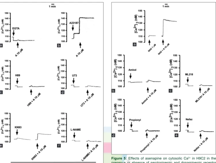
[Figures 3, 4c-f and 5b-e]. NAD-299 increased the response of H9C2 to asenapine [Figures 3c, d and 5a].
As shown in Figures 6a and 7a, b, the pool mobilized by asenapine was different from the one affected by ATP. Hence, the increase of (Ca2+) c caused by either asenapine or ATP did not significantly differ, irrespective of the sequence of addition (P > 0.05). Differentially, as reported in Figures 6b and 7c, the addition of 10 μM asenapine in costimulation with thapsigargin markedly changed the kinetics of (Ca2+) c fluctuations promoted by thapsigargin.
Finally, the effects of asenapine on (Ca2+) c were markedly reduced in H9C2 which had undergone peroxidation
[Figure 8a and b]. As shown in Figure 8a and b, in 20 min hydrogen peroxide treated cells, the increase of (Ca2+)c by asenapine reached a plateau at 100 nM asenapine. This concentration was used for the experiments performed with blockers. In this respect, it is notable that in H9C2, which were treated with hydrogen peroxide, the effects of asenapine were abolished by nefazodone, propionyl promazine, amlodipine, ML218, and L-NAME and were increased by NAD-299 [Figure 8c].
DISCUSSION
The results of this study have shown for the 1st time that in cardiomyoblasts asenapine exerts dual effects on both NO release and changes of (Ca2+)c, depending on the oxidative status of cells. Mechanisms related to D2 receptors and 5-HT receptors-dependent signaling and L- and T-type voltage Ca2+ channels opening would be involved in the response of H9C2 to asenapine.
Changes of (Ca2+) c levels are of primary importance in the regulation of NO production, eNOS being a Ca2+ dependent enzyme.[8] In the cytosol, Ca2+ is maintained at a very low level and is concentrated in intracellular calcium stores such as the endoplasmic reticulum (ER).[17] The dynamic steady state of Ca2+ in the cytosol is the result of the balance between active
Figure 1: Effects of asenapine on nitric oxide release in H9C2.
(a) Dose‑response and time‑course study. (b) Effects of 15 min asenapine (10 pM‑100 μM) in peroxidative conditions. (c) Effects of 2 min asenapine (100 nM) in the presence or absence of various agents. A = Asenapine; amlodip = Amlodipine (1 μM); NAD = NAD‑299 (1 μM); Nefaz = Nefazodone (1 μM); propionyl = Propionyl promazine (1 μM); ML218 = ML218 (1 μM); 2’5’ =2’5’ dideoxyadenosine (1 μM): H89 = H89 (1 μM); KN93 = KN93 (1 μM); U73 = U73122 (1 μM); L‑NAME = Nω‑nitro‑L‑arginine methyl ester (10 mM). Nitric oxide release is expressed as % in comparison to basal values (c). The results are the mean ± standard deviation of five experiments for each experimental protocol. (c) b, c, e, g, i, m, o, q, s, u, z P < 0.05 versus
a; d, f, h, l, n, p, r, t, v, w P < 0.05 versus b c
b a
Figure 2: Effects of asenapine on cytosolic Ca2+ in H9C2 in
nonperoxidative conditions. (a) Dose‑response and time‑course study. The results are the mean ± standard deviation of 5 experiments. (b) An example taken from one experiment
b
and passive fluxes through the cell membranes of various stores and is strictly regulated through mechanisms involving inositol-1,4,5-triphosphate-phosphate (IP3) generation, “capacitive Ca2+ entry” and the activation of voltage-gated Ca2+ channels.[9,18,19]
The results obtained in the present study have shown that in nonperoxidative conditions, asenapine can cause a persistent increase of (Ca2+) c mainly from extracellular origin by 5HT2A and through D2 receptors involvement, and L- and T-type Ca2+ channels opening. In addition, by the same way,
asenapine would increase NO release in H9C2. Mechanisms related to PKA and PLC-dependent signaling would be involved in such effects. Hence, D2-like receptors have been reported to inhibit cAMP/PKA pathway and L-type voltage-gated Ca2+ channels, and finally decrease (Ca2+)c.[20] By this way, the antagonistic effect of asenapine on D2 receptors could explain the results obtained about (Ca2+) c in H9C2. Observations about the involvement of 5-HT2A receptors in the effects of asenapine are not in agreement with previous reports. Hence, 5-HT2A receptors have been found to be responsible
Figure 3: Effects of asenapine on cytosolic Ca2+ in H9C2 in the presence or absence of various agents in nonperoxidative conditions. (a‑d)
A = 2 min asenapine (10 μM). (a and c) C = Basal. (a and b) EGTA = Ethylene glycol tetraacetic acid 50 mM; A23 = A23187 (1 μM); H89 = H89 (1 μM); KN93 = KN93 (1 μM); U73 = U73122 (1 μM); L‑NAME = Nω‑nitro‑L‑arginine methyl ester (10 mM). The results are the mean ± standard deviation of five experiments for each experimental protocol. (b and d) The effects of asenapine on Ca2+ in comparison to basal values are reported
as %. (a) b, c, e, P < 0.05 versus a; d, f, h, l, n, P P < 0.05 versus b. (b) c, b, d, e, f, g P < 0.05 versus a. (c and d) Amlodip = Amlodipine (1 μM);
ML218 = ML218 (1 μM); propionyl = Propionyl promazine (1 μM); Nefaz = Nefazodone (1 μM); NAD = NAD‑299 (1 μM). (c) b P < 0.05 versus a;
d, f, h, l, n P < 0.05 versus b. (d) b, c, d, e, f P < 0.05 versus a. One‑way ANOVA followed by Newman–Keuls post hoc test d
c
b a
for Ca2+ increase in response to serotonin by modulation of 5-HT4 subtype activity[21] and by increasing the magnitude of the L-type Ca2+ current.[22] For this reason, the finding of the abolishing effects of asenapine on Ca2+ fluctuations in H9C2 by nefazodone, would not confirm those data. That discrepancy could be related to the different cellular model and to the rather complex mechanism of action of asenapine, which acts as a high affinity antagonist of D2 receptors and 5‑HT2A receptors, but also as antagonist of 5-HT2B, 5-HT2C, 5-HT5A, 5-HT6, and 5-HT7 receptors.[2] Moreover, asenapine acts only as a partial agonist at the 5-HT1A site.[3] Regarding this issue, it is notable that the 5-HT1A receptors were found to play an inhibitory role on the effects of asenapine on Ca2+ movements, being the response of H9C2 to asenapine increased by the selective 5-HT1A antagonist, NAD-299.[5,23] Hence, since 5-HT1A have
been reported to negatively modulate the Ca2+/calmodulin pathway in neurons,[24] our results could be assumed to be in agreement with previous observations, although obtained in a different cellular model.
In addition, the Ca2+ pool mobilized by asenapine was found to be independent from that mobilized by an agent acting through IP3 generation, such as ATP,[25] but partly similar to the one affected by thapsigargin, the Ca2+-ATPase inhibitor which is able to deplete the ER Ca2+ pool.[26] These results would confirm the involvement of ‘capacitive Ca2+ entry’ in the response of H9C2 to asenapine.
The increased (Ca2+) c caused by asenapine could be the basis for the observed increase of NO release. Hence, like in CEC, asenapine was able to augment NO production in a dose-dependent and time-related way in H9C2. NO release was
Figure 4: Effects of asenapine on cytosolic Ca2+ in H9C2 in presence
or absence of Ca2+ chelator and Ca2+ ionophor, intracellular signaling
pathways, and nitric oxide synthase inhibitors in nonperoxidative conditions. An example of each experimental protocol is shown. (a) 2 min asenapine (A) with Ca2+ chelator, ethylene glycol tetraacetic
acid; (b) with Ca2+ ionophor A23187; (c) with PKA inhibitor, H89; (d) with
PLC inhibitor, U73122 (U73); (e) with CaMKII inhibitor, KN93; (f), with nitric oxide synthase inhibitor, Nω‑nitro‑L‑arginine methyl ester. The layout is the same as in Figure 3
d c b f a e
Figure 5: Effects of asenapine on cytosolic Ca2+ in H9C2 in the
presence or absence of serotoninergic and dopaminergic receptor blockers and L‑ and T‑type Ca2+ channels inhibitors. An example
of each experimental protocol is shown. (a) 2 min asenapine (A) with 5‑HT1A inhibitor, NAD‑299 (NAD); (b) with L type Ca2+ channel
blocker, amlodipine (Amlod); (c) with T type Ca2+ channel blocker,
ML218; (d) with D2 receptor inhibitor, propionyl promazine (Propionyl); (e) with 5‑HT2A inhibitor, nefazodone (Nefaz). The layout is the same
as in Figure 3 d c b a e
measured by the Griess system, which has been previously used for NO detection in endothelial cells and H9C2, as well.[5,6] Moreover, the effects of asenapine were abolished by cAMP/PKA, PLC, 5HT2A, and D2 receptors inhibitors, and L- and T-type Ca2+ channel blockers. The various antagonists were used at the same concentrations which were able to prevent the effects of asenapine in CEC or isolated arteries[5] and to abolish the nitrite release in vascular smooth muscle cells.[27] As previously observed in CEC, NO release caused by asenapine was only reduced by NAD-299.[5] Although not examined, it could be speculated that 5-HT1A receptors could play a dual role on NO release. Hence, the potentiating effects on Ca2+ movements elicited by asenapine through 5-HT1A-related mechanisms could counteract their inhibitory effects on NO production.
Overall, the results obtained in presence of various antagonists seem to suggest common mechanisms at the basis of increased NO release and Ca2+ influx in H9C2. As mentioned above, the interaction of asenapine with 5HT2A and D2 receptors would activate an intracellular PKA and PLC-dependent signaling that could increase NO release either directly through eNOS phosphorylation or indirectly through augmented Ca2+ influx from extracellular space by L- and T-type Ca2+ channels. Meanwhile, the activation of 5-HT1A receptors by asenapine could also contribute to Ca2+-dependent NO release in H9C2.
It is to note that in peroxidative conditions, the effects of asenapine on Ca2+ movements were reduced both in terms of maximum increase and as duration. As observed in nonperoxidative conditions, also in this case the effects were abolished by L- and T-type Ca2+ channels inhibitors, 5HT
2A and D2 receptors blockers and slightly increased by NAD-299. Moreover, asenapine was able to counteract the effects of hydrogen peroxide on NO release, an effect which was also observed in CEC.[5]
Thus, although not specifically examined, the reduction of NO release in peroxidative conditions could be linked to the lower Ca2+ influx, eNOS being a Ca2+ dependent enzyme, as reported above.
It should be noted that changes in NO could also be involved in modulation of Ca2+ transients. Hence, cAMP levels are regulated by NO/cGMP-dependent mechanisms related to phosphodiesterase II (PDE) inhibition.[12] By this way, changes of NO release could influence cAMP levels which would interfere with the activity of Ca2+ channels, serotonin and dopaminergic receptors, and Ca2+ pump.[12]
Interestingly, regarding this issue, in presence of L-NAME, which was able to prevent the increased NO release in H9C2, the effects of asenapine on Ca2+ movements were nearly abolished in both nonperoxidative and peroxidative conditions.
Figure 6: Effects of asenapine on ATP and thapsigargin‑dependent Ca2+ pools in H9C2. The results are the mean ± standard deviation of five
experiments for each experimental protocol. C = Basal. (a) asenapine (A) was administrated either before or after ATP. c, b P < 0.05 versus
a; e, f P < 0.05 versus d. (b) Asenapine was administrated alone or after 1 min thapsigargin (Tapsi) administration. The effects of thapsigargin
alone and after 1 min and 3 min (alone or with asenapine) are shown. b, c, d, e, f P < 0.05 versus a; c, e P < 0.05 versus b; f P < 0.05 versus d.
One‑way ANOVA followed by Newman–Keuls post hoc test
b a
Figure 7: Effects of asenapine on ATP and thapsigargin‑dependent
Ca2+ pools in H9C2. An example of each experimental protocol
is shown. (a and b) Asenapine was given before and after ATP. (c) Asenapine was given alone or in co‑administration with thapsigargin. The layout is the same as in Figure 6
Overall, the dual effects elicited by asenapine on Ca2+ in H9C2 would be of particular clinical relevance. On the one hand, in nonperoxidative conditions, asenapine would exert a beneficial role on cardiac contraction through sustained increase of Ca2+; on the whole, in peroxidation, asenapine would limit massive intracellular Ca2+ accumulation, which could be detrimental for myocytes through increased free radical generation,[13] damage of mitochondria,[11] and activation of caspase cascade.[28]
Although not specifically examined, NO as well could be involved in mediating the protective effects elicited by asenapine in H9C2. Hence, NO has been shown to modulate several aspects of “physiological” myocardial function. The effects of NO are influenced by its cellular and enzymatic source, the amount generated, the presence of reactive oxygen species, and the activation of cGMP-dependent and independent signal transduction pathways.[29]
Regarding this issue, it is to note that on the one hand, at low concentration, NO could contribute to increasing myocardial
contractility by cGMP/PDE II activation and intracellular Ca2+ increase;[30] on the other hand, at high concentration, NO could exert negative effects on cardiac performance through desensitization of cardiac contractile myofilaments to Ca2+ and peroxynitrite release.[10]
Although the results obtained in the present study evidence a role for NO in the modulation of Ca2+ transients caused by asenapine in H9C2, further studies could help clarify the relationship between NO release and Ca2+ and the implications in terms of cardiac function.
CONCLUSION
Asenapine was found for the 1st time to affect NO release and Ca2+ transients in H9C2 through its specific receptors and
Figure 8: Effects of asenapine on cytosolic Ca2+ in H9C2 in
peroxidative conditions. (a) Dose‑response and time‑course study. The results are the mean ± standard deviation of five experiments. (b) An example taken from one experiment. (c) Effects of asenapine on Ca2+ in the presence or absence of various blockers. C = Basal;
A = Asenapine; Nefaz = Nefazodone (1 μM); propionyl = Propionyl promazine (1 μM); NAD = NAD‑299 (1 μM); amlodip = Amlodipine (1 μM); ML218 = ML218 (1 μM); L‑NAME = Nω‑nitro‑L‑arginine methyl ester (10 mM). The results are the mean ± standard deviation of five experiments for each experimental protocol. (c) b, d, e P < 0.05 versus
a; c, d, e, f, g, h P < 0.05 versus b. One‑way ANOVA followed by
Newman–Keuls post hoc test c b a c b a
L- and T-type Ca2+ channels opening. Intracellular signaling involving PKA and PLC-related pathways was also shown to play a role.
Acknowledgment
We thank the Azienda Ospedaliera Maggiore Della Carità di Novara for its help.
Financial support and sponsorship Nil.
Conflicts of interest
There are no conflicts of interest.
REFERENCES
1. Bishara D, Taylor D. Asenapine monotherapy in the acute treatment of both schizophrenia and bipolar I disorder. Neuropsychiatr Dis Treat 2009;5:483‑90. 2. Samalin L, Charpeaud T, Llorca PM. Asenapine in bipolar I disorder:
Evidence and place in patient management. Ther Adv Chronic Dis 2013;4:5‑14.
3. Reynolds GP. Receptor mechanisms of antipsychotic drug action in bipolar disorder – Focus on asenapine. Ther Adv Psychopharmacol 2011;1:197‑204. 4. Gramaglia C, Rizza MC, Gattoni E, Gambaro E, Di Marco S, Coppola I,
et al. Asenapine in clinical practice: Preliminary results from a naturalistic
observational study. Riv Psichiatr 2014;49:241‑6.
5. Grossini E, Gramaglia C, Farruggio S, Bellofatto K, Anchisi C, Mary D, et al. Asenapine increases nitric oxide release and protects porcine coronary artery endothelial cells against peroxidation. Vascul Pharmacol 2014;60:127‑41. 6. Grossini E, Marotta P, Farruggio S, Sigaudo L, Qoqaiche F, Raina G, et al.
Effects of artemetin on nitric oxide release and protection against peroxidative injuries in porcine coronary artery endothelial cells. Phytother Res 2015;29. doi: 10.1002/ptr.5386. [Epub ahead of print].
7. Mizuno O, Kobayashi S, Hirano K, Nishimura J, Kubo C, Kanaide H. Stimulus‑specific alteration of the relationship between cytosolic Ca2+
transients and nitric oxide production in endothelial cells ex vivo. Br J Pharmacol 2000;130:1140‑6.
8. Moncada S, Palmer RM, Higgs EA. Nitric oxide: Physiology, pathophysiology, and pharmacology. Pharmacol Rev 1991;43:109‑42. 9. Berridge MJ, Irvine RF. Inositol phosphates and cell signalling. Nature
1989;341:197‑205.
10. Brady AJ, Warren JB, Poole‑Wilson PA, Williams TJ, Harding SE. Nitric oxide attenuates cardiac myocyte contraction. Am J Physiol 1993;265 (1 Pt 2):H176‑82.
11. Kohlhaas M, Maack C. Calcium release microdomains and mitochondria. Cardiovasc Res 2013;98:259‑68.
12. Schlüter KD, Schulz R, Schreckenberg R. Arginase induction and activation during ischemia and reperfusion and functional consequences for the heart. Front Physiol 2015;6:65.
13. Wagner S, Rokita AG, Anderson ME, Maier LS. Redox regulation of sodium and calcium handling. Antioxid Redox Signal 2013;18:1063‑77. 14. Grossini E, Caimmi PP, Molinari C, Mary DA, Uberti F, Vacca G.
Modulation of calcium movements by urocortin II in endothelial cells. Cell Physiol Biochem 2010;25:221‑32.
15. Grossini E, Molinari C, Sigaudo L, Biella M, Mary DA, Vacca G. Calcium handling in porcine coronary endothelial cells by gastrin‑17. J Mol Endocrinol 2013;50:243‑53.
16. Grynkiewicz G, Poenie M, Tsien RY. A new generation of Ca2+ indicators with
greatly improved fluorescence properties. J Biol Chem 1985;260:3440‑50. 17. Clapham DE. Calcium signaling. Cell 2007;131:1047‑58.
18. Munaron L. Intracellular calcium, endothelial cells and angiogenesis. Recent Pat Anticancer Drug Discov 2006;1:105‑19.
19. Berridge MJ. Capacitative calcium entry. Biochem J 1995;312(Pt 1):1‑11. 20. Gingrich JA, Caron MG. Recent advances in the molecular biology of
dopamine receptors. Annu Rev Neurosci 1993;16:299‑321.
21. Seitz PK, Bremer NM, McGinnis AG, Cunningham KA, Watson CS. Quantitative changes in intracellular calcium and extracellular‑regulated kinase activation measured in parallel in CHO cells stably expressing serotonin (5‑HT) 5‑HT2A or 5‑HT2C receptors. BMC Neurosci 2012;13:25.
22. Jahnel U, Nawrath H, Rupp J, Ochi R. L‑type calcium channel activity in human atrial myocytes as influenced by 5‑HT. Naunyn Schmiedebergs Arch Pharmacol 1993;348:396‑402.
23. Johansson L, Sohn D, Thorberg SO, Jackson DM, Kelder D, Larsson LG, et al. The pharmacological characterization of a novel selective 5‑hydroxytryptamine1A receptor antagonist, NAD‑299. J Pharmacol Exp Ther 1997;283:216‑25.
24. Yuen EY, Jiang Q, Chen P, Gu Z, Feng J, Yan Z. Serotonin 5‑HT1A receptors regulate NMDA receptor channels through a microtubule‑dependent mechanism. J Neurosci 2005;25:5488‑501.
25. Patel S, Joseph SK, Thomas AP. Molecular properties of inositol 1,4,5‑trisphosphate receptors. Cell Calcium 1999;25:247‑64.
26. Gamberucci A, Innocenti B, Fulceri R, Bànhegyi G, Giunti R, Pozzan T, et al. Modulation of Ca2+ influx dependent on store depletion
by intracellular adenine‑guanine nucleotide levels. J Biol Chem 1994;269:23597‑602.
27. Chou TC, Yang SP, Pei D. Amlodipine inhibits pro‑inflammatory cytokines and free radical production and inducible nitric oxide synthase expression in lipopolysaccharide/interferon‑gamma‑stimulated cultured vascular smooth muscle cells. Jpn J Pharmacol 2002;89:157‑63.
28. Mattson MP, Chan SL. Calcium orchestrates apoptosis. Nat Cell Biol 2003;5:1041‑3.
29. Shah AM, MacCarthy PA. Paracrine and autocrine effects of nitric oxide on myocardial function. Pharmacol Ther 2000;86:49‑86.
30. Takimoto E. Cyclic GMP‑dependent signaling in cardiac myocytes. Circ J 2012;76:1819‑25.