Comparison of different dose reduction system in computed tomography for orthodontic applications.
9
0
0
Testo completo
(2) research article cess should be based exclusively on image interpretation at the workstation rather than on hard copies or PACS. Dose estimated data is possible to obtain from the console of the CT scanner: • CTDI (Computer Tomography Dose Index) = dose measured in central and peripheral region of the subjects as a direct result of a CT section acquisition T millimetres thick; • DLP (Dose Length Product)= total dose deposited over the length of the acquisition.. In te rn az io na li. The CT, a radiological three-dimensional technique based on computer assessment of the radiation-absorbing characteristics of the various tissues of the body, offers many advantages (15-18): • it provides true tomography images of excellent contrast resolution; • tissue density differences of 0.5% are resolvable; • all the photons that have penetrated the body are used in the formation of the images; • X-ray beams are highly collimated into a band varying from 2-13 mm, so that very little tissue, other than that being imaged, is irradiated. The problems in adapting helical-CT scans for orthodontic use include high cost, scanning time and most importantly, high radiation exposure, which is of particular concern given that the orthodontic treatment often involves young or adolescent patients.. Techniques. ©. C IC. Ed. iz. io ni. MSCT The MDCT scanner sufficient for orthodontic applications is at 16 slice. The scanner equipped with 16 detector rows has a minimal rotation time of 0.5 sec. given a collimation between 0.75 and 1.5 mm with dose calibrated. The scanning tecnique should be applied consists of: preliminary scan performed in the anteroposterior (AP) and laterolateral (LL) projections, with the following acquisition parameters: 100 KV or 80 KV, with 200 mA (1). The subsequent scans should taken with a 1.25 mm slice thickness, 0.6 mm interval, 11.25 mm table speed/rotation, 100 mA, 13.7 cm FOV, 512x512 matrix and 0° gantry angle (Fig. 1). Data acquisition, planning of examinations and printing of axial images and standardized multiplanar reconstructions should be performed on a dedicate workstation. The radiologists should have immediate access for visualization and further multiplanar and 3D reformatting on the workstation. Additional reconstructions could be necessary to clarify the diagnosis or demanded by the referring clinician, and should be performed and printed by radiologists. The examinations and additional reconstructions should sent via a fast 100-MB/sec local area networkEthernet to the PACS. The radiologic evaluation pro-. CBCT Cone beam computed tomography (CBCT) has recently been introduced as an alternative imaging technology for acquiring 3D data for diagnostic tasks such as implant treatment planning (4, 5). This technology requires less expensive components than conventional CT and is reported to produce lower patient exposures (2). The technique also allows, in principle, to obtain the data acquisition of a volume quickly and with low dose. In the cone-beam technique is necessary to produce the X-ray photons only the number of times equal to the number of slices needed to cover the volume of interest. The system has an automatic exposure control (AEC), which allows automatic selection of the intensity of X-rays of departure in accordance with the size of the patient and changes in accordance with the thickness. Cone-beam CT scanners utilize a two dimensional detector, which allows for a single rotation of the gantry to generate a scan of the entire region of interest, as compared to conventional CT scanners whose multiple “slices”. must be stacked to obtain a complete image (9, 10). In comparison with conventional fan-beam or spiral-scan geometries, cone-beam geometry has higher efficiency in X-ray use, inherent quickness in volumetric data acquisition, and potential for reducing the cost of CT. The cone beam technique, on the other hand, requires only a single scan to capture the entire object with a cone of Xrays. Thus, the time required to acquire a single conebeam projection is the same as that required by a single fan-beam projection. The quality of the reconstructed CT image depends significantly on the qua-. ORAL & Implantology - Anno IV - N. 1-2/2011. 15.
(3) In te rn az io na li. research article. io ni. Figure 1 MSCT Image obtained with the low dose protocol of 80 KV in axial images (A), panorex recontruction (B) and volumetric reconstruction (C). In this kind of image the anatomy, the exact position, the spatial relationship and the orientation of the impacted tooth are perfectly clear.. ©. C IC. Ed. iz. lity of the data that is acquired during the scan. The characteristics of the two-dimensional detector used by a cone beam CT scanner affect therefore directly the quality of the CT scan (Fig. 2). Its high spatial resolution, smaller size and lower cost has made CBCT a natural fit dentomaxillofacial imaging. The image obtained consists of an image on a 2D axial volume acquisition. Moreover, the acquisition of a volume can not only aimed at reconstruction of the arch and the like but rework similpanoramich and Telecrane. In this way, with only one purchase you can get three types of tests (reconstruction of the arches, and ortopanoramic - Telecrane) saving the administered dose, time and obviously cost savings (11-13). Technical limits of the cone beam CT is a revised version of the images in real time, which requires at least half minute of acquisition. The processing time of images on the dedicated console appears comparable for the two techniques.. 16. The primary axial reconstructions require a few seconds. The secondary reconstructions are performed in real time by an operator or directly on a dedicated workstation. ASIR (Adaptive Statistical Iterative Reconstruction) ASIR is a revolutionary patented image reconstruction algorithm (GE medical systems) that allows to overcome the concept of reconstruction back projecton that, starting from the birth of CT technology, has characterized the principles of operation. At the base of Asir is the concept of dualism in which the trend-particle physics studies the waves elettromagnetic including X-rays, the principle is that not all the phenomena of the waves Electromagnetic you can think of a photon as a point particle, some phenomena can be explained only by modeling the photon as a wave, which is why we say that electromagnetic waves living in wave-particle duality (19).. ORAL & Implantology - Anno IV - N. 1-2/2011.
(4) io ni. In te rn az io na li. research article. Ed. iz. Figure 2 CBCT Image obtained in panorex reconstruction (A), volumetric reconstruction (B) axial images (C), and parasagital reconstruction (D). In this kind of image the anatomy, the exact position of the mandibular canal are perfectly clear.. ©. C IC. With ASIR we introduce the concept of a statistical measure of the signal produced by the photon system X detection, exceeding the simple measure of the attenuation in Hounsfield units of the photon after passing through the patient’s anatomy comes on the detector, which is why you move from a mathematical model for solving systems of linear equations a mathematical model heuristic and iterative, the operator can thus determine the value of standard deviation, that may cause the opening of the bell curve that you want. Please use as a basis for statistical model. The benefit brought by Asir, comparable to that obtained with the generation before subsidy-driven 64slice CT and demonstrated in numerous scientific studies can be summarized in: - increase of 40% of low-resolution contrast. - reduce the Xray-dose of approximately 50% - reduction in the number of artifacts - percentage applicable to all anatomic areas and all types of examination (Fig. 3). So reducing the dose of approximately 50% you get images with the same resolution as the higher KV.. Results 80 KV protocols compared with 100 KV protocols resulted in reduced total radiation dose without relevant loss of contrast and resolution of images. 80 KV protocols allow good quality of images for orthodontics diagnosis and treatment. At the Department of Diagnostic Imaging of the Uni-. ORAL & Implantology - Anno IV - N. 1-2/2011. 17.
(5) io ni. In te rn az io na li. research article. Ed. iz. Figure 3 MSCT Image obtained with the ASIR system with dose reduction but good image quality. Axial images (A), panorex recontruction (B) and parasagital reconstruction (C). In this kind of image the anatomy, the exact position, the spatial relationship and the orientation of the impacted teeth are perfectly clear.. ©. C IC. versity of Rome “Tor Vergata”, the standard setting of the equipment was modified for use in orthodontic diagnosis. The choice of the lower dose of 80 KV was made as a necessary compromise to reach a good diagnostic accuracy. According to our experiences in the 80 KV MSCT scanning we obtained a mean CTDI of 6.29 mGy and a total scan time of 19 sec. In the 100 KV scanning we obtained a mean of CTDI of 13.22 mGy and a total scan time of 20 sec. In the CBCT we obtained a mean of CTDI of 1.04mGy and a scan total scan time of 29 sec. The 80 KV scanning has a significantly lower affective radiation dose compared to the 100 KV scanning ( p< 0.05). The CBCT scanning has a significantly lower affective radiation dose compared to the 80 KV MSCT scanning ( p< 0.05) (1).. 18. No significant differents was found for image reconstruction time between 100KV and 80KV protocols. CBCT reconstruction time was least twice than MSCT. In the retroanalysis, the images of all patients were used for comparing the protocols in terms of image quality. The mean score for 80 KV scanning images was 1.1 +/- 0.5 , 1.3 +/- 0.5 for dose obtained by 100 KV scanning and 1 +/- 0.5. The median image quality was 2 (good) for MSCT both protocols and 3 (satisfactory) for CBCT. Both the protocols of examination introduce the same resolution and the same quality of the image. In the 80 KV scanning (time T0) we obtained a mean DLP of 43.1 mGy per cm, CTDI of 6.29 mGy and a total scan time of 19 sec (Table 1). The 80 KV scan-. ORAL & Implantology - Anno IV - N. 1-2/2011.
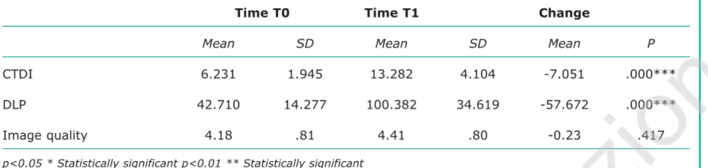
(6) research article Table 1 - Descriptive and statistical analysis of dosimetric evaluation at time T0 and T1. Time T1. Mean. SD. CTDI. 6.231. DLP Image quality. Change. In te rn az io na li. Time T0. Mean. SD. Mean. P. 1.945. 13.282. 4.104. -7.051. .000***. 42.710. 14.277. 100.382. 34.619. -57.672. .000***. 4.18. .81. 4.41. .80. -0.23. .417. p<0.05 * Statistically significant p<0.01 ** Statistically significant p<0.001 *** Statistically significant ns = Non statistically significant. ning has a significantly lower effective radiation dose (p< 0.05) compared to 100 KV scanning (time T1) (Tables 2-3 ) (1).. The data obtained indicate that the parotid glands, the thyroid and the mouth are the anatomical structures most exposed to radiation during Dentascan CT examinations, both of the upper and low jaw bone, in line with what has been reported in previous studies (7, 8). The images obtained with the 80 KV protocol were good enough for orthodontic diagnosis, so we decided to compare these images not with those obtained with 100 KV, setting typically used in Dentascan examinations, but with the low dose protocol of 100 KV (1, 6, 15,18, 21). When milliamperes are kept constant, the radiation dose delivered by an X-ray tube increases, as the Xray energy, that is the kilovoltage, increases. This effect is largely exploited in diagnostic imaging performed on babies or children, where a low radiation dose is required. As X-ray energy ( kilovoltage) decreases, the contrast between the structures crossed by the X-ray beam increases (9-11). This concept proved to be useful in our experience, as it enabled a marked reduction in the dose administered without affecting image quality. The dose value obtained are significantly lower than the minimum values reported in the literature that contains numerous studies on dosimetry in odontostomatologic radiology, often with comparisons between conventional CT, CBCT and OPT (16, 17, 20). Even when compared with the doses reported for. ©. C IC. Ed. iz. io ni. Table 2 - Dosimetric values at time T0.. Discussion. ORAL & Implantology - Anno IV - N. 1-2/2011. 19.
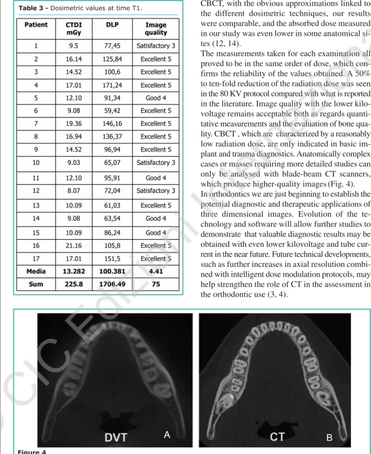
(7) In te rn az io na li. CBCT, with the obvious approximations linked to the different dosimetric techniques, our results were comparable, and the absorbed dose measured in our study was even lower in some anatomical sites (12, 14). The measurements taken for each examination all proved to be in the same order of dose, which confirms the reliability of the values obtained. A 50% to ten-fold reduction of the radiation dose was seen in the 80 KV protocol compared with what is reported in the literature. Image quality with the lower kilovoltage remains acceptable both as regards quantitative measurements and the evaluation of bone quality. CBCT , which are characterized by a reasonably low radiation dose, are only indicated in basic implant and trauma diagnostics. Anatomically complex cases or masses requiring more detailed studies can only be analysed with blade-beam CT scanners, which produce higher-quality images (Fig. 4). In orthodontics we are just beginning to establish the potential diagnostic and therapeutic applications of three dimensional images. Evolution of the technology and software will allow further studies to demonstrate that valuable diagnostic results may be obtained with even lower kilovoltage and tube current in the near future. Future technical developments, such as further increases in axial resolution combined with intelligent dose modulation protocols, may help strengthen the role of CT in the assessment in the orthodontic use (3, 4).. ©. C IC. Ed. iz. io ni. research article. Table 3 - Dosimetric values at time T1.. Figure 4 Comparation between CBCT (A) and MSCT with 80KV protocol (B). Periodontal ligament spaces easily recognizable in the dental CT but not satisfactory in the CBCT.. 20. ORAL & Implantology - Anno IV - N. 1-2/2011.
(8) research article tical bone thickness with computed tomographic scanning for orthodontic implants. Am J Orthod Dentofacial Orthop 2006;129:721.e7-721-12. 5. Espelid I, Mejàre I, Weerheijm K. EAPD guidelines for use of radiographs in children. Eur J Paed Dent 2003 Mar;4(1):40-8. 6. Fanucci E, Leporace M, Di Costanzo G, Fiaschetti V, Simonetti G. Multidetector CT and dentascan software: dosimetric evaluation and technique improvement. Radiol Med 2006;111:130-8. 7. Hajeer MY, Millet DT, Ayoub AF, Siebert JP. Current product and practices. Applications of 3D imaging in orthodontics: Part I. Journal of orthodontics 2004;31:62-70. 8. Hajeer MY, Millet DT, Ayoub AF, Siebert JP. Current product and practices. Applications of 3D imaging in orthodontics: Part II. Journal of orthodontics 2004;31:154162. 9. Hechler SL. Cone-beam CT: application in orthodontics. Dent Clin North Am. 2008 oct;52(4):809-23. 10. Katsumata A, Hirukawa A, Noujeim M, Okumura S, Naitoh M, Fujishita M, Ariji E, Langlais RP. Image artifact in dental cone-beam CT. Oral Surg Oral Med Oral Pathol Oral Radiol Endod. 2006 May;101(5):652-7. Epub 2005 Nov. 11. Kau CH, Boži M, English J, Lee R, Bussa H, Ellis RK. Cone-beam computed tomography of the maxillofacial region-an update. Int J Med Robot. 2009 Sep 23. 12. Lecomber AR, Yoneyama Y, Lovelock DJ et al. Comparison of patient dose from imaging protocols for dental implant planning using conventional radiography and computer tomography. Dentomaxillofacial Radiol 2001;30: 255-259. 13. Ludlow JB, Davies-Ludlow LE and Brooks SL. Dosimetry of two extraoral direct digital imaging devices: NewTom cone beam CT and Orthophos Plus DS panoramic unit. Dentomaxillofac Radiol 2003;32:229-234. 14. Macchi A, Carrafiello G, Cacciafesta V, Norcini A. Threedimensional digital modeling and setup. Am J Orthod Dentofacial Orthop 2006;129(5):605-610. 15. Matarese G, Portelli M, Mazza M, Militi A, Nucera R, Gatto E, Cordasco G. Evaluation of skin dose in a low dose spiral CT protocol. Eur J Paed Dent 2006;2:77-80. 16. Miller AJ, Maki K, Hatcher DC. New diagnostic tools in orthodontics. Am J Orthod Dentofacial Orthop 2004;126:395- 396. 17. Redmond WR, Huang J, Bumann A, Mah J. Three-dimensional radiographic analysis in orthodontics. JCO 2005;39(2):421- 428. 18. Scheck RJ, Coppenrath EM, Kellner MW et al. Radiation dose and image quality in spiral CT: multicentre evaluation at 6 institutions. Br J Radiol 2002;75: 140-150. 19. Silva AC, Lawder HJ, Hara A, Kujak J, Pavlicek W. Innovations in CT dose reduction strategy: application of the adaptive statistical iterative reconstruction algorithm AJR Am J Roentgenol. 2010 Jan;194(1):191-9. 20. Suomalainen A, Kiljunen T, Käser Y, Peltola J, Korte-. Ed. iz. io ni. In te rn az io na li. Three-dimensional investigations are necessary in orthodontic daily practice every time conventional Xray techniques are not enough, such as impacted maxillary canines or incisors, supernumeraries teeth, trauma and orthopaedic treatment. MSCT ASIR allows new scanning modes to set new dose standards and clinical cases showing 40% average dose reduction vs MSCT. It improve image IQ on very low dose exams and allows to decrease dose while improving image quality. With specific clinical indications whereby high-contrast lesions are surrounded by low-contrast structures, dedicated lowdose protocols have been routinely used in an effort to minimize radiation dose. For example, several studies have suggested that low-dose CT may be adequate for renal calculus evaluation, CT colonography, and pulmonary CT angiography . It can be satisfactory applied to the dento-maxillo-facial district. Although satisfactory for the dedicated evaluation of the intended anatomy, these protocols were implemented with the realization that image quality would be significantly reduced outside the area of interest. Although desirable for all patients, dose reduction is of particularly high priority for young patients (5), patients who are pregnant, and patients requiring multiple follow-up or multiphase studies. However, low-dose imaging with ASIR reconstructions appears to have potential in displaying similar or better overall image quality compared with routine-dose. New imaging methods will replace the way we look at a variety of common diagnostic and treatment issues in daily orthodontic practice.. ©. C IC. References. 1. Ballanti F, Lione R, Fiaschetti V, Fanucci E, Cozza P. Low-dose CT protocol for orthodontic diagnosis Paediatric Dentistry; Vol. 9/2-2008. 2. Bamgbose BO, Adeyemo WL, Ladeinde AL, Ogunlewe MO. Cone Beam computed tomography (CBCT): the new vista in oral and maxillofacial imaging. Nig Q J Hosp Med. 2008 Jan-Mar;18(1):32-5. 3. Chaushu S, Chaushu G, Becker A. The role of digital volume tomography in the imaging of impacted teeth. World J Orthod 2004;5:120-132. 4. Deguchi T, Nasu M, Murakami K, Yabuuchi T, Kamioka H, Takano-Yakamoto T. Quanitative evaluation of cor-. ORAL & Implantology - Anno IV - N. 1-2/2011. 21.
(9) Correspondence to:. In te rn az io na li. Prof. Ezio Fanucci Department of Odontostomatological Sciences Policlinic Tor Vergata, Viale Oxford, 81 00133 Rome, Italy. ©. C IC. Ed. iz. io ni. research article. sniemi M. Dosimetry and image quality of four dental cone beam computed tomography scanners compared with multislice computed tomography scanners. Dentomaxillofac Radiol. 2009 Sep;38(6):367-78. 21. Ziegler CM, Woertche R, Brief J and Hassfeld S. Clinical indications for Digital Volume Tomography in oral and maxillofacial surgery. Dentomaxillofac Radiol 2002;31:126-130.. 22. ORAL & Implantology - Anno IV - N. 1-2/2011.
(10)
Figura
Documenti correlati
(CF) A 5 × 13 mixed design ANCOVA with the between factor Stimulation (5 levels: P3 - 3 mA offline AtDCS, P3 - 3 mA online AtDCS, P3 - 1.5 mA offline AtDCS, P3 - 1.5 mA online
This was performed by crossing the BCR-ABL1 recombinant fly with more than 200 stocks of deletions (Figure 1 and Table S1) covering approximately 10,000 Drosophila genes and
Electro-magnetic tests conducted on particles alone, helped to understand that bigger particles are able to overcome the melting temperature of the adhesive but hot-melt
Always considering a number of 25 trains per day and a recovered energy around 350 MJ (third plot from the top) for single braking, including a charging- discharging efficiency
As shown in Figure 1 , we included all 3381 smokers aged 50 years or older who during the period from 2000 until 2010 received LDCT in the context of the two Italian screening
Based on how important TQM implementation is in Pesantren, this research was hoped to be a solution in building information system of total quality management to measure
Questo lavoro, concentratosi principalmente sull’analisi della “relazione di aiuto”, all’interno del Centro Protesi INAIL di Vigorso di Budrio, nei confronti dei
Continuità tra ascolto ed esecuzione musicale, finalizzata alla comprensione musicale Comprensione musicale Un processo di costruzione del ‘senso’ della musica
