A Gene-Alteration Profile of Human Lung Cancer Cell Lines
Raquel Blanco1,†, Reika Iwakawa2,†, Moying Tang1, Takashi Kohno2, Barbara Angulo1, Ruben Pio3, Luis M. Montuenga3, John D. Minna4, Jun Yokota2, and Montse Sanchez-Cespedes1,*
1Lung Cancer Group, Molecular Pathology Programme, Centro Nacional de Investigaciones
Oncologicas (CNIO), Madrid, Spain
2Biology Division, National Cancer Center Research Institute, Tokyo, Japan
3Division of Oncology, Centro para la Investigacion Medica Aplicada (CIMA), University of Navarra,
Pamplona, Spain
4Hamon Center for Therapeutic Oncology Research, University of Texas Southwestern Medical
Center, Dallas, Texas
Abstract
Aberrant proteins encoded from genes altered in tumors drive cancer development and may also be therapeutic targets. Here we derived a comprehensive gene-alteration profile of lung cancer cell lines. We tested 17 genes in a panel of 88 lung cancer cell lines and found the rates of alteration to be higher than previously thought. Nearly all cells feature inactivation at TP53 and CDKN2A or RB1, whereas
BRAF, MET, ERBB2, and NRAS alterations were infrequent. A preferential accumulation of
alterations among histopathological types and a mutually exclusive occurrence of alterations of
CDKN2A and RB1 as well as of KRAS, epidermal growth factor receptor (EGFR), NRAS, and ERBB2 were seen. Moreover, in non-small-cell lung cancer (NSCLC), concomitant activation of
signal transduction pathways known to converge in mammalian target of rapamycin (mTOR) was common. Cells with single activation of ERBB2, PTEN, or MET signaling showed greater sensitivity to cell-growth inhibition induced by erlotinib, LY294002, and PHA665752, respectively, than did cells featuring simultaneous activation of these pathways, underlining the need for combined therapeutic strategies in targeted cancer treatments. In conclusion, our gene-alteration landscape of lung cancer cell lines provides insights into how gene alterations accumulate and biological pathways interact in cancer.
Keywords
lung cancer; oncogenes; tumor suppressors; tyrosine kinase inhibitors
Introduction
Characterization of accumulated genetic alterations in cancer cells is important not only to understand tumor biology, but also to guide drug design and select patients who might benefit
© 2009 WILEY-LISS, INC.
*Correspondence to: Montse Sanchez-Cespedes, Cancer Epigenetics and Biology Program-PEBC, Catalan Institute of Oncology (ICO),
Bellvitge Biomedical Research Institute (IDIBELL), Hospital Duran i Reynals, Av. Gran Via s/n km 2.7, 08907 - L’Hospitalet de
NIH Public Access
Author Manuscript
Hum Mutat. Author manuscript; available in PMC 2010 July 9.
Published in final edited form as:
Hum Mutat. 2009 August ; 30(8): 1199–1206. doi:10.1002/humu.21028.
NIH-PA Author Manuscript
NIH-PA Author Manuscript
from a given targeted cancer therapy. The promise of using proteins encoded by mutated cancer genes, mainly kinases encoded by oncogenes, as molecular targets for the development of novel therapies, drives endeavors to identify novel mutated cancer genes and to create catalogues of somatic mutations in cancer [Wang et al., 2004; Sjoblom et al., 2006; Greenman et al., 2007; Thomas et al., 2007]. The paradigm of the latter is the Catalogue of Somatic Mutations in Cancer (COSMIC) database of the Wellcome Trust Sanger Institute
(www.sanger.ac.uk/cosmic) [Forbes et al., 2006], which brings together data on the mutation status of hundreds of cancer-related genes in primary tumors and cancer cell lines from a wide variety of tumor types.
In the particular case of lung cancer, several gene alterations are known to contribute to its development, including activating mutations and gene amplification at the oncogenes BRAF (MIM# 164757), epidermal growth factor receptor (EGFR) (MIM# 131550), ERBB2 (MIM# 164870), KRAS (MIM# 190070), NRAS (MIM# 164790), PIK3CA (MIM# 1171834), MYC (MIM# 190080), MYCL1 (MIM# 164850), and MYCN (MIM# 164840), as well as inactivating intragenic mutations, homozygous deletions, and promoter hypermethylation at the tumor suppressor genes BRG1/SMARCA4 (MIM# 603254), LKB1/STK11 (MIM# 602216), PTEN (MIM# 601728), CDKN2A (MIM# 600160), RB1 (MIM# 180200), and TP53 (MIM# 191170) [Sanchez-Cespedes 2007; Medina et al., 2008]. Some of these gene alterations are known to be specific to lung tumor histologies [Westra et al., 1993; Otterson et al., 1994; Kelley et al., 1995; Sanchez-Cespedes, 2007; Medina et al., 2008]. In addition, it is also well established that some gene alterations are mutually exclusive, as is the case for pairs of genes, such as
KRAS and EGFR, or CDKN2A and RB1 [Otterson et al., 1994; Lynch et al., 2004; Paez et al.,
2004], that encode proteins acting in the same signaling pathway. However, a profile of alterations at multiple well-known cancer genes in a large panel of lung cancers has never been reported. This limits our understanding of how gene alterations are distributed among lung tumors and how they interact with one another.
Here, we attempt to delineate the gene-alteration profile of lung cancer cell lines by screening for alterations of seventeen well-known cancer genes, including point mutations at AKT1 (MIM# 164730) and EML4-ALK (MIM# 607442 for EML4 and MIM# 105590 for ALK) fusions, a small inversion within chromosome 2p recently reported in a small subset of non-small-cell lung cancers (NSCLCs) [Carpten et al., 2007; Soda et al., 2007]. We examined the association between the genetic alteration profile and the response to specific small molecule inhibitors.
Material and Methods
Cell LinesCells were maintained in culture flasks in either DMEM (A549, NCI-H1299, NCI-H23, Calu-3, NCI-H522, and EBC1) or RPMI 1640 (NCI-H446, NCI-H1650, NCI-H460, and NCI-N417) (Invitrogen, Carlsbad, CA) supplemented with 10% (v/v) fetal bovine serum, 2mM L-glutamine, 50 mg/ml penicillin/streptomycin, and 2.5 µg/ml fungizone. Cultures were kept at 37°C in a humidified atmosphere of 5% CO2/95% air. DNA, RNA, and protein were extracted
using standard protocols.
Screening for Gene Mutations and Deletions
Screening for mutations in AKT1 (exon 3), BRAF (exons 11 and 15), MET (MIM# 164860) (exons 16–20), ERBB2 (exon 20), EGFR (exons 18–21), NRAS (codons 12, 13, and 61),
PIK3CA (exons 1, 9, and 20), PTEN (exons 2–9), and CDKN2A (exons 1–3) was performed
by directly sequencing PCR products using primers and conditions that have been previously described [Matsumoto et al., 2007; Angulo et al., 2008; Medina et al., 2008], or that are
NIH-PA Author Manuscript
NIH-PA Author Manuscript
available upon request. Nucleotide numbering reflects cDNA numbering with +1
corresponding to the A of the ATG transition initiation codon in the reference sequence. We considered the presence of homozygous deletions when there was a reproducible absence of PCR product of one or more consecutive exons. The mutational status of STK11, SMARCA4,
KRAS, and TP53 was either determined for those cases with incomplete/conflicting information
or gathered from previous publications [Harbors et al., 1988; Yokota et al., 1988; Otterson et al., 1994; Shimizu et al., 1994; Matsumoto et al., 2007; Angulo et al., 2008] (Supp. Table S1) or from the Wellcome Trust Sanger Institute’s Cancer Cell Line Project website
(www.sanger.ac.uk/cosmic). In those cases where mutation/deletion data were not available, cells with a reported absence of RB protein expression were classified as RB1-mutant. The presence of the EML4-ALK fusion gene was tested according to previously published conditions [Soda et al., 2007].
Promoter Hypermethylation
The determination of promoter hypermethylation at CDKN2A was evaluated by bisulfite treatment of the genomic DNA and subsequent methylation-specific PCR, using previously published protocols [Esteller et al., 2001].
Real-Time Quantitative Genomic PCR for Determining Gene Amplification
To determine MET, ERBB2, MYC, MYCL, and MYCN amplification we used quantitative real-time genomic PCR. The conditions and primers used for MYC, MYCN and MYCL have been previously described [Medina et al., 2008]. ERBB2 and MET primers and PCR conditions are available upon request. The copy number of genomic DNA was measured by SYBR green using an ABI Prism 7900 Sequence Detector (Applied Biosystems, Foster City, CA). Inhibitors and Viability Assay
Rapamycin (mammalian target of rapamycin [mTOR] inhibitor) and LY-294002 (PI3K inhibitor) were obtained from Calbiochem (La Jolla, CA) and PHA665752 (MET inhibitor) from Tocris Bioscience (Ellisville, MI). Erlotinib
(N-(3-ethynylphenyl)-6,7-bis(2-methoxyethoxy)-4-quinazolinamine) (EGFR inhibitor) was a gift from Roche Pharmaceuticals (Mannheim, Germany). Erlotinib tablets were ground to powder and dissolved in pure dimethyl sulfoxide (DMSO) to the desired concentration. For the cell-survival assays, cells were seeded at a density of 5,000 cells/well (15,000 cells/well for N417) on 96-well plates. They were allowed to recover for 12 hr before adding the drugs. Cells were exposed to various
concentrations of each drug for 48 or 72 hr, and then the viable cell number was measured by the 3-(4,5-dimethylthiazol-2)-2,5-diphenyltetrazolium bromide (MTT) assay. Briefly, 10 µl of a solution of 5 mg/ml MTT (Sigma Chemical, Zwijndrecht, The Netherlands) was added to each well. After incubation for 3 hr at 37°C, the medium was discarded, the formed formazan crystals were dissolved in 100 µl DMSO and absorbance was determined at 596 nm by means of a microplate reader (Bio-Rad, Hercules, CA). Viabilities were expressed as a percentage of the untreated controls. The 50% growth inhibition (IC50) was determined from the
dose-response curve. Results are presented as the median of at least two independent experiments performed in triplicate for each cell line and each compound.
Antibodies and Western Blot Analysis
Anti-phospho-AKT (S473), anti-AKT, anti-S6, S6 (S235/236), anti-phospho-MET (Y1234/Y1235), and anti-anti-phospho-MET were obtained from Cell Signaling Technology (Beverly, MA). For western blotting, cells were seeded in 12-well culture plates and, after incubating for 24 hr with the designated drug, were scraped from the dishes into lysis buffer. Forty micrograms (µg) of total protein were separated by SDS-PAGE, transferred to a PVDF membrane, and blotted with the appropriate antibody according to the manufacturer’s instructions.
NIH-PA Author Manuscript
NIH-PA Author Manuscript
Results
Gene Alteration Profiles of a Lung Cancer Cell Line Panel
To accurately determine the frequency of point mutations and homozygous/intragenic deletions of known cancer genes in lung cancer, avoiding the masking effect of the admixture with nonmalignant cells, we chose to screen cancer cell lines, including small-cell lung cancer (SCLC), squamous cell carcinomas (SCC), adenocarcinomas (AC), large-cell carcinomas (LCC), and carcinoids. Eighty-eight lung cancer cell lines were tested for alterations at 17 genes: AKT1, BRAF, MET, EGFR, ERBB2, KRAS, STK11, MYC, MYCL, MYCN, NRAS,
PIK3CA, PTEN, CDKN2A, RB1, and TP53, as well as the EML4-ALK fusion. Alterations were
present in all genes except AKT1. The EML4-ALK fusion was never detected. A total of 98% (86/88) of the cell lines had alterations of at least at one of the genes tested (Supp. Table S1 and Supp. Fig. S1). As expected, alterations in tumor-suppressor genes were homozygous whereas they were often heterozygous in oncogenes. Although two different heterozygous
TP53 mutations were detected in three cell lines, these mutations are likely to have occurred
in each of both alleles resulting in the complete and biallelic inactivation of the TP53 gene. The frequency of alterations when considering all histological types, from the highest to the lowest, were ranked as follows: TP53 (79%), CDKN2A (59%), RB1 (35%), STK11 (27%),
MYC-family (20%), KRAS (17%), PTEN (11%), PIK3CA (8%), EGFR (7%), NRAS (6%), MET (5%), BRAF (2%), and ERBB2 (2%). The present study does not extend to mutation
analysis at another key tumor-suppressor gene, SMARCA4, which has recently been found to be frequently altered in NSCLC [Medina et al., 2008]. Data on the mutation status of
SMARCA4 for some cell lines is also provided in Supp. Table S1.
To determine possible cell culture artifacts we compared the mutational profile of lung cancer cell lines and lung primary tumors. The mutational status of the TP53, STK11, KRAS, PIK3CA,
EGFR, and BRAF genes was available for non-small-cell lung primary tumors [Angulo et al.,
2008]. The ranking of the most commonly mutated genes in lung primary tumors (TP53 >
KRAS > ST-K11 > EGFR > PIK3CA > BRAF) was very similar to that in cell lines. However,
the frequency of mutations at any gene in primary tumors was about half that in lung cancer cell lines (Supp. Fig. S2), suggesting a reduced effectiveness in the detection of gene alterations in primary tumors, probably due to contamination by normal cells. Alternatively, it is also possible that primary tumors are more heterogeneous than cell lines with respect to the accumulated genetic alterations. Since there are models for stepwise accumulation of genetic alterations both for lung AC and SCC, we can not completely discard that these differences arise as a consequence of different progression stages between the tumors and cell lines analyzed.
Gene Alterations and Histopathological Correlations
The distribution of gene alterations among patient characteristics and tumor histopathologies are summarized in Table 1. As previously described, alterations in CDKN2A and STK11 were preferentially found in NSCLC, whereas alterations in PTEN, RB1, and in the MYC family of genes, especially MYCL and MYCN, were more common in SCLC. It is also interesting to note that mutations at other components of the EGFR/KRAS signal transduction pathway, i.e.,
EGFR, ERBB2, BRAF, and NRAS, predominate in lung AC. The differences did not reach
statistical significance probably due to the few number of cell lines with mutations at those genes. However, when combined together, mutations at any of the different components of the
KRAS pathway (EGFR, ERBB2, KRAS, NRAS, and BRAF) were significantly more frequent in
lung AC as compared to SCCs (P<0.05; Fisher’s exact test) and in NSCLC as compared to SCLC (P<0.00005; Fisher’s exact test). Alterations at TP53 were present in a similar frequency in both SCLC and NSCLC, indicating that its inactivation is required for the development of all histopathological types of lung cancer. Although very low frequency, mutations at
NIH-PA Author Manuscript
NIH-PA Author Manuscript
PIK3CA were also found in NSCLC and SCLC. The mutations found in the later correspond
to novel variants that need verification.
As previously reported, mutations at KRAS and EGFR predominate in tumors from Caucasian and Asian patients, respectively. However, a new observation that arises from our study is the accumulation of alterations at the MYC-family of gene in tumors from patients of Caucasian origin (P<0.05; Fisher’s exact test). No associations were detected between alterations at any gene and gender, or age, nor were gene alterations seen to have accumulated in tumors of older patients. Rather than a definitive observation, the lack of association between the presence of mutations at EGFR and KRAS with tumors from nonsmokers and smokers, respectively, is likely due to the lack of information on the smoking habit of many the individuals. Identification of Novel Variants
In addition to well-known somatic mutations with an oncogenic effect within the helical and kinase domains of PIK3CA [Samuels et al., 2004; Gymnopoulos et al., 2007; Angulo et al., 2008], we identified two novel variants, both located near well-characterized mutation hotspots. One of these is an insertion of 387 nt after the termination codon TGA that results in the duplication of amino acids 1,051 to 1,068 (Fig. 1B) and the other is a p.D1029Y substitution. Since no matched normal DNA was available for these cell lines, we could not test whether these mutations are germline polymorphisms or tumor-specific mutations. Four cell lines carried MET alterations, including gene amplification and two novel variants, p.L1158F (in the HCC15 cells) and p.T1259K (in the H1963 cells) (Fig. 1B and C). Again, due to the lack of normal matched DNA for these cell lines we could not verify the somatic nature of the amino acid substitutions. However, the absence of constitutive MET activation indicated by the lack of pMETY1234/Y1235 in these cell lines strongly argues against an oncogenic role for the variants (Fig. 1D). The H441, Calu3, HCC366, and HCC78 cells that were reported to have high levels of pMETY1234/Y1235 [Rikova et al., 2007] did not feature gene amplification or point mutations within the hotspots tested here.
Cooperation of Several Biological Pathways in Lung Carcinogenesis
It is widely accepted that alterations of genes in the same biological pathways are not redundant in cancer cells. Accordingly, genes that are altered in a mutually exclusive manner are likely to encode proteins that act in the same biological pathway. This hypothesis has been extensively borne out in lung cancer cells by the lack of concomitant alterations at RB1 and CDKN2A, and at EGFR and KRAS. Our data also confirm the mutually exclusive nature of these pairs of alterations (Fig. 1A). Likewise, alterations at ERBB2 and NRAS did not occur in the same cell lines or in cells carrying EGFR and KRAS mutations, consistent with their participation in the same signal transduction pathway. PTEN and PIK3CA, which are both encoding proteins that modulate the intracellular levels of the phosphoinositide-3,4,5-trisphosphate (PIP3), were also found to be mutated in a mutually exclusive manner. Only one cell line, Lu134, with a homozygous deletion at PTEN, had a concomitant change at PIK3CA. The PIK3CA variant is a p.D1029Y substitution, which has not been described before and for which there is no evidence of its somatic nature. On the other hand, there were concomitant BRAF- and NRAS-activating mutations in the H2087 lung adenocarcinoma cells. The somatic nature of the p.L597V mutation in BRAF was confirmed after sequencing the DNA of the corresponding lymphoblastoid line (BL-H2087). On the other hand, simultaneous mutations in signal transduction pathways that are known to converge in the modulation of mTOR activity, such as MET, PIK3CA/PTEN, STK11, and KRAS/EGFR/NRAS/ERBB2, were present in some cell lines, implying cooperation in cancer development. Namely, 17 (28%) of the 61 NSCLC cell lines carried single mutations, whereas 16 (26%) and two (3%) of them carried double and triple mutations, respectively, in any of this group of genes.
NIH-PA Author Manuscript
NIH-PA Author Manuscript
Correlation of Acquired Genetic Alterations With Sensitivity to Small Molecule Inhibitors To understand a possible effect of these genetic alterations on the primary resistance to tyrosine kinase inhibitors (TKIs) and other small molecule inhibitors, we selected a panel of 10 lung cancer cell lines with a known genetic background for KRAS, STK11, EGFR, PTEN,
PIK3CA, and MET, and tested the sensitivity to treatment with inhibitors of PI3K (LY294002),
mTOR (rapamycin), MET (PHA665752), and EGFR (erlotinib). As subrogate markers to test the ability of the drug to inhibit its target molecule we measured the levels of pAKTSer473 (for PI3K and EGFR inhibitors), pS6Ser235/236 (for mTOR inhibitor), and pMETY1234/Y1235 (for MET inhibitor). The calculated IC50 for the different compounds is summarized in Figure 2A.
A marked genotype–drug sensitivity association was observed for the Calu-3 and EBC-1 cells, which were highly responsive to growth inhibition triggered by erlotinib and PHA665752 compounds, respectively. The effectiveness of these treatments was also measured by their ability to decrease phosphorylation at their target molecules or at downstream effectors (Fig. 2B). We did not observe a low IC50 in response to treatment with PHA665752, in the H1963
or HCC15 cell lines (data not shown). These carry amino acid substitutions at the tyrosine kinase domain of MET, which is further indication that these variants are not functionally significant. Similarly, the Calu-3 cells that carry high levels of MET phosphorylation (Fig. 1D) but do not exhibit gene amplification or mutations were insensitive to PHA665752.
Interestingly, the H522 cells evidenced a strong sensitivity to PHA665752. These cells neither carry amplification/point mutations at MET nor MET phosphorylation. Thus, the
characterization of the gene alterations underlying the sensitivity of these cells to MET inhibitors will be of interest. Although the differences were not as marked, we also noted that sensitivity to LY294002, as indicated by the lower IC50, was increased in the H446 and N417
cell lines, both of which are PTEN-deficient. Similarly, the lowest IC50 to rapamycin was
observed for the N417, H446, EBC-1, and Calu-3 cells (Fig. 2A and B). Some of these cells carry constitutive activation of AKT due to the presence of PTEN inactivation (the N417 and H446), or to ERBB2 gene amplification (Calu-3). Intriguingly, the triple mutant
KRAS-STK11-PIK3CA (H460) and EGFR-PTEN (H1650) cells were extremely resistant to rapamycin,
LY294002, and erlotinib. Thus, we investigated the effect of the combined treatment with erlotinib and LY294002 on cell growth, and found that the addition of erlotinib significantly increased the efficiency of cell-growth inhibition of the LY294002 compound in H1650 cells, but not in H460 cells (Fig. 3A and B).
Discussion
We provide a detailed gene-alteration profile of lung cancer cells of distinct histologies. In full compliance with Knudson’s two-hit hypothesis [Knudson, 1971], mutations in tumor suppressors, but not in oncogenes, were always homozygous. We also confirmed the disproportionately high frequency of occurrence of some gene alterations in specific histological types, which probably reflects differences in the cell type of origin. The overall profile of genes mutated in lung cancer was comparable between lung primary tumors and lung cancer cell lines. However, the frequency of mutations at any gene was higher in cell lines, which strongly implies a masking effect due to the admixture of nonmalignant cells that hinders the detection of point mutations and insertions/deletions in the primary tumors. This obstacle has been noted before [Sanchez-Cespedes, 2007; Thomas et al., 2006] and is a significant problem that may be solved by the use of a novel generation of sequencers [Thomas et al., 2006], or by other technical approaches like careful microdissection of tumor cells.
TP53 was the most frequently altered gene in the lung cancer cell lines. Nearly 80% of the cell
lines carry alterations of this tumor suppressor. Similarly, alterations at the cell cycle components, either RB or CDKN2A, were also extremely common. The high frequency of
TP53 and CDKN2A/RB1 alterations in all histopathologies is a demonstration of their important
NIH-PA Author Manuscript
NIH-PA Author Manuscript
role in lung cancer development. It is tempting to speculate that TP53 and CDKN2A/RB1 inactivation in lung cancer may be universal and are thus a requisite for the evolution of lung tumors. Conversely, alterations at some oncogenes, such as BRAF, ERBB2, and MET, were infrequent.
It was remarkable the differences in the activation of components of the KRAS pathway among the lung cancer histopathologies. While alterations at any of the BRAF, EGFR, ERBB2,
KRAS, or NRAS was significantly more common in AC as compared to SCC, virtually none
of the SCLC carry alterations at any of those genes. This strongly points out towards completely different mechanisms of carcinogenesis for NSCLC and SCLC and likely accounts for the distinct clinical behavior of both types of lung cancer.
Although mutations outside the hotspots may increase the frequency of alterations at these genes to some extent, it seems certain that their contribution will be confined to a small subset of lung tumors. However, given that the encoded proteins are targets for small molecule inhibitors, the context in which these mutations arise (e.g., histological type, concomitant mutations at other genes) needs to be better understood. We confirmed the lack of concomitant mutations in those genes encoding proteins acting in the same biological pathway, such as
CDKN2A/RB1, KRAS/EGFR/ERBB2, and PIK3CA/PTEN. Apart from these, simultaneous
alterations were found in most of the other genes. Intriguingly, we also found that
BRAF-NRAS, were genetically altered in the same cells, suggesting that the collaboration of the
encoded proteins affects the development of the cancer. Similarly, it was previously reported that BRAF mutations involving codons other than 600 or 601 were highly likely to co-occur with a RAS family mutation [Thomas et al., 2007]. It is interesting to note the frequent concomitant activation of signal transduction pathways that converge in the modulation of mTOR activity upon different stimuli, such as KRAS/EGFR/ERBB2, PIK3CA/PTEN, and
STK11 [Corradetti and Guan, 2006].
Selective small inhibitors against molecules that participate in different signaling pathways have been approved or are at various stages of development for clinical use in cancer patients. In this new scenario of targeted therapies, the response to a given therapeutic drug is likely to depend on the genetic background of the tumor. Similarly to previous observations
[McDermott et al., 2007], our present results show how lung cancer cells with single alterations at MET, PTEN, or ERBB2/EGFR are sensitive to MET (PHA665752), PI3K (LY294002), and EGFR (erlotinib) inhibitors, respectively. However, this does not hold true in cells with activation of multiple signaling pathways, suggesting that there are interconnections among pathways that enable cells to bypass the negative effects on cell growth triggered by the small inhibitor. We found that in the originally resistant EGFR/PTEN double-mutant cells, erlotinib sensitized the cells to the effect of the LY294002 compound, which suggests that the use of drug combination strategies could improve sensitivity to specific therapies. Current efforts to understand the mechanisms of tumor resistance, especially to TKIs in lung cancer, further support this hypothesis [Rikova et al., 2007; Engelman et al., 2007]. Guo et al. [2008] reported that in EGFR-mutant cells which are sensitive to EGFR inhibitors, EGFR drives other receptors tyrosine kinases (RTKs) and a network of downstream signaling that collapse with drug treatment. In these cells, secondary drug resistance appears through the generation of novel gene alterations at another RTK, MET, preventing such collapse and thus bypassing the inhibitory effect of the drug. Taken together these observations are strong evidence that different signal transduction pathways assemble in networks, through the use of some common components. Beyond the contribution to the understanding of cell biology, our observations draw attention to the need to stratify tumors according to their genotype and histology and suggest that the combination of pathway-selective therapies will eventually be required for the treatment of many solid tumors.
NIH-PA Author Manuscript
NIH-PA Author Manuscript
Supplementary Material
Refer to Web version on PubMed Central for supplementary material.
Acknowledgments
We would like to acknowledge the support of Spanish Ministerio de Educación (SAF2005-00626) and Instituto de Salud Carlos III (RTICC), Grants-in-Aid from the Ministry of Health, Labor and Welfare for the 3rd-term Comprehensive 10-years Strategy for Cancer Control and for Cancer Research (16-1), a Grant-in-Aid for the Program for Promotion of Fundamental studies in Health Sciences of the National Institute of Biomedical Innovation (NiBio), NCI Lung Cancer SPORE (P50CA70907), and DOD PROSPECT (W81XWH-07-1-0306). R.B. is supported by a postdoctoral contract from the Fondo de Investigaciones Sanitarias (FIS). We also acknowledge the technical assistance of the Genomics Unit at the CNIO.
References
Angulo B, Suarez-Gauthier A, Lopez-Rios F, Medina PP, Conde E, Tang M, Soler G, Lopez-Encuentra A, Cigudosa JC, Sanchez-Cespedes M. Expression signatures in lung cancer show a profile for EGFR-mutant tumors and identifies selective PIK3CA overexpression by gene amplification. J Pathol 2008;214:347–356. [PubMed: 17992665]
Carpten JD, Faber AL, Horn C, Donoho GP, Briggs SL, Robbins CM, Hostetter G, Boguslawski S, Moses TY, Savage S, Uhlik M, Lin A, Du J, Qian YW, Zeckner DJ, Tucker-Kellogg G, Touchman J, Patel K, Mousses S, Bittner M, Schevitz R, Lai MH, Blanchard KL, Thomas JE. A transforming mutation in the pleckstrin homology domain of AKT1 in cancer. Nature 2007;448:439–444. [PubMed: 17611497]
Carretero J, Medina PP, Pio R, Montuenga LM, Sanchez-Cespedes M. Novel and natural knockout lung cancer cell lines for the LKB1/STK11 tumor suppressor gene. Oncogene 2004;23:4037–4040. [PubMed: 15021901]
Corradetti MN, Guan KL. Upstream of the mammalian target of rapamycin: do all roads pass through mTOR? Oncogene 2006;25:6347–6360. [PubMed: 17041621]
Engelman JA, Zejnullahu K, Mitsudomi T, Song Y, Hyland C, Park JO, Lindeman N, Gale CM, Zhao X, Christensen J, Kosaka T, Holmes AJ, Rogers AM, Cappuzzo F, Mok T, Lee C, Johnson BE, Cantley LC, Jänne PA. MET amplification leads to gefitinib resistance in lung cancer by activating ERBB3 signaling. Science 2007;316:1039–1043. [PubMed: 17463250]
Esteller M, Corn PG, Baylin SB, Herman JG. A gene hypermethylation profile of human cancer. Cancer Res 2001;61:3225–3229. [PubMed: 11309270]
Forbes S, Clements J, Dawson E, Bamford S, Webb T, Dogan A, Flanagan A, Teague J, Wooster R, Futreal PA, Stratton MR. COSMIC 2005. Br J Cancer 2006;94:318–322. [PubMed: 16421597] Fujita T, Kiyama M, Tomizawa Y, Kohno T, Yokota J. Comprehensive analysis of p53 gene mutation
characteristics in lung carcinoma with special reference to histological subtypes. Int J Oncol 1999;15:927–934. [PubMed: 10536175]
Greenman C, Stephens P, Smith R, Dalgliesh GL, Hunter C, Bignell G, Davies H, Teague J, Butler A, Stevens C, Edkins S, O’Meara S, Vastrik I, Schmidt EE, Avis T, Barthorpe S, Bhamra G, Buck G, Choudhury B, Clements J, Cole J, Dicks E, Forbes S, Gray K, Halliday K, Harrison R, Hills K, Hinton J, Jenkinson A, Jones D, Menzies A, Mironenko T, Perry J, Raine K, Richardson D, Shepherd R, Small A, Tofts C, Varian J, Webb T, West S, Widaa S, Yates A, Cahill DP, Louis DN, Goldstraw P, Nicholson AG, Brasseur F, Looijenga L, Weber BL, Chiew YE, DeFazio A, Greaves MF, Green AR, Campbell P, Birney E, Easton DF, Chenevix-Trench G, Tan MH, Khoo SK, Teh BT, Yuen ST, Leung SY, Wooster R, Futreal PA, Stratton MR. Patterns of somatic mutation in human cancer genomes. Nature 2007;446:153–158. [PubMed: 17344846]
Guo A, Villén J, Kornhauser J, Lee KA, Stokes MP, Rikova K, Possemato A, Nardone J, Innocenti G, Wetzel R, Wang Y, MacNeill J, Mitchell J, Gygi SP, Rush J, Polakiewicz RD, Comb MJ. Signaling networks assembled by oncogenic EGFR and c-Met. Proc Natl Acad Sci USA 2008;105:692–697. [PubMed: 18180459]
Gymnopoulos M, Elsliger MA, Vogt PK. Rare cancer-specific mutations in PIK3CA show gain of function. Proc Natl Acad Sci USA 2007;104:5569–5574. [PubMed: 17376864]
NIH-PA Author Manuscript
NIH-PA Author Manuscript
Harbors JW, Lai SL, Whang-Peng J, Gazdar AF, Minna JD, Kaye FJ. Abnormalities in structure and expression of the human retinoblastoma gene in SCLC. Science 1988;241:353–357. [PubMed: 2838909]
Horowitz JM, Park SH, Bogenmann E, Cheng JC, Yandell DW, Kaye FJ, Minna JD, Dryja TP, Weinberg RA. Frequent inactivation of the retinoblastoma anti-oncogene is restricted to a subset of human tumor cells. Proc Natl Acad Sci USA 1990;87:2775–2779. [PubMed: 2181449]
Kashii T, Mizushima Y, Monno S, Nakagawa K, Kobayashi M. Gene analysis of K-, H-ras, p53, and retinoblastoma susceptibility genes in human lung cancer cell lines by the polymerase chain reaction/ single-strand conformation polymorphism method. J Cancer Res Clin Oncol 1994;120:143–148. [PubMed: 8263009]
Kaye FJ, Kratzke RA, Gerster JL, Horowitz JM. A single amino acid substitution results in a retinoblastoma protein defective in phosphorylation and oncoprotein binding. Proc Natl Acad Sci USA 1990;87:6922–6926. [PubMed: 2168563]
Kelley MJ, Nakagawa K, Steinberg SM, Mulshine JL, Kamb A, Johnson BE. Differential inactivation of CDKN2 and Rb protein in non-small-cell and small-cell lung cancer cell lines. J Natl Cancer Inst 1995;87:756–761. [PubMed: 7563154]
Knudson AG. Mutations and cancer: statistical study of a retinoblastoma. Proc Natl Acad Sci USA 1971;68:820–823. [PubMed: 5279523]
Lynch TJ, Bell DW, Sordella R, Gurubhagavatula S, Okimoto RA, Brannigan BW, Harris PL, Haserlat SM, Supko JG, Haluska FG, Louis DN, Christiani DC, Settleman J, Haber DA. Activating mutations in the epidermal growth factor receptor underlying responsiveness of non-small-cell lung cancer to gefitinib. N Engl J Med 2004;350:2129–2139. [PubMed: 15118073]
Matsumoto S, Iwakawa R, Takahashi K, Kohno T, Nakanishi Y, Matsuno Y, Suzuki K, Nakamoto M, Shimizu E, Minna JD, Yokota J. Prevalence and specificity of STK11 genetic alterations in lung cancers. Oncogene 2007;26:5911–5918. [PubMed: 17384680]
McDermott U, Sharma SV, Dowell L, Greninger P, Montagut C, Lamb J, Archibald H, Raudales R, Tam A, Lee D, Rothenberg SM, Supko JG, Sordella R, Ulkus LE, Iafrate AJ, Maheswaran S, Njauw CN, Tsao H, Drew L, Hanke JH, Ma XJ, Erlander MG, Gray NS, Haber DA, Settleman J. Identification of genotype-correlated sensitivity to selective kinase inhibitors by using high-throughput tumor cell line profiling. Proc Natl Acad Sci USA 2007;104:19936–19941. [PubMed: 18077425]
Medina PP, Romero OA, Kohno T, Montuenga LM, Pio R, Yokota J, Sanchez-Cespedes M. Frequent BRG1/SMARCA4-inactivating mutations in human lung cancer cell lines. Human Mut
2008;29:617–622.
Mitsudomi T, Viallet J, Mulshine JL, Linnoila RI, Minna JD, Gazdar AF. Mutations of ras genes distinguish a subset of non-small-cell lung cancer cell lines from small-cell lung cancer cell lines. Oncogene 1991;6:1353–1362. [PubMed: 1679529]
Mitsudomi T, Steinberg SM, Nau MM, Carbone D, D’Amico D, Bodner S, Oie HK, Linnoila RI, Mulshine JL, Minna JD, Gazdar AF. p53 gene mutations in non-small-cell lung cancer cell lines and their correlation with the presence of ras mutations and clinical features. Oncogene 1992;7:171–180. [PubMed: 1311061]
Mori N, Yokota J, Akiyama T, Sameshima Y, Okamoto A, Mizoguchi H, Toyoshima K, Sugimura T, Terada M. Variable mutations of the RB gene in small-cell lung carcinoma. Oncogene 1990;5:1713– 1717. [PubMed: 2176283]
Murakami Y, Katahira M, Makino R, Hayashi K, Hirohashi S, Sekiya T. Inactivation of the retinoblastoma gene in a human lung carcinoma cell line detected by single-strand conformation polymorphism analysis of the polymerase chain reaction product of cDNA. Oncogene 1991;6:37– 42. [PubMed: 1992444]
Otterson GA, Kratzke RA, Coxon A, Kim YW, Kaye FJ. Absence of p16INK4 protein is restricted to the subset of lung cancer lines that retains wildtype RB. Oncogene 1994;9:3375–3378. [PubMed: 7936665]
Paez JG, Janne PA, Lee JC, Tracy S, Greulich H, Gabriel S, Herman P, Kaye FJ, Lindeman N, Boggon TJ, Naoki K, Sasaki H, Fujii Y, Eck MJ, Sellers WR, Johnson BE, Meyerson M. EGFR mutations in lung cancer: correlation with clinical response to gefitinib therapy. Science 2004;304:1497–1500. [PubMed: 15118125]
NIH-PA Author Manuscript
NIH-PA Author Manuscript
Phelps RM, Johnson BE, Ihde DC, Gazdar AF, Carbone DP, McClintock PR, Linnoila RI, Matthews MJ, Bunn PA Jr, Carney D, Minna JD, Mulshine JL. NCI-Navy Medical Oncology Branch cell line data base. J Cell Biochem Suppl 1996;24:32–91. [PubMed: 8806092]
Rikova K, Guo A, Zeng Q, Possemato A, Yu J, Haack H, Nardone J, Lee K, Reeves C, Li Y, Hu Y, Tan Z, Stokes M, Sullivan L, Mitchell J, Wetzel R, Macneill J, Ren JM, Yuan J, Bakalarski CE, Villen J, Kornhauser JM, Smith B, Li D, Zhou X, Gygi SP, Gu TL, Polakiewicz RD, Rush J, Comb MJ. Global survey of phosphotyrosine signaling identifies oncogenic kinases in lung cancer. Cell 2007;131:1190–1203. [PubMed: 18083107]
Sameshima Y, Matsuno Y, Hirohashi S, Shimosato Y, Mizoguchi H, Sugimura T, Terada M, Yokota J. Alterations of the p53 gene are common and critical events for the maintenance of malignant phenotypes in small-cell lung carcinoma. Oncogene 1992;7:451–457. [PubMed: 1312700] Samuels Y, Wang Z, Bardelli A, Silliman N, Ptak J, Szabo S, Yan H, Gazdar A, Powell SM, Riggins GJ,
Willson JK, Markowitz S, Kinzler KW, Vogelstein B, Velculescu VE. High frequency of mutations of the PIK3CA gene in human cancers. Science 2004;304:554. [PubMed: 15016963]
Sanchez-Cespedes M. Dissecting the genetic alterations involved in lung carcinogenesis. Lung Cancer 2003;40:111–121. [PubMed: 12711111]
Sanchez-Cespedes M. A role for LKB1 gene in human cancer beyond the Peutz-Jeghers syndrome. Oncogene 2007;26:7825–7832. [PubMed: 17599048]
Schauer IE, Siriwardana S, Langan TA, Sclafani RA. Cyclin D1 over expression vs. retinoblastoma inactivation: implications for growth control evasion in non-small cell and small cell lung cancer. Proc Natl Acad Sci USA 1994;91:7827–7831. [PubMed: 8052667]
Shapiro GI, Edwards CD, Kobzik L, Godleski J, Richards W, Sugarbaker DJ, Rollins BJ. Reciprocal Rb inactivation and p16INK4 expression in primary lung cancers and cell lines. Cancer Res
1995;55:505–509. [PubMed: 7834618]
Shimizu E, Coxon A, Otterson GA, Steinberg SM, Kratzke RA, Kim YW, Fedorko J, Oie H, Johnson BE, Mulshine JL, et al. RB protein status and clinical correlation from 171 cell lines representing lung cancer, extrapulmonary small cell carcinoma, and mesothelioma. Oncogene 1994;9:2441–2448. [PubMed: 8058306]
Sjoblom T, Jones S, Wood LD, Parsons DW, Lin J, Barber TD, Mandelker D, Leary RJ, Ptak J, Silliman N, Szabo S, Buckhaults P, Farrell C, Meeh P, Markowitz SD, Willis J, Dawson D, Willson JK, Gazdar AF, Hartigan J, Wu L, Liu C, Parmigiani G, Park BH, Bachman KE, Papadopoulos N, Vogelstein B, Kinzler KW, Velculescu VE. The consensus coding sequences of human breast and colorectal cancers. Science 2006;314:268–274. [PubMed: 16959974]
Soda M, Choi YL, Enomoto M, Takada S, Yamashita Y, Ishikawa S, Fujiwara S, Watanabe H, Kurashina K, Hatanaka H, Bando M, Ohno S, Ishikawa Y, Aburatani H, Niki T, Sohara Y, Sugiyama Y, Mano H. Identification of the transforming EML4-ALK fusion gene in non-small-cell lung cancer. Nature 2007;448:561–566. [PubMed: 17625570]
Sumitomo K, Shimizu E, Shinohara A, Yokota J, Sone S. Activation of RB tumor suppressor protein and growth suppression of small cell lung carcinoma cells by reintroduction of p16INK4A gene. Int J Oncol 1999;14:1075–1080. [PubMed: 10339660]
Thomas RK, Nickerson E, Simons JF, Jänne PA, Tengs T, Yuza Y, Garraway LA, LaFramboise T, Lee JC, Shah K, O’Neill K, Sasaki H, Lindeman N, Wong KK, Borras AM, Gutmann EJ, Dragnev KH, DeBiasi R, Chen TH, Glatt KA, Greulich H, Desany B, Lubeski CK, Brockman W, Alvarez P, Hutchison SK, Leamon JH, Ronan MT, Turenchalk GS, Egholm M, Sellers WR, Rothberg JM, Meyerson M. Sensitive mutation detection in heterogeneous cancer specimens by massively parallel picoliter reactor sequencing. Nat Med 2006;12:852–855. [PubMed: 16799556]
Thomas RK, Baker AC, Debiasi RM, Winckler W, Laframboise T, Lin WM, Wang M, Feng W, Zander T, MacConaill L, Lee JC, Nicoletti R, Hatton C, Goyette M, Girard L, Majmudar K, Ziaugra L, Wong KK, Gabriel S, Beroukhim R, Peyton M, Barretina J, Dutt A, Emery C, Greulich H, Shah K, Sasaki H, Gazdar A, Minna J, Armstrong SA, Mellinghoff IK, Hodi FS, Dranoff G, Mischel PS, Cloughesy TF, Nelson SF, Liau LM, Mertz K, Rubin MA, Moch H, Loda M, Catalona W, Fletcher J, Signoretti S, Kaye F, Anderson KC, Demetri GD, Dummer R, Wagner S, Herlyn M, Sellers WR, Meyerson M, Garraway LA. High-throughput oncogene mutation profiling in human cancer. Nat Genet
2007;39:347–351. [PubMed: 17293865]
NIH-PA Author Manuscript
NIH-PA Author Manuscript
Wang Z, Shen D, Parsons DW, Bardelli A, Sager J, Szabo S, Ptak J, Silliman N, Peters BA, van der Heijden MS, Parmigiani G, Yan H, Wang TL, Riggins G, Powell SM, Willson JK, Markowitz S, Kinzler KW, Vogelstein B, Velculescu VE. Mutational analysis of the tyrosine phosphatome in colorectal cancers. Science 2004;304:1164–1166. [PubMed: 15155950]
Westra WH, Slebos RJ, Offerhaus GJ, Goodman SN, Evers SG, Kensler TW, Askin FB, Rodenhuis S, Hruban RH. K-ras oncogene activation in lung adenocarcinomas from former smokers. Evidence that K-ras mutations are an early and irreversible event in the development of adenocarcinoma of the lung. Cancer 1993;72:432–438. [PubMed: 8319174]
Yandell DW, Campbell TA, Dayton SH, Petersen R, Walton D, Little JB, McConkie-Rosell A, Buckley EG, Dryja TP. Oncogenic point mutations in the human retinoblastoma gene: their application to genetic counseling. N Engl J Med 1989;321:1689–1695. [PubMed: 2594029]
Yokota J, Akiyama T, Fung YK, Benedict WF, Namba Y, Hanaoka M, Wada M, Terasaki T, Shimosato Y, Sugimura T, Terada M. Altered expression of the retinoblastoma (RB) gene in small-cell carcinoma of the lung. Oncogene 1988;3:471–475. [PubMed: 2856251]
NIH-PA Author Manuscript
NIH-PA Author Manuscript
Figure 1.
Gene alterations in lung cancer cell lines. A: Profile of genes altered in human lung cancer cell lines. The presence of alterations is indicated by gray bars. Black squares indicate no data. The black lines in the PIK3CA oncogene refer to the two variants of unknown oncogenic potential. The histopathology is also shown. B: PIK3CA and MET variants in the RERF-LC-OK and HCC15 cell lines. Nucleotide numbering reflects cDNA numbering with +1 corresponding to the A of the ATG transition initiation codon in the reference sequence C: MET gene
amplification in lung cancer cell lines revealed by quantitative PCR. The relative MET copy number was determined by comparison with an unrelated control locus, MDH2, on
chromosome 7q11. Cells with MET amplification are indicated with an arrow. D: Western blot anti-phospho-MET (pMETY1234/Y1235) and anti-MET (MET) in the indicated cell lines. Constitutive MET activation is present in the EBC-1 and Calu-3 cells, but not in the HCC15 and H1963 cells, which carry gene variants of unknown biological significance.
NIH-PA Author Manuscript
NIH-PA Author Manuscript
Figure 2.
Genotype of the cell lines and sensitivity to specific inhibitors. A: The IC50 (µM) for each
compound (RAPA, rapamycin; LY, LY294002, PHA, PHA665752; and Erlo, erlotinib) is indicated within the boxes. Treatments were applied for 72 hr. B: Immunoblotting analysis depicting the decreased phosphorylation of the indicated protein upon administering increasing concentrations of the compound. Treatments were applied for 24hr.
NIH-PA Author Manuscript
NIH-PA Author Manuscript
Figure 3.
Cell-growth inhibition upon administering combined LY294002 and erlotinib treatment. Lines represent the cell survival relative to untreated controls of the MTT assays in the H1650 and H460 cells treated with increasing concentrations of LY294002, alone or with 5 µM erlotinib for 72 hr. Error bars indicate the standard deviation of three replicates.
NIH-PA Author Manuscript
NIH-PA Author Manuscript
NIH-PA Author Manuscript
NIH-PA Author Manuscript
NIH-PA Author Manuscript
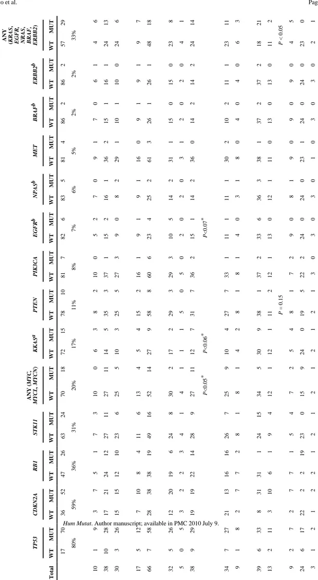
Table 1 TP53 CDKN2A RB1 STK11 ANY ( MYC, MYCL, MYCN) KKAS a PTEN PIK3CA EGFR b NPAS b MET BRAF b ERBB2 b ANY (KRAS , EGFR, NRAS , BRAF, ERBB2 ) Total WT MUT WT MUT WT MUT WT MUT WT MUT WT MUT WT MUT WT MUT WT MUT WT MUT WT MUT WT MUT WT MUT WT MUT 17 70 36 52 47 26 63 24 70 18 72 15 78 10 81 7 82 6 83 5 81 4 86 2 86 2 57 29 80% 59% 36% 31% 20% 17% 11% 8% 7% 6% 5% 2% 2% 33% 10 1 9 3 7 5 1 7 3 10 0 6 3 8 2 10 0 5 2 7 0 9 1 7 0 6 1 4 6 38 10 28 17 21 24 12 27 11 27 11 14 5 35 3 37 1 15 2 16 1 36 2 15 1 16 1 24 13 30 3 26 15 15 12 10 23 6 25 5 10 3 25 5 27 3 9 0 8 2 29 1 10 1 10 0 24 6 17 5 12 7 10 8 4 11 6 13 4 5 4 15 2 16 1 9 1 9 1 16 0 9 1 9 1 9 7 66 7 58 28 38 38 19 49 16 52 14 27 9 58 8 60 6 23 4 25 2 61 3 26 1 26 1 48 18 32 5 26 12 20 19 6 24 8 30 2 17 2 29 3 29 3 10 5 14 2 31 1 15 0 15 0 23 8 5 0 5 3 2 2 3 4 1 4 1 1 1 5 0 5 0 2 0 2 0 3 1 2 0 2 0 4 1 38 9 29 19 19 22 14 28 9 27 11 12 7 31 7 36 2 15 1 14 2 36 0 14 2 14 2 24 14 P <0.05 * P <0.06 * P <0.07 * 34 7 27 21 13 16 16 26 7 25 9 10 4 27 7 33 1 11 1 11 1 30 2 10 2 11 1 23 11 9 1 8 2 7 7 2 8 1 8 1 4 2 8 1 8 1 4 0 3 1 8 0 4 0 4 0 6 3 39 6 33 8 31 31 1 24 15 34 5 30 9 38 1 37 2 33 6 36 3 38 1 37 2 37 2 18 21 13 2 11 3 10 6 1 9 4 12 1 12 1 11 2 12 1 13 0 12 1 11 0 13 0 13 0 11 2 P = 0.15 P < 0.05 9 2 7 2 7 7 1 5 4 7 2 5 4 8 1 7 2 9 0 8 1 9 0 9 0 9 0 4 5 24 6 17 22 2 2 19 23 0 15 9 24 0 19 5 22 2 24 0 24 0 23 1 24 0 24 0 23 0 c 3 1 2 1 2 2 1 2 1 2 1 2 1 2 1 3 0 3 0 3 0 1 0 3 0 3 0 2 1
NIH-PA Author Manuscript
NIH-PA Author Manuscript
NIH-PA Author Manuscript
TP53 CDKN2A RB1 STK11 ANY ( MYC, MYCL, MYCN) KKAS a PTEN PIK3CA EGFR b NPAS b MET BRAF b ERBB2 b ANY (KRAS , EGFR, NRAS , BRAF, ERBB2 ) Total WT MUT WT MUT WT MUT WT MUT WT MUT WT MUT WT MUT WT MUT WT MUT WT MUT WT MUT WT MUT WT MUT WT MUT 61 9 52 13 48 44 3 38 23 53 8 47 13 57 4 56 5 55 6 56 5 58 3 59 2 59 2 35 29 24 6 17 22 2 2 19 23 0 15 9 23 0 19 5 22 2 24 0 24 0 23 1 24 0 24 0 23 0 P < 0.0001 P < 0.0001 P < 0.0001 P <0.05 P <0.05 P <0.05 P < 0.00005