FACULTY OF MEDICINE AND DENTISTRY
Dean Prof. Antonella Polimeni
DEPARTMENT OF ORAL SCIENCES AND MAXILLOFACIAL SURGERY
Director Prof. Ersilia Barbato
DOCTOR OF PHILOSOPHY IN INNOVATIVE TECHNOLOGIES IN THE DISEASES OF SKELETON, SKIN AND ORO-CRANIOFACIAL DISTRICT
- ODONTOSTOMATOLOGIC DISEASES - XXXII CYCLE
Director Prof. Antonella Polimeni
PHOTO-BIOMODULATION AS A PREVENTION AND MANAGEMENT MODALITY OF ORAL MUCOSITIS IN PATIENTS UNDERGOING ALLOGENEIC HEMATOPOIETIC
STEM CELL TRANSPLANTATION: CLINICAL STUDY
Tutor Candidate
Prof. Umberto Romeo Dr. Ahmed Mohsen
Supervisors Matricola Nr. 1572186
Prof. Alessandro Del Vecchio Prof. Gianluca Tenore
ACADEMIC YEARS 2019 - 2020
“We can know only that we know nothing. And that is the highest degree of human wisdom.”
Acknowledgements
Immeasurable appreciation and deepest gratitude for the help and support are extended to the following persons who in one way or another have contributed in making this study possible.
Foremost, I would like to express my sincere gratitude to Professor Umberto Romeo for the continuous support of my study and research, for his patience, motivation, enthusiasm and immense knowledge. His guidance helped me in all the time of research and writing this thesis. I could not have imagined having a better advisor and mentor for my PhD study.
I would like to express my special thanks to Professor Alessandro Del Vecchio, for his help and constant support; not to mention his advice and unsurpassed knowledge that has contributed to the success of this research. I would also like to acknowledge Professor Gianluca Tenore; as his collaboration, support and perpetual enthusiasm were of a great help for me.
Special appreciation for my family for the encouragement which helped me in completion of this thesis. My parents has given me their unequivocal support, as always, for which my mere expression of thanks likewise does not suffice. My supportive brother, Mohamed, for his unceasing encouragement and support.
I also place on record, my sense of gratitude to one and all who, directly or indirectly, have lent their helping hand in this research.
Abstract
Aim
To observe the effectiveness of a Photo-Biomodulation (PBM) protocol as a prevention and management modality for Oral Mucositis (OM) in patients undergoing conditioning regimen for allogeneic Hematopoietic Stem Cell Transplantation (aHSCT).
Introduction
OM is one of the major debilitating complications of aHSCT. PBM has been recommended as a prophylactic intervention for OM in patients undergoing aHSCT. The absence of a standardized protocol and technical parameters for prevention of OM is still the principal limit. Materials and Methods
Forty-nine patients undergoing aHSCT were included in this study and divided into three groups;
Observational Group (OG): 9 patients (3 females and 6 males) were subjected to PBM five sessions a week when the OM was developed till the complete resolution.
Preventive Group (PG): 20 patients (7 females and 13 males) were subjected to PBM five sessions a week starting at one day before the conditioning regimen till the 10th day after transplantation (D+10).
Retrospective Control Group (CG): 20 patients (10 females and 10 males) were selected to compare the obtained results.
At each session, the OM score, pain value, count of blood cells, and the morphine dosage were recorded.
Results
The mean duration of OM in PG (4.7 days) was significantly lower than CG (15 days) (p<0.05). Only 40% of PG showed severe OM, while it was shown in 85% of the CG (p<0.05). OM was not developed in 8 patients of the PG (with grade 0).
Conclusion
The study demonstrates that the preventive PBM protocol reduced the severity and duration of OM in patients undergoing aHSCT.
Contents
ACKNOWLEDGEMENTS ...II ABSTRACT ...III CONTENTS ...IV STATEMENT OF ORIGINAL AUTHORSHIP ...V
1.CHAPTER I : ORAL MUCOSITIS ...1
1.1. CLINICAL FEATURES ...2
1.2. MECHANISM AND PATHOBIOLOGY OF ORAL MUCOISITIS ...4
1.3. RISK FACTORS ...9
1.4. PREVALENCE ...11
1.5. STAGING AND GRADING OF ORAL MUCOSITIS ...13
1.6. NON-MUCOSITIS LESIONS AND DIFFERENTIAL DIAGNOSIS ...19
1.7. MORBIDITY AND IMPACT OF ORAL MUCOSITIS ...21
2.CHAPTER II : MANAGEMENT OF ORAL MUCOSITIS ...23
2.1. MANAGEMENT CLINICAL GUIDELINE ...23
2.2. LASER TISSUE INTERACTIONS ...28
2.3. PHOTO-BIOMODULATION (PBM) ...30
2.4. DIFFERENT APPLICATIONS OF PBM ...34
2.5. OBJECTIVES OF THE STUDY ...36
3.CHAPTER III : MATERIALS AND METHODS ...37
4.CHAPTER IV : RESULTS ...44
4.1. STATISTICAL RESULTS ...51
4.2. CLINICAL RESULTS ...55
5.CHAPTER V : DISCUSSION AND CONCLUSIONS ...62
6.CHAPTER VI : REFERENCES ...71
Statement of Original Authorship
The work contained in this thesis has not been previously submitted to meet requirements for an award at this or any other higher education institution. To the best of my knowledge and belief, the thesis contains no material previously published or written by another person except where due reference is made.
Signature: _________________________
Chapter I: Oral Mucositis
“Stomatitis” was the nomenclature of any mucosal damage associated with cancer treatments. All oral infections associated with cancer treatment were considered and referred as “stomatitis”. In 2007, the term mucositis was introduced and widely recognised for lesions associated with cytotoxic cancer therapy. [1]
Recognising Oral Mucositis (OM) as one of the acute complications of cancer therapy started days before World War I with the introduction of radiation as an anticancer therapy. Later on, the chemotherapy also was introduced to the medical field. In 1940s, mucositis was reported as one of the possible acute complications of cancer treatment for both targeted radiation or chemotherapy or a combination of both. Since then, many studies had been performed and investigations had been carried out to understand the pathogenesis, risk factors, and impact of this complication. The only thing that remained since the first observation of this complication, is the therapeutic modalities that can prevent or treat this condition (OM). [1]
Not only is the oral mucosa targeted by the chemotherapeutic agents and/or radiation therapy, but they may also target many healthy cells. Therefore, many other acute adverse effects are reported; such as: dysphagia, dermatitis, infections, and dysgeusia. [2] Also, many chronic adverse effects to cancer therapy are reported; such as: xerostomia, neuropathies, tooth demineralization, rampant caries, osteonecrosis, progressive periodontitis, muscular fibrosis, trismus, voice alterations, and dermatitis. [2]
However, OM is the most acute debilitating adverse effect of cancer therapy. Mucosal cells are usually targeted by cytotoxic anticancer agents due to having high mitotic rates causing the development of OM. This may explain that the collateral toxic effect of treatment is a dose limiting toxicity. OM is mostly obvious in patients with head and neck cancer
treated by chemo-radiation, and/or patients receiving conditioning regimens for Hematopoietic Stem Cell Transplantation (HSCT). [3]Also, many chemotherapeutic agents have been introduced to the chemotherapy field that may increase the toxicity of radiation; such as cetuximab and rapamycin (mTOR) inhibitors. [3 - 6]
1.1 CLINICAL FEATURES
In the initial stages, OM manifests a diffuse mucosal erythema accompanied by a burning sensation. The feeling of burning is not similar to other clinical conditions; such as severe food burn. In the advanced stages, deep and painful ulcers develop. The associated pain in this stage is more significant in comparison to other lesions such as aphthous stomatitis or traumatic lesion. The pain in the advanced stage may lead to the inability to tolerate normal food. [7]
This kind of ulcers has two unique characteristics and key elements for diagnosis: their location and their clinical form and course. They usually develop on movable mucosa involving: buccal mucosa, ventral and lateral border of the tongue, soft palate, the inner mucosal surface of lips, and floor of the mouth. (Fig. 1) They mainly involve non-keratinized areas of the oral cavity. [1]
Also, the ulcers typically manifest without peripheral erythema. This would be a key point to distinguish them from other ulcerative lesions; such as aphthous stomatitis. The ulcer can be localized and focal or diffused with poor defined borders. [7]The clinical course of OM differs according to many factors that would be discussed later; such as the kind and dose of treatment. For example, OM may not proceed to advanced ulcerative stages in patients receiving chemotherapy for a treatment of solid tumors; such as breast or colorectal cancers. [1]
Figure 1 OM on ventral surface of the tongue
1.1.1 Radiotherapy-Induced Oral Mucositis (RIOM)
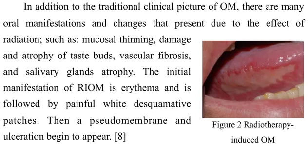
In addition to the traditional clinical picture of OM, there are many oral manifestations and changes that present due to the effect of radiation; such as: mucosal thinning, damage
and atrophy of taste buds, vascular fibrosis, and salivary glands atrophy. The initial manifestation of RIOM is erythema and is followed by painful white desquamative patches. Then a pseudomembrane and ulceration begin to appear. [8]
The severity of RIOM depends on many factors: the administrated radiation dose, dose fractions, the volume of targeted tissues, and the type of radiation. In comparison to Chemotherapy-Induced Oral Mucositis (CIOM), the healing process and the clinical course are slightly slower and more chronic. (Fig. 2) Complete resolution occur 2 to 3 weeks after the end of conventional fractionated radiotherapy. [8]
1.1.2 Chemotherapy-Induced Oral Mucositis (CIOM)
The clinical picture and symptoms are similar to that of RIOM. The most common chemotherapeutic agents causing the development of CIOM, are the anti-tumor antibiotics and the antimetabolites; such as Methotrexate (Meth) and 5-Flourouracil. CIOM is usually present at the end of the first week of drugs administration. Advanced stages (ulcerative stages) occur lately in comparison to that of radiation therapy, and the condition becomes a sever event. (Fig. 3) [8]
The severity of OM depends on the type of malignancy, patient, and type and dose of drugs. As OM in treated patients from solid tumors develops less frequently than patients with leukaemia. Myelo and
Figure 2 Radiotherapy-induced OM
Figure 3 Chemotherapy-induced OM
immunosuppressive drugs used for transplantation; such as patients undergoing HSCT, are more supposed to experience OM than others. This kind of patients (with hematological malignancies) are more likely subjected to intensive therapies of immunosuppresive drugs, so they have a higher risk of OM. [8]
1.2 MECHANISM AND PATHOBIOLOGY OF ORAL MUCOISITIS
1.2.1 Mechanisms of Oral Mucositis
Firstly, mucositis was thought to be solely a consequence of epithelial injury. [9, 10]It was thought that the non specific targeting of the proliferating basal cells of epithelium by radiation or chemotherapy, and the consequential loss of the ability of tissue to regenerate, were the only cause of mucositis. Proposing that trauma and oral microorganisms assist these events and facilitate the formation of mucositis. [10]
The non specific damage to the basal cells associated to RIOM was identified as an “outside-in” process, where the radiation cause breaks of DNA strands of these cells; while in CIOM, the damage of cells is resulted from permeating the drugs to these cells through the submucosal blood supply and/or saliva-borne chemotherapeutic agents. Considering that atrophy, thinning and ulceration associated with mucositis are a consequence of these events. [9]
With the increase of information and advancement of science about tissue and cells response to these kind of treatments (pathobiology of regimen-related mucosal injury), this concept or hypothesis has been challenged. [9-11]Mucositis should be considered as a complex sequence of biological events rather than being only a result of direct non-specific cell damage. Believing in this concept may create chances for developing prevention modalities of OM through the mechanism-based interventions, and the indirect modulation of radiation or chemotherapy-initiated pathways. [10]
Sonis S.T., [9] found through several clinical and biological observations that there are many non epithelial factors that play a role in the pathogenesis of OM. Confirming that OM is a more complex sequence of biologic events than being considered as a tissue injury. These non epithelial factors can be observed in:
-
The changes occur in the submucosal endothelium and connective tissue before the epithelial damage. It is found that after radiation in animals, a damage of endothelial walls occurs 5 days before epithelial damage. [12]The same observation has been noticed with several anti-neoplastic drugs. [9]-
Platelet aggregation may play a role in OM. Whereas the increase of cytokine platelet-activating factors has been detected in the saliva in patients with OM after radiation therapy. [9]-
A damage to the submucosal tissue, resulted from the apoptosis of fibroblasts, is found with radiation and stomatotoxic anti-neoplastic drugs. This submucosal damage is confirmed to have happened before epithelial damage. While for only anti-neoplastic drugs (chemotherapy), it is not confirmed till now. [9, 12]-
Several histological studies are not with considering OM as an inflammatory response or considering that the inflammatory cytokines; such as Tumour Necrosis Factor-α (TNF-α) and Interleukin-6 (IL-6), are playing a role in initiating OM. [9]-
It is found that ceramide, that is generated through the sphingomyelin pathway, might play a role in endothelial apoptosis and the initiation of OM after radiation. [9]-
Activator protein-1 transcription-factor family including FOS and c-JUN are found to be associated with the initiation of OM after both radiotherapy and chemotherapy. Interestingly, c-JUN can be found in all nuclei of all epithelial cells except for the basal cells. And the c-FOS is mainly localized in the nuclei of spinous cells. [13]1.2.2 Pathobiology of Oral Mucoisitis
The pathogenesis of RIOM, CIOM, and chemo-radiotherapy associated with OM are the same. It can be summarised in five phases (Fig. 4):
•
Initiation PhaseThis phase consists of two events. The first is the direct damage of DNA strands causing breaks of DNA leading to the death of basal cells. The second and the more significant event in tissue damage is the production of Reactive Oxygen Species (ROS).
During this phase, the DNA strand breaks and lipid per-oxidation leads to the activation of the transduction pathways and triggering at least 14 canonical pathways. [11, 14]
The Nuclear Factor Kappa-B (NF-kB) pathway is the most investigated pathway and the most relative pathway to mucositis. This pathway can be activated directly by radiation and/or chemotherapy or indirectly by ROS. [10, 15]
An expression of 200 genes occurs due to the activation of NF-kB pathway. These genes are related to the production of pro-inflammatory cytokines; such as IL-6 and TNF-α, cytokine modulators, stress responders (such as Cyclooxygenase-2 (COX-2)), and cell adhesion molecules. The activated NF-kB pathway can also lead directly to cell death. [15]
Other pathways; such as ceramide pathway, may indirectly lead to cell damage, are activated after radiation and chemotherapy. [16]A stimulation of macrophages occurs with a production of matrix metallo-proteinases due to the occurrence of fibrinolysis in connective tissue. [10]All of these events that consequently lead to direct or indirect cell death occur immediately after radiation and/or chemotherapy.
•
Signalling and Amplification PhasesIn these two phases, the molecules induced in the previous phase can give a positive or negative feedback. (Fig. 5) TNF-α may give a positive feedback on NF-kB to amplify its impact through initiating Mitogen-Activated Protein Kinase (MAPK) signalling. [10]
•
Ulceration PhaseThis is the most important phase for both the clinician and patient; in which the ulceration occurs. The formed ulcers are usually deep and supra-infection of bacteria may occur. Bacteria may play a role in the mucositis process. As the cell wall products of these bacteria can penetrate deeply in the submucosal tissues stimulating the macrophages for further secretions of the pro-inflammatory cytokines which consequently worsen the situation. In some cases, the supra-infection of bacteria may lead to bacteraemia or sepsis due to its invasion to the submucosal blood vessels. [10]
•
Healing PhaseOM in majority of cases heals spontaneously, because signalling the submucosa’s Extracellular Matrix (ECM) usually induce the proliferation, migration, and differentiation of the epithelium. This mechanism has been extensively studied and it has been found that the disruption of the submucosal ECM leads to delay of healing and rarely inhibits the healing phase. [17]
The initial symptoms of CIOM begin 3 - 5 days after drug infusion. Ulceration starts two days later and healing occurs within 2 weeks. CIOM is characterized by being considered as an acute complication, while RIOM is characterized by being a more chronic event. Usually, the patients are treated with incremental doses of radiation. Ulcers usually remain up to 3 - 4 weeks after the completion of radiation therapy. [10]
1.3 RISK FACTORS
Risk factors of OM can be classified into two categories. Factors that are associated with the treatment type, can be called “treatment-associated risk factors”. And factors that are “treatment-associated with the patient himself, can be called “patient-associated risk factors”. [18]
1.3.1 Treatment-Associated Risk Factors
These risk factors include type of the administrated drugs, their dosage, the site and route of administration, and the delivery schedule. The most significant risk factors are the type and dosage of treatment (radiotherapy and/or chemotherapy). [10]
Many studies have demonstrated the increase of risk with the increase of therapy intensity. Multiple cycles of myelosuppressive therapy may increase the risk. It would have a cumulative effect in particular in patients with a history of OM (in the first cycle of chemotherapy). Using a combination of radiotherapy and chemotherapy also increases the risk of OM. [15]The type of the chemotherapeutic agent may influence the risk. Whereas, 5-Fluorouracil, cisplatin, Cyclophosphamide (Cyclo), and Meth are reported to be high risk agents of OM. [15, 18]
Regarding the site of administration as a risk factor, it has been found that 100% of patients with tongue cancer are supposed to experience OM after chemo-radiotherapy. The percentage may drop to 50% in patients with hypo-pharyngeal cancer who are undergoing to receive the same dose of chemo-radiotherapy. [15]
For the type of therapy as a risk factor, it has been found that patients undergoing HSCT, who are usually subjected to conditioning regimens of total body radiotherapy and/or high doses of stomatotoxic drugs, are considered to be at high risk of mucositis. [10]
Many investigations have been performed by a less toxic protocols, especially in patients receiving head and neck radiation or undergoing HSCT, to evaluate these risk factors and their impact on the development of mucositis. Interestingly, it has been found that the drop of risk of mucositis has been in only 19% of the total annual patients with OM. Which confirm the importance to study other risk factors that are associated to the patient himself. [10]
1.3.2 Patient-Associated Risk Factors
Patients’ age, weight, gender, alterations in salivary secretion, poor oral health, and mucosal trauma have been proposed to be risk factors of OM.
Few consistent data are available to prove this association with the development of OM. [10, 18]Chansky K. et al, have tried to evaluate the differences in toxicity between men and women treated with 5-fluorouracil therapy for colorectal carcinoma. They found that females may confront a higher risk of OM after receiving 5-Fluorouracil and Meth. [19]
Many conditions; such as: psoriasis, diabetes mellitus, Addison’s disease, and impaired renal function, may have an impact on the risk of OM. [3, 10]Interestingly, in a study carried out on patients with hematological malignancies, it has been found that the patients with psoriasis had less risk in comparison to patients with Addison’s disease. Explaining that the epithelial proliferation associated with psoriasis might have caused the decrease of risk, while the presence of high levels of pro-inflammatory cytokines in patients with Addison’s disease might have increased the risk of OM. [10, 15]
Genetic factors have been suggested to be considered as a risk factor, and have a role in the development of OM. [10, 15, 20]Schwab, M. et al, have performed a study to evaluate the relation between the mucositis risk and the value of many polymorphisms that are associated with the metabolism of Fluorouracil. They found that there is a relation between specific nucleotide polymorphism and the development of OM. [20]These may propose that the risk of OM is genetically controlled.
The type of tumor is also proposed as a risk factor of OM. The parenchyma and stroma of the tumor may affect the cells reaction during the treatment and consequently may impact the risk of OM. [10]The peptides and protein products that are derived from the tumor may affect the cell response to the treatment. [21]Other factors related to the tumor itself; such as the interaction of tumors with the host and the impact of tumor products after cell death on the risk of OM, have the need of further investigations. [10]
Oral microflora is also suggested as a risk factor. Whereas, the supra-infection of bacteria may increase the severity and delay the healing process. The neutrophil count is also reported as a risk factor especially in patients undergoing HSCT after neutrophil engraftment; however, there is still no consistent data to prove it. [3]
1.4 PREVALENCE
About 43% of new cancer patients will be at low risk of OM. Since the treatment protocol will be curative surgery, peripheral radiotherapy, or low dose of chemotherapy. While the rest of new cancer patients have an intermediate risk of OM at some point during treatment. [10, 15]
In literature, the absence of standardized scoring of OM, the presence of different treatment protocols, and different tumor location influence negatively the actual estimation of OM prevalence. The presence of many side effects and complications to cancer therapy result in underreporting OM as a crucial condition and consequently have also influenced the estimation of OM prevalence. [15, 22]
In general, the prevalence range of OM is between 30 to 40% and increase to 100% if all gradings of mucositis would be considered. Its prevalence is 100% in patients undergoing Head and Neck Radiotherapy (H&N RT). 85% of patients receiving high dose of chemotherapy as a conditioning regimen for HSCT suffer from OM. Mucositis develop in 20 to 40% of patients receiving conventional chemotherapy. [15]
Patients undergoing treatment of the common solid tumor in breast, lung, rectum, and colon are usually considered as intermediate risk patients. In which, the prevalence at the first cycle of conventional chemotherapy, will be 20%. The risk increase to 60% with the second cycle due to the cumulative effect of chemotherapy. [15]
OM in patients undergoing HSCT can be predicted 3 to 4 days after the start of infusion of conditioning regimen with a peak of severity with ulcer formation 7 to 14 days later. Healing and resolution occur after that in the next 7 days. [15]
In patients undergoing standard regimens of concomitant Chemo-Radiotherapy (CT-RT), the regimens usually include cisplatin or carboplatin and fractioned radiotherapy (2 Gy five times a week for 6 weeks with a total dose of 60-70 Gy). OM is characterized in these kind of patients by being prolonged. It starts with erythema and soreness at the end of the first week. A loss of mucosal integrity and ulcers start by the end of the second week. Then, the developed ulcers persist for 2 - 4 weeks after the completion of treatment. [15]
1.5 STAGING AND GRADING OF ORAL MUCOSITIS 1.5.1 Importance of Scoring Oral Mucositis
The importance of scoring OM is derived from two main significant issues. The first is the need of accurate and standardized documentation of local complications associated with cancer therapy. This would provide means of comparing different therapeutic modalities. It would also provide an accurate data to help the health-giver in evaluating the relation between the complication and the different existing variants of therapy; such as: dosage, delivery method, etc. [1, 8]
The second is that it would help to investigate accurately the impact of this kind of treatment and these complications on patient morbidity, mortality, and the quality of life. [8]
1.5.2 Scoring Systems
There are two criteria that should be concerned to produce a valid: scoring system, content, and inter/intra user reliability. The available scoring systems in the literature are categorized into three categories: scoring systems that assess the toxicity through evaluating the oral mucosa by clinical examination. The second is those that evaluate a number of parameters related to oral function and patient morbidity factors. The third is scoring systems that have been produced in mucositis research studies. [1, 8]
1.5.2.1 Toxicity Assessment Scales
These scoring scales are the most common used scales. They assess the severity of OM and describe the overall toxicity in relation to a specific chemotherapeutic regimens or radiotherapy dose. All of them are based on clinical examination of oral mucosa and the observation of clinical changes. [1]This category of scoring systems include:
•
National Cancer Institute Common Toxicity Criteria (NCI-CTC): There are three types of NCI-CTC for different situations to assess the toxicity grading of OM, including: [1]1. Grading system to assess chemotherapy-induced stomatitis/ pharyngitis. (Table 1)
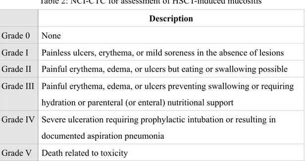
2. Grading system to assess mucositis associated with HSCT (stomatitis/
pharyngitis). (Table 2)
Description Grade 0 None
Grade I Painless ulcers, erythema, or mild soreness in the absence of lesions Grade II Painful erythema, edema, or ulcers but eating or swallowing possible Grade III Painful erythema, edema, or ulcers requiring intravenous hydration Grade IV Severe ulceration, requiring parenteral, enteral nutritional support, or
prophylactic intubation Grade V Death related to toxicity
Table 1: NCI-CTC for assessment of chemotherapy-induced mucositis
Description Grade 0 None
Grade I Painless ulcers, erythema, or mild soreness in the absence of lesions Grade II Painful erythema, edema, or ulcers but eating or swallowing possible Grade III Painful erythema, edema, or ulcers preventing swallowing or requiring
hydration or parenteral (or enteral) nutritional support
Grade IV Severe ulceration requiring prophylactic intubation or resulting in documented aspiration pneumonia
Grade V Death related to toxicity
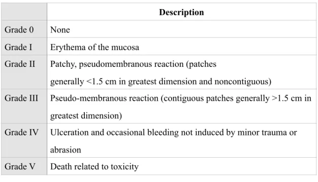
3. Grading system to assess mucositis associated with radiotherapy. (Table 3)
•
Radiation Therapy Oncology Group (RTOG) [1]: This grading system is mainly constructed for acute oral mucous membrane toxicity caused by radiotherapy. (Table 4)•
World Health Organization (WHO): This grading system is the most widely used for scoring of the toxicity grade of OM. (Table 5)Description Grade 0 None
Grade I Erythema of the mucosa
Grade II Patchy, pseudomembranous reaction (patches
generally <1.5 cm in greatest dimension and noncontiguous)
Grade III Pseudo-membranous reaction (contiguous patches generally >1.5 cm in greatest dimension)
Grade IV Ulceration and occasional bleeding not induced by minor trauma or abrasion
Grade V Death related to toxicity
Table 3: NCI-CTC for assessment of radiotherapy-induced mucositis
Description Grade 0 No change over baseline
Grade I Injection, may experience mild pain not requiring analgesic Grade II Patchy mucositis that may produce inflammatory serosanguinitis
discharge; may experience moderate pain requiring analgesia Grade III Confluent, fibrinous mucositis, may include severe pain requiring
narcotic
Grade IV Ulceration, hemorrhage, or necrosis
1.5.2.2 Patient Management Scoring Systems
This kind of scales have been proposed mainly for daily evaluation of patients’ oral health and function including speech quality, salivary function, oral hygiene, swallowing, and gingival health. The goal of these scales is to evaluate the overall oral health to formulate treatment plans. This scales include: [1]
•
Oral Assessment Guide (OAG). (Table 6)Description Grade 0 None
Grade I Oral soreness, erythema
Grade II Oral erythema, ulcers, solid diet tolerated Grade III Oral ulcers, liquid diet only
Grade IV Oral alimentation impossible
Table 5: WHO grading score for assessment of mucositis. [23]
Item
Score
1 2 3
Voice Normal Deeper or raspy Difficulty talking or
painful
Swallow Normal Some pain on
swallowing
Unable to swallow
Lips Smooth, painful, and moist
Dry or cracked Ulcerated bleeding
Tongue Pink, moist, and papillae present
Coated of loss of papillae with a shiny appearance with or without redness
Blistered or cracked
Saliva Watery Thick or ropy Abscent
Mucous membrane (buccal mucosa, palate)
Pink and moist Reddened or coated without ulceration
Ulceration with or without bleeding
Mucous membrane (labial mucosa)
Pink and moist Reddened or coated without ulceration
Ulceration with or without bleeding
Gingiva Pink, stippled, and firm Oedematous with or without redness
Spontaneous bleeding or bleeding with pressure Table 6: OAG. [24]
•
Western Consortium for Cancer Nursing Research (WCCNR). (Table 7)•
MacDibbs Scales for patients treated with radiotherapy. (Table 8)Stage Erythema Colour Bleeding
0 none pink none
1 1- 4 slightly red none
2 more than 4 moderately red with eating and oral hygiene
3 coalescing very red spontaneous
Table 7: WCCNR. [25] Patient Information Pain 0 = None 1 = Mild 2 = Moderate 3 = Severe Dryness Eat Talk Swallow Taste Saliva Total Sore Examination Nr of Ulcers
Size of largest ulcers - mm Vesicles
+ or -Red areas
White patches
KOH smear Results + or
-Not done
HSV culture Results + or
-Not done
•
Nijmegen Nursing Mucositis Scoring System (NNMSS). (Table 9)1.5.2.3 Research Related Scales
These scales have been developed and used in research studies concerned about mucositis. They assess OM through evaluating different strict parameters. There are two mostly common used scales in the literature. [1]
•
Oral Mucositis Index (OMI). (Table 10)Characteristics 0 point 1 point 2 points 3 points
Characteristics based on inspection by
nurse
Erythema Pink and moist
Mild/moderate Severe
Edema Abscent Mild/ Print of teeth in tongue
edge, gingiva swollen and red
Mild/ Print of teeth in tongue edge, gingiva swollen and white Sever/ swollen tongue / gingival swollen and shining white/ elapse ulceration Characteristics based on patients information Lesions Abscent 1 to 4 > 4 Pain None VAS score < 3 VAS score
4,5,6
VAS score > 6 Dryness of mouth Normal Mild Moderate Severe
Saliva Viscosity Normal Slimy Thick
Table 9: NNMSS. [1] Erythema 0 = Normal 1 = Mild 2 = Moderate 3 = Severe Atrophy Hyperkeratosis Lichenoid Edema Ulceration 0: None 1: > 0cm2 but ≤ 1cm2 2: > 1cm2 but ≤ 2cm2 3: > 2cm2 Pseudomembrane
Pain Visual Analogue Scale (from 0 to 10)
Mouth dryness Visual Analogue Scale (from 0 to 10) Table 10: OMI. [27]
•
Oral Mucositis Assessment Scale (OMAS). (Table 11)1.6 NON-MUCOSITIS LESIONS AND DIFFERENTIAL DIAGNOSIS
In addition to OM, there are many oral lesions that can be developed in treated cancer patients; in particular patients treated with myelosuppressive chemotherapy. Oral lesions include opportunistic infections and Graft-Versus-Host Disease (GVHD); in particular in patients undergoing allogeneic HSCT (aHSCT).
There are two key elements to differentiate between OM and non-mucositis lesions. The first is the clinical picture and course of OM, as they differ from other lesions. The second is the location of lesions. OM can be observed on the movable non-keratinized mucosa; such as: buccal mucosa, lateral and ventral surface of the tongue, floor of the mouth, soft palate, and the inner mucosal surface of lips.
In contrast, opportunistic infections are mainly observed on the keratinized mucosa; such as: hard palate, dorsal surface of the tongue, and gingiva. GVHD can be observed on both keratinized and non-keratinized mucosa. [1, 15]
1.6.1 Necrotizing Gingivitis
Necrotizing gingivitis is one of the possible bacterial infections associated with n e u t r o p e n i c p a t i e n t s r e c e i v i n g myelosuppressive chemotherapy. It manifests as a necrosis of the gingival margins and
papilla. It results in the loss of gingival architecture. (Fig. 6) [1, 15]
Ulceration Erythema 0 = no lesion 0 = none 1 = < 1cm2 1 = not severe 2 = 1-3cm2 2 = severe 3 = > 3cm2 Table 11: OMAS. [28] Figure 6 Necrotizing Gingivitis
1.6.2 Herpes Viruses Infection
The clinical course of this kind of infection is different. It starts with a clustering blisters mainly on the hard palate. These vesicles rupture and ulcers develop without the presence of pseudomembrane. The viral infection includes Herpes Simplex Virus (HSV) and Herpes Zoster Virus (HZV). The difference between them is that the infection of HSV is usually bilateral while for HZV is unilateral in linear pattern. [1]
1.6.3 Oral Candidiasis
It is a common oral infection in patients immunocompromised with a history of cancer therapy. Its prevalence increases when the cancer treatment alter the oral environment; such as salivary secretion and function. The common type
of oral candidiasis in cancer patients is pseudomembranous candidiasis. It manifests as an erythema, hyperplasia, and angular chelitis. (Fig. 7) It can be distinguished from OM by the clinical picture and oral swap. [1]
1.6.4 Graft-Versus-Host-Disease (GVHD)
This kind of lesion have two types: Chronic type that occurs after 100 days of transplantation; therefore, it can not be considered as a differential diagnosis. The acute type starts approximately 4 days after the start of the conditioning regimen. Its peak is at the 10th day of infusion. It
heals after 3 - 4 weeks. It manifests as a combination of hyper-keratotic and erosive lesions with the lichenoid criteria. (Fig. 8) It can be also localized on the dorsal surface of the tongue. The clinical appearance is different from OM and can be easily distinguished. [15]
Figure 8 Erosive form of GVHD
1.7 MORBIDITY AND IMPACT OF ORAL MUCOSITIS
OM is the most debilitating adverse effect of cancer therapy. [3] Many studies have investigated its impact on patients treatment course and Quality Of Life (QOL).
1.7.1 Pain
Pain is the main complication of OM. Its peak is in the ulceration period. Systemic opioids are one of the main modalities for pain management of ulcerative OM. Many side effects of narcotic systemic therapy have been demonstrated; such as: increase of risk of dependence, constipation, and alteration of mental state. [29]It has been found that in a study carried out in patients undergoing HSCT, the increase of one point of mucositis scoring may lead to an increase of the need of adding approximately 3 days of systemic opioids. [30]In addition, the increase of severity of OM may hinder the normal nutrition. That lead to seeking other solutions to overcome this complication through parenteral nutrition or gastrostomy tube. Consequently, these may result in loss of weight, healing compromise, and reduction of resistance to infections. It is found that patients with OM induced by H&N RT are liable to lose more than 5% of their weight. [29, 31]
1.7.2 Quality of Life
Many studies have demonstrated the impact of OM on the QOL. Severe OM result in worsening of the QOL. Some studies have observed that even mild grades of OM might influence negatively the QOL. [1, 32]In a study performed by Bellm, L.A. and his colleagues to evaluate the outcome of HSCT, it has been concluded that the most debilitating complication of HSCT treatment protocol is OM. [29, 33]
1.7.3 Cancer Therapy Course
OM may have a negative impact on the cancer prognosis. In many situations, interruption or reduction of dose may occur due to the severity of OM. In a study, it has been demonstrated that the unplanned reduction of dose of chemotherapy cycle might increase the risk of mucositis two
times in patients undergoing chemotherapy for solid tumors or lymphoma. The interruption of radiotherapy occur in 11% of patients undergoing H&N RT due to OM. [29, 34]
1.7.4 Infection
OM may be complicated with supra-infections; such as HSV infections or candidal infection. These supra-infections may lead to systemic sepsis which may be life threatening due to the immunosuppression that occur with chemotherapy. In the literature, the rate of infection is reported to be directly related to OM severity and to be increased two times more in patients with OM than patients without. [35]Also, it has been reported that infection related deaths are more in patients with OM. The transplant-related mortality and systemic infection are reported to be related to severity of OM in patients receiving high dose of chemotherapy for HSCT. [29]
1.7.5 Oral Health
OM may negatively impact the oral and dental health of cancer patients. It impairs the patient to carry out the routine oral hygiene measures. This may increase the risk of dental caries and periodontal diseases; in particular with the presence of transient or permanent hypo-salivation that occur in patients undergoing cancer therapy. [29]
1.7.6 Increase of Treatment Cost
Pain management, parenteral nutrition, gastrostomy tubes with diet supplements, hospitalisation, and secondary infections-associated with the management of OM, increase the cost significantly. For example, an increase of cost from $1700 to $6000 can be predicted with the increase of the OM severity in patients receiving H&N RT. [29, 30]In patients undergoing HSCT, an increase of severity of OM one grade may increase the hospitalization time to 2.6 days, which in turn increase the hospital charges with about $25000. [29, 31]
Chapter II: Management of Oral Mucositis
2.1 MANAGEMENT CLINICAL GUIDELINE
Many organizations have constructed guidelines for the management of OM; such as: the American Society of Clinical Oncology (ASCO), [36]the Multinational Association of Supportive Care in Cancer (MASCC), [37]and the National Comprehensive Care Network (NCCN). [38]Although, there is a general agreement of these guidelines and a lot of studies are carried out, the prevention and management modalities are sill not completely evidenced. [1]
The most popular guideline for the management of OM is that of MASCC and the International Society of Oral Oncology (ISOO). The guideline has been demonstrated in two levels; recommendations and suggestions. These levels have been divided into in-favour and against usage of agents for the management of OM. The proposed agents and elements for management of OM have been categorized into: basic oral care, growth factors and cytokines, anti-inflammatory agents, antimicrobial, coating agents, anaesthetics and anti-analgesics, cryotherapy, natural and miscellaneous agents, and laser therapy. [37] The MASCC/ISOO guideline has had recommendations against:
•
The use of PTA paste (Polymyxin, Tobramycin Amphotericin B) and BCoG lozenges (Bacitracin, Clotrimazole, Gentamicin B) to prevent OM in Head and Neck (H&N) cancer patients receiving H&N RT (with a level of evidence grade II).•
The use of iseganan antimicrobial mouthwash to prevent OM in H&N cancer patients receiving chemo-radiotherapy or patients undergoing HSCT receiving high dose of chemotherapy with or without radiotherapy (with a level of evidence grade II).•
The use of sucralfate mouthwash to prevent or to treat OM in H&N cancer patients receiving chemotherapy (with level of evidence grade I), radiotherapy (with level of evidence grade I), or chemo-radiotherapy (with level of evidence grade II).•
The use of intravenous glutamine to prevent OM in patients undergoing HSCT receiving high dose chemotherapy with or without radiotherapy (with level of evidence grade II). [37]Moreover, the MASCC/ISOO guideline has had suggestions against:
•
The use of chlorhexidine mouthwash to prevent OM in H&N cancer patients receiving radiotherapy (with level of evidence grade III).•
The use of granulocyte macrophage colony stimulating factor mouthwash to prevent OM in patients undergoing autologous or allogenic HSCT receiving high dose of chemotherapy (with level of evidence grade II).•
The use of misoprostol mouthwash to prevent OM in H&N cancer patients receiving radiotherapy (with level of evidence grade III).•
The oral administration of systemic pentoxifylline for patients undergoing HSCT (with level of evidence grade III).•
The oral administration of systemic pilocarpine to prevent OM in H&N patients receiving H&N RT (with level of evidence grade III) or patients undergoing HSCT receiving high dose chemotherapy with or without radiotherapy (with level of evidence grade II). [37]While the MASCC/ISOO clinical recommendations and suggestions are as follow;
2.1.1 Basic Oral Care
It has been considered for a long time that achieving a sound and good oral hygiene might decrease the risk and even the severity of OM. [39]Since the sound oral hygiene may increase the potential of oral environment to reduce the possible traumatic injuries, and reduce the risk of oral infections that may contribute in the development of OM. [39, 40] The basic oral care can be achieved through two strategies: professional oral hygiene measures; including dental care before the cancer treatment, and oral care protocols during the cancer therapy. [39]
The professional oral hygiene measures can be carried out through an oral examination prior the beginning of the cancer treatment. In which a complete elimination of risk factors, optimization of oral status, and patient education should be performed. [39]Although, there is no evidence to support the professional oral hygiene measures for the prevention of OM, all the guidelines support it even for the general well-being of patients. [37, 39]
According to the MASCC/ISOO clinical practice guideline, the oral care protocols consist of tooth brushing, flossing, and the use of mouthwash to maintain the oral status. There is no adequate evidence to support the oral care protocols. MASCC/ISOO guideline has suggested them in-favour for the prevention of OM even with the absence of evidence supporting them. [37]
In patients treated by chemotherapy, the efficacy of different bland mouthwashes; such as: saline, sodium bicarbonate, mixed medication rinses, calcium phosphate, and chlorhexidine is not adequately evidenced in the literature. [37, 39]
2.1.2 Pailfermin (Keratinocyte Growth Factor-1)
There are many growth factors that are introduced for the management of OM including: palifermin, granulocyte macrophage colony stimulating factor, granulocyte colony stimulating factor,
velafermin, fibroblast growth factor-20, and sargramostim. [15]The only approved growth factor for OM by the US Food and Drug Administration (FDA) and the European Medicines Agency is palifermin. [37, 39]Palifermin has an effect on the glutathione activity and up-regulates Nrf2 which consequently reduce the oxygen free radical damage and prevent the DNA strand breaks. [15]
The MASCC/ISOO panel has included palifermin as a recommended in-favour in their guideline for the prevention of OM in patients with haematological malignancies undergoing high-dose chemotherapy and/or radiotherapy for autologous HSCT. [37]
For other growth factors, the MASCC/ISOO panel has included the granulocyte macrophage colony stimulating factor in the suggestion against category with a sufficient evidence for the prevention of OM in patients undergoing HSCT, and in patients with H&N cancers due to the evidence of its negative impact on local tumor control. [41]
While, the rest of all introduced growth factors have not been included in the guideline due to the insufficient data. [37]
2.1.3 Benzydamine Hydrochloride (BZD)
BZD is a non-steroidal anti-inflammatory mouthwash. [15]It can inhibit the production of pro-inflammatory cytokines including TNF-ɒ and IL-6. Animal studies have demonstrated the anti-apoptotic activity of BZD in epithelium and connective tissues. [15, 42]
The MASCC/ISOO panel has included it in the recommended section for the prevention of OM in patients of H&N cancer receiving radiation > 50 Gy. [37]The BZD is administrated intravenously within 30 minutes of each radiation session. [15]
2.1.4 Amifostine
Amifostine (Ethyol) is a free radical scavenger. It has been approved in many countries for the the prevention of xerostomia induced by H&N RT for caner patients. [1]Many studies have been carried out for testing its efficacy against OM. It is usually used as a mucoprotective agent. The results are conflicting and the MASCC/ISOO did not include in its guideline. [37]
2.1.5 N-Acetyl Cysteine
N-Acetyl Cysteine is an anti-oxidant that have a potentiality to affect the NF-kB. [15, 43]It has been demonstrated that n-acetyl cysteine can decrease the duration of severe OM. It is applied in parenteral formulation of 100mg/kg/day. [43]The MASCC/ISOO did not include it in its guideline.
2.1.6 Oral Cryotherapy
It is the placement of ice chips for 30 - 60 minutes in the mouth during the delivery of chemotherapy. [15]It has been evaluated in many studies and has been recommended for the prevention of OM in patients receiving bolus dose of 5-Fluorouracil. [22]It has been also suggested for the prevention in patients undergoing HSCT with conditioning regimens with or without radiation.
2.1.7 Analgesic Agents
There are many analgesic agents that have been suggested by MASCC/ISOO clinical practice guideline for the pain management of OM; such as: morphine, transdermal fentanyl, morphine mouth rinse, and doxipine. 2% morphine mouthwash has been suggested for pain control in patients receiving chemo-radiotherapy for H&N cancer. Transdermal fentanyl has been suggested for patients receiving conventional or high dose chemotherapy with or without radiotherapy. And 0.5% doxipine mouthwash can be suggested generally for pain of OM. [37]
2.1.8 Photo-Biomodulation (PBM)
According to MASCC/ISOO clinical guideline, two specific treatment settings of PBM have been inserted in the guideline. PBM is recommended for the prevention of OM in patients undergoing HSCT receiving high dose of chemotherapy with or without radiotherapy (with a level of evidence grade II). Also, PBM is suggested for the prevention of OM in H&N cancer patients receiving only radiotherapy (with a level of evidence grade III). [37]
2.2 LASER TISSUE INTERACTIONS
There are four different interactions between the light energy of laser and the target tissue. (Fig. 9) These interactions mainly depend on the optical characteristics of the targeted tissues.
2.2.1 Reflection
The laser beam is directed off the tissue surface without any effect. The light beam may remain narrow or become diffused. Mostly, the increase of the distance between the hand-piece and the target tissue increases the divergence of the light beam, e.g. the reflection occur between CO2 laser and titanium implants.
2.2.2 Transmission
The laser beam transmits through the target tissue without any effect. This occurs when the laser used is diode and the medium is water. [44]
2.2.3 Scattering
It is a distortion of beam with a decrease of the laser energy. The laser rays travel in an uncontrolled manner through the medium.
2.2.4 Absorption
It is the absorption of laser energy in the targeted tissue. It mainly depends on the chromophores, water contents, and the laser wavelength. It is the desirable interaction of laser in the medical field and cause mainly one of five photo-biologic effects. These photo-biologic effects are:
•
Photo-thermal effect: It is the main and the principal laser tissueinteractions. The laser energy is absorbed and transformed into heat. The resulted heat; depending on the laser parameters, light energy, and time of laser application, can be used for incision, ablation (vaporisation), and coagulation (haemostasis).
•
Photo-chemical effect: It may occur in two patterns. First, the laserstimulate chemical reactions, like the light used for curing composite resin. Second, the laser break chemical compounds, like laser applied in periodontal pockets or pulp canals filled with photosensitising agent for disinfection through the resulted singlet oxygen.
•
Fluoresce: It occurs when certain biologic pigments absorb the laserlight.
•
Photo-acoustic (spallation): It is the production of shock wavescausing surface ablation. This effect is used mainly in operative dentistry.
•
Bio-stimulation: It is a non surgical effect of laser resulting in astimulation of tissue regeneration, reduction of inflammation, and control of pain. The bio-stimulation effect occurs in the different laser applications with all kinds of dental lasers (cutting lasers and soft lasers). [44]
2.3 PHOTO-BIOMODULATION (PBM)
PBM is the use of laser for therapeutic purposes. It is the use of light in low power to stimulate tissue regeneration, reduce inflammation, and control pain.
Many nomenclatures have been suggested to refer to this therapeutic modality. [2]The joint conference between North American Association for Laser Therapy (NAALT) and World Association For Laser Therapy (WFLT) in 2014 recommended the use of “Photo-Biomodulation” term and defined it as “The therapeutic use of light (e.g. Visible, Near Infrared (NIR), Infrared (IR)) absorbed by endogenous chromophores, triggering non thermal, non cytotoxic, biological reactions through photo-chemical or photo-physical events, leading to physiological changes”. [45]
2.3.1 PBM parameters
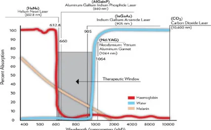
PBM can be accomplished through the use of therapeutic lasers with a power density between 5 and 150mW/cm2 applied for 30 - 60
seconds per point. [2]The therapeutic lasers can be typically found in “the therapeutic window” part of the electromagnetic spectrum in the visible and NIR part (from 600 -1000nm). (Fig. 10)
Many laser devices have been utilised for PBM applications; such as: Helium-Neon gas laser (HeNe), LED arrays, visible light, Indium Gallium Aluminum Phosphide (InGaAlP) diode laser, Gallium Aluminum Arsenide (GaAlAs) diode laser, Gallium Arsenide (GaAs) laser, Neodymium doped Yttrium Aluminum Garnet (Nd:YAG), and non-thermal, non-ablative Carbon dioxide (CO2) laser. [2]
The PBM irradiation parameters are crucial factors that should be concerned to achieve the desirable effects and outcome. [46]They include wavelength, delivered energy, energy density, pulse mode, mode of application, power density, and time. [47]The response to PBM is reported to be biphasic; as the decrease of dosage than the optimal needed value may lead to a reduced or negative effect, while the increase of the dosage may affect the therapeutic outcomes. [2, 46, 47]Other factors may affect the response to PBM; such as: the underlying pathology, cell type, the redox state of cells, site of application (intra-oral or extra-oral), and individual patient associated factors. [2, 46]
In the literature, there are a diversity and inconsistency of the reporting parameters of PBM. The standardization of measuring the irradiation parameters and dose calculation for each indication is needed to be able to get a reproducible and consistent outcomes. These have pushed Zecha, J.A.et al., to propose a table of all important and crucial parameters that should be demonstrated and concerned to achieve a standardization of treatment settings and outcomes. [2]
2.3.2 Mechanism of Action of PBM
Several effects of PBM have been demonstrated; including pain relief, enhancement of wound healing, nerve regeneration, stimulation of cell proliferation, and anti-inflammatory effect. [48, 49, 50]Since the introduction of PBM in 1967, a large amount of clinical and laboratory studies have been carried out to understand the PBM mechanism of action that explain these positive effects. [2]
However, PBM mechanism of action is still controversial, it may be due to the lack of uniform reporting physical and biological variables; such as: type of laser, output power, frequency of pulse, time of application, distance of source from the irradiated tissue, and histological differences of the targeted tissues. [51]The PBM effects; such as enhancement of wound healing, are mostly referred to the direct influence on the injury resolution phases; inflammatory phase, proliferative phase, and remodelling phase. [2, 51]
The current data propose that, PBM modulates the biological function of cells through the predominate action on the Cytochrome c Oxidase (CcO) in the mitochondrial respiratory chain. It facilitates the electron transportation leading to an increase of transmembrane proton gradient which consequently lead to production of Adenosine Triphosphate (ATP). The increase of ATP production, even with a small amount means an increase of source of energy in cells and consequently an enhancement of the biological functions and metabolism of cells. (Fig. 11) [2]
Figure 11 Schematic graph shows the cellular mechanism of PBM designed by Huang YY, et al [46]
PBM may lead to a transient burst of ROS resulting in an adaptive decrease of oxidative state of cells. The reduction of ROS production influence cellular processes and the activation of transcription factors; such as NF-kB. That lead to expression of stimulatory and protective genes, and generation of growth factors; such as: fibroblast growth factors, pro-inflammatory cytokines, and chemokines that promote tissue repair. [2]
In case of presence of hypoxic or stressed cells, a binding between the CcO and the produced mitochondrial Nitric Oxide (mtNO) occur in the mitochondria and leading to the displacement of oxygen. [2]That lead to cellular respiration inhibition, decrease of ATP production, and increase of the oxidative state of cells. So, an activation of the intra-cellular signalling pathways and several transcription factors would occur. These transcription factors include NF-kB, p53, Redox factor-1 (Ref-1), Activator Protein-1 (AP-1), Hypoxia Inducible Factor-1 (HIF-1), etc.[52]These induce the production of anti inflammatory mediators and inflammatory mediators; such as: IL-1, IL-6, TNF-α Prostaglandin E2 (PGE-2), and COX-2. [2, 47, 52]
It is suggested that the appropriate administration of PBM on the stressed cells may lead to the dissociation of mtNO binding to CcO, increase of ATP production and balance between pro-oxidant and antioxidant mediators that consequently lead to decrease of the oxidative state of cells. (Fig. 12) [2, 53]
PBM associated pain reduction is likely to be due to other mechanisms rather than the increase of ATP and the reduction of oxidative state of cells. It is found that PBM with a relatively high power (more than 300mW/cm2) is absorbed by nociceptors that may lead to an
inhibitory effect on A and C neuronal pain fibres. These would lead to a decrease of neural conduction velocity, a reduction of the amplitude of compound action potentials, and a decrease of neurogenic inflammation. [2, 54]
2.4 DIFFERENT APPLICATIONS OF PHOTO-BIOMODULATION
Many in-vitro and in-vivo studies have demonstrated the different desirable effects of PBM; including enhancement of wound healing, stimulation of cell proliferation, pain relief, nerve regeneration, and anti-inflammatory effect.
An in-vitro study have demonstrated a change of the metabolic activity of human fibroblasts after laser irradiation. [55]A study by Pereira et al., have reported an elevation of cell growth and synthesis of pro-collagen of fibroblasts after being irradiated with a diode laser in low power and power density ranges between 3 to 5J/cm2. [56]Similar results
have been observed with PBM for stimulating the proliferation of cartilaginous cells, [57]promoting formation of lymphatics and blood capillaries, [58]stimulating the accumulation of reparative new bone, [59]and accelerating the re-epithelization. [51, 60]
These several studies and promising results have promoted performing several investigations on PBM for different oral applications. The beneficial use of PBM has been highlighted in many clinical studies in different oral and dental applications; such as the application of PBM to minimize the edema after third molar surgery, [61]as a reinforcing modality of conventional periodontal treatments, [62]as an adjunctive
Figure 12 Schematic graph shows a possible mitochondrial mechanism of PBM designed by Huang YY, et al [46]
modality for the surgical approach of Medication Related Osteonecrosis of the Jaw (MRONJ), [63]to decrease the pain in myofacial pain dysfunction syndrome, [64]and to increase the primary stability of implants. [51, 65]
2.5 OBJECTIVES OF THE STUDY
Observing the effectiveness of a PBM protocol as a prevention and management modality for OM in patients undergoing conditioning regimen for aHSCT.
Evaluating the impact of PBM on OM incidence, severity, duration, and pattern or course.
Observing the influence of PBM on pain associated with OM.
Evaluating the influence of other different patient characteristics; such as: age, gender, and type of transplant on OM with and without PBM.
Observing the relation between the value of different blood cells count and the development of OM; including White Blood Cells (WBCs), Platelets (PLTS), and Neutrophils (N).
Chapter III: Materials and Methods
Attending patients receiving aHSCT at the Department of Cellular Biotechnology and Hematology, Sapienza University of Rome, were included in this study. The study was a part of “MoMax” project (Oral Medicine and Maxillofacial) of the Department of Oral Sciences and Maxillofacial Surgery at Sapienza University of Rome. The project is a task force designed to provide cancer patients and high risk patients with a multidisciplinary team care through the cooperation between different health providers. The main target of the project is to accelerate and customise the treatment plan which may have a positive impact on the patients’ survival rate.
The inclusion criteria of this study were as follow; patients receiving conditioning regimens for aHSCT with an age range of 6 to 80 years. Non cooperative patients, patients with systemic diseases that hinder the wound healing (e.g. uncontrolled Diabetes Mellitus, etc.), patients with oxygen mask or orogastric catheters that may interfere the PBM administration, and patients with suspicious lesions within the oral cavity, were excluded from the study.
All the selected patients were subjected to a standardized care protocol for the prevention of GVHD through the administration of prophylaxis agents; such as cyclosporine or Meth.
All the study procedures were performed in accordance with the ethical standards of the institutional and/or national research committee and with the 1964 Helsinki declaration and its later amendments or comparable ethical standards.
All the patients were subjected to a comprehensive oral examination with panoramic radiographs prior the start of chemotherapy conditioning regimen administration. The aim of the oral examination was to optimize, treat, and eliminate all the potential
sources of oral infection that might compromise the transplantation procedures. An informed consent was signed by all the patients.
Patient education about the possible oral collateral effects of the conditioning regimen and aHSCT was performed; including OM prevalence, course, and prognosis. All the patients were informed with a standardized basic oral hygiene practices during the transplantation procedures, which included flossing and teeth brushing 3 times daily using soft brushes. In case of difficulties, they were informed to use a wet gauze. A mixture of saline and sodium bicarbonate was used as a mouthwash by all the patients.
General information; such as: name, age, gender, medical and dental history, underlying pathology, planned conditioning regimen, and type of transplantation, for all the patients were gathered.
In the beginning, an Observational Group (OG) of 9 patients fulfilling the above criteria was subjected to PBM in case of development of OM. (Fig. 13) The purpose of PBM was treating the existing conditioning-induced OM.
PBM was administered over the ulcerated areas and repeated five times a week (Monday to Friday) till the complete resolution of lesions.
The results of this observational group were promising. Then, it was decided to develop the study for the evaluation of the prevention purposes following the recent prevention clinical practice guideline of MASCC/ISOO. [37, 66] The study was designed as follows:
Control Group (CG): 20 patients (10 females and 10 males) received aHSCT; who were not subjected to laser therapy and fulfilling the decided inclusion and exclusion criteria, were selected retrospectively from the patient record data of the department between December 2014 and October 2016.
Preventive Group (PG): 20 patients (7 females and 13 males) received aHSCT and fulfilling the inclusion and exclusion criteria were selected between April 2017 and September 2019.
The PG patients were subjected to 5 sessions of an intra-oral PBM per week (Monday to Friday), starting one day before the conditioning regimen and continued till the 10th day after
transplantation (D+10).
All the procedures were performed by the same operator. A custom-made clinical chart was used to register all the recordings. At each session, the blood cells count (WBCs, PLTS, N) and the morphine dosage were recorded. (Fig. 14) In case of development of OM during the preventive therapy, the laser parameters were changed to the treatment laser parameters of OG till the complete resolution of OM.
Patients were always photographed with the same equipment (Nikon D200, Nikon Corporation, Tokyo, Japan).
3.1 Assessment of OM
In all the PBM visits (five times a week) for all the laser patients (OG and PG), an evaluation and scoring of the oral tissues using the grading scale of WHO were performed. (Fig. 15)
The grade of OM was considered grade 0; in case of absence of signs and symptoms, grade I; in case of presence of localized or diffused erythema, grade II; in case of presence of ulcers that did not hinder eating solid food, grade III; in case of presence of ulcers that hindered eating solid food but the patient was able to swallow liquids, and grade IV; in case the ulcers impair eating or swallowing liquids. [23]
The assessment and the evaluation of pain were included using the Numeric Rating Scale (NRS). The site of the developed OM was also registered. In the retrospective CG, the routine patient care included the assessment of OM with the same OM grading scale of WHO.
3.2 PBM parameters
The laser device used in both OG and PG was a Double diode laser (Lumix2®; FISIOLINE, Verduno, Cuneo, Italy) emitting simultaneously two wavelengths 650nm and at 904 - 910nm. (Fig. 16) Before commencing each PBM session, cleaning of the device and covering the hand-piece with a plastic wrap (to avoid direct contact with patients) were carried out. Appropriate safety glasses were
used by both the operator and all patients subjected to PBM.
Figure 15 WHO grading scale used for this study
In the OG, the laser was applied point per point to cover all the ulcerated area in a defocused (non-contact) mode with the following parameters per point: energy of 6J, power of 90.9mWatt, fluence of 12J/cm2, frequency of 13kHz, application time of 66 seconds, spot
diameter of 8mm, and point area of 0.5cm2. (Fig. 17)
In the PG, all the at risk mucosal surfaces (10 points); including buccal mucosa, tip, ventral and marginal surface of the tongue, floor of the mouth, soft palate, and inner surfaces of the lip, were irradiated in each session of PBM. The laser was applied point per point in a defocused (non-contact) mode with the following parameters per point (Fig. 18): energy of 4J, power of 89mWatt, fluence of 8J/cm2 ,
frequency of 13kHz, application time of 45 seconds, spot diameter of 8mm, and point area of 0.5cm2. (Fig. 19) The total energy per session
was 40J. (Table 12)
41 Figure 17 PBM parameters for OG
Figure 18 PBM application modality for prevention
![Figure 4 Five phases of OM pathogenesis by Sonis ST [10]](https://thumb-eu.123doks.com/thumbv2/123dokorg/2887208.11003/14.892.243.720.266.646/figure-phases-om-pathogenesis-sonis-st.webp)
![Figure 5 Signalling and amplification phases by Sonis ST [9]](https://thumb-eu.123doks.com/thumbv2/123dokorg/2887208.11003/15.892.281.680.763.1051/figure-signalling-amplification-phases-sonis-st.webp)
![Table 5: WHO grading score for assessment of mucositis. [23]](https://thumb-eu.123doks.com/thumbv2/123dokorg/2887208.11003/24.892.177.792.105.320/table-grading-score-assessment-mucositis.webp)
![Table 8: MacDibbs Scales. [26]](https://thumb-eu.123doks.com/thumbv2/123dokorg/2887208.11003/25.892.172.789.525.1137/table-macdibbs-scales.webp)
![Table 9: NNMSS. [1] Erythema 0 = Normal 1 = Mild 2 = Moderate 3 = SevereAtrophyHyperkeratosisLichenoid Edema Ulceration 0: None 1: > 0cm 2 but ≤ 1cm 2 2: > 1cm 2 but ≤ 2cm 2 3: > 2cm 2Pseudomembrane](https://thumb-eu.123doks.com/thumbv2/123dokorg/2887208.11003/26.892.167.789.162.512/table-nnmss-erythema-normal-moderate-severeatrophyhyperkeratosislichenoid-ulceration-pseudomembrane.webp)
![Figure 11 Schematic graph shows the cellular mechanism of PBM designed by Huang YY, et al [46]](https://thumb-eu.123doks.com/thumbv2/123dokorg/2887208.11003/40.892.225.717.701.1102/figure-schematic-graph-shows-cellular-mechanism-designed-huang.webp)
