Alma Mater Studiorum - Università di Bologna
Dottorato di Ricerca in
Scienze Farmacologiche, Tossicologiche, dello Sviluppo e del Movimento Umano Ciclo XXIX
Settore Concorsuale di afferenza: 03/D1 Settore Scientifico disciplinare: CHIM/08
Design, synthesis and biological evaluation of new
Transglutaminase 2 inhibitors and
zetaCOP1-gammaCOP1 interaction disrupters
Presentata da Chen Huanhuan
Coordinatore Dottorato Relatore
Chiar.ma Prof.ssa Patrizia Hrelia Chiar.mo Prof. Vincenzo Tumiatti
Esame finale Anno 2017
Contents
Abstract………... Section I- Transglutaminase 2 inhibitors based multitarget ligands ……….
1. Introduction……….. 1.1 Functions of transglutaminase 2………... 1.2 Transglutaminase 2- Implications in human diseases………... 1.3 Transglutaminase 2 and Alzheimer’s disease………... 1.4 Transglutaminase 2 inhibitors ....……….. 1.4.1 Irreversible TG2 inhibitors……… 1.4.2 Reversible TG2 inhibitors………. 1.5 Multitarget ligands approach in drug discovery………
1.5.1 Rational design of a multitarget ligand………. 1.5.2 Multitarget ligands for the treatment of Alzheimer’s disease………... 2. Dual inhibitors targeting transglutaminase 2 and histone deacetylase………
2.1 HDAC’s role in the pathogenesis of Alzheimer’s disease……… 2.2 HDAC inhibitors………... 2.3 Drug design………... 2.4 Methods……….
2.4.1 Synthetic methods……….. 2.4.2 In vitro TG2 inhibition assay………. 2.5 Results and discussions……….. 2.6 Conclusions……… 3. Cinnamoyl structure based transglutaminase 2 inhibitors with potential antioxidant
activities……… 3.1 Oxidative stress in Alzheimer’s disease……….
3.1.1 Metal ions and Aβ toxicity………. 3.1.2 Tau hyperphosphorylation and oxidative stress………. 3.2 Cinnamic acid derivatives as antioxidants………... 3.2.1 General antioxidant mechanisms of polyphenols……….. 3.2.2 Classes of polyphenols……… 3.2.3 Antioxidant activity of hydroxycinnamic acids………. 3.3 Drug design……… 3.4 Methods……….. 3.4.1 Chemical synthesis………. 3.4.2 Evaluation of antioxidant activity and in vitro TG2 inhibition assay……… 3.4.3 In vitro evaluation of inhibition on Aβ self-induced aggregation and GSK3β
activity……….... 3.5 Results and discussions……….. 3.6 Conclusions……….... 4. Fluorescent probe-linked polyamine derivatives for subcellular imaging studies on the role of transglutaminase 2 in Alzheimer’s disease……….. 4.1 Fluorescent process……… 4.2 Fluorophores in cell biology……….. 4.3 Classes of fluorophores……….. 1 4 5 5 7 8 10 10 12 15 16 16 20 20 22 25 26 26 28 29 30 31 31 32 32 33 33 34 36 37 39 39 40 40 41 43 44 44 46 47
4.3.1 Coumarins………. 4.3.2 Fluoresceins……….. 4.3.3 BODIPY………... 4.3.4 Rhodamines……….. 4.3.5 Cyanines………... 4.3.6 Benzoxadiazoles………... 4.4 Drug design……….. 4.5 Methods……… 4.5.1 Chemical synthesis………... 4.6 Conclusions………..
Section II- Disrupters of ζCOP1/γCOP1 protein-protein interaction……… 1. Introduction………..
1.1 COPI secretary pathway………. 1.2 Mechanism of coatomer recruitment to membranes……….. 1.3 ζCOP……….. 1.4 ζCOP as potential cancer target……….. 2. Drug design……….. 3. Methods……….... 3.1 Chemical synthesis………. 3.2 Cell viability assay………. 3.3 Co-immunoprecipitation assay………... 3.4 Docking studies……….. 3.5 Cell fluorescence imaging……….. 4. Results and discussions………. 5. Conclusions……….. Experimental section……… 1. Chemistry………. 2. Biology………. List of Figures/Tables……….. Acknowledgements……….. References………. 48 48 49 50 50 51 52 54 54 56 57 58 58 60 61 62 64 66 67 69 69 69 69 70 73 75 75 90 95 96 97
1
Abstract
Section I
Over the last decades, the classic “one disease-one target” drug discovery approach has encountered new challenges by newly established concepts of drug design. Numerous disease systems are generally characterized by disregulation of multiple biological pathways; this observation fosters the formation of “multifactorial disease” concept. Several efforts were devoted to integrate the complex disease systems to drug discovery process. A new strategy was identified and defined as polypharmacology, which is the treatment of diseases by modulating their multiple targets. In particular, the more recently consolidated “multitarget ligands” (MLs) drug design, designated for the first time by Morphy et al., proposed the development of single chemical entities capable of simultaneously modulating multiple targets to achieve the desired therapeutic effect.
Alzheimer’s disease (AD) is a neurodegeneration disorder, defined as multifactorial disease for the reason of multiple pathogenesis pathways involved in the disease onset and progress; in fact, it is correlated with protein misfolding which leads to hallmarks such as amyloid plaques and neurofibrillary tangles, as well as with events including oxidative stress. The event of oxidative stress in AD promotes amyloid-β (Aβ) toxicity and has been proved to increase tau hyperphosphorylation, through a mechanism involving GSK3β as a key enzyme. In addition, AD has been correlated with disfunctions at epigenetic level, involving a set of epigenetic factors as causes of its pathogenesis, including the histone deacetylases. Among numerous potential AD targets, transglutaminase 2 (TG2) is one of the most recently identified targets, given its overexpression observed in this neurodegenerative disease. Being responsible of protein cross-linking reactions, TG2 may play an important role in disfunction of proteins, such as Aβ and protein tau. Moreover, TG2 has been colocalized with AD hallmarks in samples of both animal and human origins. Thus, TG2 inhibitors could represent a promising therapeutic agent for AD’s treatment.
This section illustrates the drug design and development of multitarget ligands targeting simultaneously TG2 and other targets/events implicated in AD’s pathology: a core structure of TG2 inhibitor was taken into consideration and coupled to hydroxamic acid moiety to give potential TG2-HDAC dual inhibitors; by merging the same core structure to the polyphenol moiety of hydroxicinnamic acid derivatives, potential TG2 inhibitors bearing antioxidant activity have been developed. The first series of compounds showed interesting activity profile in in vitro assays, with compound 3 being a low micromolar inhibitor of TG2 and good inhibitor for HDACs. The series of antioxidants were additionally evaluated for their potential inhibition toward GSK3β and amyloid
2
aggregation, and compound 22 turned out to be the most promising derivative with good antioxidant activity and a balanced inhibition profile on both GSK3β and Aβ self-induced aggregation.
At the attempt of achieving a further elucidation of TG2’s role in AD, fluorescent probes targeting this enzyme could offer a glimpse into the complexity of this disease. Thus, fluorophores have been conjugated to TG2 inhibitors to construct probes for cell fluorescence imaging studies.
Section II
Coat protein I (COPI) is a protein complex referred to as coatomer complex. It represents the major component of vesicles that mediate the retrograde transport from endoplasmic reticulum to the Golgi apparatus in eukaryotic cells. In the absence of COPI, cells experience Golgi fragmentation, immature autophagosome accumulation, and ultimately death. A recent function-based genomic screen identified COPZ1, a gene encoding the coatomer complex subunit ζCOP1, as a promising cancer target. In fact, COPZ1 knockdown using siRNA causes tumor-specific growth inhibition without affecting the viability of normal cells. The mechanism of the tumor selectivity of COPZ1 knockdown was explained by remarkably reduced mRNA levels of ζCOP2, a functionally redundant isoform of ζCOP1. As a result, while normal COPZ2-expressing cells survive COPZ1 depletion, COPZ2-deficient tumor cells are killed by COPZ1 knockdown. Therefore, a correct function of ζCOP1 is crucial for the survival of tumor cells and could be a promising target for the development of anticancer drugs. Compounds capable of interfering with the function of ζCOP1, by inhibiting the association of ζCOP1 to the coatomer complex, could exert selective antitumor activity. Since the main function of ζCOP1 is to form a dimer with γCOP1 within the coatomer complex, disrupters of this ζCOP1/γCOP1 protein-protein interaction (PPI) could impair the correct function of ζCOP1, thus, being potential anticancer drugs.
Therefore, a cyclic peptide-based compound was designed from in silico studies of the interface ζCOP1/γCOP1 as potential disrupter of this PPI; moreover, a library of compounds was docked in
silico to critical areas of the interface and a series of potential small-molecule disrupters of this PPI
4
SECTION I
5
1.
Introduction
Transglutaminase 2 (TG2, tissue transglutaminase) is the most well studied and biologically characterized enzymatic isoform within the transglutaminase family. In addition to its role as Ca2+ -dependent catalyst of post-translational modification of proteins, TG2 plays a role in a number of diseases, such as celiac sprue, Alzheimer’s disease, Huntington’s disease and certain types of cancer. In particular, numerous evidences from both in vitro and in vivo studies support the hypothesis that TG2 is involved in the complex pathogenesis of Alzheimer’s disease (AD). AD is considered a multifactorial disease and a more appropriate approach for its therapy is based on the design of new multitarget ligands able to interact with the different pathological targets which characterize AD. Hence, potent and selective TG2 inhibitors may provide important indications concerning the chemical features of phamarcophores potentially useful for designing multitarget ligands as new therapeutic agents against AD.
1.1 Functions of transglutaminase 2
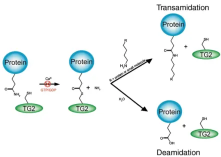
Transglutaminases were discovered in 1950s as enzymes that catalyze the post-translational modification of proteins by the formation of isopeptide bond. To date, nine members have been characterized within the transglutaminase family, including transglutaminases 1-7, factor XIII A and erythrocyte-band4.2, which prevalently exert functions in blood clotting, wound healing, cell envelope formation, cell matrix stabilization and epidermal differentiation. TG2 is one of these nine members and is one of the most investigated and biologically characterized isoform, being the most frequently occurring member in eukaryotes and present in almost all mammalian cells. As main catalytic activity, TG2 is able to catalyze the Ca2+-dependent modification of protein-bound glutamine side chain. In the presence of calcium, the ɤ-glutaminyl side chain of a protein is acylated by active site cysteine (Cys277) of TG2 to form a thioester intermediate with the release of ammonia. The intermediate thioester is attacked by a primary amine, being either a small molecule amine or the ɛ-amino group of protein-bound lysine residue1
. This trans-amidation reaction results in the formation of a relatively stable isopeptide bond. TG2 can catalyze also a deamination reaction, where water acts as nucleophile with the consequent conversion of glutamine into glutamate2.
Given the abundance of glutamine and lysine residues on the proteins in an organism, those residues represent the most important substrate for TG23. Therefore, the enzymatic activity is tightly regulated by different mechanisms to prevent an excessive protein aggregation through crosslinking reaction. Guanosine triphosphate (GTP) and guanosine diphosphate (GDP) can both bind to TG2 and
6
inhibit the transamidase activity4, whereas calcium ionophores capable of transporting free calcium across cell membrane can increase intracellular calcium levels enough to activate intracellular TG2 which is enzymatically latent under physiological conditions5. Nitric oxide can abolish the transamidase activity through Ca2+-dependent S-nitrosylation of multiple cysteine residues6.
Figure 1. TG2 transamidation and deamidation catalytic mechanism7.
Under physiological conditions, intracellular TG2 exists in its inactive conformation, due to the low intracellular Ca2+ concentration. During situations of unbalanced Ca2+ homeostasis, such as apoptosis or wound healing, TG2 undergoes rapid activation upon binding of Ca2+ ions. It has been suggested that activated cytosolic TG2 may crosslink intracellular proteins to prevent them from spilling out of a dying cell and causing inflammation8. In the extracellular environment, TG2 is mainly transamidase-inactive despite the presence of Ca2+. In the extracellular environment, TG2 once associated with cell surface integrin and fibronectin, can transmit signals to intracellular environment affecting cell adhesion, migration and signaling functions, through a mechanism independent of crosslinking activity9.
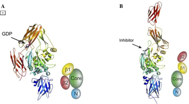
TG2 has been well characterized by X-ray crystallography, in particular in conformations bound with GDP10 and with a peptidic inhibitor11. It consists of four distinct domains: 1) N-terminal containing the fibronectin binding site, 2) catalytic core containing the substrate binding pocket and catalytic triad, 3) a β-barrel domain with binding pocket for GTP and interaction site with α1 adrenergic
receptor, 4) C-terminal containing interaction site with phospholipase. This multidomain structure reflects diverse catalytic activities and signaling pathways in which TG2 is involved.
The crystal structures confirm the existence of at least two conformations for TG2: the “closed” ones and the “open” ones. In fact, the binding of GTP or inhibitors to TG2 causes significant
7
conformational shifts. Although it is clear that multiple TG2 conformations exist, few information is available about the biological relevance of each conformation.
Figure 2. X-ray crystal structures of two conformations (open and closed) of human TG2 in complex with GDP in
β1-barrel (A) and with peptidic inhibitor in the active site (B)11.
1.2 Transglutaminase 2- Implications in human diseases
TG2 is implicated in a number of human diseases, such as celiac sprue, neurodegenerative disorders, diabetes, liver cirrhosis, fibrosis and certain types of cancer. Importantly, the pathology or etiology of most of the aforementioned diseases is likely ascribable to the enzymatic function of TG2.
Celia sprue is an autoimmune disease characterized by an impairment of the small intestine caused by a class of digestion-resistant proteins called prolamines, characterized by high content of proline and glutamine. TG2 exerts two main functions in the disease pathogenesis: 1) the enzyme deaminates specific glutamine residues in the prolamine proteins leading to an increased binding affinities of these proteins to the human leukocyte antigen (HLA) serotypes, which mediate the T-cell response12; 2) The covalent complex TG2-prolamine can finally lead to an autoimmune response by generation of anti-TG 2 antibodies13.
There is accumulating evidence that TG2 contributes to the process of cataractogenesis in the eye, through cross-linking of crystallins, the proteins constituting the eye lens, leading to lens opacification and cataract. This seems to correlate with the formation of bis(γ-glutamyl)spermidine cross-links between the crystallin proteins14.
A
A
8
A number of experimental observations support the hypothesis that TG2 is implicated in tumor progression. Multiple studies showed that TG2 protein is up-regulated in various cancerous tissues including pancreatic carcinoma15, breast carcinoma16 and malignant melanoma17. A positive correlation between the drug resistance and metastatic potential of certain cancers with TG2 expression levels has been demonstrated. A unifying molecular mechanism that explains the way how TG2 promotes oncogenesis has not been discovered. At this regard, there are mainly two hypotheses: first, TG2 causes activation of nuclear factor κB (NF-κB) probably through a crosslinking reaction, thus, consequently promotes expression of anti-apoptotic proteins, such as Bcl-xL; second, TG2 activates the focal adhesion kinase (FAK) with the subsequent activation of anti-apoptotic pathways, such as the P13K/Akt pathway. Furthermore, in a recent study on cancer cell-derived microvesicles, TG2 was shown to play a role in the communication between tumor cells and with normal cells, by inducing transformed characteristic of cancer cells onto normal fibroblasts through the release of microvesicles18.
The hallmarks of several neurodegenerative disorders are extensive neuronal loss and progressive formation of insoluble protein aggregates in the affected cerebral regions, such as amyloid plaques and neurofibrillary tangles in Alzheimer’s disease, Lewy bodies in Parkinson diseases and huntingtin protein in the homonymous disease. Importantly, all of the disease-related proteins in the aforementioned disorders are good substrates of TG2 in vitro19. Furthermore, it has been proven that both TG2 and its transamidase activity are increased in the affected cerebral regions of persons suffering from these disorders20. These observations suggest that TG2 may be significantly involved in the pathogenesis of neurodegenerative diseases.
1.3 Transglutaminase 2 and Alzheimer’s disease
Protein misfolding and the formation of insoluble protein complexes play a central role in the pathogenesis of Alzheimer's disease (AD), characterized by the aggregation of amyloid-β (Aβ) protein in cerebral tissue as senile plaques and the hyperphosphorilation of protein tau as neurofibrillary tangles21. However, the underlying mechanisms that trigger the misfolding of self-interacting proteins that eventually results in formation of neurotoxic aggregates remain unclear. Elucidation of the driving forces of protein complex formation in AD has crucial importance for the development of therapies for this disease.
Accumulating evidences support the hypothesis that TG2 may be involved in the abnormal protein aggregation observed in AD. As aforementioned, TG2 is a calcium-dependent enzyme that induces the formation of covalent γ-glutamyl-ε-lysine isopeptide bonds, which results in protein cross-links.
9
TG2-catalyzed cross-linking induces stable and protease-resistant protein complexes. Inhibition of TG2-catalyzed cross-linking counteracts the formation of protein aggregates, as observed in disease-models of other protein misfolding diseases, in particular, Parkinson's and Huntington's diseases19b. With regard to AD, there is compelling evidence from in vitro studies, as well as from studies on postmortem human brain tissue of AD patients and animal models that demonstrates a crucial role for TG2 in AD pathology:
1) Aβ and protein tau are good substrates for transglutaminase 2. In AD, Aβ shifts from soluble monomers to toxic oligomers and eventually forms insoluble mature fibrils. Brain transglutaminases could play a role in the Aβ accumulation. Using a synthetic partial-length of Aβ protein, Ikura et al. demonstrated that this peptide was subject to transamidation catalyzed by transglutaminase resulting in formation of multimeric peptides. The same experiment showed that in the presence of EDTA which chelates calcium ions, multimeric peptides were not produced and the monomer peptide remained unchanged22. Protein tau is a microtubule-binding phosphoprotein and functionally regulated by site-specific phosphorylation. In regard to the correlation between TG2 activity and protein tau aggregates, it has been reported that bovine tau protein is an excellent substrate for TG2 through the polyamine incorporation assays, whereas recombinant human tau presents different levels of affinity toward TG2 based on isoform types. This study also demonstrated that TG2-catalyzed cross-links on protein tau is specific only for a small number of glutamine residues, in fact, only few segments on tau are susceptible to the modification induced by the enzyme23. Additionally, a study that used co-immunoprecipitation and immunocytochemistry methods confirmed the actual interaction between TG2 and protein tau and their colocalization at intracellular levels24.
2) TG2 levels in cerebrospinal fluid are increased in AD. In vivo analysis of cerebrospinal fluid (CSF) in patients affected by AD has revealed that the levels of TG2 are significantly higher in comparison with the control group, whereas CSF tau protein concentration does not change significantly, suggesting that CSF TG2 concentration could be an important biomarker for this neurodegeneration disease25.
3) TG2 colocalizes with amyloid deposits in the brains of AD patients. Several in vitro immunochemical studies showed the presence of TG2 in both isolated amyloid plaque cores and cortex tissue sections from patients affected by AD. These results provide evidence that insoluble amyloid deposits may involve TG-mediated cross-linked Aβ-peptide polymer26.
4) TG2 activity is increased in AD brain. In a study conducted by Johnson G.V. et al. that used frozen prefrontal cortex and cerebellum samples from Alzheimer's disease, a total transglutaminase activity significantly higher in the AD prefrontal cortex compared to control was
10
observed. In addition, the levels of TG2, as determined by quantitative immunoblotting, were elevated approximately 3-fold in AD brain with respect to control. In contrast, in the cerebellum which usually is not impaired in AD, no significant increment in transglutaminase activity or levels has been detected27.
5) Catalytically active TG2 colocalizes with Aβ in AD animal models. A recent in vitro study investigated the role of TG2 in AD pathology using two mouse models that represent early and late stages of pathology development for this purpose. According to the results, the general distribution pattern of TG2 in transgenic mice is similar to the observations in human brains. In fact, there was association of both TG2 and in situ active TG2 with Aβ plaques and vascular Aβ, indicating that alike human AD cases, TG2 was associated with Aβ depositions in the animal models. Although spatial overlay of TG2 with Aβ pathology observed in the mouse models was substantially different from observations in human samples, this finding provide evidence for a role of TG2 in Aβ pathology28.
In summary, given these evidences on the role of TG2 in the initiation and development of protein aggregates in AD, its distribution overlay with the disease hallmarks and its increased activity in the same pathology, it is highly suggested the possibility to use inhibitors of the cross-linking activity of TG2 as a new therapeutic approach for AD.
1.4 Transglutaminase 2 inhibitors
The range of pathologies in which TG2 is implicated demonstrated the need for potent TG2-specific inhibitors. To date, TG2 inhibitors have been generally divided into two classes based on the inactivating mechanism of the enzyme: reversible (competitive and non-competitive) and irreversible inhibitors.
1.4.1 Irreversible TG2 inhibitors
Most irreversible TG2 inhibitors are designed to target the active site cysteine (Cys277) using electrophilic functional groups. These inhibitors prevent enzyme activity by covalently modifying the enzyme, thereby preventing substrate binding.
Halomethyl carbonyl inhibitors
This class of inhibitors has been developed based on the structure of iodoacetamide, which was one of the first irreversible inhibitors and was able to inactivate guinea pig liver TG229. More recently, the Keillor group made a series of chloromethyl ketones and combined them with a Cbz-Phe
11
scaffold, where Cbz functional group was previously assessed to be essential for active site recognition30. The best inhibitor (compound A) showed a Ki = 6μM and a good specificity profile with very low reactivity toward glutathione, the most abundant physiological thiol present in living cells31.
3-Halo-4,5-dihydroisoxazole inhibitors
Acivicin is a natural product that is known to inhibit several cysteine-dependent enzymes. The scaffold 3-halo-4,5-dihydroisoxazole showed to be able to alkylate the active site cysteine of two bacterial glutamine amidotransferases32. A number of TG2 inhibitors structurally containing the 3-halo-4,5-dihydroisoxazole warhead have been developed. In particular, Khosla group synthesized a large series of 3-bromo analogs where the electrophilic warhead is linked to a Cbz moiety through a peptidic spacer. The dihydroisoxazole compounds were assayed against recombinant human TG2 using an enzyme-coupled assay, where glutamate dehydrogenase was used to couple ammonium ion, a product of the TG2-catalyzed reaction. The most potent derivative was compound B, which showed high specificity for human TG2 but essentially no reactivity toward physiological thiols, and exhibited good oral bioavailability in mice33. Importantly, compound B showed excellent synergism with antitumor drug carmustine, against refractory glioblastoma in mice, through a mechanism that involves disruption of fibronectin assembly in extracellular environment34.
α,β-unsaturated amide
α,β-unsaturated amide, termed “Michael acceptors”, have been used to inhibit many cysteine proteases showing good outcome35. Keillor and coworkers synthesized novel TG2 irreversible inhibitors by attaching an α,β-unsaturated amide to the typical Cbz-Phe scaffold and varying the chain length of the spacer. It was noted that a spacer length of four methylene units (compound C) provided a potent inhibitor with inhibitory efficiency of 42-fold more efficient than the same Cbz-Phe scaffold bearing a halo-dihydroisoxazole group31.
12
Compound D is identified as a potent TG2 inhibitor during an extensive drug-screening against the enzyme conducted by a biomedical foundation for Huntington’s disease. This compound exhibited an IC50 of 10nM against recombinant human TG2 and showed an enormously higher selectivity
against other TG isoforms, such as FXIIIa, TG6, TG1 and TG336. Low plasma stability of compound D leaded to the development of two newer compounds (E and F) which demonstrated superior plasma stability and resistance to glutathione conjugation36.
1.4.2 Reversible inhibitors
The first reversible inhibitors used in TG2 related research were amine-bearing molecules, typically characterized by a medium to long saturated aliphatic chain with a terminal amine group. Some of the most commonly used competitive amine inhibitors include putrescine, monodansylcadaverine, 5-biotinamidopentylamine and fluorescein cadaverine37. These inhibitors exert their activity by competing with the natural amine substrate in the cross-linking reaction. Thus, TG2 is still enzymatically active in the presence of competitive amine inhibitors. One compound of this class is represented by cystamine, which is a diamine capable of competitive amine inhibition through its reduced form, cysteamine38. However, lack of selectivity has been observed for cystamine, since it inhibits also the thiol-dependent protease caspase 3 and causes increased production of glutathione inside of cells39. The amine competitive inhibitors were extensive used in the early research studies on transglutaminases, although useful for in vitro studies, they have limited applications since the resulting aminated protein could elicit autoimmune response in vivo40.
13
Recenty developed TG2 reversible inhibitors with competitive inhibition mechanisms include: 1) acylideneoxoindoles derivatives, based on the structure of isatin whose analogs are widely used as reversible inhibitors of Cys-dependent proteases41; 2) 3-alkynyl quinoxaline derivatives identified through screening of 1000 small molecules as TG2 inhibitors using a cancer cell proliferation inhibition assay42.
TG2 is allosterically deactivated upon binding of guanine nucleotides, as aforementioned, thus, there has been an effort to develop reversible TG2 inhibitors targeting the GDP/GTP pocket on the enzyme. Non-hydrolysable GTP analogs, such as GTPγS and GMP-PCP, have been used as reversible inhibitors4. A new class of allosteric TG2 inhibitors, discovered during high throughput screening, is structurally characterized by the thieno[2,3-d]pyrimidin-4-one acylhydrazides backbones43. In particular, the compound G showed to be a slow-binding inhibitor that likely does not bind at the enzyme’s active site but rather at the enzyme’s GTD binding site and that was non-competitive with the standard acyl-donor and acyl-acceptor substrates44.
Trans-cinnamoyl inhibitors of TG2
One of the most promising TG2 reversible inhibitors recently discovered is represented by the trans-cinnamoyl derivatives inhibitors. As aforementioned, starting from a comparative study between Cbz-Gln-Gly versus Boc-Gln-Gly-binding modes via molecular modeling, the group of Keillor discovered the importance of Cbz protecting group to confer active site recognition and improve affinity. Further investigation of the rigidity of the Cbz group led to the identification of trans-cinnamoyl derivatives as competitive and reversible inhibitors with IC50 value for guinea pig liver
transglutaminase as low as 18µM45. The enzyme inhibition assay was performed using the kinetic analysis by Michaelis-Menten equation, based on UV-visible detection of a chromogenic TG2 substrate. According to the structure-activity relationship (SAR) study, the most potent inhibitors are
14
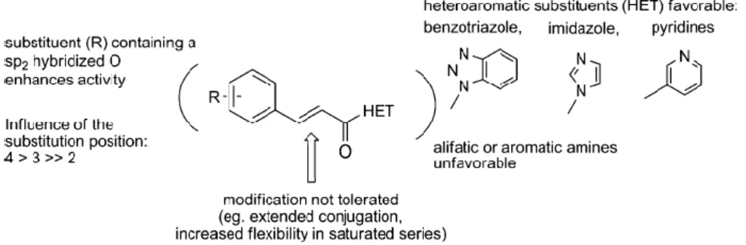
divided into two subclasses, depending on the nature of the heteroaromatic substituent (HET) moiety: cinnamoyl benzotriazolyl amide and 3-substituted cinnamoyl pyridines, referred to more commonly as azachalcones. Initial SAR investigations on the aromatic scaffold of cinnamoyl moiety showed that substituents in para position improved the affinity with enzyme. In particular, substituents with a sp2-hybridized oxygen, such as NO2, BOCNH, FmocNH and MeO2C, conferred
the best inhibitory activity. While the HET moiety seemed involved in hydrogen bond connection inside the enzyme, thus, aromatic moiety possessing hydrogen bond acceptor like benzotriazolyl and pyridinyl group is mandatory for a good affinity for this series of compounds. Finally, evaluation on the importance of the length and flexibility of the linker between the two aromatic moieties showed that introduction of a supplementary conjugated alkene was detrimental to the affinity for TG2, in comparison with the cinnamic double bond. Moreover, the reduction of the alkene or replacement with methylene/ether led to a loss of activity. For the selectivity studies of this new family of TG2 inhibitors, several representative compounds were tested as inhibitors on Factor XIIIa, a transglutaminase isoform, and Caspase 3, a cysteine protease with acyl transfer mechanism similar to that of TG2. As results, most of these compounds showed a good selectivity profile, since no inhibitory effect was detected on these enzymes even at concentration approaching the limits of their solubility.
Figure 3. General structure of cinnamoyl derivatives and their SAR tendencies.
Subsequent labeling experiments with an azachalcone derivative (compound H) were performed to determine the binding mode of the cinnamoyl inhibitors46. This inhibitor with IC50 = 28μM was
designed to incorporate the photolabile diazirine moiety. Photoactivation of diazirine generates diazo and carbene derivatives that react rapidly to covalently label residues in their proximity. The following LC-MS analysis indicated that Cys230 located inside the hydrophobic groove was primarily labeled. Modeling simulations suggest that Cys230 is more exposed in the “closed” conformation of TG2 rather than the “open” conformation, in agreement with the result of docking
15
studies. These findings suggest the hypothesis that trans-cinnamoyl inhibitors favorably interact with the “closed” conformation of TG2.
A more recently developed class of 4-nitrocinnamoyl triazoles by Keillor group47 highlighted an inhibitor (compound I) which exhibited IC50 value of 2.1µM, representing the most potent inhibitor
of this class and provided a promising scaffold for building novel TG2 inhibitors.
1.5 Multitarget ligands approach in drug discovery
Over the last decades, most drug discovery efforts were aimed at single-target compounds. However, not all diseases succumb to this “one disease-one target” approach. Disease systems are generally characterized by the dysregulation of multiple biological pathways and efforts of integrating the complex disease systems to drug discovery process leaded to a new concept, termed polypharmacology: the treatment of diseases by modulating multiple targets. Besides the approaches of drug cocktail and multicomponent drugs, the more recently consolidated strategy of polypharmacology is the development of multitarget ligands (MLs). Multitarget ligands, designated firstly by Morphy et al.48, are single chemical entities capable of modulating simultaneously multiple targets involved in a certain disease. MLs are gaining increasing attention from the drug discovery community and a plethora of multitarget ligands have been developed for the treatments of various diseases, by offering a number of potential advantages over the drug cocktail or multicomponent drugs: 1) less costly and complicate in clinical development because predicting the pharmacokinetics of a single compound is much easier than a drug cocktail; 2) improved efficacy due to the synergistic effect of simultaneously inhibiting multiple targets; 3) improved safety with reduced risk of drug-drug interactions; 4) simplified therapeutic regime and improved compliance.
16
1.5.1 Rational design of a multitarget ligand
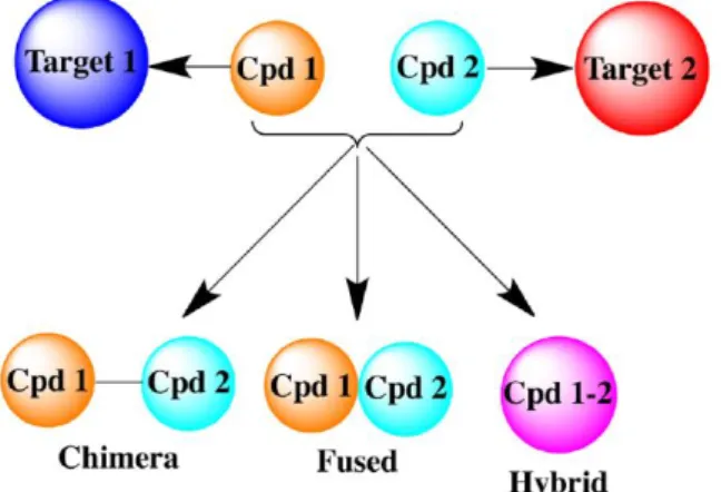
A general drug design for MLs starts with identification of two pharmacologically relevant targets, A and B, located on complementary pathological pathways, followed by verifying the potential synergistic effect from hitting the two targets simultaneously. Afterwards, the pharmacophores responsible for binding to target A and B must be identified, and finally, the pharmacophores can be linked together in one single entity to obtain hybrid, fused or chimeric dual-target ligands.
Figure 4. Dual-target ligand design strategy49.
However, there are two main challenges correlated to the development of MLs. First, suboptimal physicochemical properties are frequently encountered with the MLs. In fact, MLs currently developed tend to be larger and more lipophilic than marketed drugs and are also less efficient than a broad set of preclinical compounds in terms of their binding energy per unit of molecular weight or lipophilicity, with detrimental consequences for oral bioavailability50. A recent paper from Hopkins et al. showed that there is a reverse correlation between the mean molecular weight of compounds and the total number of targets that those compounds were found to show a minimal activity. This finding suggests that smaller molecules are more likely to establish interactions with multiple biological targets51. Secondly, the lead-to-drug optimization is much more complex for multitarget compounds: the binding affinity profiles must be balanced with multiple biological targets. Therefore, the ratio of activities at the different target must be adjusted to achieve a good pharmacological profile for the MLs.
1.5.2. Multitarget ligands for the treatment of Alzheimer’s disease
As aforementioned, AD pathogenesis involves a complex interplay of genetic and biochemical factors and is phenotypically characterized by hallmarks like Aβ peptide accumulation and increased phosphorylation of the microtubule-associated tau protein. Multitarget ligands represent a promising
17
approach for the treatment of this multifactorial disease. To date, a number of MLs toward different AD’s pathological targets were developed and showed interesting biological profiles. Some potential MLs for AD treatment are reported below.
The dimer bis-(7)-tacrine was one of the first homodimers reported in the literature with increased acetylcholinesterase (AChE) affinity, exhibiting a 1000-fold higher inhibitory potency compared to tacrine. The inhibitory profile is due to a simultaneous interaction with both active site and peripheral site of AChE, furthermore, the compound is capable to inhibit the AChE-induced Aβ aggregation with an IC50=41.7μM52. Minarini and coworkers proposed modifications on the structure of
bis-(7)-tacrine by replacing the aliphatic linker with cystamine, which has shown important neuroprotective properties53. The resulting dimer cystamine-tacrine was able to inhibit AChE (IC50=5.04 nM),
BuChE (IC50=4.23 nM), self-Aβ aggregation (IC50=24.2μM) and AChE-induced Aβ aggregation
(52.6%), exhibiting additional neuroprotective effect on SH-SYS5 cell line against H2O2-induced
oxidative injury54.
The current standard of care for AD is a combination of an AChE inhibitor with the non-competitive
N-methyl-D-aspartate receptor (NMDAR) antagonist, memantine55. The reason for this combination therapy is that the NMDAR antagonist would stop or delay neurodegeneration, while the AChE inhibitor would improve memory and cognition by stimulating the surviving neurons. Simoni et al. combined in single new chemical entities the pharmacophoric moieties of memantine and galantamine, two marketed drugs for the treatment of AD, with the latter one being an AChE inhibitor56. The new chimera compounds were initially investigated by docking simulations based on the crystallographic structure of AChE complexed with an inhibitor. The derivatives with various
18
spacer lengthes were then tested against AChE and NMDAR. The most potent derivative, endowed with a spacer of eight methylene units, showed an IC50 value of 0.52nM against AChE, albeit it
turned out to be a weak binder toward NMDAR with IC50 in micromolar range. This series of
compounds has highlighted the inherent difficulty of balancing the affinity profiles against different targets, when discovering and developing MLs. Interestingly, these molecules showed to be active also toward NMDAR containing the NR2B subunit, a receptor subunit particularly implicated in pathological process linked to overexcitation of glutamatergic pathways. Finally, several derivatives of this series exhibited a remarkable neuroprotective profile by inhibiting the NMDA-induced neurotoxicity.
Lu and coworkers57 have reported some multitarget directed ligands by combining the stilbene moiety of resveratrol with clioquinol. Resveratrol, a natural product with a stilbene structure, has been shown to function as an anti-AD agent through the inhibition of Aβ aggregation by scavenging oxidants and exerting anti-inflammatory activities58, while clioquinol is a well-known metal chelator which significantly decreased the rate of cognitive decline in moderately severe AD patients in a phase II clinical trial59. Two derivatives of these fused MLs exhibited significant inhibition of Aβ aggregation, metal-chelating ability, disaggregation of Aβ fibrils generated by self- and Cu(II)-induced Aβ aggregation, antioxidant activity, acceptable MAO-A and MAO-B inhibition, moderate AChE inhibition and low neurotoxicity. One derivative of the series was able to cross the blood-brain barrier in vitro and did not exhibit acute toxicity in mice at doses up to 2000 mg/kg.
20
2. Dual inhibitors targeting Transglutaminase 2 and Histone Deacetylase
This chapter focuses on the development of inhibitors designed to hit simultaneously two important targets implicated in the pathogenesis of Alzheimer’s disease (AD): Transglutaminase 2 (TG2) and histone deacetylase (HDAC). The role that TG2 plays in AD pathology has been elucidated in the first chapter, while HDAC, a class of enzymes importantly involved in epigenetic modification of gene expression by influencing the status of histone acetylation, has been proved to play a key role in various diseases of central nervous system (CNS), including the AD. The inhibitors were designed following the frame-combination method to achieve a multitarget activity, in particular by combination of the trans-cinnamoyl moiety of a potent TG2 inhibitor and the hydroxamic acid functionality, an excellent zinc chelating agent demonstrated to be responsible of HDAC’s inhibition. The synthesized multitarget ligands have been evaluated for in vitro inhibition activity on hTG2 and all compounds of the series showed good inhibition activities towards this AD target, while the results on HDACs inhibition will be ready shortly.
2.1 Histone acetylation’s role in the pathogenesis of Alzheimer’s disease
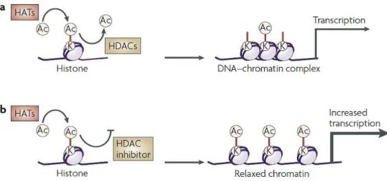
Epigenetic processes include histone modifications (acetylation, phosphorylation, methylation, ubiquitination and ADP ribosylation), DNA methylation and gene silencing by non-coding RNAs. Histone acetylation and deacetylation regulate gene transcription by altering the chromatin structure and the accessibility to transcription factors. Levels of histone acetylation depend on the activities of histone acetylases (HATs) and histone deacetylases (HDACs), which add or remove acetyl groups from histone proteins, respectively. Although transcriptional regulation is highly complex and dynamic, in general an increase in histone acetylation causes remodeling of chromatin from a tightly packed configuration to a loosely packed one, which subsequently leads to transcriptional activation; conversely, a decrease in histone acetylation generally results in transcriptional silencing. Therefore, an upregulation of transcription can be achieved by either stimulating HATs or inhibiting HDACs.
21
Figure 5. Effect of HDAC inhibitors on chromatin remodelling and transcription60.
HDACs repress transcription by removing an acetyl group from lysine residues on histone tails and thus compacting chromatin. In addition, a number of non-histone cellular proteins are substrates for HDACs, which mediate diverse biological functions via transcriptional-dependent as well as independent mechanisms.
Mammalian HDAC enzymes are classified into four major classes: (1) Class I HDACs contain ubiquitously expressed HDACs 1, 2 and 3, and the muscle-specific HDAC8; (2) Class II HDACs further separated into two subclasses: class IIa including HDACs 4, 5, 7 and 9 which show distinct tissue-specific patterns of expression, predominantly in muscle and heart; class IIb includes the unusual HDAC6, which deacetylates α-tubulins and alters microtubule stability, and HDAC10 not yet well characterized; (3) Class III HDACs are named sirtuins and includes SirT1-7, sharing their homology sequence to the yeast Sir2; (4) The class IV enzyme HDAC11 is structurally different from the class deacetylases I and II, and its function is poorly known. Class I, II, and IV HDACs are Zn2+-dependent enzymes, while Class III HDACs enzymes are dependent on nicotinamide adenine dinucleotide (NAD+) to carry out their catalytic functions.
The superfamily of HDACs has been recognized as an important therapeutic target for a broad range of human diseases, particularly for cancer treatment. More recently, increasing studies point out another important application of HDAC-based therapeutics: as potential treatment for human CNS disorders. Genetic evidences suggest crucial roles for HDACs and HATs in the maintenance of CNS homeostasis and a possible correlation between mutations in gene encoding histone-binding proteins and neurological disorders. In fact, HDAC inhibitors showed interesting results in several in vitro studies for the investigation of their potential therapeutic effects in CNS diseases, such as psychiatric disorders, Rubinstein-Taybi syndrome, motor-neuron disease and Huntington’s disease60. In
22
particular, HDACs have been correlated directly to AD in several studies. The isoform HDAC2 is widely expressed in CNS and negatively regulates memory and synaptic plasticity, it is overexpressed in the post-mortem brain samples of AD patients and its knockdown by shRNA restores the synaptic plasticity and decreases memory impairments in AD model mice61. These findings highlight the important role of HDAC2-regulated chromatin modification in regulating the synaptic plasticity and memory formation in the cognitive impairment context of AD. Furthermore, an increased HDAC6 protein level is observed in the cortices and hippocampus of AD postmortem brain samples62 and reducing endogenous HDAC6 results in the restore of learning and memory deficits in AD mouse model63. The mechanism is likely correlated with the involvement of HDAC6 in tau metabolism, in fact, the selective inhibitor of HDAC6, tubacin, represses tau phosphorylation62. This enzymatic isoform has been observed to act as regulatory protein of the ubiquitin-proteasome system as well, thus, is intimately linked to the aggregation of misfolded mutant proteins which represents a hallmark of many neurological disorders64.
HDAC inhibitors exhibited neuroprotective, neurotrophic and anti-inflammatory properties, and improve neurological performance -learning and memory- in neurodegenerative disease models. Particularly, studies demonstrated that treatment with HDAC inhibitors could affect the AD pathology: 1) Sung et al. demonstrated that a mercaptoacetamide-based class II HDAC inhibitor and a hydroxamide-based class I and II inhibitor can reduce Aβ levels65; 2) In an AD mice, daily injections of 4-phenylbutyrate reversed spatial memory deficits by normalizing tau hyperphosphorylation in the hippocampus without affecting Aβ levels66; 3) As aforementioned, HDAC6 inhibition by tubacin reduces the protein tau phosphorylation.
2.2 HDAC inhibitors
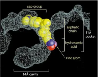
Trichostatin A (TSA) is the first natural hydroxamate discovered to inhibit HDACs. The crystal structure of this inhibitor complexed with a bacterial HDAC-like protein (HDLP) illustrated the chemical structural features of the HDAC catalytic core, as well as the configuration of interaction inhibitor-enzyme. According to the revealed structure, the active site of HDAC consists of a tubular pocket, a zinc-binding site and two Asp-His charge-relay system. TSA binds the enzyme by inserting its long aliphatic chain into the HDLP pocket making multiple contacts to the tube-like hydrophobic portion of the pocket. The hydroxamic acid group coordinates the zinc in a bidentate fashion resulting in a penta-coordinated Zn2+, while the aromatic dimetylamino-phenyl group of the TSA chain makes contacts at the pocket entrance capping the pocket67. The human HDACs crystal
23
structures has been revealed later and its structural analysis showed the common active site features in agreement with the ones observed in bacterial HDLP.
Figure 6. Representation of TSA in the active-site of HDLP67.
Generally, the pharmacophore of HDAC inhibitors is composed of three regions: a ‘cap’ region or ‘surface recognition domain’, which occludes the entrance of the active site pocket; a ‘zinc-binding group’ which chelates the zinc ion in the active site and is required for catalytic function; and a ‘linker’ region, which connects the two moieties.
Figure 7. TSA and general structural illustration of HDAC inhibitors.
HDAC inhibitors can be divided into several classes based on the chemical structures: hydroxamates, benzamides, aliphatic acids and cyclic peptides.
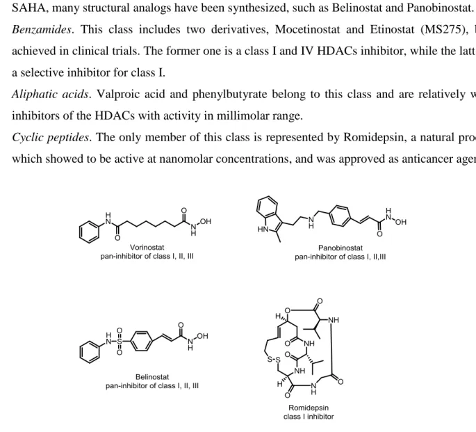
- Hydroxamates. The most important compounds of this class are represented by TSA and SAHA, structurally characterized by the presence of a hydroxamic acid function, which is an optimal chelating agent for Zn2+ present in the catalytic active site of HDACs. SAHA, or vorinostat, is a pan-inhibitor of class I, II and IV HDACs and demonstrated activity in low nanomolar range68.
24
It is the first HDAC inhibitor to be approved for anticancer treatment by FDA. Following SAHA, many structural analogs have been synthesized, such as Belinostat and Panobinostat. - Benzamides. This class includes two derivatives, Mocetinostat and Etinostat (MS275), both
achieved in clinical trials. The former one is a class I and IV HDACs inhibitor, while the latter is a selective inhibitor for class I.
- Aliphatic acids. Valproic acid and phenylbutyrate belong to this class and are relatively weak inhibitors of the HDACs with activity in millimolar range.
- Cyclic peptides. The only member of this class is represented by Romidepsin, a natural product which showed to be active at nanomolar concentrations, and was approved as anticancer agent.
Figure 8. FDA approved HDACs inhibitors.
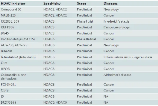
An important drawback of most HDAC inhibitors is the lack of isoform-selectivity, which makes it difficult to determine whether the biological outcome is due to inhibition of a specific HDAC or by inhibition on multiprotein complexes, besides the undesirable clinical effects due to off-target activity. Hence, new trend in the field of HDAC inhibitor drug discovery is the development of isoform-specific HDAC inhibitors, and to date, a handful of these selective inhibitors has emerged.
25
Table 1. Isoform-specific HDAC inhibitors.
2.3 Drug design
AD pathogenesis involves a complex interplay of genetic and biochemical factors and a therapy aimed at hitting diverse pathological pathways is strongly recommended. Given the evidences that both transglutaminase 2 and HDACs play important role in the pathology of AD and both overexpressed in this neurodegenerative disease, inhibition of these two enzymes could produce additive or synergic amelioration of the pathology. This hypothesis is supported by a study (unpublished results) which demonstrated that treatment with Vorinostat and cystamine on neuronal cells results in a synergic neuroprotective effect against excitotoxicity induced by glutamate; additionally, according to a study on Huntington’s disease (HD), combinatorial treatment of SAHA, cystamine and Congo red improved survival of Drosophila HD model69. Therefore, the aim is to apply the ‘multitarget ligands’ approach to develop an inhibitor active on both two biological targets, through combination of pharmacologically important fragments for each target to give a single chemical entity.
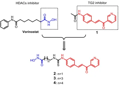
To the purpose of designing a TG2-HDAC dual inhibitor, we selected a potent TG2 inhibitor bearing the trans-cinnamoyl structure, the compound 1, which was found to be a reversible competitive inhibitor with IC50=28μM. According to the structure-activity relationship studies, the large
substituents on the cinnamoyl aromatic ring in the para position gave the best results46. Thus, by replacing this substituent on compound 1 with an aliphatic chain bearing hydroxamic acid, the zinc
26
chelating group responsible of HDACs inhibition exhibited by Vorinostat, we designed a series of new compounds 2-4 where the two fragments are connected through a linker.
Figure 9. Drug design of compounds 2-4.
The conjugates 2-4 are structural analogs with difference in the linker length, which is composed of 2, 4 and 5 methylene units, respectively, along with a thiourea bond, chosen for the reason of synthetic accessibility. The derivative characterized by three methylene units as linker was designed as well, but the chemical synthesis could not be accomplished despite numerous synthetic attempts. We postulated that these new chemical conjugates would maintain inhibition activity on HDACs and TG2 by the reason of the presence of both pharmacophores; additionally, we assume that the bulky
trans-cinnamoyl moiety would serve as ‘surface recognition group’ for HDACs, allowing favorable
orientation of hydroxamic acid inside the active site pocket.
2.4 Methods
Compounds 2-4 were synthesized and studied in in vitro inhibition assays on TG2, while the inhibition activities on HDACs are still under investigation. According to the preliminary data, the compounds showed good inhibition activities on TG2 in an assay conducted on human recombinant transglutaminase 2 using a chromogenic donor substrate.
2.4.1 Synthetic methods
The compounds 2-4 were synthesized following the scheme 1: The trans-cinnamoyl moiety was synthesized starting from an aldol condensation reaction between 3-acetylpyridine and para-nitro substituted benzaldehyde in basic conditions to give compound 5, followed by reduction of nitro
27
group into the correspondent amine using Tin(II) chloride to afford compound 6. The amine group was then transformed into isothiocyanate (compound 7) by reacting with 1,1′-Thiocarbonyldi-2(1H)-pyridone.
For the synthesis of the aliphatic chain bearing hydroxamic acid, commercially available bromo-alkyl carboxylic acids were subjected to nucleophilic substitution to replace the halogen with azide group by reacting with sodium azide to give compounds 8-10. Afterwards, the carboxylic acid was reacted with O-Tritylhydroxylamine in a coupling reaction to give the correspondent protected hydroxamic acids 11-13. The azide group was then converted to amine through Staudinger reaction using triphenylphosphine.
The two moieties were finally linked together in a coupling reaction where the electrophilic carbon of isothiocyanate reacts with the nucleophilic amine to form a thiourea bridge connecting the two fragments.
Finally, the trityl group of hydroxamic acid was cleaved in acidic conditions in the presence of triethylsilane as scavenger to afford the final products 2-4. The derivative characterized by a linker of three methylene units was not obtained despite numerous synthetic attempts.
28
Scheme1. a) KOH, MeOH/H2O, 30min, r.t.; b) SnCl2, EtOH, 30min, 70°C; c) 1,1′-Thiocarbonyldi-2(1H)-pyridone,
DCM, overnight, r.t.; d) NaN3, DMF, 5h, reflux; e) O-Tritylhydroxylamine, IBCF, NMM, THF, 2h, r.t.; f) PPh3, DCM,
overnight, r.t.; g) DCM, overnight, r.t.; h) TFA, Et3SiH, r.t., 30 min.
2.4.2 In vitro TG2 inhibition assay
According to a study conducted by Keillor group, TG2 reacts with N-Cbz-Glu(γ-p-nitrophenylester)Gly, a TG2 acyl-donor substrate analog, to undergo rapid acylation that can be followed spectrophotometrically at 400nm. The inhibition activity of the synthesized compounds 2-4 was evaluated using this method on human recombinant TG2. The IC50 values were determined
based on the inhibition of the enzymatic reaction of 2.2mM of N-Cbz-L-Glu(γ-p-nitrophenylester)Gly with 0.5mU of human recombinant TG2.
Briefly, kinetic runs were recorded at 25°C on a UV-visible spectrophotometer at 405nm, in a buffer composed of 3-(N-morpholino)propanesulfonic acid (MOPS, pH = 7.0), CaCl2 and EDTA. Each
N-Cbz-29
Glu(γ-p-nitrophenylester)Gly, 10μL of a solution of hTG2 and 0 to 5μL of a DMSO stock solution of inhibitor. IC50 values were obtained as the negative x-intercept of a Dixon plot.
2.5 Results and discussions
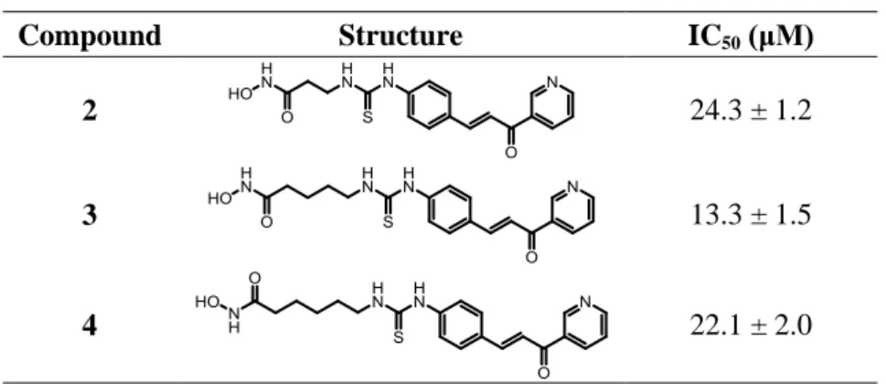
The test compounds were subject to preliminary studies for in vitro evaluation of the inhibition activity on hTG2 enzymatic function and the IC50 values are reported in Table 2.
From an analysis of the results, the compound 3 exhibited the best activity with IC50=13μM. Among
the class of trans-cinnamoyl inhibitors, endowed with a competitive reversible mechanism of inhibition, the IC50 for the most potent inhibitor was determined to be 2.1μM. This inhibitor belongs
to the subclass of cinnamoyl-triazoles (see chapter 1); while the best inhibitor of the subclass of cinnamoyl-pyridine showed an IC50 value of 21μM, as reported in literature. All three synthesized
derivatives 2-4 of the series are characterized by cinnamoyl-pyridine structure, and showed a good TG2 inhibition activity. In particular, the compound 3 could be considered the most potent cinnamoyl-pyridine inhibitor reported so far in literature.
The structural difference within this series of compounds resides in the chain length of the linker. An observation on inhibition potency of these compounds could drive to the conclusion that the presence of a bulky moiety in para position on the aromatic ring is not harmful in terms of inhibition activity, in agreement with observations reported in literature. Furthermore, the results show that the chain length can affect the inhibitor’s activity and the derivative with four methylene units probably fits better than other analogs in the active site of the TG2 enzyme.
Table 2. IC50 values of the TG2 inhibitors 2-4.
Compound Structure IC50 (μM)
2 24.3 ± 1.2
3 13.3 ± 1.5
30 2.6 Conclusions
The series of derivatives 2-4 were designed to be dual inhibitors targeting simultaneously HDACs and TG2, by incorporating the trans-cinnamoyl fragment for TG2 inhibition and hydroxamic acid moiety for the inhibition of HDACs.
At the time of edition for the present manuscript, we cannot elaborate final conclusions. However, preliminary in vitro data showed that the compounds of this series are good TG2 inhibitors. The best inhibitor was compound 3, which exhibited an IC50 value of 13µM and could be the most potent
inhibitor within the subclass of trans-cinnamoyl pyridines currently developed and reported. The mode of inhibition of these compounds is currently under investigation; while studies on the inhibition activity on HDACs are in progress.
31
3. Trans-cinnamoyl structure based transglutaminase 2 inhibitors with
potential antioxidant activities
Oxidative stress is one of the key factors in the AD’s pathology, and among various cell damage mechanisms, it promotes Aβ toxicity and increases tau hyperphosphorylation where the enzyme GSK3β could be involved in the pathological mechanisms. Natural antioxidants like hydroxycinnamic acids, including caffeic acid, ferulic acid etc., exert neuroprotective activities against neuronal cell damage induced by oxidative stress. Based on the trans-cinnamoyl structure which is essential for TG2 inhibition, addition of hydroxyl groups on the aromatic ring allowed the obtainment of a series of compounds designed as‘fused’multitarget ligands, which could be TG2 inhibitors and at the same time antioxidant agents. In vitro antioxidant evaluation outlined one compound of the series as a good antioxidant and endowed with interesting biological activity toward Aβ self-aggregation process and GSK3β enzyme activity. Against expectation, the slight structural modifications determined an almost complete loss of inhibition toward TG2.
3.1 Oxidative stress in Alzheimer’s disease
Oxidative stress is a mechanism of cell damage characterized by excessive production of highly reactive free radicals. It is the result of an imbalance in pro-oxidant/antioxidant homeostasis, which leads to the generation of toxic reactive oxygen species (ROS). The instability of free radicals is due to the presence of an unpaired electron on the outer orbital. This is responsible for the high reactivity of these species. ROS can potentially react with many cellular components including lipids, proteins and DNA, resulting in impaired cellular functions and formation of toxic species, such as peroxides, aldehydes and cholesterol oxide.
Unsaturated lipids are particularly susceptible to oxidative modification and lipid peroxidation is a sensitive marker of oxidative stress. Lipid peroxidation, which is the result of the attack by radicals on the double bond of unsaturated fatty acids, generates lipid peroxy radicals which initiates a chain reaction and breakdown products such as 4-hydroxy-2,3-nonenal (HNE), acrolein and malondialhedyde, molecules highly toxic for cells and all increased in neurodegenerative models70. The excessive production of ROS leads to disregulation of intracellular calcium signaling and such disregulation has been widely observed in neurodegenerative diseases71. One of the downstream events that occurs in response to an ROS-induced calcium influx is an excitotoxic response72, that is activation of glutamate receptors that triggers a cascade of events leading to cell death. Excitotoxic
32
response has been implicated in several neurological conditions as well as neurodegenerative diseases such as AD, Parkinson Disease (PD) and Huntington’s disease73.
The neurons are particularly susceptible to oxidative stress for several reasons. The first one is that the membrane of neuron cells contains elevated amount of unsaturated fatty acids, which are excellent substrate for the lipid peroxidation chain reactions; the second one, is that neuronal functions require a large amount of energy generated through oxidative phosphorylation reactions; the third one, is that the brain is an organ that concentrates metal ions, such as copper and iron. Therefore, the vulnerability of neuron cells to the oxidative stress is strongly related to the pathogenesis of neurodegenerative diseases.
3.1.1 Metal ions and Aβ toxicity
The hypothesis that oxidative stress could be an important factor to AD’s pathogenesis and progression is widely accepted. A support to this hypothesis is the correlation found to exist between metal ions and amyloid-β peptide (Aβ).
As a general principle, the chemical origin of the majority of ROS is the reaction of molecular oxygen with the redox-active metals, copper and iron. The ability of these metal ions of initiating redox cycling and activating molecular oxygen is the basis of function for most of enzymes.
Aβ is the major component of amyloid plaques and studies showed that there were remarkably high concentrations of Cu, Zn and Fe in the amyloid deposit in AD-affected brains74. The cause of the neuronal cell loss in AD might be related to oxidative stress from excessive free-radical generation and the major source of oxidative stress in the brain of AD is likely the transition metals Cu and Fe when bound to Aβ75. According to a study by Opazo et al., when Cu2+ or Fe3+ coordinate Aβ, extensive redox chemical reactions take place that reduce the oxidation state of both metals and produce H2O2 from O2 in a catalytic manner76. The formal reduction potential of Cu2+ to Cu+ by Aβ42
is highly positive and typical of strongly reducing cupro-proteins. The toxicity of Aβ against neuronal cells is enhanced by the presence of Cu, which is usually present in culture media. Aβ possesses histidine residues at positions 6, 13 and 14 forming a structural element that enable Aβ to coordinate transition metal ions77; in fact, studies confirmed that histidine is the principal site of metal coordination.
3.1.2 Tau hyperphosphorylation and oxidative stress
Tau hyperphosphorylation appears to be a critical event leading to abnormal aggregation and disrupted function of tau in affected AD neurons. As a prominent early event during AD
33
pathogenesis, oxidative stress is believed to have important connections with tau phosphorylation and the formation of neurofibrillary lesions.
Several studies performed on cellular or animal models of tau pathology have established that overexpression of mutant forms of human tau increases both expression of oxidative markers and the sensitivity of neurons to oxidative molecules. In fact, following overexpression of wild-type tau in a neuroblastoma cell line, an increased susceptibility to H2O2 was observed78. Furthermore, using
transgenic mice carrying human tauP301L protein, biochemical analysis showed an increased ROS production and lipid peroxidation in the brain, elevated activities of antioxidant enzymes and evidence of mitochondrial dysfunction79. In another mice model expressing human tauP301S protein, tau hyperphosphorylation and tangle formation were detected and associated with GSK3β activation. In these mice, also elevated protein carbonyl levels, an indicator of protein oxidation, were detected in the brain80. These studies establish that expression of tau mutants in animal models induces oxidative stress in neurons.
From the other hand, there are evidences suggesting that accumulation of ROS can directly stimulate tau hyperphosphorylation and aggregation. 4-hydroxy-2-nonenal (HNE), a product of lipid peroxidation, facilitates aggregation of phosphorylated tau in vitro81 and induces tau hyperphosphorylation82. Also oxidized fatty acids have been shown to stimulate tau polymerization
in vitro83. However, the mechanism by which oxidative stress affects tau phosphorylation remains in
discussion. GSK3β, a serine/threonine kinase, is involved in tau hyperphosphorylation. Lovell et al. reported that the treatment of primary rat cortical neuron cultures with a combination of cuprizone, a copper chelator, Fe2+ and H2O2 significantly increased GSK3β activity and pathologic tau
hyperphosphorylation. Treatment of the same cultures with LiCl, an inhibitor of GSK3β, remarkably decreased abnormal tau phosphorylation, suggesting that GSK3β could be the enzyme involved in tau hyperphosphorylation in oxidative conditions84. Moreover, treatment of neuronal cells exposed to H2O2 with GSK3β inhibitors protected the cells against oxidative stress. By contrast, higher
concentrations of the same inhibitors induce opposite effects85.
These studies suggest that GSK3β plays important roles in tau pathologies and mild inhibition of its activity may prevent tau phosphorylation induced by oxidative stress.
3.2 Cinnamic acid derivatives as antioxidants
Polyphenols are naturally occurring compounds found largely in the fruits, vegetables, cereals and beverages. They are secondary metabolites of plants and are generally involved in defense against ultraviolet radiation or aggression by pathogens. They consist of a large variety of molecules, but most of these compounds arise from a common structural origin: the amino acids phenylalanine or