Index
ABBREVIATIONS’ INDEX ... 4
INTRODUCTION TO TORPOR ... 8
Torpor general aspects ... 8
Principal torpor behaviours: Hibernation and Daily torpor ... 12
Advantage and disadvantage of torpor use in different species ... 16
THERMOREGULATION AND METABOLISM ... 19
Thermoregulation in physiological conditions ... 19
Thermoregulation ... 19
General aspects of thermoregulation control ... 21
Thermal sensitivity and TRP receptors ... 23
Thermoregulatory control network ... 24
Thermogenesis in brown adipose tissue ... 26
Metabolism in physiological conditions ... 28
Interactions between metabolism and torpor ... 29
Body temperature and metabolism ... 29
Circulating and cellular fuels ... 31
Glucose contributes to the torpid state ... 33
SLEEP AND TORPOR ... 35
Physiology of Sleep ... 35
Sleep and torpor ... 36
Common Physiological Changes in NREM Sleep and Torpor ... 39
OREXIN, SLEEP AND TORPOR ... 41
Physiology of Orexin system ... 41
Orexins and circadian cycle ... 45
Orexin regulation of glucose level and metabolism ... 46
Body temperature regulation and orexins ... 51
Interactions between Torpor and orexins ... 52
AIMS ... 57
FIRST EXPERIMENT ... 60
MATERIAL AND METHODS ... 61
Sleep, orexins and daily torpor in mice ... 61
Experimental procedures ... 63
Caloric restriction protocol ... 64
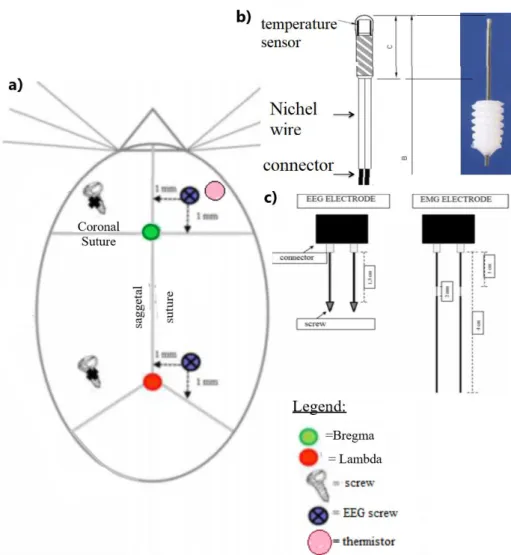
Surgery ... 65
Torpor induction ... 67
Experimental protocol ... 67
Data analysis ... 68
RESULTS ... 70
DISCUSSION... 75
SECOND EXPERIMENT ... 79
MATERIAL AND METHODS ... 80
Glucose dynamics during daily torpor in mice ... 80
Ethical approvals ... 81
Experimental procedures ... 82
Surgery ... 82
Experimental protocol ... 83
Data analysis ... 85
RESULTS ... 87
DISCUSSION... 90
CONCLUSION ... 94
FIGURES ... 99
BIBLIOGRAPHY ... 113
ABSTRACT ... 128
Abbreviations’ Index
A
Activity (ACT)
Slope of the ACT during cooling
(ACT-c)
Slope of the ACT during rewarming (ACT-R)
Autonomic nervous system (ANS) Arterial pressure (AP)
Agouti-related peptide (AgRP) Ambient temperature (Ta)
B
brown adipose tissue (BAT) The baseline (B)
Basal metabolic rate (BMR) Body weight (BW)
C
Core body temperature (CBT) Central nervous system (CNS) Cutaneous vasoconstriction (CVC)
D
Mouse model missing a functional leptin receptor (db/db mice)
Dorsal raphe (DR)
Dorsomedial hypothalamus (DMH)
E
Electroencephalographic (EEG) Electromyographic activity (EMG)
G
Blood glucose concentration or glycemia (GLC)
G-protein-coupled cell surface receptors (GPCRs)
H
Hypocretin 1 and 2 (HCRT-1, HCRT-2) Slope of HP during cooling (HP-c) Heart period (HP),
Slope of HP during rewarming (HP-R) Heart rate (HR)
I
Intracerebroventricular (ICV) Percentage of time spent in indeterminate state (%IND)
K
Knock-out mice for the pre-pro-orexin gene (KO-HCRT)
5
L
Lateral hypothalamic area (LHA) the locus coeruleus (LC)
length of the cooling period (L-c) Length rewarming period (L-r)
M
Median preoptic nucleus (MnPO)
N
Neuropeptide Y (NPY)
Non-rapid eye movement sleep (NREM)
Percentage of time spent in NREM sleep (%NREM)
O
Orexin A and B (ORX A, ORX B) Mice carrier of a mutation in the leptin gene (Ob/ob mice)
Orexins A and B (ORXS) Orexin A receptor (ORX-1R) Orexin B receptor (ORX-2R)
P
Parabrachial nucleus (PBN)
Parasympathetic nervous system (PNS) Preoptic area (POA)
Preoptic anterior hypothalamus (POAH)
Thalamus para-ventricular-nucleus (PVNT)
R
Rapid-eye movement sleep (REM) Percentage of time spent in REM sleep (%REM)
Resting metabolic rate (RMR) Raphe pallidus (RPa)
S
Systolic blood pressure (SBP)
Slope of SBP during cooling (SBP-c) Slope of SBP during rewarming (SBP-r) Suprachiasmatic Nucleus (SCN)
Sympathetic nervous system (SNS) Slow wave activity (SWA)
6
T
Deep torpor (T)
Body temperature (Tb)
Slope of Tb during cooling (Tb-c) Slope of Tb during rewarming (Tb-r) Torpor metabolic rate (TMR)
Tuberomammillary nucleus (TMN) Thermoneutral zone (TNZ)
Transient receptor potential channels (TRP)
Hypothalamic temperature set point (Tset)
U
Uncoupling protein-1 (UCP1)
V
Ventrolateral preoptic nucleus (VLPO) Ventromedial hypothalamus (VMH) Ventral tegmental area (VTA)
W
Wakefulness (W), Control mice (WT)
8
Introduction to Torpor
Torpor general aspects
The large seasonal and daily environmental changes in temperature and food availability obligated organisms to develop adaptation during evolution (Geiser 2004). Endotherms, like mammals and birds, can maintain a constant body temperature of 37°C over a wide range of ambient temperatures. This is the result of their high metabolic rate that generates heat, even during rest, and maintains body temperature (Tb) often well above ambient temperature (Ta). Thus, endothermy allows maintaining internal temperatures at the level of maximum physical performance (Heldmaier, Ortmann et al. 2004; Ruf and Geiser 2015). For all endotherms living in temperate climates maintaining Tb has high energy costs (Geiser 2004). Therefore, the life of endotherms depends upon a continuous high supply of food (Heldmaier, Ortmann et al. 2004).
In case of an adverse environmental condition and scarce food availability, reducing thermoregulatory costs allows animals to redirect energy to different tasks and so optimize growth and reproduction (van der Vinne, Gorter et al. 2015). This is more crucial for small endotherms because the surface area/volume ratio increases as size decreases, so they must produce substantial amounts of endogenous heat to compensate for high heat loss during cold exposure (Geiser 2004).
Therefore, a widespread solution for endotherms, who cannot escape harsh environmental conditions by migration, is to suspend the maintenance of high Tb (torpid state) and then re-warm and return to a normal level of activity when the environment becomes favorable (Melvin and Andrews 2009).
Four conditions are required for a euthermic animal to undergo in a torpid state, and the mechanisms of none of them are currently clearly known (Sunagawa and Takahashi 2016). First, the animal has to turn off, or at least suppress, the thermoregulatory system, producing a marked decrease in temperature. In normal conditions, the
9
lowering of Tb would trigger a response by the thermoregulatory mechanism, but such a response is abolished during torpor (Sunagawa and Takahashi 2016).
Second, the animal must be able to endure low respiration levels or low oxygen supply. In torpid animals, the oxygen consumption is reduced to the 2–3% of that in an aroused state (Andrews 2007).
Third, the animal must be resistant to hypothermia because during torpor hibernators typically have a minimum Tb of 2–10 °C (Carey, Andrews et al. 2003).
Fourth, the animal must be able to return to a normal metabolic state by producing heat starting from a hypometabolic state and to tolerate this rise in heat (Sunagawa and Takahashi 2016).
Animals that can display torpor are a wide and uneven group of species and are present in most of the orders of mammals (Melvin and Andrews 2009), including primates (Dausmann, Glos et al. 2004). This suggests that the ability to suppress metabolic rate was probably a trait present in the ancestral mammal. The trait was later lost in many mammals but remain present in many small species with a high energy expenditure during normothermia. Particularly, many of these animals eat food that becomes seasonally unavailable (Geiser 1998; Cerri 2017).
Laboratory mouse and chipmunk represent two good examples to highlight that the expression of torpor is responsive to the availability of food. Laboratory mouse (Mus Musculus) shows daily torpor only when fasted in cold ambient temperature (Hudson and Scott 1979). During the hibernation season, Eastern chipmunk (Tamias Striatus) whose food hoard was greater had a higher frequency of normothermia and a higher mean Tb than the ones whose food hoard are smaller (Landry-Cuerrier, Munro et al. 2008) (Melvin and Andrews 2009).
Today, an unequivocal and agreed definition of torpor is still missing, even if this topic is deeply investigated in the physiological research field since the beginning of XX century.
Indeed, torpor is a physiological state of temporary inactivity characterized by a profound reductions of basal metabolic rate (BMR) and Tb (Melvin and Andrews 2009;
10
Bouma, Verhaag et al. 2012; Careau 2013; Ruf and Geiser 2015), it is also characterized by a lowering of heart rate (HR) and respiration (reduce current volume, respiration frequency and ventilation)(Carey, Andrews et al. 2003). Although metabolic rate during torpor may be a fraction in respect to normothermic individuals the regulation of Tb during torpor is not abandoned. Tb is regulated at or above a species- or population-specific minimum (Geiser 2004).
It is difficult to pinpoint accurately the absolute value reached by physiological variables involved in torpor because there is great variability intra and inter-species (Hudson and Scott 1979). For example, Tb during torpor bouts falls from normothermic values (35 to 38 °C) (Melvin and Andrews 2009) to an extreme value of -2.7°C in arctic ground squirrel, while in Ursus Americanus it decreases to 28-30°C (Ruf and Geiser 2015; Silvani, Cerri et al. 2018), commonly Tb decrease under 25°C and BMR is reduced by the 70 to 95% (Carey, Andrews et al. 2003; Geiser 2004; Storey and Storey 2010).
Laboratory mouse (Mus Musculus) is one of the most widely studied laboratory mammals, it can enter bouts of daily torpor upon caloric restriction or food deprivation. During torpor Tb in mice fall from 36.6°C (euthermic values) to a minimum of 20 °C (Silvani, Cerri et al. 2018), with a concomitant fall in heart rate from 600 beats per minute (bpm) to a minimum of 160 bpm; the blood pressure also significantly declines during torpor (Swoap and Gutilla 2009).
Circadian periods of slight hypometabolism are regularly observed during the 24 h. During the resting phase, the metabolic rate of mammals decreases by about 20% below the level of metabolic rate during the active phase. This shallow circadian hypometabolism is often associated with a 0.5 - 2 °C decrease in core body temperature (Heldmaier, Ortmann et al. 2004).
The multifactorial nature of the torpor mechanisms still not well characterized in literature; the environmental, physiological, metabolic, and molecular changes altogether can induce the animal to undergo torpor, but how they are connected still not clear (Melvin and Andrews 2009).
11
It would be important to discover the trigger to induce torpor because the drug-induced torpor (synthetic torpor) has a large number of applications. For example, in medicine, the outcome of this discovery would be great because, despite the dramatic decreases in Tb, metabolic rate, and, consequently, reduced blood flow, animals recovering from torpor bouts do not exhibit indications of tissue damage (Bouma, Verhaag et al. 2012) (Dave, Christian et al. 2012). The absence of injury upon rewarming and reperfusion makes torpor a good model to investigate protective mechanisms that could be employed as treatments for different trauma states as cardiac arrest, organ transplantation, major cardiac and brain surgery. Also, in transplantation medicine, reducing metabolism in donor organs may provide a better preservation method giving a greater time window for transplantation of organs to recipients (Bouma, Verhaag et al. 2012).
Like-wise Synthetic Torpor could be applied in long-duration space exploration because the reduction in metabolism would diminish the amount of food required for crew survival for long periods. Moreover, disuse atrophy of skeletal muscle and bones would be cut down (Cerri 2017).
For all the characteristics pointed above Torpor is the closest state to death which mammals can reach and at the same time allows animals to survive in arduous environmental conditions (Ruby, Barakat et al. 2004).
12
Principal torpor behaviours: Hibernation and Daily torpor
Torpor works as a basic element for more complex behaviors (Cerri 2017), depending on the timing, duration, and depth, it can be categorized as seasonal and non-seasonal. Seasonal torpor is represented by two behaviors: estivation and hibernation.
The former is characterized by food store caches and variable lengths/depths of torpor episodes, or sometimes no Torpor, depending on food availability and the severity of environmental conditions, a common example of estivating animal is the chipmunk (Storey and Storey 2010).
Hibernation usually lasts from autumn to early spring and is typified using body the fuel reserves, mainly represented by the fat, to survive the winter. Thereby, this species is called fat-store hibernators, they are the most common hibernator category, and, in this paragraph, when you will find the term Hibernator it will be referred to them. Hibernators, which include many mammals but only a single bird species, are generally small, and most weigh between 10 and 1000 g, with a median mass of 85 g (Geiser and Ruf 1995). However, the entire mass range of hibernators, for which metabolic data are available includes black bears (Ursus americanus), and it is from 5 to 80,000 g (Geiser 2004). During the weeks leading up to the hibernating season, hibernators undergo hyperphagia and build up huge white adipose depots that can increase body mass by 50% (Storey and Storey 2010).
Most of these species exhibit extensive fattening prior to the season in which they display torpor, this behavior is not observed in most of the non-seasonal torpid animals that typically enter torpor at times in which body mass is low, accordingly large fat stores is proved to inhibit daily torpor (Geiser 1998).
Lipids provide to hibernators the primary fuel source for the whole winter although some amino-acid catabolism also occurs, often in support of gluconeogenesis since carbohydrate stores are minimal and reserved in just few tissue types (Dark 2005).
13
Even hibernator brain can switch to a lipid-based economy by oxidizing ketone bodies that are synthesized by the liver (Storey and Storey 2010).
Hibernators do not remain torpid throughout all the hibernation season, but they present a hibernation pattern characterized by bouts of torpor, which are characterized by four different states: 1) entrance into hibernation; 2) maintenance of deep hibernation for several days; 3) arousal that terminates each torpor bouts; 4) a euthermic resting periods of 1-2 days, with normal Tb and high energy turnover that conclude the torpor bout (Heldmaier, Ortmann et al. 2004). Thus, the entire hibernation season of 6–7 months consists out of a sequence of 15 through 20 hibernation bouts (Heldmaier, Ortmann et al. 2004).
This hibernation pattern is similar trough the different hibernating species.
Entrance into torpor is characterized by a rapid reduction of metabolic rate until the minimum is reached. The metabolic reduction is paralleled by a rapid decrease of Tb, which continues into deep hibernation, the second and longer phase. Deep hibernation is terminated with the arousal, when animals raise their Tb to euthermic level, in a time lapse of some hours, through a burst of heat production (Heldmaier, Ortmann et al. 2004). During arousal from torpor bout are used two main power source: non-shivering thermogenesis, mainly in brown adipose tissue, and shivering thermogenesis, produced by skeletal muscles, that is only a supplement to the former (Cannon and Nedergaard 2004). The percentage of time spent in arousal and interbout is short, because of its high energetic cost (Storey and Storey 2010).
In order to have a prolonged period of hypometabolism during hibernation season, the circadian system seems to be inactivated, so, torpid bouts can give a reliance on energy stores (Ruf and Geiser 2015).
The length of torpor season (e.g., up to 8.5 months in the Richardson's ground squirrel (Wang 2011)) and the significant decrease of Tb during torpor, allow a very high energy savings that could be as high as 88% in comparison with animals that remain euthermic in the same period (Wang and W. Wolowyk 2011). However, the repeated arousals and the euthermic periods are energetically very costly. For example, in marmots, 72% of all energy reserves required for the entire hibernation season are
14
consumed during arousals (17%) and the euthermic periods (57%) (Heldmaier, Ortmann et al. 2004).
An example of hibernator is ground squirrel, during torpor can drop his heartbeat from >200 beats/min in interbout to <10 beats/min in torpor (Milsom, Zimmer et al. 1999).
Daily torpor, is a widely used pattern of torpor in mammals and, in contrast to hibernation, also in birds (Geiser 2004). This form of torpor is usually not as deep as hibernation (Heldmaier, Ortmann et al. 2004), in fact, it lasts only for hours rather than days or weeks and it is usually interrupted by daily foraging and feeding (Geiser 2004). Daily heterotherms are unable to express multiday torpor bouts. Many daily heterotherms may employ torpor throughout the year, although torpor use often increases in winter (Geiser 2010). In order to facilitate foraging, mating, migrations etc. daily heterotherms employ the circadian system to control torpor timing to stay entrained with the light-dark cycle (Ruf and Geiser 2015). Daily torpor is usually restricted to the night, whereas, in nocturnal mammals and birds, it is common in the second part of the night and the early morning.
In daily torpor the extent of hypometabolism and hypothermia is usually less pronounced if compared to hypometabolism in hibernation; consequently, also less energy is saved (Heldmaier, Ortmann et al. 2004).
Recent evidence from the field, suggests that daily torpor may also be used to restrict foraging times to only few hours per day in the early evening, with torpor bouts lasting for most of the day. Together with passive rewarming in the sun, this reduces daily energy expenditure by up to 80% (Geiser 2010; Jastroch, Giroud et al. 2016).
A daily torpor bout follows the same events sequence observed in hibernation, i.e. entrance into torpor, maintenance of deep torpor, arousal, and return to the euthermic state (Heldmaier, Ortmann et al. 2004).
The minimum Tb reached in daily torpor strongly depends on species and ambient temperature (Bouma, Verhaag et al. 2012). The lowest tolerable range of Tb is from 10 to 22°C with a mean value of 17 °C (Geiser 1998); even though there is a large variation among species, it stays considerably higher than a hibernator’s.
15
On average, daily heterotherms are smaller than hibernators weigh between 5 and 50 g, with a median of 19 g, in a complete range of from 2 to 9000 g (Geiser 2004). The metabolic rate during daily torpor is on average reduced to about 30% of the BMR (Geiser 2010) although this percentage is strongly affected by body mass and other external factors.
The onset of daily torpor is typically facilitated by food deprivation, although spontaneous torpor in presence of an adequate food supply has also been observed in many species (Wang and W. Wolowyk 2011). Daily torpid species and hibernators have a different reaction to food deprivation: small daily heterotherms will perish if food is absent for several days (Kennedy and Macfarlane 1971), whereas hibernators can survive for months.
So, the main energy supply of daily heterotherms remains ingested food rather than stored body fat, and they appear to balance energy expenditure and uptake on a daily basis (Geiser 2004).
As proof of it, daily torpor can be induced in laboratory mouse with a caloric restriction protocol, which induce replicable torpor bouts, with a mean length of 2-4 hours (Vicent, Borre et al. 2017).
Therefore, daily torpor is not simply a breakdown of thermoregulation due to starvation, these small mammals maintain control of their Tb, so they remain endotherms because they can alternate between a euthermic tachy-metabolic state and a torpid brady-metabolic state with largely reduced body functions. They can even use endothermic thermoregulation when torpid (Hudson and Scott 1979; Jastroch, Giroud et al. 2016).
Another fundamental difference between Hibernating and daily torpid animals is the mechanisms of the metabolic rate reduction, that will be analyzed more specifically in the chapter titled “Interaction between metabolism and torpor”. Also, the effect of torpor on subsequent sleep pattern is different. In hibernating animals who display deep torpor, regulation of temperature and sleep seems to be reduced to a level that currently cannot be measured reliably. In contrast, animals who display daily torpor appear to
16
make use of the same processes, although applied in an extreme way, which reduces body temperature at the onset of sleep in humans (Kräuchi 2011).
For all these reasons it has been discussed whether daily torpor, estivation and hibernation are based on different physiological mechanisms or are different branches of the same adaptation. At present no clear qualitative differentiation is known. The physiological properties of daily torpor, estivation, and hibernation are very similar. All are characterized by major reductions of metabolic rate, heart rate, ventilation, body temperature, and in the torpid state thermoregulatory control is maintained. This suggests that they are based on a common paradigm of physiological inhibition. The classifications of hibernation, daily torpor, or estivation simply represent gradual differences in the timing, the duration, and the amplitude of physiological inhibition (Heldmaier, Ortmann et al. 2004).
Advantage and disadvantage of torpor use in different species
Differences and similarities between daily torpor and hibernation has been extensively discussed in Ruf T. and F. Geiser’s paper titled “Daily torpor and hibernation in birds and mammals” (Ruf and Geiser 2015).
Daily torpor allows to keep entrain the light-dark cycle using short (less than a day) torpor bouts; and daily heterotherms continue to remain active and forage above ground, as opposed to hibernators that show multiday torpor bouts. Species that employ multiday torpor, on the other hand, benefit from larger body mass facilitating higher body energy stores; moreover, they reach a lower metabolic rate during deep torpor bouts, to maximize energy savings.
Hibernation and daily torpor also differ in their primary source of energy: during hibernation, animals can use either body fat or food stores reserve, whereas in daily torpor animals need to continue foraging. This factor more likely explains the significant difference in body mass between daily heterotherms and hibernators.
17
Indeed, a small body mass holds down the size of body fat stores, not only in absolute storage amounts but also in terms of the percentage of body fat, so hibernators which seem to rely on endogenous energy stores benefit from an increased body size.
Daily heterotherms, on the other hand, which continue to forage, should benefit from a functional circadian system that keeps them entrained with the light-dark cycle and serves to optimize times of daily activity and rest.
Torpor state has a crucial role in hibernating and daily torpid animals, it facilitates migration in certain birds because it allows to balance the equilibrium between energy supply and energy demand (Doucette, Brigham et al. 2012). Torpor is an integral part of reproductive strategies that involve sperm storage in certain bats and other mammals, it can primarily serve as a water conservation mechanism, and it was found to lower the risk of extinction (Ruf and Geiser 2015). The latter finding is a consequence of hibernation behavior: typically, during torpor the animal retreats into secluded areas like underground burrows, and this reduces predation risk and results into higher survival rates than during active season (Geiser and Turbill 2009).
A recent experiment demonstrated that a single torpor bout in mice improves the memory process, and protect the memory against from the ambient stressors (e.g. cool ambient temperature and food shortage) (Nowakowski, Swoap et al. 2009).
Indeed, the animals try to reduce torpor when it is possible even though there are a lot of benefits derived from it. A clear example is fat-storing hibernators in with high body energy reserves reduce torpor and increase euthermic episodes during winter (Claudia, Karin et al. 2014; Zervanos, Maher et al. 2014).
The reasons why animals reduce entrance into torpor is still not clear but there are many hypotheses.
The sleep deprivation hypothesis claims that torpor could be an evolution of sleep but during torpor the time spent in a sleep state is deeply reduced or absent, as will be discussed in chapter “Sleep and Torpor”. Actually, the relationship between sleep and torpor is complex, and it appears to be species specific (Kräuchi 2011).
The immune-suppression state is induced in torpor by low Tb, and it is reversed during periodic arousals (Bouma, Carey et al. 2010). As a matter of principle,
immune-18
suppression could be benefic because it saves energy, protects from inflammatory processes, and typically involves little risks since microbes proliferate very slow at low temperatures (Bouma, Carey et al. 2010). However, impaired immune function during torpor may increase the risk of contracting viral or fungal diseases that can be lethal (Prendergast, Freeman et al. 2002; Bouma, Carey et al. 2010).
A new theory claims that an adverse effect of torpor could be a rise of oxidative stress and a consequent increase in costly up-regulation of antioxidant defenses (Ruf and Geiser 2015); this hypothesis is controversial, because despite the presence of a pro-apoptotic and oxidative environment during hibernation and the consequent accumulation of DNA damage, the apoptosis is suppressed and necrotic tissue injury is minimal (Sunagawa and Takahashi 2016).
19
Thermoregulation and Metabolism
Thermoregulation in physiological conditions
Thermoregulation
Thermoregulation is a complex physiological regulatory process that allows the animals to maintain their constant Tb level. This is managed by the body through the regulation of loss and production of heat. The body is highly sensitive to changes in Ta. Animals have a thermo-neutral zone that is defined as the range of Ta without regulatory changes in metabolic heat production or evaporative heat loss (Kingma, Frijns et al. 2012).
Heat can be lost from the surface of the body through radiation, conduction, convection and evaporation. Radiant heat transfer occurs in the absence of contact between objects. Conductive heat transfer occurs between physical objects in each physical state: solid, liquid or gas and they must be in contact with one another. Radiation and conduction are fixed physical phenomena that cannot be manipulated, but are a function of the temperature differential between the body surface and the environment.
Convection is the heat transfer associated with the movement of a fluid (either liquid and gaseous), and evaporation is the conversion of a material from a liquid state to a gaseous state, an example is the heat transfer from the surface of the body. Convection and evaporation in biological systems can be manipulated by adjusting fluid movement (convection) or delivering an expandable fluid to the interface surface (evaporation) (Dennis Grahn 2004).
More than 50 years ago it was theorized that the human body could be divided into two thermo-physiologic compartments: the heat-producing region identified in the homeothermic core, and the heat-loss-regulating poikilothermic shell (Kräuchi 2011). The shell temperature is influenced by skin blood flow, which is increased when core
20
temperature rises, and by environmental temperature. Usually, the distal skin temperature is measured in hands and feet’s (Tansey and Johnson 2015).
The shell size depends directly on Ta, in a warm environment the shell is small, while in a cool environment it is larger and therefore it acts as a bulwark to protect the body core from cooling.
All the peripheral tissues (fat, skin, legs and arms, skeletal muscles) can contribute substantially to the shell size, provided that peripheral blood flow is low (Kräuchi 2011).
Core body temperature (CBT) is the temperature within the “deep” body tissues and organs, which have a high level of basal metabolism. The main ones are the brain and all the abdominal cavity inner organs (e.g., liver, heart, kidney) (Krauchi 2007; Tansey and Johnson 2015).
So, Tb, which depends on CBT, is a critical homeostatic parameter influencing cellular function and organismal survival. Increasing of Tb induces protein denaturation and reduction in membrane fluidity, ion fluxes, and enzyme performance that could dangerously affect the organism’s health.
The central neural circuits for thermoregulation orchestrate behavioral and autonomic repertoires that maintain Tb during thermal challenges. Tb is sensed by thermal receptors, either in the external or the internal (e.g., during exercise) environment (Morrison 2016).
In the majority of mammals CBT standard value is set around 37° C (Mekjavic and Eiken 2006; Tansey and Johnson 2015) and the brain CBT is the main target for homeothermy, thus all the behavioral and physiologic processes are regulated on it over a broad environmental temperature range (Mekjavic and Eiken 2006).
21
General aspects of thermoregulation control
Ta is sensed primarily by the skin, but there are also thermoreceptors in the oral cavity and in the upper respiratory airways (Cliff and Green 1996; Cerri 2017).
The temperature inputs are then elaborated by the central nervous system, mainly by the preoptic area and parabrachial nucleus, to correct any impairment by activating adequate effector mechanisms.
The effector mechanisms for the cold defense, which cause an increasing in the energy costs, includes thermoregulatory behavior to reduce heat loss, cutaneous vasoconstriction (CVC), to conserve heat in the body core, piloerection, non-shivering thermogenesis in brown adipose tissue (BAT) and shivering thermogenesis in skeletal muscle (Morrison and Nakamura 2011).
Animals also show thermoregulatory behaviors that are defined as stereotypical somatic motor acts and are performed in to minimize or optimize heat transfer from the body to the environment. In rodents, such behaviors include postural changes (huddling in the cold or stretching the limbs in the heat), the movement to a more comfortable environment (cold seeking moves to hot environment), or the spreading of saliva on the fur in a hot environment (Morrison and Nakamura 2011).
Since the skin is the first organ that receives the indication of a potential threat to brain and core temperature homeostasis, it is not surprising that thermoregulatory behavior in animals is triggered primarily by cutaneous thermal receptors (Morrison and Nakamura 2011).
There are many theories about the regulation of CBT: the first interpretative theory is named “the open loop theory of thermoregulation” (Romanovsky 2007), which asserts that every thermal effector operates independently from the others, and it is turned on or off by its own neural controller at different temperature thresholds. From this point of view, Tb is just the result of the parallel activity of all the open loops controlling the thermal effectors (Cerri 2017). The main theoretical framework of the thermoregulation physiology is the “set point theory”. According to this theory, the
22
brain controls Tb by changing the reference value of CBT, the so-called set point (Boulant 2006). The thermal effectors will, therefore, work in a coordinated manner to reduce the difference between the core temperature and the set point (Cerri 2017). Thermoregulation in mammals is under the control of the autonomic nervous system (ANS) which modulates the thermogenic and thermo-dissipative activity of organs to maintain an appropriate Tb for each of the animal’s activities (Cerri 2017).
CBT is regulated with thermo-effector thresholds, which are subject to circadian oscillations (Tayefeh, Plattner et al. 1998). Circadian rhythms in mammals are produced by the self-sustaining central pacemaker in the suprachiasmatic nuclei (SCN) of the hypothalamus (Moore and Danchenko 2002). A rostral projection from the SCN to the preoptic anterior hypothalamus (POAH) conveys the circadian signal to the thermoregulatory system (Moore and Danchenko 2002).
The regulation of CBT results from the concerted action of the homeostatic and circadian processes (Kräuchi 2011). In humans, the daily decline of CBT in the evening results from a regulated decline in the thermoregulatory thresholds of heat production and heat loss; the inverse happens in the morning. When heat production is higher than heat loss, body heat content increases and vice versa.
Depending on Ta, about 70 to 90% of body heat content is located in the body core CBT (Kräuchi 2011). The balance in heat production and loss are changed by activities such as muscular exertion and fluid and food intake that is not randomly distributed over the circadian cycle.
Under resting conditions, about 70% of heat production depends on the metabolic activity of inner organs (Kräuchi 2011), whereas body heat loss is initiated by heat redistribution from the core to the shell through blood flow to the distal skin region.
23
Thermal sensitivity and TRP receptors
The location of thermoreceptors is not homogeneous in all the organs and their role is different in each body part.
Thermal sensitivity can also be divided into peripheral-thermoception and central-thermoception.
As previously stated, Ta is primarily sensed by the peripheral thermoreceptors located in the skin, and warm receptors are less abundant than cold receptors (Cliff and Green 1996). Warm central thermoreceptors are located in the hypothalamus, spinal cord, viscera, and great veins and they are in a higher number than cold thermoreceptors. The activation of central thermoreceptor can modify the CBT inhibiting cold thermoreceptors (Tansey and Johnson 2015).
Major advances in the transduction processes in peripheral thermal sensation have been made since the discovery of the large family of transient receptor potential (TRP) channels in the past fifteen years. Thermoreceptors are part of this family, and different TRP channels provide information regarding specific ranges of temperature, they can be expressed in cell membranes and in membranes of internal structures (Wang and Siemens 2015). Many TRP receptors are polymodal in their activation, but all result in cation influx. TRP channels are involved in thermal sensation and they may contribute by direct activation of sensory fibers (Tansey and Johnson 2015). Nine of them are sensitive to temperature thresholds and their roles have been extensively reviewed (Morrison, Sved et al. 1999; Caterina 2007). In literature the temperature receptors are subdivided into cold noxious, cold, warm, and hot noxious subpopulations on the basis of the temperature stimuli which discriminate (Tansey and Johnson 2015).
Among the most interesting of these channels is the TRPM8 channel, whose natural agonist is menthol; this channel mediates the feeling of cold and it is increasingly activated as temperature drops below 27°C. So it’s likely to presume that TRPM8 could mediate innocuous cold sensation (Peier, Moqrich et al. 2002; Tansey and Johnson 2015). Further, TRPA1 channel is activated at temperatures below 17°C and may
24
contribute to noxious cold sensation, although his role is not completely clear (Cliff and Green 1996).
The feeling of warmth is sensed primarily by the TRPV1 channel (Chen 2015), whose natural agonist is capsaicin, but the TRPV3 channel is also involved (Cliff and Green 1996). TRPV1 and TRPV2 were among the first to be identified with temperature sensitivity. They are activated by temperatures higher than 43 and 52°C, respectively, and may mediate noxious hot sensation (Nakamura and Morrison 2010). TRPV4 and TRPV3 are activated by temperatures above 25 and 31°C, so is likely to assume that they mediate innocuous warm sensation (Smith, Gunthorpe et al. 2002).
Thermoregulatory control network
In non-anesthetized animals, the core and brain temperature doesn’t drastically change if they are exposed to a cold environment (Bratincsak and Palkovits 2005). This is an example of a rapid thermo-defensive response that is evoked by detecting changes in environmental temperature through thermoreceptors in primary sensory nerve endings which are distributed across the skin.
The central thermoregulatory control of the sympathetic outflows mediates CVC and BAT thermogenesis. Moreover, regulates the somatic motoneurons that produce shivering, which is effected through parallel but distinct, effector-specific, integrative/efferent circuits (Morrison 2016).
The cutaneous thermoceptors information is transmitted in a feedforward manner to the preoptic area (POA), a sensory-motor integrative site for thermoregulation, located rostral to the hypothalamus (Morrison and Nakamura 2011; Morrison 2016; Cerri 2017). It has been proposed that spontaneous activity in POA neurons could be a pacemaker able to produce action potentials. This neurons are temperature sensitive , this reflects the temperature sensitivity of these membrane currents (Zhao and Boulant 2005; Wechselberger, Wright et al. 2006).
25
Neurons in the POA are postulated to integrate ascending peripheral signals with local signals to regulate the output of BAT and shivering thermogenesis- promoting neurons in the dorsomedial hypothalamus (DMH) (Nakamura and Morrison 2007) and of CVC-promoting neurons in the median preoptic nucleus (MnPO) (Morrison 2016) (Morrison and Nakamura 2011).
Thermal information from core body receptors is transmitted to the parabrachial nucleus (PBN) in the brainstem (Cerri 2017); the dorsolateral part receives the cold-activated fibers, whereas the dorsomedial part receives the warm ones (Cerri 2017). PBN transmits the thermal information to the ventrolateral portion of the thalamus that delivers the signals to the cerebral cortex, which is the anatomical substrates for the effective cold or warm sensation.
At the same time, thermal information is also sent to the hypothalamus, especially to the MnPO, providing the anatomical substrates for the autonomic control of temperature (Schmieg, Mercer et al. 1980; Xue, Yang et al. 2016; Cerri 2017).
The core body thermoregulatory mechanisms involve many organs like the brain, spinal cord and abdomen (Morrison 2016).
The afferent fibers from cold and warm abdominal thermoreceptors are included among the splanchnic and vagus nerves afferent fibers and their responses to temperature changes are similar to those of cutaneous thermoreceptors (Morrison and Nakamura 2011). It’s reasonable to suppose that thermal information derived from core body structures would rise a thermoregulatory response only for extreme thermal situations (Morrison and Nakamura 2011).
Therefore, warm-sensitive POA neurons integrate cutaneous and local thermal information, and project to the medial preoptic region of the hypothalamus (MPO) with has an inhibitory function. MPO neurons are tonically activated at thermoneutral temperatures and suppress both shivering and non-shivering thermogenesis (Morrison and Nakamura 2011). Moreover, it’s a current hypothesis that the MnPO neurons integrate all the information (J., T. et al. 2015)
26
Furthermore, there are many organs that play a direct role in heat production, such as the BAT, liver, and skeletal muscle. Others, such as the heart or the thyroid, play an indirect role (Cerri 2017). The activity of most of the thermoregulatory organs is controlled by the sympathetic branch. The sympathetic preganglionic neurons to these organs are in the intermediolateral column within the spinal cord, whereas the sympathetic premotor neurons are in the brainstem area of the raphe pallidus (RPa) (Cerri 2017).
The activation of neurons within the RPa stimulates thermogenesis having little or no effect on arterial pressure. Thermoregulation is also in relationship with osmoregulation, in particular, dehydration in hot environments suppresses heat dissipation (Doris and Baker 1981). Other conditions that can modulate thermoregulation include the hypoxia (Cadena and Tattersall 2014), diving reflex (Hill, Schneider et al. 1987) and sleep (Parmeggiani 2003).
Nocturnal secretion of melatonin, a pineal hormone, which is under central nervous system (CNS) control, contribute to downregulation of CBT in the evening (Krauchi, Cajochen et al. 2006). Moreover melatonin, acting directly on blood vessel receptors and indirectly through modulation of sympathetic nerve activity, induces distal vasodilation in humans (Krauchi, Cajochen et al. 2006).
Thermogenesis in brown adipose tissue
In consequence to a fall in CBT due to a cold environment or to the presence of pyrogenic cytokines, CNS thermoregulatory networks can stimulate thermogenesis primarily in BAT, skeletal muscle (shivering) and heart (Morrison and Nakamura 2011). Non-shivering or adaptive thermogenesis in BAT is the specific metabolic function of this tissue and it is actuated by the high expression of uncoupling protein-1 (UCPprotein-1) in its mitochondrial membranes, which permit to uncouple oxidative metabolism from ATP production and energy expense (Tansey and Johnson 2015).
27
BAT is a crucial thermoregulatory effector in rodents and small mammals but also for adult humans (Nedergaard, Bengtsson et al. 2007). Recently, it has been confirmed the role of the BAT in the thermoregulation for human adults as well as neonates (van der Lans, Hoeks et al. 2013).
Moreover, BAT has a central role in the process of metabolic balance and it has been recognized to be a potential site for drugs aimed at controlling energy expenditure, so it could potentially be a therapeutic site for the treatment of obesity (Tseng, Cypess et al. 2010).
In response to inputs from peripheral and central thermoreceptors SNS activity can stimulate BAT thermogenesis by catecholamines which bind on β3-adrenergic receptors and activates UCP1. POA neurons are supposed to have an inhibitory action on BAT thermogenesis (Morrison and Nakamura 2011).
In addition, BAT thermogenesis can be modulated by a number of nonthermal factors, including hypoxia, infection, hypoglycemia, and psychological stress (Morrison and Madden 2014).
28
Metabolism in physiological conditions
BMR indicates the energy supply required to perform the fundamentals metabolic functions, thermoregulation and exercise (Dennis Grahn 2004). Tb is regulated with a proportional increase in heat production that compensates for heat loss at or above a species- or population-specific minimum (Geiser and Ruf 1995). When the heat produced by basal metabolic activity is scarce to provide for the resting animal thermal demands, additional endogenous heat could be generated through different voluntary or involuntary activities, for example with shivering, voluntary exercise or non-shivering thermogenesis (Dennis Grahn 2004).
Likewise, when Tb is higher than the dissipation capacity metabolic efforts will increase to enhance heat dissipation by evaporative heat loss. The thermoneutral zone (TNZ) is limited by a lower critical Tb, below which the resting metabolic rate (RMR) is not enough to compensate for heat loss. Therefore, with acute exposure to severe cold or prolonged exposure to moderate cold, heat loss from the body will be higher than the capacity to produce metabolic heat and the Tb will fall (Dennis Grahn 2004). The entrance into torpor is facilitated with low Ta usually well below the TNZ. The difference between Tb and Ta is usually small 1–3 °C (Geiser 2004) so Ta has a crucial role in determining the value reached by Tb in torpid animals. Another crucial factor promoting the torpid state occurrence is the food scarcity, that in laboratory is mimed with the caloric restriction protocol (see experiment one methods chapter) (Swoap and Gutilla 2009).
If any external interference occurs the hypothalamic temperature set point (Tset) can rise in torpid animals starting thermoregulation, so, the thermoregulation is maintained during torpor. In placental heterotherms BAT is one of the major players involved in endogenous heat production during arousal from torpid states (Geiser 2004). The mean normothermic life parameter values of the laboratory mouse (Mus Musculus) are a body temperature is 37.4 °C and a BMR of 1.47 ml O2/(gh) that decreases respectively
29
to 19 °C during torpor bouts with a mean torpid metabolic rate value of 0.3 ml O2/(gh) and a Q10 value of 2,4 (Geiser 2004).
Interactions between metabolism and torpor
Body temperature and metabolism
The mechanism that rule different phases of a torpid state isn’t clear in literature, in the past 20 years many theories have been formulated by scientists all over the world. Although many experts agree that the reduction of MR during torpor is substantial and is crucial for survival in many species, the mechanisms of how MR is reduced remains controversial.
Several mutually exclusive hypotheses on MR reduction during torpor have been put forward. Several mutually exclusive hypotheses on MR reduction during torpor has been proposed. The traditional, and obsolete, view assert that Tb and MR fall together at the moment of torpor entry, this affirmation is based on the Q10 value, which is the measure of biological system changing rate consequently of a temperature range variation of 10 °C. This theory suggests that the MR reduction during torpor below BMR can be explained by temperature effects, because a Q10 value of 2 was found in torpid animals (Snapp and Heller 1981; Guppy and Withers 1999). Therefore, in this theory the reduction in metabolic rate seems to be explained solely by the temperature effect on the biochemical processes in the body (Strijkstra and Daan 1998) as, for example, the reduction of the enzyme-catalysed reactions rate (the so-called Arrhenius or Q10 effect) (José 2002).
In successive experiments, higher values of Q10 (>3) have found in different species during torpor entry, so the temperature effects itself isn’t enough to explain the entrance process into torpor. Therefore, a physiological regulation must be involved in the
30
reduction of MR (Geiser 1988; Storey and Storey 1990). Others scientist propose that TMR isn’t influenced by Tb. They argue that MR is down-regulated at torpor entry and the fall of Tb is a consequence and not the reason for the MR reduction (Storey and Storey 1990; Heldmaier and Ruf 1992; Heldmaier G 1993). Finally, it has been suggested that, as during normothermia, MR during torpor is a function of the Tb-Ta differential (Heldmaier G 1993; Sunagawa and Takahashi 2016).
All the more recent theories agree that Tb fall after MR, this suggests an active, regulated metabolic suppression (Geiser 2004; Staples 2014; Cerri 2017). To better understand the MR-Tb relationship we have to consider that the Tset for Tb is down-regulated during the torpor entrance (Heller, Colliver et al. 1977). The fall of Tset results in a fall of MR from the RMR (that is calculated adding together the energetic cost of thermoregulation and BMR) to BMR because the thermoregulation is supposed to cease during the torpor entry (Geiser 2004). Only when Tb reach the lower Tset during the cooling phase is the metabolic heat production increased in order to keep Tb at or above torpor Tset (Geiser 2004).
An important consideration is that small variation in Tset can radically reduce energy expenditure, because of the thermal inertia, so the animal doesn’t need large changes in Tb to save energy during the cooling phase. From this point of view, the transient fall of MR is not due to the fall of Tb but to the fall of Tset. Thus, it is correct to say that the MR has to decrease before the falling of Tb (Geiser 2004).
We have also to consider, that this mechanism is proved to be different between daily heterotherms and hibernators because the main energy supply is different between these two groups. Daily heterotherms, even during the torpor season, remains food collectors and, so, torpor is interrupted to have daily foraging, as opposed to hibernators, whose main fuel source is fat (Geiser 2004). Small daily heterotherms have higher BMRs, and the effects of a reduction of Tb by about 20°C, as commonly observed in daily heterotherms, results in a maximum reduction of the torpor metabolic rate (TMR) to about 25% of BMR assuming a value Q10 around 2 (Geiser 2004).
31
The reduction in metabolic rate in animals who display daily torpor is largely determined by the decrease in Tb, hibernators seem to apply some kind of extra reduction in metabolic rate (Kräuchi 2011).
Small hibernators undergo prolonged periods of torpor and can survive on stored fat for months.
If hibernators exhibit the same MR reduction as small daily heterotherms their fuel stores would be depleted in a very short term, well before the end of winter. Thus, it is not surprising that the reduction of TMR below BMR in small hibernators is much more pronounced than in daily heterotherms and that the relationship between Tb and TMR of the two groups differs (Geiser 2004).
Circulating and cellular fuels
In the last decade, the improvement of gene analysis technologies allowed to discover new links between gene expression and torpor. Consequently, new theories involving circadian clock genes and torpor have been postulated: new experiments on 43 birds and 141 mammals (Ruf and Geiser 2015) confirmed that Per1, Per2 and Bmal1, are expressed rhythmically during hibernation rather as opposed to constantly (Revel, Herwig et al. 2007; Melvin and Andrews 2009).
The theories described below (about the decrease in MR/Tb) and the new ones about gene expression mechanisms may not be entirely mutually exclusive, because any metabolism-driven changes may still be modulated by the endogenous clock that influences, particularly, the arousal from torpor (Ruf and Geiser 2015).
In any case, it should be mentioned that the endogenous clock modulating Tb and MR in hibernators, if it exists, must differ functionally and anatomically from the circadian clock controlling daily torpor (Malan 2010).
The researches on circulating and cellular fuels are a fundamental part of torpor metabolism study.
32
In fact, both hibernation and daily torpor episodes are preceded by a period of time in with shortage of food alter the animal nutrient status (Geiser 2004; Swoap and Gutilla 2009). So, the scarcity of fuels in blood and cells could be a trigger for torpor.
A study in mice showed a connection between cellular nutrient status and the circadian clock (Ramsey, Yoshino et al. 2009). An important parameter to measure the cellular nutrient status are the ratios of intracellular [5’-AMP] to [ATP] and [NAD+] to [NADH] (Rodgers, Lerin et al. 2005). These ratios increase proportionally to the decrease of cellular nutrient status.
Shortage of food is a stressful situation leading the cells to switch their metabolism to lipids as the primary energetical source in order to recover the normal nutrient status (Tashima, Adelstein et al. 1970).
So, new theories arose, which links reduced the availability of food in the environment, the energy status of the cell, the mammalian circadian clock and the response to oxidative stress.
R. G. Melvin and Matthew T. A. (Melvin and Andrews 2009) supposed that in a fasted animal occurs a depletion of cellular nutrients and this would increase intracellular concentrations 5’-AMP and NAD+.
Increased [NAD+] would stimulate the activation of the SIRT1 deacetylase that controls the activation of SERT1 itself. SIRT1 can enter into the nucleus and deacetylate lots of targets including the core circadian clock protein, BMAL1 (Takahashi, Hong et al. 2008). SIRT1 could also activate nuclear receptor proteins that promote transcription of enzymes active during lipolysis and in the response to hypoxic stress like HIF-2α(Dioum, Chen et al. 2009). Activation of lipolytic enzymes is part of the switch from carbohydrate to lipid-based metabolism.
Nutrient depletion provokes also increased intracellular [AMP] and results in increased activation of adenosine monophosphate kinase that phosphorylates the nuclear receptor carbohydrate responsive element binding protein, this down-regulate glycolysis and, so, concur to the switch from carbohydrate to lipid based metabolism (Postic, Dentin et al. 2007).
33
Glucose contributes to the torpid state
As previously mentioned, in mice daily torpor occurs in response to decreased energy availability. The most recent theories, not in order of importance, assume that potential contributors towards daily torpid state are all the players that can cause a drop of power levels. Could be energy-sensitive hormones such as leptin and insulin, as well as the availability of circulating fuels, such as glucose, free fatty acids, and ketones (Melvin and Andrews 2009).
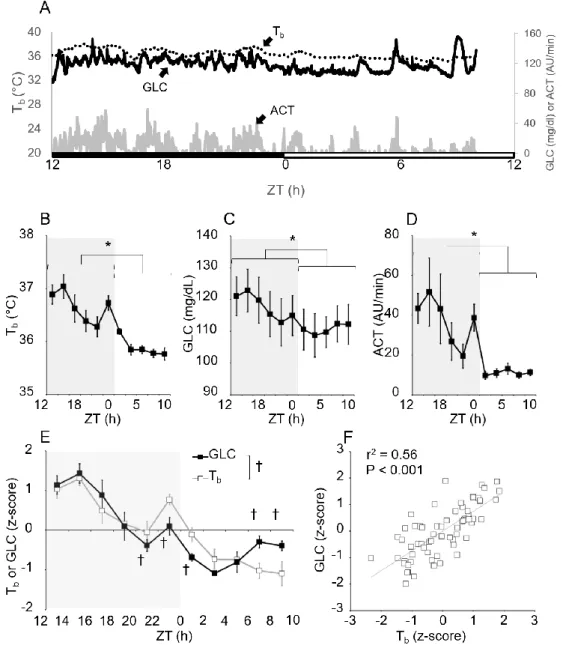
Glucose is the primary fuel used by the brain metabolism, so its deficiency may play an important regulating role in initiating or maintaining torpor. This idea is not new, many experiments on daily torpid animals showed a gradual decline in plasma glucose which precedes or is combined with daily torpor onset (Nestler 1990; Heldmaier, Klingenspor et al. 1999; Dark 2005; Franco, Contreras et al. 2013)
Hibernators similarly to daily torpid animals during deep hibernation shows a reduction in plasma glucose simultaneously with the fall of body temperature and metabolic rate (Sarajas HSS 1967; Galster and Morrison 1970; Atgie, Nibbelink et al. 1990), although this is not true for all the species of hibernators (Musacchia and Deavers 1981; Zimmerman 1982). The role of glucose into torpor triggering emerges also in the pharmacological experiment with the administration of 2-deoxy-glucose (2-DG). 2-DG is an analogue of glucose that can’t be metabolized by the enzyme phospho-gluco-isomerase and block the glycolysis cycle (Wick, Drury et al. 1957). The administration of 2-DG in pharmacological in vivo studies on two daily torpid species, the Siberian hamster and the deer mouse, causes a temporary decrease in Tb (Dark, Miller et al. 1994; Stamper and Dark 1996). However, it’s important to note that administration of 2-DG induces low body temperature also in rat and human which are not able to undergo in daily torpor (Freinkel, Metzger et al. 1972; Penicaud, Thompson et al. 1986).
34
Moreover, the autonomic nervous system (ANS) is deeply involved in the regulation of the different phases of the Torpor bout. There is a lot of evidence of a sympathetic downregulation while parasympathetic is upregulated during the torpor entrance and the deep torpor (Morhardt 1970; Milsom, Zimmer et al. 1999; Zosky 2002; Horwitz, Chau et al. 2013).
Even though, sympathetic activity to white fat increases during caloric-restriction-induced torpor and causes the decreasing in clearing leptin and elevates lipolysis. Consequently, the sympathetic activity increases free fatty acid releasing in the blood (Rayner 2001; Swoap, Gutilla et al. 2006; Swoap and Weinshenker 2008).
Moreover, glucose deficiency leads to the inhibition of sympathetic tonus in rats (Young and Landsberg 1979) and garden dormice (Atgie, Nibbelink et al. 1990). The sympathetic nervous system appears to be activated during arousal from torpor when the heart rate is maximal and brown adipose tissue is activated to produce heat (Hudson and Scott 1979; Swoap and Weinshenker 2008; Oelkrug, Heldmaier et al. 2011).
So, the hepatic glucose dynamics are modified in daily torpor because the liver is involved by the autonomic changes described before.
In physiological conditions the sympathetic activity on the liver provokes the release of glucose while repressing glycogen synthesis; the opposite effect is exerted by the parasympathetic activation via the vagus nerve (Mizuno and Ueno 2017).
Reasonably, the decrease in blood glucose during a day of scarce food availability is a consequence of reduced carbohydrate intake and the extensive reduction of sympathetic outflow that occur during daily torpor episodes, especially for decreased sympathetic outflow to the liver (Lo Martire, Valli et al. 2018).
35
Sleep and Torpor
Physiology of Sleep
The wake-sleep cycle is the sequence of three principal behavioural states: wakefulness (W), non-rapid eye movement sleep (NREM) and rapid-eye movement sleep (REM). From a physiological point of view, sleep can be defined as a reversible behavioural state of perceptual disengagement form and unresponsiveness to the environment. However, this definition is simplistic because it is also true that sleep is a complex mixture of behavioural and physiologic processes (Dement 2011).
The distinction of REM and NREM sleep is based on numerous physiologic parameters, these two states exist in all mammals and birds yet studied, and they are as distinct from one another as each is from wakefulness.
In polysomnography, W is defined by a high electromyographic activity (EMG) and a desynchronized electroencephalographic (EEG) activity with low-voltage rapid waves. NREM sleep (or slow wave sleep) is easily distinguishable from both wakefulness and REM sleep by high voltage and synchronous EEG rhythms, including sleep spindles, K complexes and high-voltage slow waves, associated with low muscle tones and minimal physiological activity. NREM sleep, in human sleep, can be divided into four stages using the EEG (Dement 2011).
Stages 1, 2, 3 and 4 sleep and approximately parallel a depth-of-sleep continuum during NREM sleep, the arousal threshold is generally lower in stage 1 and increase in each consequent stage. NREM sleep is usually associated with minimal or fragmentary mental activity. A shorthand definition of NREM sleep is a relatively inactive yet actively regulating brain in a movable body (Rechtschaffen 1968, repr. 1973.; Dement 2011).
36
REM sleep, or paradoxical sleep, is identified by episodic bursts of rapid eye movements, suppression of EMG and EEG activation. A relatively low voltage waves characterizes the EEG pattern with a mix of frequency (frequency in θ range is common during REM sleep, particularly in proximity to eye movements). The activity in REM sleep’ EEG can be in theαrange (1-2 Hz slower than waking activity). There are not any stages in REM sleep, and it is characterized by the spinal motor neurons inhibition which provokes the suppression of postural motor tonus during REM sleep, although tonic phases may occur in this state.
A normal human adult enters sleep by the NREM state, this principle is important because reflects a highly reliable finding of normal human sleep and this knowledge permit to consider normal versus pathologic sleep. A significant example can be the abnormal entry into sleep through REM sleep that permits to diagnostic in adult patients with narcolepsy (Dement 2011).
Sleep and torpor
Sleep and torpor have blatant common aspects: they are highly regulated, adaptive, and reversible behaviours with a stick out the tendency towards energy conservation (Berger 1984).
Nocturnal hypothermia is the most common pattern of non-seasonal torpor, representing a survival strategy of many small mammals, which allow their metabolic rate to decrease during sleep by 20–30%, and a decrease in Tb of few degrees Celsius. This strategy allows to increases the interval from one meal to the next (Storey and Storey 2010). For instance, the fruit-eating manakins from the tropical rain forest and several species of chickadees and artic willow tits show nightly shallow depressions of body temperature to approximately 27-34°C (REINERTSEN 1983) for few hours. The daily energy savings derived from nocturnal hypothermia range from about 10% in the willow tit to about 30% in the manakin (REINERTSEN 1983).
37
Usually, torpid episodes happen in the animals’ nest, suggesting that they look for a safe environment to undergo into torpor, this is true also for sleep episodes.
Moreover, animals in torpor seem to be asleep because they maintain a sleep-like posture for the duration of the bout, with elevated arousal thresholds, accompanied by a reduction in muscle tone (Kräuchi 2011; Silvani, Cerri et al. 2018).
Furthermore, they are both characterized by a reduction in Tb, greater in torpor than in sleep, but the mechanism implied in the resetting of the central regulation of Tb during the transition from wakefulness to NREM sleep appears to be homolog with the one of torpor entrance (Heller and Glotzbach 1977).
Analysis of the EEG patterns shows that rodents during torpid bouts stay more in NREM sleep than REM sleep, which is reduced or not present (Walker, Garber et al. 1979; Harris, Walker et al. 1984; Deboer and Tobler 1994; Deboer and Tobler 1995). In particular, recordings in the deep hibernator hypothalamus indicate that these animals keep alternating between long NREM sleep bouts and short waking bouts (Krilowicz, Glotzbach et al. 1988). These findings support the hypothesis that NREM sleep is an adaptive behaviour for energy conservation, and its function is strengthened during torpor (Berger 1984; Obal, Rubicsek et al. 1985; Heller 1988).
Accordingly, torpor is usually entered through sleep, as demonstrated by the studies conduct by Walker and colleagues (Walker, Glotzbach et al. 1977) in ground squirrels. They showed that Tb starts to decrease during either REM or NREM sleep but never during wakefulness. Walker and colleagues were able to record the EEG during entrance in torpor only if the brain temperature was maintained above 25°C. To correctly analyse the EEG pattern, we have to consider the temperature effect on EEG frequency as Deboer at all proves in hamsters (Deboer and Tobler 1995). They show a systematic downward shift of EEG frequency bands as cortical temperature decreased. Therefore, the EEG slow waves during torpor occurred at frequencies lower than those during euthermia. This is crucial because the lack of physiological EEG slow wave during torpor can represent a long period of sleep deprivation. During the arousal, the animals emerging from torpor immediately enter into deep NREM sleep, irrespective
38
of whether animals emerge from deep hibernation (Daan, Barnes et al. 1991; Trachsel, Edgar et al. 1991) or daily torpor (Deboer and Tobler 1994).
Therefore, during deep torpor it is likely that the restoring function of sleep cannot be fulfilled, so animals must awaken to recover from the hypothermic sleep deprivation. This statement was confirmed in Djungarian hamster (Deboer and Tobler 2000). Experiment conducted on golden-mantled ground squirrels showed that the slow-wave activity during NREM sleep, following arousal from torpor, is related only to the minimum brain temperature reached during torpor. So, the sleep debt accumulates during torpor may be a brain-temperature dependent effect (Larkin and Heller 1996). The synthetic torpor experiment in rats shows a massive increase in EEG slow-wave activity in the euthermic period after deep hypothermia (22°C) (Cerri 2017).
The power of the EEG possibly increases proportionally to the magnitude of the metabolic effort made by the animal to rise from torpor and recover to euthermia. In light of this results in the rat, a great part of the sleep debt could be accumulated during the rewarming phase. In conclusion it is possible that the neural circuits that generate and sustain torpor and NREM are shared, at least in part, mainly because NREM sleep and torpor have in common the outcome of reducing energy expenditure (Silvani, Cerri et al. 2018).
39
Common Physiological Changes in NREM Sleep and Torpor
The daily temperature trend is characterized by a drop in Tb during the resting phase, which results from an increase in heat dissipation due to cutaneous vasodilation and a concurrent decrease in heat production (Krauchi and Wirz-Justice 1994). NREM sleep in humans is the prevalent component of the rest period and reduces energy expenditure by approximately one-third (Bouma, Kroese et al. 2011; Kayaba, Park et al. 2017). The energy saving characteristic of NREM sleep is observed in small animal models, such as mice, particularly when are considered long, consolidated wake-sleep episodes (Zhang, Zeitzer et al. 2007). Moreover, NREM sleep is characterized, in human and small animal model, by a fall in MR accompanied in concomitance to a decrease in arterial pressure (AP) and heart rate (HR) (Silvani, Bastianini et al. 2014).
It’s still not completely clear how AP can decrease but there are two currently main theories in literature: the first assert that the decrease in HR is due to a balance of cardiac sympathetic withdrawal and parasympathetic activation, which cause the AP decrees (Lo Martire, Silvani et al. 2018) in absence of a compensatory increase in stroke volume and/or peripheral resistance (Khatri and Freis 1967). The second theory is based on a recent evidence obtained in mice, which suggest that the diminish AP during NREM sleep depends from a reduction in sympathetic vasoconstriction (Lo Martire, Silvani et al. 2018), itself due to a decrease in sympathetic activity to the vasculature of skeletal muscles (Somers, Dyken et al. 1993), skin (Takeuchi, Iwase et al. 1994), and kidneys (Yoshimoto, Sakagami et al. 2004).
The profound reduction of MR during torpor results in greater energy savings compared with NREM sleep. As in NREM sleep the autonomic nervous system is deeply involved in all stages of a bout of torpor. During entrance and the maintenance of a torpor bout, parasympathetic activity to the heart is markedly increased causing a diminishing in HR. Periodic increases in HR coupled to ventilation are present in torpor bouts due to the instability of parasympathetic action (Morhardt 1970; Milsom,
40
Zimmer et al. 2001; Zosky 2002; Zosky and Larcombe 2003; Horwitz, Chau et al. 2013). Entrance into torpor is characterized by a sympathetic activity withdraws in concomitance to the parasympathetic dominance (Atgie, Nibbelink et al. 1990). Similarly, in humans NREM sleep decrease in energy expenditure is associated with a decrease in minute ventilation due to the parasympathetic activity, which leads to a rise in the partial pressure of carbon dioxide and a fall in the partial pressure of oxygen (Douglas, White et al. 1982). These effects of NREM sleep on respiratory control are also seen in small model organisms: in mice NREM sleep decreases the respiratory rate and blunts the chemoreflex responses to hypoxia (Nakamura and Morrison 2007). Approaching the onset of torpor, the ventilation in mice occurs in concert with the spike of EMG activity and tachycardia (Milsom and Jackson 2011; Swoap, Kortner et al. 2017). Likewise in NREM sleep the ventilation during torpor decreases more than metabolic rate does, leading to a modest respiratory acidosis (Milsom, Zimmer et al. 2001; Milsom and Jackson 2011).