ALMA MATER STUDIORUM UNIVERSITA’ DI BOLOGNA
_____________________________________________________
DOTTORATO DI RICERCA IN
Scienze Cardio Nefro Toraciche
Ciclo XXXII
Settore Concorsulae: 06/I1 - DIAGNOSTICA PER IMMAGINI, RADIOTERAPIA E NEURORADIOLOGIA
Settore Scientifico Disciplinare: MED/36 - DIAGNOSTICA PER IMMAGINI E RADIOTERAPIA
TITOLO TESI
Prediction Nomogram for
68Ga-PSMA-11 PET/CT
in different clinical settings of PSA failure after radical treatment
for Prostate Cancer
Presentata da:
Dott. Francesco Ceci
Coordinatore Dottorato
Supervisore
Chiar.mo Prof.
Chiar.mo Prof.
Gaetano Domenico Gargiulo
Nicolò Daddi
Table of Contents
1. Introduction ………...pag. 4
1.1 Imaging in Prostate Cancer: State of the Art ………..pag 4 1.2 PSMA as target for PET imaging and radionuclide therapy ……….pag. 5 1.3 PSMA based PET imaging in Primary Staging of Prostate Cancer ………..pag. 7 1.4 PSMA based PET imaging to localize the site(s) of Biochemical Recurrence ………...pag. 11 1.5 Comparison of PSMA based PET imaging with other imaging modalities ………pag. 14 1.6 Salvage therapy PSMA-guided and clinical impact in patient management …………...pag. 16 1.7 The Theranostic (Therapy and Diagnostic) Approach ………pag. 21
2. Scientific Study ………pag. 23
2.1 Background ………..pag. 23 2.2 Materials and Methods ……….pag. 24 2.3 Results ……….pag. 28 2.4 Discussion ………pag. 43 2.5 Conclusion ………...pag. 46
3. References ………...pag. 47
4. Professional and academic results achieved by the candidate during the PhD pag 56
4.1 Professional Experiences ……….pag. 56 4.2 Academic Experiences ……….pag. 56 4.3 List of peer-reviewed Publication in indexed journals ……….pag. 57 4.4 List of Book Chapters..……….pag. 62 4.5 List of peer-reviewd congress Abstracts ……….pag. 62
ABSTRACT
Objective
The primary objective of this study was to develop a clinical nomogram aimed to predict gallium-68 prostate-specific membrane antigen positron emission tomography/computed tomography (68
Ga-PSMA-11-PET/CT) positivity in relapsed prostate cancer (PCa) after radical treatment, presenting with different clinical settings of PSA failure. Secondary objective was to assess the most informative nomogram-derived cutoff in order to establish a predictive threshold for 68Ga-PSMA-11-PET/CT positivity.
Materials and Methods
The cohort of patients included in this analysis was enrolled through an open-label, single-center, prospective registry study performed at our institution (Prot. PSMA-PROSTATA; Eudract: 2015-004589-27 OsSC). All patients provided signed informed consent prior to PET/CT scan. Among all patients referred to our center for 68Ga-PSMA-11-PET/CT imaging in restaging setting (n = 1128), we included in this analysis only patients who had all clinical, pathological, imaging, and follow-up data available. Seven hundred three (n = 703) PCa patients with confirmed PSA failure after radical therapy, who performed 68Ga-PSMA-11-PET/CT at single referral center (Nuclear Medicine, University of Bologna, Italy) between March 2016 and November 2018, were considered in the present analysis. Patients were stratified according to different clinical settings (first time biochemical recurrence [BCR]: group 1; BCR after salvage therapy: group 2; biochemical persistence after radical prostatectomy [BCP]: group 3; advanced stage PCa before second-line systemic therapies: group 4). First, we assessed 68Ga-PSMA-11-PET/CT positivity rate. Second, multivariable logistic regression analyses were used to determine predictors of positive scan. Third, regression-based coefficients were used to develop a nomogram predicting positive 68Ga-PSMA-11-PET/CT result and 200 bootstrap resamples were used for internal validation. Fourth, receiver operating characteristic (ROC) analysis was used to identify the most informative nomogram’s derived cutoff. Decision curve analysis (DCA) was implemented to quantify nomogram’s clinical benefit.
Results
68Ga-PSMA-11-PET/CT overall positivity rate was 51.2% (CI95% 46.8% - 71.3%), while it was
40.3% in group 1, 54% in group 2, 60.5% in group 3, and 86.9% in group 4 (p < 0.001). Multivariable regression analysis revealed that ISUP groups 3 and 5 (all p ≤ 0.04), PSA ≥ 0.5 ng/ml (all p ≤0.003), PSAdt ≤6 months (all p ≤ 0.001), and the presence of a PSA progression before second-line treatments (group 4) were independent predicting factors of positive imaging. A nomogram based on covariates included in the multivariate model demonstrated a bootstrap-corrected accuracy of 82% (AUC = 0.82; 95%CI = 0.79–0.85). The calibration plot of predicted probabilities against observed positive 68Ga-PSMA-11 PET/CT indicated good concordance. The nomogram-derived best cutoff
value was 40%. The sensitivity, specificity, and NPV associated with 40% as cutoff were 84.7%, 66.2%, and 80.5%, respectively. In DCA, the nomogram revealed clinical net benefit of > 10%.
Conclusions
This novel nomogram proved its good accuracy to predict positive 68Ga-PSMA-11-PET/CT results. Forty percent was the most informative probability threshold providing the most accurate cutoff in counselling patients to 68Ga-PSMA-11 PET/CT. This tool might be important as a guide to clinicians in the best use of PSMA-based PET imaging, in order to select the best treatment option.
1.
INTRODUCTION
1.1 Imaging in Prostate Cancer: State of the Art
Prostate cancer (PCa) is the most common solid neoplasm in men and while definitive treatment of clinically localized PCa is highly successful, up to 50% of the patients treated with radical prostatectomy (RP) or external-beam radiotherapy (EBRT) will experience biochemical recurrence during follow-up [1]. Once biochemical recurrence (BCR) occurred, patients are usually investigated with conventional radiological imaging in order to identify the site(s) of recurrence. However, conventional imaging, including CT, bone scintigraphy (BS) and MRI, are insensitive for detecting recurrent PCa especially in patients presenting low serum PSA levels [2].
As precision medicine evolves, the contribution of molecular imaging to the management of PCa patients is gaining importance. Especially, positron emission tomography (PET) provided good results in this field of investigation. 18F-FDG PET has been already coined as “useless” for
investigating PCa [3]. While this characterization maybe accurate for initial assessments it is likely insufficient for describing its potential for metabolic phenotyping (high risk vs. low risk cancers) or for monitoring therapy responses in patients with advanced disease. During the last decade, the role of choline for PET imaging, as other metabolic agents, including acetate or fluciclovine, was investigated. However, only choline was implemented in the clinical routine practice. Taken up via a specific transporter its cellular retention reflects the activity of choline kinase, the rate limiting enzyme in the Kennedy pathway to generate cell membrane lipids [4]. Despite high expectations, neither 11C nor 18F-choline analogues have been routinely implemented in the clinical practice. The limited success of these probes is explained by good but not excellent performance for initial and subsequent management decisions. In the initial staging setting, choline PET imaging was completely outplayed by the performance of multi-parametric MRI (mp-MRI), with the implementation of dynamic contrast-enhanced (DCE) and diffusion weighted imaging (DWI) sequences [5,6]. In the recurrent setting the main drawback is represented by the lack of sensitivity to identify the site(s) of disease recurrence, in patients with low PSA levels (PSA<2 ng/mL). Accordingly, the role of choline
PET to guide salvage therapies, including salvage radiotherapy or metastasis-directed therapy, remains uncertain. Furthermore, the inability to widely distribute 11C labeled compounds due to their short half-life likely contributed to the limited enthusiasm [6,7]. Therefore, there was a significant unmet need to develop PET probes that improve the initial staging of PCa and aid in identifying recurrence sites in patients with BCR. Finally, patients with metastatic castration-resistant prostate cancer (mCRPC) have a poor prognosis, and those patients with metastases are expected to survive ≤19 months. As patient disease progresses, quality of life deteriorates, and until recently, few treatment options were available [8].
1.2 PSMA as target for PET imaging and radionuclide therapy
Recently, highly successful approaches to measure the expression of PSMA have been introduced recently. PSMA, the glutamate carboxypeptidase II (GCP-II), is a membrane bound metallo-peptidase. PSMA expression and localization in the normal human prostate is associated with the cytoplasm and apical side of the epithelium surrounding prostatic ducts, but not basal epithelium, neuroendocrine or stromal cells [9]. In malignant tissue, PSMA has been suggested to be involved in angiogenesis, as increased PSMA expression was found to be expressed in the stroma adjacent to neo-vasculature of solid tumors [10]. Due to its selective over-expression in 90-100% of PCa cell, PSMA is a reliable tissue marker for PCa and is considered an ideal target for tumor specific imaging and therapy [11,12]. Several studies showed that PSMA expression levels increase according to the stage and grade of the tumor as well as aneuploidy and BCR thus potentially allowing PSMA-imaging to account for prognosis [13]. More important PSMA-expression is upregulated when tumors become androgen-independent, showing upregulated PSMA expression after anti-androgen therapy (ADT) in up to 100% of the cases [14]. This characteristic makes PSMA particularly valuable, since it has potential as an early indicator of tumor progression after ADT.
The precise localization of the catalytic site of PSMA in extracellular domain allowed for the development of small, highly specific urea-based inhibitors that are internalized inside the cell after
ligand binding [15]. The first agent released, labeled with 68Ga (PSMA-11), quickly evolved to the most commonly used radiotracer for PSMA-PET imaging of PCa [16]. The radionuclide 68Ga is introduced to PSMA-11 via the chelator N,N´-bis[2-hydroxy- 5-(ethylene-β-carboxy)benzyl]ethylenediamine-N,N´-diacetic acid (HBED-CC). However, the chelating agent HBED-CC cannot form stable complexes with the trivalent therapeutic radionuclides 177Lu, 90Y, and 225Ac. Thus, a compound bearing a DOTA-chelator while maintaining the binding properties of 11 was of high clinical interest [17]. The structural key element of this compound, PSMA-617, is its linker design, which triggers binding and internalization of the substance by a presumed interaction of the lipophilic (tranexamic acid) and aromatic (2-naphthylalanine) amino acids with lipophilic parts of the PSMA binding pocket. Although prospective clinical trials are still pending, the PSMA-617 already proved its therapeutic potential [18]. PSMA-11 and PSMA-617 are adequate to cover the diagnostic and therapeutic needing of clinical PCa care. Nevertheless, regarding 68Ga the major drawback is related to the availability of the radionuclide: currently commercially available 68Ge/68Ga generators can offer a maximum activity of 1.85 GBq [68Ga] (88.9% β+, 67.71 min t1/2), limiting the average batch production of the desired tracer to approximately 2 to 4 patient doses, depending on the usage of the radionuclide generator. The cyclotron production of 68Ga using a liquid target can be considered an alternative [19]. However, this technique has so far not been established as standard for large scale production and thus cannot guarantee a reliable provision of the tracer. As a consequence, there is a strong demand for 18F-labeled PSMA-targeting radiotracers (96.7% β+, 109.77 min t1/2). The search for such an agent making use of its adjusted linker design, mimicking the biodistribution of labeled PSMA-617 and offering a complete theranostic tandem with PSMA-617, resulted in the development of 18F-PSMA-1007 [20]. Together with the advantages of 18F labelling, PSMA-1007 showed a more favorable biodistribution compared to PSMA-11. Due to the lipophilic characteristics of PSMA-1007, the hepatobiliary clearance can be observed while urinary excretion is minimal. As a consequence, the low pelvic background activity is an advantage as it enables excellent identification of the prostate or prostate bed relapse. However, the amount
activity of PSMA-expression needed to provide an adequate imaging signal on PSMA-1007 PET after injection of the study probe is unknown. The direct comparison of this 18F labeled agent with 68Ga-PSMA-11 is still missing as well.
1.3 PSMA based PET imaging in Primary Staging of Prostate Cancer
The initial staging of PCa can address the question if the disease is organ confined or extents outside of the prostate into the periprostatic fat/tissue or into the seminal vesicles, resulting in locally advanced disease. This is a key information in order to tailor local treatment such as surgery with neurovascular bundle sparing, or radiotherapy and its association with ADT. Initial staging can also address the question if the disease already involved pelvic lymph-nodes, resulting in loco-regional disease that might still be curable by surgical pelvic lymph node dissection (PLND) or radiotherapy with an extended field to the regional lymph nodes. In addition, the initial staging can address the question if the disease has spread to distant lymph nodes such as the retroperitoneal lymph nodes or to the bone or viscera such as lung or liver, resulting in initially metastatic disease that would need early systemic treatment by either ADT or chemotherapy. Hence, all the information acquired during initial staging has significant impact in clinical decision making. The most reliable staging is related to the best individual treatment and with the best intermediate and long-term control of the disease [21]. The role of PET/CT imaging for intraprostatic detection of PCa is very limited. The lack of accuracy for choline PET/CT for evaluating the intraprostatic lesion was assessed in several studies during the last decade and, according to the literature, the main drawback for choline PET/CT in the evaluation of the intraprostatic cancer is represented both by the sub-optimal sensitivity, related with the presence of small lesions, and by the sub-optimal specificity, related with the presence of benign disease that may show increased choline metabolism (e.g. benign prostatic hyperplasia (BPH), prostatitis) [22,23].
Recently, PSMA based imaging was proposed to investigate PCa prior to RP. Fendler et al. evaluated the accuracy of PET/CT with PSMA-11 to localize cancer in the prostate and surrounding tissue at
initial diagnosis in cohort of 21 patient [24]. The calculated performance was: sensitivity= 0.67, specificity= 0.92 an accuracy= 0.72, positive predictive value (PPV)= 0.97 and negative predictive value (NPV)= 0.42. Histopathology-positive segments (100/126; 79%) demonstrated a significantly higher mean SUVmax than histopathology-negative segments. However, despite better values for specificity and PPV if compared to choline PET/CT, the sensitivity still remains sub-optimal. Thus, it was recently proposed the combination of PSMA based PET with MRI to improve the performance of both methodologies. In a small cohort of patients Zamboglou et al. proved that the combination of both methods performed even better in terms of sensitivity (0.82) and specificity (0.89). Eiber et al confirmed these results, and compared the diagnostic performance of simultaneous PSMA-11 PET/MRI for localization of primary PCa with mpMRI and PET alone in a cohort of 53 patient [25]. Simultaneous PET/MRI outperformed mpMRI and PET imaging alone detecting the lesion in the 98% of cases with a sensitivity of 0.76 and a specificity of 0.98 (MRI alone 0.43, 0.98; PET alone 0.58, 0.82) [26]. Moreover, according to the data present at the moment in literature, it seems reasonable to assume that PSMA based imaging is able to distinguish with good accuracy between intraprostatic PCa lesion and BPH [24,26]. In the staging setting mpMRI still remains the standard of reference. However, preliminary results support hybrid PSMA based PET/MRI as an alternative technique for staging localized PCa prior to surgery.
The detection of lymph node metastasis using CT or MRI allows only to address morphological criteria. Generally speaking, the sensitivity for correct identification of lymph node metastasis with conventional radiological imaging ranges between 0.20-0.60 and the specificity between 0.78-0.92 [27,28]. Even in patients with a very high risk of lymph node metastasis according to a nomogram the performance remained poor with a sensitivity and specificity of 0.24 and 0.95 [29]. Considering this poor performance of cross sectional imaging for lymph node staging it is clear that surgical PLND remains the gold standard in lymph node staging for PCa. The indication for lymph node staging as well as for pelvic lymph node dissection is depending on the risk of lymph node invasion that can be estimated based on pretreatment variables such as clinical stage, PSA, biopsy Gleason score and
biopsy information of cancer volume [30]. Currently the most reliable approach is the use of prediction models that allow to systematically assign a certain risk of lymph node metastasis to an individual patient.
The role of PSMA PET/CT for initial lymph-node staging currently remains investigational. The first results presented by Budäus et al. in a cohort of 30 patients, revealed a poor sensitivity for PSMA-11 PET/CT for identifying metastasis. The sensitivity assessed in the per-patient analysis was 0.33, while the sensitivity assessed in the per-side analysis was 0.27 [31]. Conversely, authors assessed an optimal specificity and PPV, both resulted to be 1.0 in the per-patient and in the per-side analysis. Nevertheless, this study presented several limitations including the retrospective design of the study, the low incidence of lymph nodes metastasis in the enrolled population. In the study presented by Maurer et al. the diagnostic performance of PSMA-11 PET/CT was tested in a cohort of 130 patients [32]. The performance of PSMA-11 PET/CT to detect lymph node metastasis was calculated on a patient based analysis. The sensitivity, specificity and accuracy of PSMA-11 were 0.66, 0.99 and 0.89, and those of morphological imaging were 0.44, 0.85 and 0.72, respectively. Of 734 dissected lymph node samples, 117 (15.9%) showed metastases. On a per-side based analysis the sensitivity, specificity and accuracy of PSMA-11 were 0.68, 0.99 and 0.95, and those of morphological imaging were 0.27, 0.97 and 0.87, respectively. These results were confirmed by van Leeuwen et al., where they assessed the accuracy of PSMA-11 PET/CT for lymph node staging in a cohort of 30 intermediate- and high-risk prostate cancer patients [33]. The 37% of patients presented lymph nodes metastasis. 180 lymph nodes fields were analyzed and 26 lymph nodes metastasis were identified in the histological analysis. Patient analysis showed that PSMA-11 PET/CT had a sensitivity of 0.64, and a specificity was 0.95. In the region-based analysis, the sensitivity was 0.56, and the specificity was 0.98.
The metastatic spread of PCa is either to the regional or distant lymph nodes or to the bone. Metastasis to visceral organs, as lung or liver, are rare events. However, patients presenting Gleason pattern 5 are more likely to have visceral metastasis [34]. The bone staging is usually performed by bone
scintigraphy. Nevertheless, bone metastases are the result of an infiltration of the bone marrow by PCa cells due to expressions of adhesion molecules similar to those found on hematopoietic stem cells [35]. This is of importance to explain differences in sensibility to detect bone metastasis between modern molecular imaging techniques and classic imaging modalities as CT or bone scintigraphy. In order to detect bone metastasis with conventional radiological imaging, changes in the bone mineralization are necessary and usually become visible at a more advanced stage relative to the stage where bone scintigraphy could detect the lesions [36]. However, these modalities still hold low sensitivity and low specificity to correctly identify small and initial lesions [37]. PET/CT with sodium 18F-fluoride or choline can detect more skeletal lesions than bone scintigraphy. Currently, the role of PSMA imaging to assess the presence of bone metastases during stating work-up has not been tested yet. However, according to the data published in patients who experienced BCR, PSMA PET/CT showed an optimal performance to detect the presence of bone lesions. In particular, in the largest patient-series published so far it was demonstrated an optimal tumor-to-background ratio for PSMA-11 PET/CT allowing a proper visualization of the suspected bone metastases [16,38]. Moreover, in a direct comparison between PSMA-11 PET/CT and choline PET/CT, it was confirmed a major detection rate for PSMA over choline regardless of the PSA level [39]. Within this patient cohort, a total of 16 bone lesions were identified by PSMA while only 9 lesions with choline. There is no study that provides a head to head comparison between whole body MRI, PET/CT and bone scintigraphy in the same patient cohort.
1.4 PSMA based PET imaging to localize the site(s) of Biochemical Recurrence
After radical therapy patients may experience BCR, defined as rising PSA level becomes detectable after achieving a nadir following treatment with curative intent. Relapse might come from local recurrence in the prostatic bed or the prostate, from loco-regional lymph nodes, form extra-pelvic lymph nodes or from distant systemic lesion(s) such as bone metastasis and/or visceral metastasis. Local or loco-regional relapse can be treated with salvage, possibly curative treatment. On the
contrary, systemic disease (e.g. bone metastasis) will be less likely to be curable and would need systemic treatment instead of local salvage treatment [40]. Therefore, once BCR occurred the assessment of local or distant recurrence or both become crucial for patient management. Classically, the staging of recurring disease is done by CT and bone scintigraphy, both being associated with a very low diagnostic yield and a poor diagnostic performance unless the PSA is elevated (i.e. >20 ng/mL) [2]. It is of note that if a local salvage treatment for local recurrence after radical prostatectomy or radiotherapy (i.e.: salvage radiotherapy or salvage prostatectomy) is considered, it needs to be done early at low PSA levels. The results for salvage radiotherapy after RP show a long-term disease-free survival rate of roughly 50% when the PSA at the time point of salvage radiotherapy is <0.5 ng/mL. The long-term outcome drops to 30% if the PSA is between 0.5-1 ng/mL and to 10% if the PSA is >1 ng/mL [41]. Ideally, the decision in favor or against salvage radiotherapy should be taken at when the PSA level become detectable (0.2 ng/mL). As a consequence, if salvage treatment is considered, early treatment is key to be effective and PSA is one of the main drivers for prognosis in this situation as it can be considered as a proxy for cancer volume.
Currently, the performance of choline-PET in this setting is limited as this imaging technique has low sensitivity (<25-30%) when PSA level is <2.0 ng/mL [6]. Unfortunately, the optimal timing for salvage treatments to obtain the best chance of cure in case of recurrence would be when the PSA level is low or very low, which reflect a still limited cancer burden [42]. Furthermore, even if the study resulted positive there is a non-negligible risk of disease underestimation. Recently, it was demonstrated that patients with one positive lymph node on choline-PET and treated with salvage lymph node dissection showed in 61% of cases positive nodes in other regions that were not detected by choline PET [43]. Several efforts have been done over the last years to develop new probes able to provide to detect PCa recurrence with high accuracy, especially in the early stage of BCR. The development of radiotracers designed to specifically target the extracellular domains of substrates overexpressed in PCa cells, lead to a sensible improvement for PET radiopharmaceuticals. In one of the largest patient-series published so far, Eiber et al. reported about the performance of PSMA
PET/CT in a population of 248 recurrent PCa with BCR (median PSA 1,99 ng/mL) [16]. The authors observed a promising overall positivity rate of 89.5% for PSMA PET/CT. More in detail, it was observed a considerably high positivity rate with low PSA levels with positivity rate of 93.0% (67 of 72) for a PSA value between 1- 2 ng/mL, 72.7% (24 of 33) between 0.5-1 ng/mL, and 57.9% (11 of 19) for a PSA value between 0.2-0.5 ng/mL [37]. In the largest patient-series so far published (1007 recurrent PCa patients enrolled between January 2104 to January 2017), authors observed positive PSMA-11 PET/CT in 79.5% of cases with at least one lesion with characteristics suggestive of recurrent PCa detected [44]. The patient-based detection rate was therefore 79.5% with a 95% confidence interval of 77.0% to 81.9%. The pathological PET/CT scan was significantly associated with PSA level and ADT. In the range values of PSA between 1-2 ng/mL, the detection rate was 80%, between 0.5-1 ng/mL was 73%, and for a PSA value between 0.2-0.5 ng/mL was 50%. These results confirmed those previously reported by Eiber et al. The association between PSMA PET/CT and PSA and PSA kinetics was described also by Ceci et al [45]. In a cohort of 70 patients (median PSA 1.7 ng/mL), it was described a positivity rate of 74.2%. A PSA level of 0.83 ng/mL and a PSA doubling time of 6.5 months were found to be valuable cut-off values for predicting with high probability a positive or negative scan result. Moreover, PSA at the time of the scan and PSA doubling time were associated significantly with an increased probability of a positive PSMA PET/CT result.
Several PSMA PET studies in recurrent PCa have been released over the last 4 years and, recently, most of them were summarized in a systematic review and meta-analysis performed in accordance with Cochrane and PRISMA criteria [46]. In this review were included studies evaluating the utility of 68Ga-PSMA-11 PET in detection of metastatic disease in advanced PCa. Study designs considered for inclusion were clinical trials, prospective studies, and retrospective cohorts or comparative series. Studies assessing the diagnostic utility of 68Ga-PSMA PET in PCa staging (before definitive treatment) or staging for recurrent disease (following therapy) were included for assessment. The primary outcome was to identify predictors of 68Ga-PSMA-11 PET positivity. The secondary outcome measure was the sensitivity and specificity of 68Ga-PSMA-11 PET-positive lesions in
advanced PCa. In total were analyzed 16 studies involving 1309 patients who underwent a 68Ga-PSMA-11 PET scan. In an overall meta-analysis by cohort type, 746 patients presenting recurrent disease after radical therapy were analyzed and the 76% (95% CI: 66-85%) of PET scans resulted positive. In assessing disease recurrence, PSMA PET positivity increased with the PSA category. For patients with PSA <0.2 ng/mL, the pooled detection rate was 42%, which increased to 58%, 76%, and 95% for the 0.2-0.99, 1.00-1.99, and >2.00 ng/mL PSA subgroups, respectively. In meta-regression analysis, the predicted positivity was 48% (95% CI: 38-57%) for PSA of 0.2 ng/mL, 56% (95% CI: 49-64%) for 0.5 ng/ml, and 70% (95% CI: 63-76%) for 1.0 ng/mL. A similar finding was observed for PSAdt: the pooled PSMA positivity was 64% for PSAdt ≥6 months and 92% for PSAdt <6 months. Five studies met the inclusion criteria for the histopathologic correlation. In the per-lesion analysis, the pooled sensitivity and specificity were 80% and 97%, respectively. In the per-patient analysis, the pooled sensitivity and specificity were 86% and 86% respectively, although the confidence intervals are especially wide because of the low patient numbers. The calculated pooled sensitivity assessed by Perera et al. was confirmed in a second systematic review [47]. Nine studies reported restaging with 68Ga-PSMA PET/CT for patients with persisting and rising PSA after initial treatment were analyzed. The mean of the mean/median restaging PSA levels was 2.3/1.4 ng/mL (range 0.21-4.6 ng/mL). PSMA-11 PET detected sites of recurrence in 799 of 983 imaged patients (81%). It was calculated that the 10% patients had PET positivity in the prostate bed, 22% had pelvic lymph nodes, 13% patients had sites in distant organs, and 36% had local and distant sites of recurrence.
Concluding, the absence of accurate radiological imaging for detection of small volume metastases in relapsed PCa has prompted the introduction of PSMA based imaging PET. PSA and the associated kinetics (PSAdt and PSAvel) predict good accuracy the risk of metastatic disease diagnosed by PSMA-11 PET. PSMA-11 PET/CT already proved its high accuracy to detect cancer recurrence when the PSA level is low or very low, which reflect a still limited cancer burden still curable with salvage therapies, including metastases directed therapies.
1.5 Comparison of PSMA based PET imaging with other imaging modalities
Generally, once a new radiopharmaceutical become available for research and clinical purposes should be tested and compared with the reference standard or the best competitor. This helps to clarify if the new agent is superior to other techniques already available in the clinical practice. PSMA-11 PET/CT has been compared so far with direct competitors, such as choline and fluciclovine based PET/CT. The first study published by the Heidelberg group compared the performance of PSMA-11 PET/CT to that of 18F-Choline PET/CT in a cohort of thirty-seven patients with biochemical relapse (mean PSA 11.1±24.1 ng/mL, range 0.01-116) [48]. The two procedures were performed within 30 days each other. A total of 78 lesions characteristic for PCa were detected in 32 patients using PSMA-11 PET/CT and 56 lesions were detected in 26 patients using choline PET/CT. The higher detection rate in PSMA-11 PET/CT was statistically significant (P=0.04). All lesions detected by choline PET/CT were also seen by PSMA-11 PET/CT. In PSMA-11 PET/CT SUVmax was clearly (>10%) higher in 62 of 78 lesions (79.1%) and the tumor to background ratio was clearly (>10%) higher in 74 of 78 lesions (94.9%) when compared to choline PET/CT. These results were later confirmed by Morigi et al. in a prospective trial performed in a cohort of 38 recurrent patients [39]. The mean PSA level was 1.74±2.54 ng/mL. The scan results were positive in 26 patients (68%) and negative with both tracers in 12 patients (32%). Of the 26 positive scans, 14 (54%) were positive with PSMA-11 alone, 11 (42%) with both choline and PSMA-11, and only 1 (4%) with choline alone. When PSA was below 0.5 ng/mL, the detection rate was 50% for PSMA-11 vs. 12.5% for choline PET. When PSA was 0.5-2.0 ng/mL, the detection rate was 69% for PSMA-11 vs. 31% for choline, and when PSA was >2.0 ng/mL, the detection rate was 86% for PSMA-11 vs. 57% for choline. On lesion-based analysis, PSMA-11 detected more lesions than choline PET and the tumor-to-background ratio in positive scans was also higher for PSMA-11 PET. The implemented value of PSMA-11 PET/CT in patient presenting negative choline PET scan was assessed by Bluemel et al [49]. In a cohort of 125 recurrent PCa patients investigated with 18F-choline PET/CT authors were able to demonstrate that
PSMA-11 PET/CT identified sites of recurrent disease in 43.8% of patients with negative 18F-choline PET/CT scans.
Recently the accuracy of PSMA-11 PET/CT to stage recurrent PCa was compared to that of 18F-fluciclovine PET/CT [50]. A retrospectively comparison was performed in a small case-series of 10 patients presenting BCR after radical therapy. The median PSA value was 1.0 ng/ml (mean 4.7 ng/mL; range 0.13-18.1) and 1.1 ng/mL (mean 6.2 ng/mL; range 0.24-31.3) at the time of 18F-Fluciclovine and PSMA-11 PET/CT, respectively. Five out of 10 patients (50%) were negative on 18F-Fluciclovine PET/CT but showed positive results on PSMA-11 PET/CT. Two out of 10 patients (20%) had both positive 18F-Fluciclovine and PSMA-11 PET/CT, but PSMA-11 PET/CT identified additional lymph nodes metastasis. Three out of 10 patients (30%) had both negative 18F-Fluciclovine and PSMA-11 PET/CT. This case series suggests superior detection rate of PSMA-11 PET/CT than 18F-Fluciclovine PET/CT in recurrent PCa.
Concluding, PSMA-11 PET/CT appears to provide superior accuracy compared to alternative PET imaging techniques, like choline or fluciclovine PET/CT. However, especially considering the comparison with fluciclovine PET/CT, these data were obtained in small and heterogeneous cohort of patients. Hence, while far from definitive evidence for superiority, these observations strongly support the initiation of prospective trials to directly compare the performance of 68Ga-PSMA-11 to other PET radiopharmaceuticals.
1.6 Salvage therapy PSMA-guided and clinical impact in patient management
Recently the concept of oligo-metastatic disease emerged as a sub entity of metastatic disease, regrouping patients with a low load and number of metastasis that might also be amenable to treatment with, maybe, curative intent be combining local with systemic and image targeted ablative treatments [51]. Its emergence comes especially from the clinical situation of relapsing disease after initial local treatment with curative intent, but the concept can be extended to patients with primary diagnosis of PCa and possible low volume metastatic disease. As PET seems to be more sensitive than conventional imaging, it might play a major role for this new clinical situation in the future. Salvage
radiotherapy (SRT) offers long-term biochemical control in about 50% of patients, depending on pre-SRT PSA levels, administrated dose and risk group [41]. However, pre-SRT is only curative if recurrent disease is completely encompassed by the irradiated volumes. Therefore, accurate estimation of the location of recurrent disease is crucial. In the clinical practice, SRT is commonly performed in patients with serum PSA levels <1 ng/mL, a threshold at which standard of care imaging is insensitive for detecting recurrence. As a consequence, SRT target volumes are usually drawn without radiographically visible disease. The potential impact of PSMA-11 PET/CT on RT planning has been assessed in several inhomogeneous patient groups with primary and recurrent disease. These studies established that PSMA-11 PET/CT is able to identify PCa recurrence at low serum PSA values and potentially impact radiotherapy planning. Limitations include inconsistent descriptions of anatomic relapse patterns and the pooling of patients with a wide range of serum PSA values and clinical disease states. Recently, a multicenter retrospective study investigated a large cohort of 270 patients who underwent PSMA-11 PET/CT at a PSA <1 ng/mL after radical prostatectomy [52]. This cohort of patients was representative of those who are routinely offered SRT in the absence of radiographically visible disease. The study aimed to map the PSMA-11 PET/CT recurrence pattern of early BCR after prostatectomy, to evaluate how often SRT based on consensus contouring guidelines fails to cover PSMA expressing disease, and to assess the potential impact of PSMA-11 PET/CT on SRT planning for patients with PCa early BCR. The 49% of patients (median PSA 0.48 ng/ml) had a positive PSMA-11 PET/CT. The 19% had at least one PSMA-positive lesion not covered by the consensus clinical target volume (CTV). The 12% had extra-pelvic PSMA-positive lesions and the 7% had PSMA-positive lesions within the pelvis but not covered by consensus CTV. The two most common PSMA-11 PET positive lesion locations outside the consensus CTV were bone (44%) and perirectal lymph nodes (31%). According to this multicenter study post-hoc analysis, PSMA-11 PET/CT implies a major impact on SRT planning in the 19% (52/270) of patients presenting early recurrence of the disease after radical surgery. Other studies explored the potential role of PSMA based imaging to guide salvage therapy such as SRT or S-PLND. Pfister et al. compared the
usefulness of PSMA PET/CT vs. choline PET/CT as diagnostic tool to guide S-PLND. They reported better sensitivity and specificity for PSMA (0.87; 0.93) compared to choline (0.71; 0.86) in the detection of lymph node metastasis using histology as standard of reference [53]. These results are consistent with data presented by Rauscher et al. who evaluated the accuracy of PSMA PET/CT compared with morphologic imaging for the assessment of lymph node metastasis in patients with BCR, using histopathology as standard of reference [54]. They observed that PSMA was much more accurate to guide salvage lymph-node dissection than conventional morphological imaging with CT and/or MRI. In detail they observed, for the assessment of lymph node metastasis, a sensitivity of 0.78 and specificity of 0.97 for PSMA vs. a sensitivity of 0.27 and specificity of 0.99 for conventional morphologic imaging.
According to these publication, PSMA based PET imaging has a crucial role in recurrent PCa, since can be considered as gatekeeper for salvage therapies, including metastases directed therapies. Recently, the impact of PSMA PET/CT on the management of patients with BCR was investigated [55] in a cohort of 131 consecutive prostate cancer patients (median PSA 2.2 ng/mL) with an overall detection rate of 75% for PET/CT. Authors observed an impact on subsequent management in 99/131 patients (76%). The main modifications included continuing surveillance, hormonal manipulations, stereotaxic radiotherapy, SRT and S-PLND. However, Health Care providers and government agencies frequently judge the value of novel diagnostic tests by measuring their impact on patient management. Often, this impact has been estimated from survey information after the index test information becomes available to treating physicians. However, intended management early after imaging results becomes available do not necessarily translate into actually implemented management. Recently, the rate of implemented management changes related to PSMA-11 PET/CT has been investigated prospectively [56]. In this prospective survey of referring physicians was investigated whether and how PSMA-11 PET/CT affects the actually implemented management of patients presenting BCR. Referring physicians completed one questionnaire prior to the scan to indicate the treatment plan without PSMA-11 PET/CT information (Q1; N.=101); one immediately
after the scan to denote intended management changes (Q2; N.=101); and one 3 to 6 months later to document the final implemented management (Q3; N.=56). Complete documented management strategy was available in 101/161 patients (63%). The actually implemented management differed from the pre-scan intended treatment plan (Q1) in the 53% of patients. The post-scan intended management (Q2) differed from the pre-scan intended management (Q1) in the 61% of patients. However, these intended changes were not implemented in 29/62 patients (47%). Pelvic nodal and extra-pelvic metastatic disease on PSMA-11 PET/CT were significantly associated with implemented management changes.
Concluding, PSMA-11 PET/CT can be potentially considered as valuable imaging procedure in all those patients scheduled for salvage treatment. The accurate identification of the site(s) of recurrence has a positive impact both for addressing personalized treatments and on decision-making process. Future cost effectiveness analysis for PSMA PET/CT are needed in order to assess the incremental cost effectiveness ratio for accurate staging comparing PSMA and conventional imaging in both biochemical recurrent and primary high risk PCa setting.
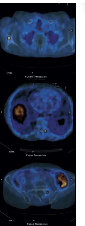
Figures 1 and Figure 2 display typical clinical cases of PCa patients who received PSMA-11 PET/CT in the course of diagnostic work-up evaluation.
Figure 1.
72-year-old patient treated in September 2013 for PCa with radical prostatectomy and pelvic lymphoadenctomy (Gleason Score 4=4). Biochemical recurrence occurred in March 2016 with PSA 0.73 ng/mL and PSAdt of 4.7 months. PSMA-11 PET/CT identified 2 small lymph nodes. The patient subsequently received SRT, with dedicated boost on PSMA positive findings. PSA levels were not detectable after SRT.
Figure 2. This is the same patient reported in Figure 1. Twelve months after SRT PSA values rapidly
raised up to 1.7 ng/mL (PSAdt, 3 months). Bicalutamide was administrated without partial PSA response. Considering subsequent PSA progression, the patient was treated with Abiraterone acetate and referred to PSMA-11 PET/CT to restage the disease. PET imaging demonstrated disease progression, with 3 bone metastases identified.
1.7 The Theranostic (Therapy and Diagnostic) Approach
Patients with mCRPC have a poor prognosis, and those patients with metastases are expected to survive ≤19 months [21]. As patient disease progresses, quality of life deteriorates, and until recently, few treatment options were available. Several new therapies have shown an improvement in overall survival for patients with mCRPC who have already received chemotherapy with docetaxel [8]. The most used agents include novel hormonal therapy targeting the androgen receptor (enzalutamide or abiraterone), second generation chemotherapy (cabazitaxel), bone targeting alpha particles (radium-223) and immunotherapy (sipuleucel-T and cabozantinib) [8]. Despite these therapies provided promising results in terms of life expectancy or pain palliation, the impact of these new data on clinical practice, treatment sequencing and best care for individual patients is not yet fully established. Over the past several decades, numerous combined diagnostic and therapeutic radioligands (Theranostics) were designed to target receptors on the cancer cell surface. Antibodies (whole or small fragments), small molecules, peptides with affinities to receptors (agonist or antagonist) have demonstrated in vivo efficacy for targeting cancers based on up-regulated antigens or receptor populations. This approach, also called radioligand therapy (RLT), presents several advantages over conventional chemotherapy. The expression of the antigens or special receptors can be identified by a diagnostic probe before exposing patients to therapeutic doses of these agents allowing identification of suitable subjects for therapeutic procedures and preventing unnecessary exposure of the patients to radiation without significant benefit. This approach allows the physician to select only those patients with high expression of the target prior to treatment. Since the unused radioactive materials are excreted from the body, RLTs are generally well tolerated with no significant or generally reversible or manageable side effects as has been demonstrated for 177Lu-DOTATATE treatment in patients with neuroendocrine tumor [57].
PSMA is an ideal target for RLT. It shows significant (100-1000 fold) over-expression on >90% of all PCa cells and its expression is further increased in advanced stages and even more in mCPRC [10,14]. Furthermore, PSMA extracellular domain contains an internalization motif resulting in its
internalization and endosomal recycling which increases the deposition of binding substrates leading to enhanced tumor uptake and retention and subsequent high image quality for diagnostic procedures and a high local dose for therapeutic applications [17]. The agent used so far used in preclinical studies and clinical trials is 177 Lu-DO TA-PSMA commercially known as PSMA-617.
Currently, several phase II-III trials exploring the safety and the efficacy of PSMA-617 (both labeled with 177Lu and 225Ac) in mCRPC patients are on-going across Europe, United States and Australia, demonstrating favorable safety and efficacy of PSMA-617 RLT in mCRPC patients. 177Lu-PSMA-617 RLT might exceed the performance of other third-line systemic therapies reported in the literature. Once completed, these trials will clarify the potential of this new targeted RLT especially considering the toxicity, efficacy and the impact of PSMA-617 RLT on cancer-specific survival and overall survival.
2. SCIENTIFIC STUDY
2.1 Background
The management of patients experiencing PSA failure (namely biochemical persistence [BCP] and biochemical recurrence [BCR]) after radical therapy PCa includes treatments based on the risk of local vs systemic recurrence as identified by clinical nomograms [60]. However, clinical models cannot reliably predict recurrence sites and the extent of metastatic disease. Recent innovation imaging technologies significantly influenced the clinical management of recurrent PCa, leading to novel “imaging-guided” approaches [51]. These techniques may potentially improve patient outcome and avoid or delay the toxicity associated with the use of systemic therapies [61]. The use of 68Ga-PSMA-11-PET/CT revealed favorable sensitivity and specificity profiles when compared with choline or fluciclovine PET imaging techniques. Recently, the use of PSMA PET imaging was introduced by the EAU guidelines, which in its current form suggests performing it in all men with BCR [62]. However, whether such an approach is really cost-effective remains largely unknown. This is a key point since men with recurrent/persistent disease reflect different clinical settings and highly heterogeneous population, carrying different prognosis and different profiles of disease aggressiveness. Therefore, selecting the most suitable candidates for 68Ga-PSMA-11-PET/CT is critical to optimize its use and to spare lower-risk patients by expensive and potentially unnecessary staging procedures.
We hypothesized that the likelihood of a positive 68Ga-PSMA-11-PET/CT can be determined by means of other identifying factors. To test our hypothesis, we developed and internally validated a clinical nomogram able to assess the likelihood of each patient, in different settings of PSA failure after primary treatment, to result in positive 68Ga-PSMA-11- PET/CT. This tool might be important as a guide to clinicians for the best use of PSMA-based PET imaging.
2.2
Materials and Methods
2.2.1 Study Population
The cohort of patients included in this analysis was enrolled through an open-label, single-center, prospective registry study performed at our institution (Prot. PSMA-PROSTATA; Eudract: 2015-004589-27 OsSC). All patients provided signed informed consent prior to PET/CT scan. Among all patients referred to our center for 68Ga-PSMA-11-PET/CT imaging in restaging setting (n = 1128), we included in this analysis only patients who had all clinical, pathological, imaging, and follow-up data available (n = 703). All patients analyzed received 68Ga-PSMA-11-PET/CT at single referral center (Nuclear Medicine, University of Bologna, Italy) between March 2016 and November 2018 due to BCR (n = 627) or BCP (n = 76) after definitive therapy. BCR was defined as two consecutive PSA assays ≥ 0.2 ng/ml in patients treated with radical prostatectomy (RP) as primary treatment with/ without post-operative radiotherapy (RT) and as PSA ≥ 2 ng/ml above the nadir in patients treated with primary RT, in accordance with the Phoenix criteria [63]. BCP was defined as a PSA ≥ 0.1 ng/ml at 6 weeks after RP [64,65].
2.2.2 Definition of Clinical Settings
Different clinical settings of PSA relapse were identified by referring physicians (urologist, radiation oncologist, and clinical oncologist) in a single-center multidisciplinary tumor board (Prostate Cancer Unit) and the overall population was grouped into 4 different categories of PSA recurrence, namely, first-time BCR (group 1, n = 325) defined as patients who had PSA nadir < 0.1 ng/ml after RP and subsequently experienced first recurrence (group 1); PSA recurrence after salvage therapies (group 2, n = 241); BCP (group 3, n = 76); and advanced-stage PCa [19] defined as patients with PSA progression under ADT and candidate to second-line systemic therapies (including taxane based chemotherapy and new androgen receptor targeted agents [ARTA], such as abiraterone, enzalutamide, apalutamide, or darolutamide; group 4, n = 61). No patients included in this analysis ever received any chemotherapy or ARTA.
2.2.3
68Ga-PSMA11 synthesis and PET/CT acquisition
68Ga-PSMA was synthesized in the radiopharmacy of the Service of Nuclear Medicine of the S.Orsola Malpighi University Hospital of Bologna. 68Ga-PSMA-HBED-CC(Glu-NH-CO-NH-Lys-(Ahx)-[[68Ga]Ga(N,N′-bis-[2-hydroxy-5-(carboxyethyl)benzyl]ethylenediamine-N,N′-diacetic acid]) (68Ga-PSMA-11) was prepared using a procedure similar to that described by Eder et al. [66], transferred to a cassette-based automated synthesis module (Modular-Lab, PharmTracer; Eckert & Ziegler, Berlin) based on acetone-free cation exchange post-processing.
68Ga-PSMA-11 PET/CT imaging was performed on either a Discovery MI or a Discovery 710 PET/CT hybrid system (GE Healthcare) after intravenous injection of 68Ga-PSMA–ligand complex at a mean dose of 2 MBq/kg of 68Ga-PSMA-11 (dose range 150 ± 50 MBq). The tomographs were validated for proper quantification and quality of the images recorded. Daily quality control procedures were performed. Patients did not need any preparation before the procedure. An attenuation-corrected whole-body scan (skull base to mid-thighs) in 3D (emission time 2 min per bed position with an axial field of view of 15.6 cm per bed position) starting 60 min after tracer injection was acquired. A low-dose CT scan was performed for attenuation correction of the PET emission data and contrast medium was not used. Emission data were also corrected for scatter and random coincidence events by dedicated software. All 68Ga-PSMA-11 PET/CT images were analyzed with dedicated software (eNTEGRA; GE Healthcare).
2.2.4 Images interpretation
All 68Ga-PSMA-11-PET/CT images were analyzed with dedicated software (eNTEGRA; GE Healthcare) and were independently interpreted with central reading by two nuclear medicine physicians with more than 8 years of experience in PET imaging (Francesco Ceci and Paolo Castellucci). Readers were aware of all clinical data. In case of disagreement or undetermined lesion (event which occurred in 23 cases), a final diagnosis was reached by consensus considering the opinion of a third reader (Stefano Fanti) (majority rule in case of reader disagreement 2:1). Images
were interpreted according to procedure guidelines. In brief, any focal tracer uptake higher than the surrounding background and not associated with physiological uptake was considered suspicious for malignant lesion. PET-positive lesions were classified as suspected local relapse (prostate/prostate bed recurrence), nodal relapse (including both pelvic and extra-pelvic lymph nodes), and skeletal and visceral metastases (including soft tissue metastases and other systemic localization of recurrence). Based on PET/CT results, oligo-metastatic state includes patients with 1 to 3 detected lesions, while multi-metastatic stage includes men with more than 3 lesions.
2.2.5 Outcomes and Statistical Analysis
The primary outcomes of the study were to investigate the independent predictors of positive 68Ga-PSMA-11-PET/CT results in patients with PSA relapse after primary treatment and to develop a clinical nomogram aimed to assess the likelihood of each patient to have a positive scan. Secondary outcome was to assess the most informative nomogram-derived cutoff in order to establish a predictive threshold for 68Ga- PSMA-11-PET/CT positivity.
Statistical analyses first consisted of descriptive statistics on the overall positivity rate of 68Ga-PSMA-11-PET/CT and the positivity rate of pelvic/distant recurrence and oligo-metastatic/ multi-metastatic disease. The population was stratified according to different clinical stages of PSA relapse (groups 1 to 4). Chi-squared test and ANOVA test were used to compare categorical variables and continuous variables, respectively. Second, univariable and multivariable logistic regression analyses were performed to assess independent predictors of positivity on patient-based analysis. We included the following covariates: pathologic International Society of Urological Pathology (ISUP) group (namely, 1 vs 2 vs 3 vs 4 vs 5) in men referred to RP or clinic ISUP group in patients who underwent primary RT, PSA at 68Ga-PSMA-11-PET/CT (≤ 0.2 ng/ml vs 0.21–0.49 ng/ml vs 0.5–0.99 ng/ml vs 1–1.99 ng/ml vs ≥ 2 ng/ml), PSA doubling time (PSAdt; < 3 months vs 3–5.99 months vs 6–11.9 months vs ≥12 months), ongoing ADT (yes vs no), time to recurrence (> 12 months vs ≤ 12 months), and clinical setting of PSA relapse (group 1 vs group 2 vs group 3 vs group 4). Third,
regression-based coefficients were used to develop a nomogram predicting positive 68Ga-PSMA-11-PET/CT result. The predictive accuracy of the nomogram was quantified using the Harrell concordance index (discrimination) and the extent of over- or underestimation of the observed positive PET/CT result was explored graphically in logistic calibration plots. The nomogram was subjected to 200 bootstrap resamples to reduce overfit bias and for internal validation. Fourth, we systematically analyzed specificity, sensitivity, positive predictive value (PPV), and negative predictive value (NPV) for each nomogram-derived cutoff. Receiver operating characteristic (ROC) analysis and the area under curve (AUC) analyses were used to assess the predictive accuracy of each nomogram’s derived cutoff and to identify the most informative value among them in predicting a positive scan. Moreover, we implemented with decision curve analysis (DCA), in order to quantify the nomogram’s clinical outcome on daily clinical practice [24]. DCA investigates the theoretical relationship between the threshold probability of positive PET/CT and the relative value of false-positive and false-negative findings to assess the net benefit of the predictive multivariable model. Finally, to investigate which ideal patients at different stages of PSA relapse are best suited for referral to 68Ga-PSMA-11- PET/CT, dedicated multivariate logistic regression models were performed to predict positive scan in each group of PSA relapse (namely, group 1, group 2, group 3, and group 4). All statistical tests were performed using the R statistical package (R Foundation for Statistical Computing, Vienna, Austria) with a 2-sided significance level set at p < 0.05.
2.3
Results
2.3.1 Overall Population
Table 1 depicts the baseline characteristics of patients included in the study (n = 703). Median PSA at 68Ga-PSMA-11-PET/ CT and median PSAdt were 0.7 ng/ml (interquartile range [IQR] 0.4–1.3) and 6 months (3.5–9.6), respectively. Out of 703 individuals, 116 (16.5%) were receiving ADT at the time of PET/CT. Median time from primary therapy to PSA recurrence was 27 months (IQR 11.1– 54.3). Overall, 325 (46.2%), 241 (34.3%), 76 (10.8%), and 61 (8.7%) patients were referred to 68Ga-PSMA-11-PET/CT due to first-time BCR (group 1), PSA recurrence after salvage therapies (group 2), BCP (group 3), and advanced-stage PCa with PSA recurrence prior to second-line systemic therapy (group 4), respectively. The overall positivity rate of 68Ga-PSMA-11-PET/CT was 51.2% (95% confidence interval [CI] 46.8–71.3%).
29
Table 1
Population Characteristics (n= 703)
Characteristics Mean±SD Median (IQR)
Age (years) 67.6±6.8 68 (63-73)
Initial PSA (ng/mL) 13.0±16.9 8.7 (6.3-12.6) PSA at PET/CT (ng/mL) 1.3± 3.0 0.7 (0.4-1.3) PSA doubling
time (months) 8.0 ± 7.5 6 (3.5-9.6) PSA Velocity (ng/mL/yr) 1.5± 4.4 0.7 (0.3-1.5) Time to BCR* (months) 38.5± 36.7 27.5 (11.1-54.3) Frequency % <T3a 257/703 36.5% ≥T3a 446/703 63.5% N1 127/703 18.0% R1 285/703 40.5%
ISUP Grade Group < 4 419/703 59.6%
ISUP Grade Group ≥ 4 284/703 40.4%
Primary Therapy Frequency %
RP 148/703 21.0%
RP + LND 536/703 76.3%
Primary Radiotherapy 19/703 2.7%
Adjuvant Treatment Frequency %
Adjuvant Radiotherapy 182/703 25.9%
Adjuvant ADT 126/703 17.9%
Therapies during BCR Frequency %
Salvage Radiotherapy 182/703 25.9%
Salvage LND 19/703 2.7%
ADT naïve 420/703 59.7%
ADT at the time of the scan 116/703 16.5%
Clinical setting of PSA
Relapse Frequency %
First-time BCR (Group-1) 3sd25/703 46.2% PSA recurrence after salvage
therapy (Group-2) 241/703 34.3%
BCP (Group-3) 76/703 10.8%
PSA progression before second line systemic therapies (Group-4)
2.3.2 Clinical Nomogram
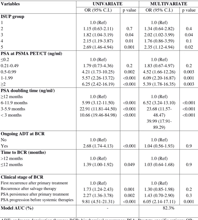
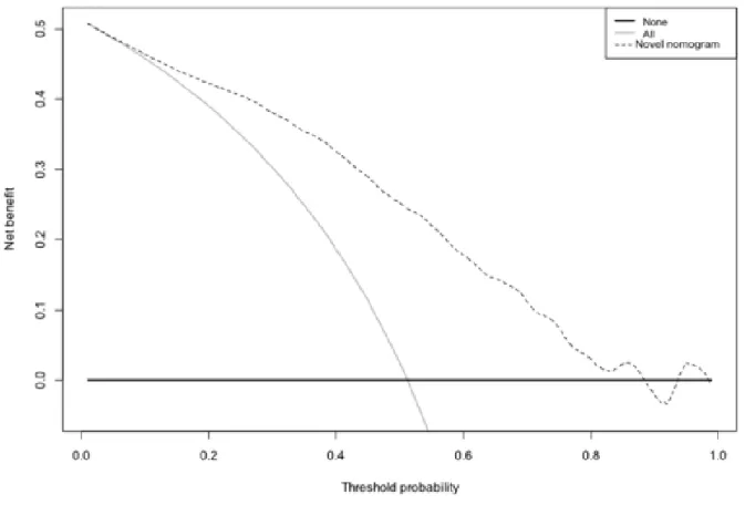
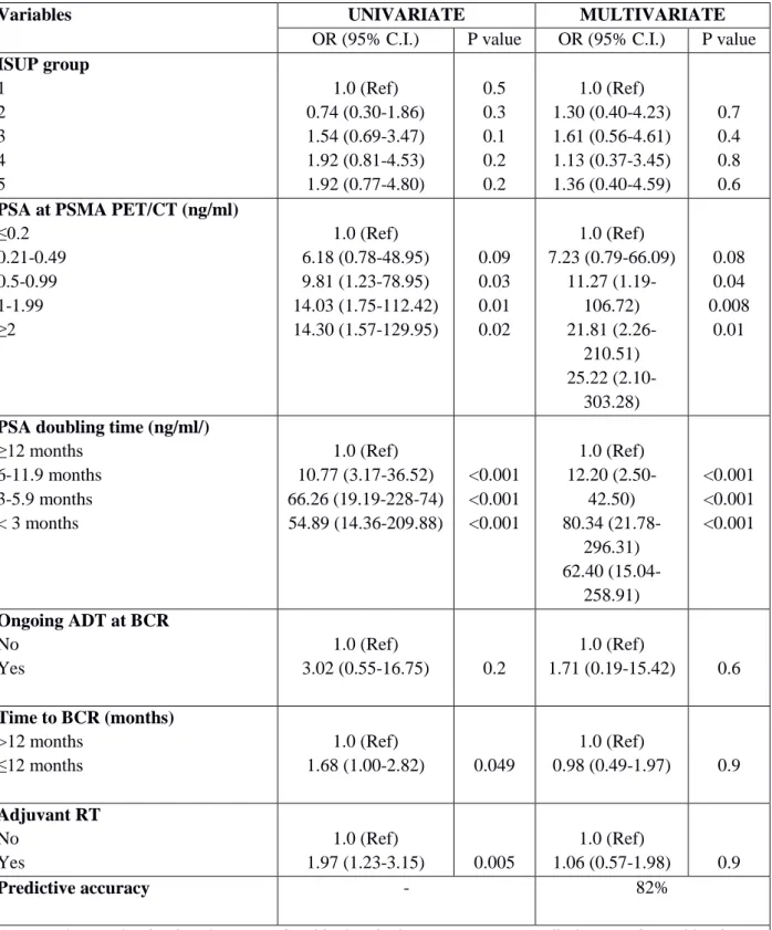
Multivariable regression analysis revealed that ISUP groups 3 and 5 (all p ≤ 0.04), PSA ≥ 0.5 ng/ml (all p ≤0.003), PSAdt ≤6 months (all p ≤ 0.001), and the presence of a PSA progression before second-line treatments (group 4) were independent predicting factors of positive imaging (Table 2; all p ≤ 0.04). Multivariable derived coefficients were used to develop a novel nomogram to predict positive 68Ga-PSMA-11-PET/CT result (Fig. 1). After bootstrap correction, the discrimination accuracy of the model was 82% (AUC = 0.82; 95%CI = 0.79–0.85; Figure 2). The calibration plot of predicted probabilities against observed positive 68Ga-PSMA-11- PET/CT indicated good concordance (Fig. 3). Furthermore, nomogram-derived predicted probabilities of positive 68Ga-PSMA-11-PET/CT are categorized into each nomogram’s derived cutoff. The numbers of patients having negative 68Ga-PSMA-11-PET/CT and those with positive 68Ga-68Ga-PSMA-11-PET/CT as well as sensitivity, specificity, PPV, NPV, and accuracy are depicted, for each cutoff value, in Table 3. At ROC analysis, the best cutoff value to reliably predict positive 68Ga-PSMA-11-PET/CT was 40% (AUC = 0.76; 95%CI = 0.72 – 0.79; Figure 4). Accordingly, assuming a nomogram cutoff of 40%, 282 out of 703 patients (40.1%) might have avoided unnecessary 68Ga-PSMA-11-PET/ CT scanning, while on the other hand, 68Ga-PSMA-11- PET/CT would be missed in 55 patients (15.3%). The sensitivity, specificity, and NPV associated with 40% as cutoff were 84.7%, 66.2%, and 80.5%, respectively (Table 3). Finally, in DCA, the nomogram revealed clinical net benefit when the threshold probability of positive 68Ga-PSMA-11-PET/CT is > 10% (Fig. 5). With a nomogram-derived probability threshold ≥ 40%, the use of the nomogram would result in a net benefit gain of 13. This net benefit, when compared with the scenario of treating none, would be 33. The net benefit of 33, considering the nomogram-derived probability threshold ≥ 40%, might be interpreted in terms that use of the model leads to the equivalent of a net 33 true-positive results per 100 patients without increasing the number of false-positive results (Figure 5).
Table 2 - Univariate and multivariate logistic regression to predict positive findings.
Variables UNIVARIATE MULTIVARIATE
OR (95% C.I.) p value OR (95% C.I.) p value
ISUP group 1 2 3 4 5 1.0 (Ref) 1.15 (0.63-2.11) 1.82 (1.04-3.19) 2.15 (1.19-3.87) 2.69 (1.46-4.94) 0.7 0.04 0.01 0.001 1.0 (Ref) 1.34 (0.64-2.82) 2.02 (1.02-3.99) 1.76 (0.86-3.59) 2.35 (1.12-4.94) 0.4 0.04 0.1 0.02
PSA at PSMA PET/CT (ng/ml)
≤0.2 0.21-0.49 0.5-0.99 1-1.99 ≥2 1.0 (Ref) 1.79 (0.73-4.36) 4.21 (1.73-10.25) 5.57 (2.26-13.72) 6.25 (2.42-16.19) 0.2 0.002 <0.001 <0.001 1.0 (Ref) 1.83 (0.67-4.97) 4.52 (1.66-12.26) 6.09 (2.20-16.87) 5.39 (1.78-16.35) 0.2 0.003 0.001 0.003
PSA doubling time (ng/ml/)
≥12 months 6-11.9 months 3-5.9 months < 3 months 1.0 (Ref) 5.99 (3.12-11.50) 22.91 (11.81-44.50) 10.66 (19.46-84.98) <0.001 <0.001 <0.001 1.0 (Ref) 6.52 (3.24-13.10) 23.68 (11.57-48.47) 39.99 (17.91-89.29) <0.001 <0.001 <0.001 Ongoing ADT at BCR No Yes 1.0 (Ref) 2.68 (1.74-4.13) <0.001 1.0 (Ref) 1.04 (0.56-1.93) 0.9 Time to BCR (months) >12 months ≤12 months 1.0 (Ref) 1.39 (1.00-1.92) 0.049 1.0 (Ref) 1.03 (0.64-1.68) 0.9 Clinical stage of BCR
First recurrence after primary treatment Recurrence after salvage therapy PSA persistence after primary treatment PSA progression before systemic therapies
1.0 (Ref) 1.73 (1.24-2.43) 2.27 (1.36-3.78) 9.81 (4.51-21.31) 0.001 0.002 <0.001 1.0 (Ref) 1.30 (0.85-1.98) 1.43 (0.70-2.90) 6.05 (2.14-17.11) 0.2 0.3 0.001 Model AUC (%) - 82.3%
ADT: androgen deprivation therapy; BCR: biochemical recurrence; PSA: Prostate-specific antigen; OR: odd ratio; C.I.: Confidence Interval; Ref: reference. ISUP: International Society of Urological Pathology; AUC: Area Under the Curve
Figure 1 - Nomogram predicting the likelihood of positive 68Ga-PSMA-11-PET/CT for patients with
different clinical settings of PSA failure after radical treatment for prostate cancer. Instructions: locate, for instance, the patient’s PSA on the PSA group axis. Draw a line straight upward to the point axis to determine how many points toward the probability of positive PSMA PET/CT the patient receives for his PSA group. Repeat the process for each additional variable. Sum the points for each predictor. Locate the final sum on the total-point axis. Draw a line straight down to find the patient’s probability of having positive PSMA PET/CT
Figure 2 - Receiver-operator characteristic (ROC) and area under curve (AUC) of the nomogram in
overall population to predict positive 68Ga-PSMA-11-PET/CT results (AUC=0.82; 95%CI=0.79-0.85).
Figure 3 - Nomogram calibration plot. The predicted probability of the multivariable model is shown
on the x-axis, and the observed proportion of men with positive 68Ga-PSMA-11-PET/CT is shown on the y-axis. The 45° line indicates location of the ideal nomogram, in which the predicted probability and the observed proportion of men with positive 68Ga-PSMA-11-PET/CT are identical. Broken line indicates actual nomogram performance.
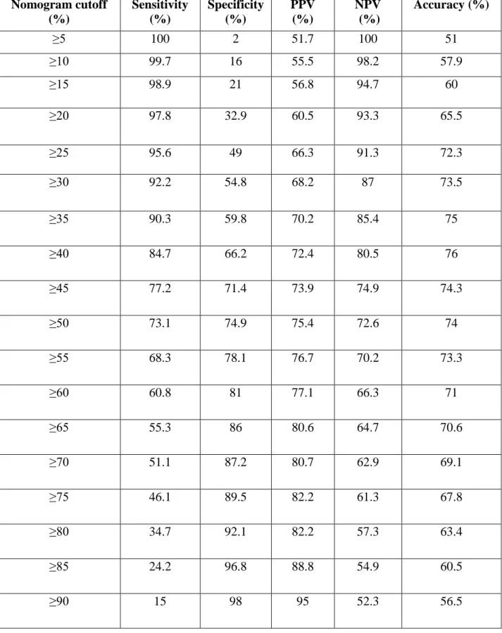
Table 3 – Nomogram’s cut-off Nomogram cutoff (%) Sensitivity (%) Specificity (%) PPV (%) NPV (%) Accuracy (%) ≥5 100 2 51.7 100 51 ≥10 99.7 16 55.5 98.2 57.9 ≥15 98.9 21 56.8 94.7 60 ≥20 97.8 32.9 60.5 93.3 65.5 ≥25 95.6 49 66.3 91.3 72.3 ≥30 92.2 54.8 68.2 87 73.5 ≥35 90.3 59.8 70.2 85.4 75 ≥40 84.7 66.2 72.4 80.5 76 ≥45 77.2 71.4 73.9 74.9 74.3 ≥50 73.1 74.9 75.4 72.6 74 ≥55 68.3 78.1 76.7 70.2 73.3 ≥60 60.8 81 77.1 66.3 71 ≥65 55.3 86 80.6 64.7 70.6 ≥70 51.1 87.2 80.7 62.9 69.1 ≥75 46.1 89.5 82.2 61.3 67.8 ≥80 34.7 92.1 82.2 57.3 63.4 ≥85 24.2 96.8 88.8 54.9 60.5 ≥90 15 98 95 52.3 56.5
Table 3 - Performance characteristics of various nomogram's cut-off for discriminating between
patients with positive 68Ga-PSMA-11-PET/CT and those with negative 68Ga-PSMA-11-PET/CT and the quantified number of avoidable 68Ga-PSMA-11-PET/CT vs. the number of potentially missed patients with positive 68Ga-PSMA-11-PET/CT findings.
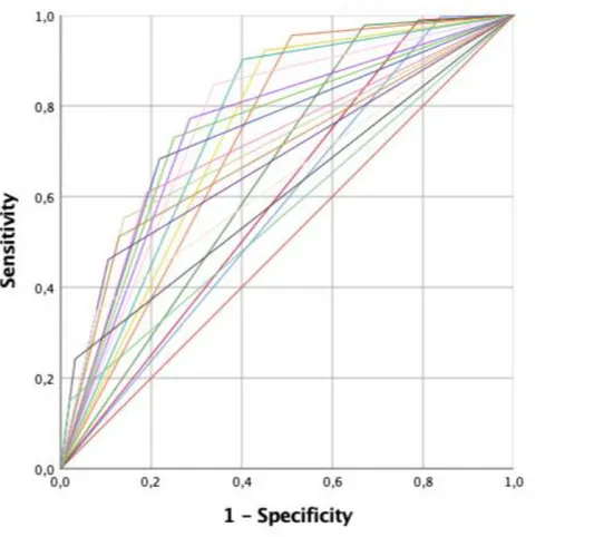
Figure 4 – Receiver-operator characteristic (ROC) and area under curve (AUC) of each nomogram
derived cut-off to predict positive 68Ga-PSMA-11-PET/CT results. The best nomogram’s cut-off to predict positive scan was 40% (AUC=0.76; 95%CI=0.72-0.79).
Figure 5 – Decision curve analysis (DCA) demonstrating the net benefit associated with use of the
nomogram for the prediction of men with positive 68Ga-PSMA-11-PET/CT. The net benefit is represented by the gap between the continuous line and the dotted line.
2.3.3 Subgroup Analyses
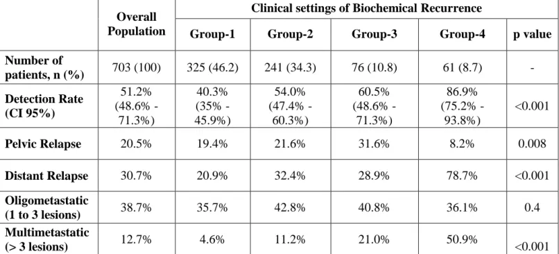
Biochemical features in each subgroup are reported in Table 4. The detection rate of 68Ga-PSMA-11-PET/CT was significantly different between the four groups: 40.3%, 54%, 60.5%, and 86.9% in groups 1, 2, 3, and 4, respectively (Table 5; p < 0.001). However, we observed no significant differences in its detection rate when applied to oligo-metastatic disease (≤ 3 lesions), even considering different clinical settings.
In patients with first-time BCR (group 1), only PSA ≥ 0.5 ng/ml (all p ≤ 0.01) and PSAdt ≤ 6 months (all p < 0.001) were independent predictors of positive 68Ga-PSMA-11-PET/CT at multivariate analysis (Table 6). In patients with PSA recurrence after salvage therapy (group 2), only PSAdt ≤6 months (all p ≤ 0.001) was independent predictor of positive PET/CT at multivariate analysis (Table 7). In BCP (group 3), only PSA (p=0.04) and ISUP group 5 (p =0.04) were independent predictors of
positive scan Table 8). Finally, in patients with advanced PCa with PSA relapse prior to second-line systemic therapy (group 4), only PSAdt (p = 0.01) was independent predictor of positive PET/CT (Table 9).
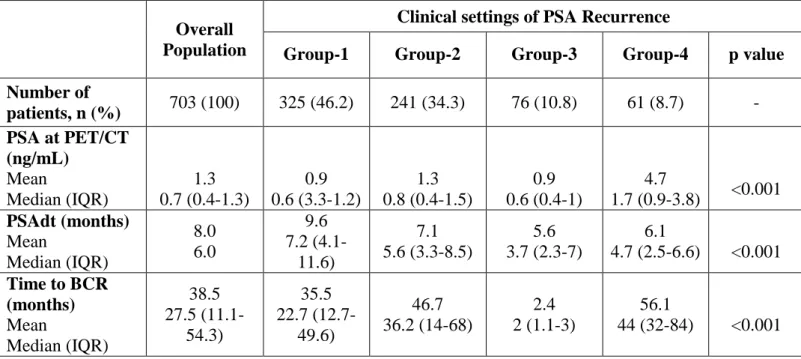
Table 4 - Biochemical features in overall population and between the different clinical settings of
biochemical recurrence.
Overall Population
Clinical settings of PSA Recurrence
Group-1 Group-2 Group-3 Group-4 p value
Number of patients, n (%) 703 (100) 325 (46.2) 241 (34.3) 76 (10.8) 61 (8.7) - PSA at PET/CT (ng/mL) Mean Median (IQR) 1.3 0.7 (0.4-1.3) 0.9 0.6 (3.3-1.2) 1.3 0.8 (0.4-1.5) 0.9 0.6 (0.4-1) 4.7 1.7 (0.9-3.8) <0.001 PSAdt (months) Mean Median (IQR) 8.0 6.0 9.6 7.2 (4.1-11.6) 7.1 5.6 (3.3-8.5) 5.6 3.7 (2.3-7) 6.1 4.7 (2.5-6.6) <0.001 Time to BCR (months) Mean Median (IQR) 38.5 27.5 (11.1-54.3) 35.5 22.7 (12.7-49.6) 46.7 36.2 (14-68) 2.4 2 (1.1-3) 56.1 44 (32-84) <0.001
Table 5 - 68Ga-PSMA-11 PET/CT performance (positivity rate) in overall population and between
the different clinical settings of biochemical recurrence.
Overall Population
Clinical settings of Biochemical Recurrence
Group-1 Group-2 Group-3 Group-4 p value
Number of patients, n (%) 703 (100) 325 (46.2) 241 (34.3) 76 (10.8) 61 (8.7) - Detection Rate (CI 95%) 51.2% (48.6% -71.3%) 40.3% (35% -45.9%) 54.0% (47.4% -60.3%) 60.5% (48.6% -71.3%) 86.9% (75.2% -93.8%) <0.001 Pelvic Relapse 20.5% 19.4% 21.6% 31.6% 8.2% 0.008 Distant Relapse 30.7% 20.9% 32.4% 28.9% 78.7% <0.001 Oligometastatic (1 to 3 lesions) 38.7% 35.7% 42.8% 40.8% 36.1% 0.4 Multimetastatic (> 3 lesions) 12.7% 4.6% 11.2% 21.0% 50.9% <0.001
Group-1: first-time BCR; Group-2: PSA recurrence after salvage therapy; Group-3: BCP; Group-4: PSA progression before second line systemic therapies.
Table 6 - Univariate and multivariate logistic regression to predict positive findings at
68Ga-PSMA-11-PET/CT in Group-1 (n=325).
Variables UNIVARIATE MULTIVARIATE
OR (95% C.I.) P value OR (95% C.I.) P value
ISUP group 1 2 3 4 5 1.0 (Ref) 0.74 (0.30-1.86) 1.54 (0.69-3.47) 1.92 (0.81-4.53) 1.92 (0.77-4.80) 0.5 0.3 0.1 0.2 0.2 1.0 (Ref) 1.30 (0.40-4.23) 1.61 (0.56-4.61) 1.13 (0.37-3.45) 1.36 (0.40-4.59) 0.7 0.4 0.8 0.6
PSA at PSMA PET/CT (ng/ml)
≤0.2 0.21-0.49 0.5-0.99 1-1.99 ≥2 1.0 (Ref) 6.18 (0.78-48.95) 9.81 (1.23-78.95) 14.03 (1.75-112.42) 14.30 (1.57-129.95) 0.09 0.03 0.01 0.02 1.0 (Ref) 7.23 (0.79-66.09) 11.27 (1.19-106.72) 21.81 (2.26-210.51) 25.22 (2.10-303.28) 0.08 0.04 0.008 0.01
PSA doubling time (ng/ml/)
≥12 months 6-11.9 months 3-5.9 months < 3 months 1.0 (Ref) 10.77 (3.17-36.52) 66.26 (19.19-228-74) 54.89 (14.36-209.88) <0.001 <0.001 <0.001 1.0 (Ref) 12.20 (2.50-42.50) 80.34 (21.78-296.31) 62.40 (15.04-258.91) <0.001 <0.001 <0.001 Ongoing ADT at BCR No Yes 1.0 (Ref) 3.02 (0.55-16.75) 0.2 1.0 (Ref) 1.71 (0.19-15.42) 0.6 Time to BCR (months) >12 months ≤12 months 1.0 (Ref) 1.68 (1.00-2.82) 0.049 1.0 (Ref) 0.98 (0.49-1.97) 0.9 Adjuvant RT No Yes 1.0 (Ref) 1.97 (1.23-3.15) 0.005 1.0 (Ref) 1.06 (0.57-1.98) 0.9 Predictive accuracy - 82%
ADT: androgen deprivation therapy; BCR: biochemical recurrence; RT: Radiotherapy; OR: odd ratio; C.I.: Confidence Interval; Ref: reference. ISUP: International Society of Urological Pathology