R E S E A R C H
Open Access
Immunocompromised patients with acute
respiratory distress syndrome: secondary
analysis of the LUNG SAFE database
Andrea Cortegiani
1*, Fabiana Madotto
2, Cesare Gregoretti
1, Giacomo Bellani
3,4, John G. Laffey
5,6,7, Tai Pham
6,7,
Frank Van Haren
8,9, Antonino Giarratano
1, Massimo Antonelli
10, Antonio Pesenti
11,12, Giacomo Grasselli
11,
LUNG SAFE Investigators and the ESICM Trials Group
Abstract
Background: The aim of this study was to describe data on epidemiology, ventilatory management, and outcome
of acute respiratory distress syndrome (ARDS) in immunocompromised patients.
Methods: We performed a post hoc analysis on the cohort of immunocompromised patients enrolled in the Large
Observational Study to Understand the Global Impact of Severe Acute Respiratory Failure (LUNG SAFE) study.
The LUNG SAFE study was an international, prospective study including hypoxemic patients in 459 ICUs from 50
countries across 5 continents.
Results: Of 2813 patients with ARDS, 584 (20.8%) were immunocompromised, 38.9% of whom had an unspecified
cause. Pneumonia, nonpulmonary sepsis, and noncardiogenic shock were their most common risk factors for ARDS.
Hospital mortality was higher in immunocompromised than in immunocompetent patients (52.4% vs 36.2%;
p < 0.0001), despite similar severity of ARDS. Decisions regarding limiting life-sustaining measures were significantly
more frequent in immunocompromised patients (27.1% vs 18.6%; p < 0.0001). Use of noninvasive ventilation (NIV) as
first-line treatment was higher in immunocompromised patients (20.9% vs 15.9%; p = 0.0048), and immunodeficiency
remained independently associated with the use of NIV after adjustment for confounders. Forty-eight percent of the
patients treated with NIV were intubated, and their mortality was not different from that of the patients invasively
ventilated ab initio.
Conclusions: Immunosuppression is frequent in patients with ARDS, and infections are the main risk factors for ARDS
in these immunocompromised patients. Their management differs from that of immunocompetent patients,
particularly the greater use of NIV as first-line ventilation strategy. Compared with immunocompetent subjects, they
have higher mortality regardless of ARDS severity as well as a higher frequency of limitation of life-sustaining measures.
Nonetheless, nearly half of these patients survive to hospital discharge.
Trial registration: ClinicalTrials.gov,
NCT02010073
. Registered on 12 December 2013.
Keywords: Acute respiratory failure, ARDS, Immunocompromised patients, Mechanical ventilation, Noninvasive ventilation
* Correspondence:[email protected]
1
Department of Biopathology and Medical Biotechnologies (DIBIMED), Section of Anesthesia, Analgesia, Intensive Care and Emergency, Policlinico Paolo Giaccone, University of Palermo, Via del vespro 129, 90127 Palermo, Italy
Full list of author information is available at the end of the article
© The Author(s). 2018 Open Access This article is distributed under the terms of the Creative Commons Attribution 4.0 International License (http://creativecommons.org/licenses/by/4.0/), which permits unrestricted use, distribution, and reproduction in any medium, provided you give appropriate credit to the original author(s) and the source, provide a link to the Creative Commons license, and indicate if changes were made. The Creative Commons Public Domain Dedication waiver (http://creativecommons.org/publicdomain/zero/1.0/) applies to the data made available in this article, unless otherwise stated.
Background
In recent decades, significant advances in the
manage-ment of immunocompromised patients have led to
im-proved survival rates [
1
–
3
]. Hence, intensive care unit
(ICU) admission and invasive life-sustaining treatments
are offered with increasing frequency to these patients
[
3
,
4
]. However, several studies show that the prognosis
of critically ill patients with active malignancies or
im-munodeficiency remains poor, especially when the cause
of ICU admission is acute respiratory distress syndrome
(ARDS) requiring invasive mechanical ventilation (IMV)
[
5
–
11
]. Data about incidence, causes, management, and
outcomes of ARDS in immunocompromised patients are
scarce. The best ventilatory strategy in this population is
still uncertain, and available literature data on the role of
noninvasive ventilation (NIV) are conflicting [
12
–
22
].
Recently, researchers in the Large Observational Study
to Understand the Global Impact of Severe Acute
Respiratory Failure (LUNG SAFE study) investigated the
incidence, management, and clinical outcomes in
pa-tients with acute hypoxemic respiratory failure (AHRF)
requiring ventilatory support, with a specific focus on
ARDS [
23
]. This aim of this post hoc subgroup analysis
was to describe the epidemiology, clinical characteristics,
ventilatory management (with particular attention to the
use of NIV), and outcomes of ARDS in the subset of
pa-tients with clinically significant immunodeficiency.
Methods
LUNG SAFE: patients, study design, and data collection
LUNG-SAFE was an international, multicenter,
pro-spective observational cohort study conducted in a 459
ICUs worldwide. During 4 consecutive weeks in the
win-ter of 2014 (February–March 2014 in the Northern
Hemisphere and June–August 2014 in the Southern
Hemisphere),
participating
ICUs
enrolled
patients
undergoing IMV or NIV. Participating ICUs obtained
ethics committee approval and either patient consent or
waiver of consent as per local guidelines. National
coordi-nators, site investigators, and endorsing societies are listed
in Additional file
1
. Exclusion criteria were age < 16 years
or lack of informed consent when required. Patients were
screened daily for AHRF, defined as follows: (1) ratio of
partial pressure of arterial oxygen to fraction of
in-spired oxygen (PaO
2/FiO
2)
≤ 300 mmHg while
receiv-ing IMV or NIV with positive end-expiratory pressure
(PEEP)
≥ 5 cmH
2O and (2) new radiological pulmonary
parenchymal abnormalities. In patients with AHRF, a more
detailed set of data was collected to determine whether
they met the Berlin definition criteria for ARDS. Data on
comorbidities, etiology of AHRF, and risk factors for ARDS
were recorded. Data on arterial blood gases, ventilatory
support, use of adjunctive therapies (e.g., prone
position-ing, extracorporeal membrane oxygenation, neuromuscular
blockade), severity of ARDS, and other organ involvement
by modified nonpulmonary Sequential Organ Failure
Assessment (SOFA) score [
24
] were collected on selected
days. The following clinical endpoints were assessed: ICU
and hospital survival, censored at 90 days after enrollment;
duration of mechanical ventilation; changes in ARDS
se-verity; and decision to withhold or withdraw life-sustaining
therapies. A full description of the methods of the LUNG
SAFE study, including the full study protocol, case report
form (CRF), sample size, and quality control, can be found
in the original study paper [
23
].
Immunocompromised patient cohort and definitions
We defined
“immunocompromised” patients as all
pa-tients with at least one of the following conditions listed
in the LUNG SAFE CRF: (1) immunosuppression (defined
as viral immunosuppression, neoplastic disease,
immuno-suppressive drugs including steroids, chemotherapy, or
congenital immunosuppression), (2) active hematologic
malignancy (i.e., still requiring treatment), and (3) active
neoplasm (i.e., a neoplasm that has not been resected, still
requires treatment, or with metastasis). Patients without
these conditions were classified as
“controls.” For the
pur-poses of this analysis, the study population was restricted
to the subset of patients fulfilling ARDS criteria on day 1
or 2 following the onset of AHRF.
In regard to management, patients were subdivided in
three ventilation subgroups: (1) IMV, defined as patients
invasively ventilated from day 1, independently of the
type of support received after the eventual extubation;
(2) NIV, defined as patients treated exclusively with NIV
from day 1 to study exit (i.e., ICU discharge or death);
and (3) NIV failure, defined as patients initially treated
with NIV and subsequently intubated during the study
period. The term
NIV encompassed all forms of NIV
modes and interfaces (including continuous positive
airway pressure). ARDS severity was assessed from the
first to the second day from ARDS onset, according to
the Berlin definition criteria: mild (PaO
2/FiO
2201–
300 mmHg), moderate (PaO
2/FiO
2101–200 mmHg),
and severe (PaO
2/FiO
2≤ 100 mmHg). Changes in ARDS
severity were evaluated in patients staying in the ICU for
at least 2 days, and they were classified into four
categories: (1) no change, (2) worsening (shift to a more
severe category), (3) improvement (shift to a less severe
category), and (4) resolution. Duration of invasive
venti-lation was computed as the number of days that the
pa-tient required IMV up to day 28. Survival was evaluated
at ICU and hospital discharge or at day 90, whichever
event occurred first.
Statistical analysis
Continuous variables were expressed as mean (SD) or
median (IQR), and categorical variables were presented
as count and percent. No assumptions were made for
missing data, which were rare [
23
]. To assess differences
between groups, we used Student’s t test or the
Wilcoxon rank-sum test (according normality
distribu-tion of data) for continuous variables and the
χ
2or
Fisher’s exact test (according sample size) for proportions.
We used analysis of variance or the Kruskal-Wallis test
(as appropriate) and the
χ
2test (or Fisher’s exact test) to
assess differences among the NIV, NIV failure, and IMV
groups. The Bonferroni correction was applied to
deter-mine significance in the setting of multiple comparisons.
To evaluate factors associated with the use of NIV, we
applied a multivariable logistic regression model, and the
independent predictors (demographic characteristics,
co-morbidities, ARDS risk factors, and clinical parameters
concerning the illness severity of ARDS onset) were
identified through a stepwise regression approach. This
approach combines forward and backward selection
methods (combined with a significance level of 0.05 for
both entry and retention) in an iterative procedure to
select predictors in the final multivariable model. This
approach was also applied to identify factors associated
with hospital mortality in immunocompromised
pa-tients. In this case, the stepwise approach also evaluated
as possible predictors ventilator setting variables
mea-sured at ARDS onset.
Survival analysis was performed according to the
Kaplan-Meier method. We assumed that patients
dis-charged alive from the hospital before 90 days were alive
on day 90. The log-rank test was used to compare
sur-vival curves among groups.
All
p values were two-sided, and values less than 0.05
were considered significant. Statistical analyses were
car-ried out with R version 3.3.3 (R Project for Statistical
Computing;
https://www.r-project.org/
) and SAS version
9.4 software (SAS Institute, Cary, NC, USA).
Results
Baseline patient characteristics
A total of 459 ICUs from 50 countries enrolled patients
in the LUNG SAFE study. Among 12,906 mechanically
ventilated patients, 4499 had AHRF, and of these, 2813
fulfilled the Berlin criteria for ARDS on day 1 or 2.
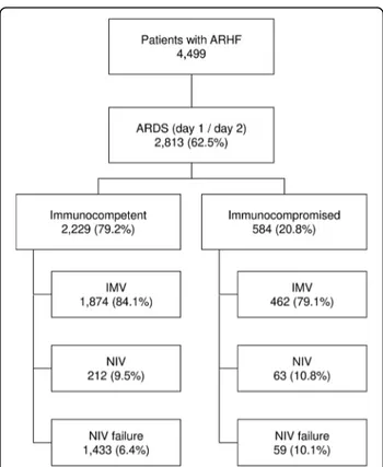
Among ARDS patients, 584 (20.8%) were
immunocom-promised (Fig.
1
). Of these, 232 (39.7%) had an active
neoplasm and 138 (23.6%) had a hematologic
malig-nancy, whereas the causes of immunosuppression were
not specified in 38.7%. Table
1
shows baseline
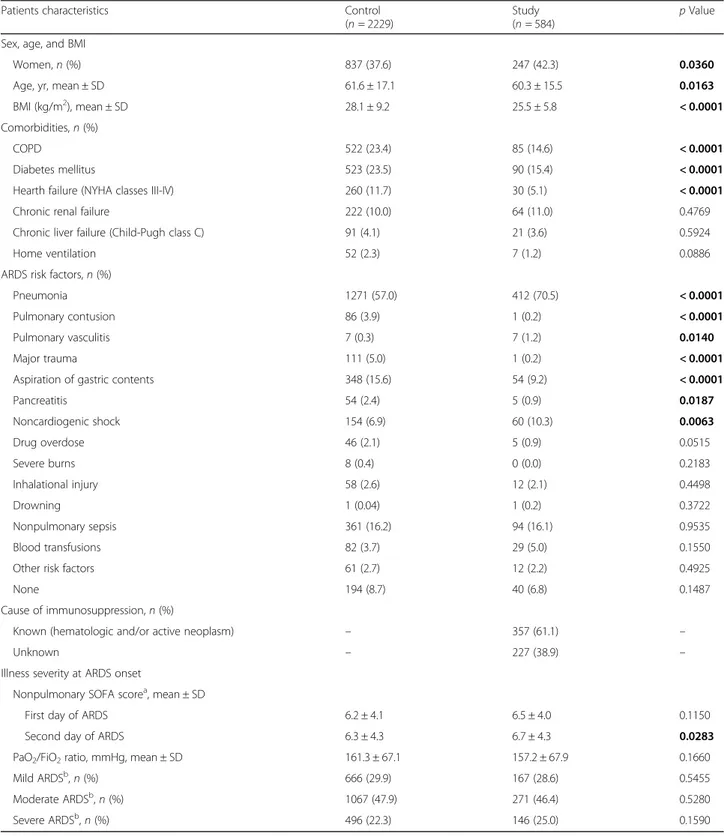
characteris-tics of immunocompromised and control patients.
Im-munocompromised subjects were younger than controls
(60.3 vs 61.6 years;
p = 0.0163) and had a lower body mass
index (BMI) (25.5 ± 5.8 vs 28.1 ± 9.2 kg/m
2;
p < 0.0001).
They had a lower prevalence of chronic obstructive
pulmonary disease, diabetes mellitus, and heart failure
(New York Heart Association classes III–IV) and a higher
incidence of pneumonia, pulmonary vasculitis, and
non-cardiogenic shock. Among immunocompromised
pa-tients, 28.6% had mild, 46.4% moderate, and 25.0% severe
ARDS, and the most common risk factors for ARDS were
pneumonia (70.5%), nonpulmonary sepsis (16.1%), and
noncardiogenic shock (10.3%). Nonpulmonary SOFA
score at day 1 and ARDS severity were similar between
immunocompromised and controls. Additional file
2
com-pares comorbidities, ARDS severity, and nonpulmonary
SOFA score in the three ventilation subgroups (IMV, NIV,
and NIV failure) among immunocompromised patients.
Mean patient age in the NIV subgroup was older than in
the other two subgroups, and the difference between NIV
and IMV was statistically significant (65.2 vs 59.7 years;
p = 0.0045). Comorbidities and PaO
2/FiO
2were not
differ-ent among the subgroups. There was a marked difference
in the mean nonpulmonary SOFA score, which was
sig-nificantly higher in IMV than in NIV (7.0 ± 3.9 vs 3.7 ± 3.1;
p < 0.0001) and NIV failure subgroups (7.0 ± 3.9 vs
5.3 ± 3.6;
p = 0.0023).
Fig. 1 Flow diagram of the study. Flow diagram showing the distribution of patients included in this analysis dataset, according to ventilation subgroup. AHRF acute hypoxemic respiratory failure, ARDS Acute respiratory distress syndrome, IMV Patients invasively ventilated from day 1, independently of the type of support received after the eventual extubation, NIV Patients treated exclusively with noninvasive ventilation, from day 1 to study exit, independently of outcome, NIV failure Patients initially treated with noninvasive ventilation and subsequently intubated during the study period
Table 1 Patients characteristics in immunocompetent (Control) and immunocompromised (Study) groups
Patients characteristics Control
(n = 2229)
Study (n = 584)
p Value Sex, age, and BMI
Women, n (%) 837 (37.6) 247 (42.3) 0.0360
Age, yr, mean ± SD 61.6 ± 17.1 60.3 ± 15.5 0.0163
BMI (kg/m2), mean ± SD 28.1 ± 9.2 25.5 ± 5.8 < 0.0001
Comorbidities, n (%)
COPD 522 (23.4) 85 (14.6) < 0.0001
Diabetes mellitus 523 (23.5) 90 (15.4) < 0.0001
Hearth failure (NYHA classes III-IV) 260 (11.7) 30 (5.1) < 0.0001
Chronic renal failure 222 (10.0) 64 (11.0) 0.4769
Chronic liver failure (Child-Pugh class C) 91 (4.1) 21 (3.6) 0.5924
Home ventilation 52 (2.3) 7 (1.2) 0.0886
ARDS risk factors, n (%)
Pneumonia 1271 (57.0) 412 (70.5) < 0.0001
Pulmonary contusion 86 (3.9) 1 (0.2) < 0.0001
Pulmonary vasculitis 7 (0.3) 7 (1.2) 0.0140
Major trauma 111 (5.0) 1 (0.2) < 0.0001
Aspiration of gastric contents 348 (15.6) 54 (9.2) < 0.0001
Pancreatitis 54 (2.4) 5 (0.9) 0.0187 Noncardiogenic shock 154 (6.9) 60 (10.3) 0.0063 Drug overdose 46 (2.1) 5 (0.9) 0.0515 Severe burns 8 (0.4) 0 (0.0) 0.2183 Inhalational injury 58 (2.6) 12 (2.1) 0.4498 Drowning 1 (0.04) 1 (0.2) 0.3722 Nonpulmonary sepsis 361 (16.2) 94 (16.1) 0.9535 Blood transfusions 82 (3.7) 29 (5.0) 0.1550
Other risk factors 61 (2.7) 12 (2.2) 0.4925
None 194 (8.7) 40 (6.8) 0.1487
Cause of immunosuppression, n (%)
Known (hematologic and/or active neoplasm) – 357 (61.1) –
Unknown – 227 (38.9) –
Illness severity at ARDS onset
Nonpulmonary SOFA scorea, mean ± SD
First day of ARDS 6.2 ± 4.1 6.5 ± 4.0 0.1150
Second day of ARDS 6.3 ± 4.3 6.7 ± 4.3 0.0283
PaO2/FiO2ratio, mmHg, mean ± SD 161.3 ± 67.1 157.2 ± 67.9 0.1660
Mild ARDSb, n (%) 666 (29.9) 167 (28.6) 0.5455
Moderate ARDSb, n (%) 1067 (47.9) 271 (46.4) 0.5280
Severe ARDSb, n (%) 496 (22.3) 146 (25.0) 0.1590
Abbreviations: BMI Body mass index, ARDS Acute respiratory distress syndrome, COPD Chronic obstructive pulmonary disease, NYHA New York Heart Association, SOFA Sequential Organ Failure Assessment, PaO2/FiO2Ratio of partial pressure of arterial oxygen to fraction of inspired oxygen
Note: Bold p values represent a statistically significant difference between the two groups
a
Nonpulmonary SOFA score adjusted for missing values
b
Type of ventilatory support, ventilator setting, and
adjunctive measure/therapies
Figure
1
summarizes the type of ventilatory support in
enrolled patients. On day 1 of ARDS, IMV was the most
frequent type of ventilatory approach in both groups;
however, NIV use as first-line treatment was significantly
more frequent in immunocompromised than in
im-munocompetent patients (20.9% vs 15.9%;
p = 0.0044).
The proportion of patients remaining on NIV from day
1 to study exit (NIV subgroup) was similar, whereas the
incidence of NIV failure was significantly higher in
im-munocompromised patients (10.1% vs 6.4%;
p = 0.0021).
A multivariable logistic regression model revealed that,
adjusting on confounders, immunodeficiency was
inde-pendently associated with the use of NIV (OR, 1.567;
95% CI, 1.217–2.017; p = 0.0005). Other factors
associ-ated with NIV are shown in Additional file
3
.
Additional file
4
compares the ventilator settings on
the first day of ARDS between immunocompromised
and control patients: FiO
2and respiratory rate and PEEP
were statistically significantly higher in
immunocom-promised patients, but the difference for PEEP was not
clinically relevant. There was no difference in tidal
volume, peak and plateau pressures, and the proportion
of patients with spontaneous ventilation. No significant
dif-ferences were observed in adjunctive therapies, except for
a significantly higher use of continuous neuromuscular
blocking agents in the immunocompromised group (22.6%
vs 18.5%;
p = 0.0266) (Additional file
5
). Additional file
6
describes ventilator settings in immunocompromised and
immunocompetent (control) patients, stratified by the type
of ventilator support (IMV, NIV, NIV failure).
Clinical endpoints
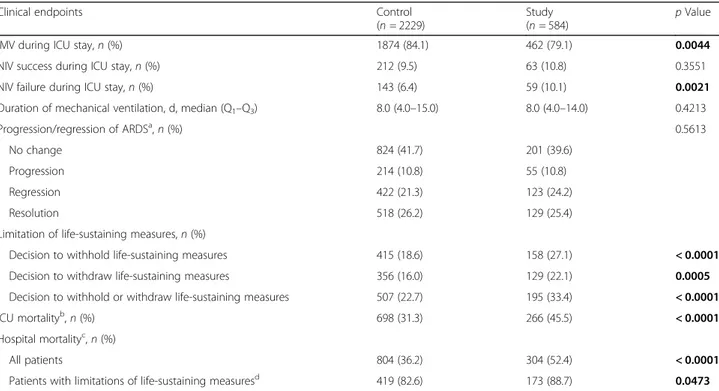
Table
2
compares selected clinical endpoints in
im-munocompromised and control patients. Hospital
mor-tality and ICU mormor-tality were significantly higher in
immunocompromised subjects (respectively, 52.4% vs
36.2%,
p < 0.0001; and 45.5% vs 31.3, p < 0.0001),
whereas there was no difference in duration of
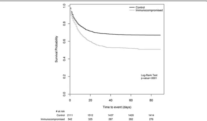
mechan-ical ventilation and changes in ARDS severity. Survival
curves for hospital (or 90-day) mortality are shown in
Fig.
2
. The decision to withhold and/or withdraw
life-sustaining measures was significantly more frequent
in immunocompromised patients (Table
2
). The same
clinical endpoints were also analyzed in the cohort of
Table 2 Clinical endpoints in immunocompetent (Control) and immunocompromised (Study) patients
Clinical endpoints Control
(n = 2229)
Study (n = 584)
p Value
IMV during ICU stay, n (%) 1874 (84.1) 462 (79.1) 0.0044
NIV success during ICU stay, n (%) 212 (9.5) 63 (10.8) 0.3551
NIV failure during ICU stay, n (%) 143 (6.4) 59 (10.1) 0.0021
Duration of mechanical ventilation, d, median (Q1–Q3) 8.0 (4.0–15.0) 8.0 (4.0–14.0) 0.4213
Progression/regression of ARDSa, n (%) 0.5613
No change 824 (41.7) 201 (39.6)
Progression 214 (10.8) 55 (10.8)
Regression 422 (21.3) 123 (24.2)
Resolution 518 (26.2) 129 (25.4)
Limitation of life-sustaining measures, n (%)
Decision to withhold life-sustaining measures 415 (18.6) 158 (27.1) < 0.0001
Decision to withdraw life-sustaining measures 356 (16.0) 129 (22.1) 0.0005
Decision to withhold or withdraw life-sustaining measures 507 (22.7) 195 (33.4) < 0.0001
ICU mortalityb, n (%) 698 (31.3) 266 (45.5) < 0.0001
Hospital mortalityc, n (%)
All patients 804 (36.2) 304 (52.4) < 0.0001
Patients with limitations of life-sustaining measuresd 419 (82.6) 173 (88.7) 0.0473
Abbreviations: ARDS Acute respiratory distress syndrome, IMV Invasive mechanical ventilation, ICU Intensive care unit, NIV Noninvasive mechanical ventilation, Q1First quartile, Q3Third quartile
a
Change in ARDS severity (according Berlin definition) was not evaluable for 327 pients (251 immunocompetent and 76 immunocompromised patients)
b
Mortality is defined as mortality at ICU discharge or at the 90th day in the ICU after onset of acute hypoxemic respiratory failure, whichever event occurred first
c
Mortality is defined as mortality at hospital discharge or at the 90th day in the hospital after onset of acute hypoxemic respiratory failure, whichever event occurred first
d
Mortality assessed on patients with a decision to withhold or withdraw life-sustaining measures Note: Bold p values represent a statistically significant difference between the two groups
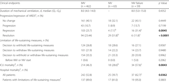
immunocompromised patients according to the
ventila-tion subgroup (Table
3
). Duration of mechanical
ventila-tion and decisions of limitaventila-tion (both withholding and
withdrawal) of life-sustaining measures were not different
among the subgroups. ICU mortality was significantly
lower in NIV patients than in the IMV (28.6% vs
46.3%;
p = 0.0078) and NIV failure (28.6% vs 57.6%;
p = 0.0012) subgroups. Of the NIV patients who died, 68%
had a limitation of life-sustaining measures. The incidence
of NIV failure was 48%. ICU and hospital mortality of
pa-tients with NIV failure were significantly higher than those
of patients managed exclusively with NIV (respectively,
57.6% vs 28.6%,
p = 0.012; and 62.7 vs 39.7%, p = 0.011),
whereas they did not differ from those of IMV patients.
Survival curves for hospital (or 90-day) mortality of
im-munocompromised patients stratified by ARDS severity
and by ventilation subgroups are shown in Additional files
7
and
8
, respectively. In a multivariable logistic regression
model, factors independently associated with hospital
mor-tality in immunocompromised patients were higher
non-pulmonary SOFA score (OR, 1.079; 95% CI, 1.026–1.134;
p = 0.0032), higher peak inspiratory pressure level
(OR, 1.028; 95% CI, 1.007–1.051; p = 0.0097), lower PaO
2/
FiO
2ratio (OR, 0.995; 95% CI, 0.992–0.998; p = 0.0022),
lower degree of improvement in PaO
2/FiO
2ratio between
day 1 and day 2 of ARDS (OR, 0.996; 95% CI, 0.993–0.999;
p = 0.0058), and lower BMI (OR, 0.944; 95% CI, 0.91–0.98;
p = 0.0023) (Additional file
9
). According to the investigators’
clinical judgment, in immunocompromised patients,
the most common main factor leading to death in
ICU was respiratory failure (51.5%), followed by
car-diovascular failure. In contrast, carcar-diovascular failure
was the most common factor in the control group
(Additional file
10
).
Additional file
11
describes patient characteristics and
clinical endpoints in immunocompetent (control)
pa-tients according to type of ventilator support. Because
the lack of a precise definition of the cause of
immuno-suppression in a relevant proportion of patients may
affect the strength of our findings, we compared baseline
patient characteristics and clinical outcomes in patients
with a
“known” cause of immunosuppression (i.e., those
with active hematologic malignancy or active neoplasm)
and in patients with an unspecified (“unknown”) cause
of immunosuppression (i.e., those indicated in the CRF
with the generic term
immunosuppression). The results of
this analysis are reported in Additional file
12
. Briefly,
pa-tients with an unspecified cause of immunosuppression
were significantly younger (55.7 ± 15.6 vs 63.1 ± 14.8 years;
p < 0.0001) and had lower hospital mortality (41.6% vs
59.3%;
p < 0.0001) and fewer limitations of life-sustaining
measures (27.3% vs 37.3%;
p = 0.013).
Fig. 2 Kaplan-Meier curve for hospital survival. Mortality was defined as mortality at hospital discharge or at 90 days after onset of acute hypoxemic respiratory failure, whichever event occurred first. We assumed that patients discharged alive from the hospital before 90 days were alive on day 90. Note: The number of patients at risk reported at the bottom of the figure is referred to as the end of the corresponding day
Discussion
The main findings of this analysis can be summarized as
follows: immunosuppression is frequent in ARDS
pa-tients; causes of ARDS in immunocompromised patients
are mainly related to infection; immunocompromised
patients are more likely to receive NIV as first-line
venti-latory treatment; and the outcome of ARDS is worse and
limitation of life-sustaining measures is more frequent in
immunocompromised patients. Among the 2813 ARDS
patients included in the LUNG SAFE study, one-fifth
were immunocompromised, and 62.7% of them had an
active malignancy. In line with recent literature [
18
,
21
],
a diagnosis of active malignancy is considered as a cause of
immunosuppression, owing to the negative effects on
im-mune function of anticancer treatments and of the
malig-nancy itself. All other causes of immune deficiency were
identified by the generic variable
“immunosuppression.”
To the best of our knowledge, this was the first
prospect-ive, multicenter study conducted on a large cohort of
immunocompromised patients with a diagnosis of ARDS
according to Berlin definition criteria [
25
], whereas
previous
studies
relied
on
the
definition
of
the
American-European Consensus Conference on ARDS [
26
].
As expected, ARDS in immunocompromised patients was
associated mainly with an infectious cause. Pneumonia,
noncardiogenic shock, and pulmonary vasculitis were
sig-nificantly more frequent in immunocompromised patients,
whereas other risk factors for ARDS were more frequently
represented in the control group.
Immunocompromised subjects had a
significantly
higher ICU and hospital mortality, despite similar ARDS
severity and nonpulmonary SOFA score. Another
subana-lysis of the same database confirmed that active neoplasm,
hematologic malignancies, and immunosuppression are
independently associated with increased mortality [
27
]. In
addition, the higher frequency of limitation of
life-sustain-ing measures (probably as a result of perceived futility)
may have contributed to the increased mortality of
im-munocompromised patients [
27
]. This is in line with the
results of Laffey et al., who found that
immunosuppres-sion and cancer were among the factors associated with
increased likelihood of limitation of life-sustaining
therapies [
27
].
Hospital mortality of our patients was lower than that
(64%) reported by Azoulay et al. in a large retrospective
analysis of 1004 patients with cancer and ARDS
admit-ted to the ICU over a period of 21 years [
28
]. In that
study, mortality did drop from 89% in the 1990
–1995
period to 52% in the 2006–2011 period, matching the
mortality rate of our population.
Table 3 Clinical endpoints in immunocompromised (Study) patients according to ventilation subgroup
Clinical endpoints IMV
(n = 462) NIV (n = 63) NIV failure (n = 59) p Value
Duration of mechanical ventilation, d, median (Q1–Q3) 8.0 (4.0–14.0) – 8.0 (5.0–15.0) 0.4352
Progression/regression of ARDSa, n (%)
No change 161 (40.1) 18 (32.1) 22 (43.1) 0.4449
Progression 43 (10.7) 5 (8.9) 7 (13.7) 0.7199
Regression 103 (25.7) 4 (7.1)b 16 (31.4)c 0.0045
Resolution 94 (23.44) 29 (51.8)b 6 (11.8)c < 0.0001
Limitation of life-sustaining measures, n (%)
Decision to withhold life-sustaining measures 124 (26.8) 18 (28.6) 16 (27.1) 0.9587
Decision to withdraw life-sustaining measures 101 (21.9) 14 (22.2) 14 (23.7) 0.9480
Decision to withhold or withdraw life-sustaining measures 154 (33.3) 21 (33.3) 20 (33.9) 0.9962
Before IMV or NIV start 1 (0.6) 0 (0.0) 1 (5.0) 0.2062
ICU mortalityd, n (%) 214 (46.3) 18 (28.6)b 34 (57.6)c 0.0043
Hospital mortalitye, n (%)
All patients 242 (52.8) 25 (39.7) 37 (62.7)c 0.0362
Patients with limitations of life-sustaining measuresf 137 (89.0) 17 (81.0) 19 (95.0) 0.3803
Abbreviations: ARDS Acute respiratory distress syndrome, IMV Patients invasively ventilated from day 1, independently of the type of support received after the eventual extubation, NIV Patients treated exclusively with noninvasive ventilation, from day 1 to study exit, independently of outcome, NIV failure Patients initially treated with noninvasive ventilation and subsequently intubated during the study period, Q1First quartile, Q3Third quartile
a
Change in ARDS severity (according Berlin definition) was not evaluable for 76 immunocompromised patients (61 IMV, 7 NIV, and 8 NIV failure)
b
Statistically significant different from the IMV group
c
Statistically significant different from the NIV group
d
Mortality is defined as mortality at ICU discharge or at the 90th day in ICU after onset of acute hypoxemic respiratory failure, whichever event occurred first
e
Mortality is defined as mortality at hospital discharge or at the 90th day in hospital, after onset of acute hypoxemic respiratory failure, whichever event occurred first
f
Mortality assessed in patients with a decision to withhold or withdraw life-sustaining measures Note: Bold p values represent a statistically significant difference among the three groups
The actual advantage of ICU admission of
immuno-compromised patients remains debated [
3
,
4
,
10
,
29
,
30
].
Our results demonstrate that almost 50% of
immuno-compromised patients with ARDS survive to hospital
discharge, and this may support the decision to offer
them at least an
“ICU trial” [
31
]. Interestingly, our data
show that once immunocompromised patients are
intu-bated and invasively ventilated, they are managed very
similarly to the general ARDS population with regard to
ventilator settings and use of adjunctive therapies. As an
example, the use of advanced
“rescue” treatments (such
as prone positioning and even extracorporeal membrane
oxygenation) was similar between immunocompromised
and control patients.
Two important questions on the optimal ventilatory
management of AHRF in immunocompromised patients
remain unanswered. First, is NIV the optimal first-line
ventilatory support? Two randomized controlled trials
conducted almost 20 years ago showed that NIV,
com-pared with standard oxygen therapy, significantly
re-duces the rate of intubation and mortality [
16
,
17
], but
these findings have not been confirmed in more recent
studies. A recent randomized trial on 374
immunocom-promised patients with AHRF did not find any benefit of
NIV over standard oxygen therapy [
18
]. Similarly, a post
hoc analysis on immunocompromised patients enrolled
in a large randomized trial comparing different
noninva-sive oxygenation strategies showed that first-line NIV
was associated with the highest risk of intubation and
mortality compared with standard oxygen and high-flow
nasal cannula (HFNC) oxygen [
21
]. In our study, 20.9%
of immunocompromised patients received NIV as
first-line ventilatory approach compared with 15.9% of
controls, and the multivariable analysis revealed that
im-munodeficiency was independently associated with the
use of NIV. This frequency of use of NIV equals exactly
that reported by Gristina in a large population of
pa-tients with hematologic malignancies admitted to ICU in
the years 2002–2006 [
26
]. In Azoulay’s study, the global
rate of NIV application was 38.6%, but it decreased over
the years, dropping to 26% in the period 2006–2011
[
28
]. In our study, patients treated exclusively with NIV
had significantly lower mortality than patients requiring
in-vasive ventilation. Of note, the majority of NIV patients
who died had a decision of limitation of life-sustaining
mea-sures. Importantly, whereas ARDS severity was not different
among ventilation subgroups, NIV patients had a markedly
lower nonpulmonary SOFA score (Additional file
2
:
Table S1), indicating that patients with more severe organ
failures were more frequently treated with invasive
ventila-tory support. However, the multivariable regression
ana-lysis did not identify the need for invasive ventilation as an
independent predictor of death. This represents a major
difference with Azoulay’s study, where IMV (especially
after failure of NIV) was a strong predictor of poor
out-come [
28
]. Again, this can be explained at least in part by
considering that Azoulay’s study included patients from
the year 1990, when the techniques of mechanical
ventila-tion were completely different and protective ventilaventila-tion
was certainly not the standard of care.
The impact of NIV failure on patients’ outcomes is the
second important issue. We observed a significantly
higher incidence of NIV failure in immunocompromised
patients than in controls, in keeping with the
observa-tion of Thille that a diagnosis of active cancer is
inde-pendently associated with NIV failure [
32
]. In our study,
in 48% of immunocompromised patients initially treated
with NIV, NIV failed, and they had a significantly worse
mortality than patients successfully managed with NIV,
as previously reported by Bellani et al. [
23
]. Less
ex-pected was the finding that mortality of NIV failure
pa-tients was not different from that of the papa-tients
managed
ab initio with IMV. However, two factors may
limit the relevance of this observation: the relatively low
number of patients in the NIV failure subgroup and the
lack of information on the actual duration (i.e., in hours
rather than days) of the NIV period before intubation,
which would be important to know in the light of
litera-ture data showing that delaying endotracheal intubation
after a prolonged NIV trial may negatively impact
pa-tient survival [
19
,
20
]. In line with our data, Demoule et
al. recently observed a progressive reduction of the
im-pact of NIV failure on mortality in a large population of
AHRF patients also including immunocompromised
subjects [
33
]. Taken together, these data probably
sug-gest that better patient selection, earlier recognition of
failure, and improvement in ventilation techniques may
have contributed to limit the impact of NIV failure on
mortality in recent years.
Limitations
The present study has several limitations. First, it is a
post hoc analysis of a prospective multicenter
observa-tional trial, and unknown confounders associated with
the subgroup analysis may bias the results. Although the
data were prospectively collected from a high number of
centers from 50 countries, different approaches to
clin-ical decisions from different centers (e.g., decisions on
withholding or withdrawing of life-sustaining measures)
may have influenced the outcomes. Second, the criteria
used to define the immunocompromised cohort were
quite heterogeneous. It was impossible to stratify the
pa-tients according to the prognosis of baseline disease and
to the severity of immune deficiency. Indeed, causes of
immunosuppression, other than malignancies, were not
specified in nearly 40% of the whole cohort of
immuno-compromised patients. Moreover, in patients with
can-cer, no information was available on the type of cancan-cer,
its staging, and the nature and timing of anticancer
treatments. This lack of information, related to the
LUNG SAFE original CRF, should be considered a major
limitation because the outcome of immunocompromised
patients is strictly dependent on the type of underlying
diseases and associated therapeutic approach. All these
factors may limit the generalizability of our findings.
Third, to limit the burden on investigators, data were
collected once daily only, and information on the actual
hours of duration of ventilatory treatments was not
available. This is particularly relevant for patients treated
with NIV because the precise duration of NIV before
the eventual intubation might be important to
under-stand the impact of NIV failure on outcome [
24
]. Fourth,
no information were provided on the type of interface
used for NIV, a factor that can affect the outcome of
NIV [
34
]. Moreover, patients treated with HFNC oxygen
therapy were excluded from the LUNG SAFE study
be-cause they did not fulfill criteria for ARDS. Fifth, the
LUNG SAFE study was conducted in a very large
num-ber of ICUs with different experience in the treatment of
ARDS. This may be particularly relevant for
immuno-compromised patients, who may have better outcomes if
treated in highly experienced, dedicated units [
35
].
Conclusions
Immunocompromised patients represent an important
proportion of ARDS patients in the ICU. Compared with
immunocompetent subjects, they had higher mortality,
regardless of ARDS severity, and a higher frequency
of limitation of life-sustaining measures. Nonetheless,
nearly half of these patients survive to hospital discharge.
They were more likely to receive NIV as the first ventilator
strategy, and those who did not require invasive
ventila-tion had a lower mortality. Mortality of
immunocom-promised patients who failed NIV was not different from
that of patients treated
ab initio with IMV. These data
should be considered in light of the nonspecific criteria
used to define the immunocompromised population and
the potentially heterogeneous approaches to clinical
decision making in the participating centers.
Additional files
Additional file 1:List of LUNG SAFE investigators. Names and affiliations of the LUNG SAFE investigators. (PDF 172 kb)
Additional file 2:Table S1. Patient characteristics of immunocompromised patients according to the type of ventilator support. This table shows patient characteristics, including comorbidities, ARDS risk factors, and illness severity at ARDS onset of immunocompromised patients according to the type of ventilator support. (PDF 74 kb)
Additional file 3:Table S2. Factors associated with the use of noninvasive ventilation. Multivariate logistic regression model describing the factors associated with the use of noninvasive ventilation. (PDF 49 kb)
Additional file 4:Table S3. Ventilator settings during the first day of ARDS in the immunocompetent (Control) and immunocompromised (Study) groups. This table shows ventilator settings during the first day of ARDS in the immunocompetent (Control) and immunocompromised (Study) groups. (PDF 50 kb)
Additional file 5:Table S4. Adjunctive measures/therapies during at least one day during follow-up in immunocompetent and immunocompromised patients. This table shows the proportions of adjunctive measures/therapies during at least one day during follow-up in immunocompetent and immunocompromised patients. (PDF 97 kb)
Additional file 6:Table S6. Ventilator settings during the first day of ARDS in immunocompetent (Control) and immunocompromised (Study) patients, stratified by the type of ventilatory support (IMV, NIV, NIV failure). (PDF 60 kb)
Additional file 7:Figure S1. Kaplan-Meier curve for hospital survival in immunocompromised patients according to ARDS severity. Kaplan-Meier curve for hospital survival in immunocompromised patients according to ARDS severity. Mortality is defined as mortality at hospital discharge or at 90 days after onset of acute hypoxemic respiratory failure, whichever event occurred first. We assumed that patients discharged alive from the hospital before 90 days were alive on day 90. Severity of ARDS was evaluated at the day of onset according to the Berlin definition. Note: The number of patients reported in the bottom of figure is referred to as the end of the corresponding day. (PDF 402 kb)
Additional file 8:Figure S2. This figure shows a Kaplan-Meier curve for hospital survival of immunocompromised patients according to the ventilation subgroup. This figure shows a Kaplan-Meier curve for hospital survival of immunocompromised patients according to the ventilation subgroup. Mortality is defined as mortality at hospital discharge or at 90 days after onset of acute hypoxemic respiratory failure, whichever event occurred first. We assumed that patients discharged alive from the hospital before 90 days were alive on day 90. Type of ventilator support: IMV Patients invasively ventilated from day 1, independently of the type of support received after the eventual extubation; NIV Patients treated exclusively with noninvasive ventilation, from day 1 to study exit, independently of outcome; NIV failure Patients initially treated with noninvasive ventilation and subsequently intubated during the study period. Note: The number of patients reported in the bottom of the figure is referred to as the end of the corresponding day. (PDF 396 kb)
Additional file 9:Table S5. Factors associated with hospital mortality in immunocompromised patients. Multivariate logistic regression model describing the factors associated with hospital mortality in immunocompromised patients. (PDF 49 kb)
Additional file 10:Table S9. The most important factors leading to death in the ICU in immunocompetent and immunocompromised patients. (PDF 44 kb)
Additional file 11:Table S7. Patient characteristics and clinical endpoints of immunocompetent patients, according to the type of ventilatory support. (PDF 88 kb)
Additional file 12:Table S8. Patients’ characteristics and clinical endpoints of immunocompromised (study) patients, according to the cause of immunosuppression (known, unknown). (PDF 79 kb) Acknowledgements
LUNG SAFE investigators and ESICM Trial Group email address: [email protected] Study coordination: Guy M. Francois (European Society of Intensive Care Medicine, Brussels, Belgium).
Data revision and management: Francesca Rabboni (University of Milan-Bicocca, Monza, Italy), Fabiana Madotto (University of Milan-Bicocca, Monza, Italy), Sara Conti (University of Milan-Bicocca, Monza, Italy). LUNG SAFE executive and steering committees: John G. Laffey, Giacomo Bellani, Tai Pham, Eddy Fan, Antonio Pesenti, Laurent Brochard, Andres Esteban, Luciano Gattinoni, Frank van Haren, Anders Larsson, Daniel F. McAuley, Marco Ranieri, Gordon Rubenfeld, B. Taylor Thompson, Hermann Wrigge, Arthur S. Slutsky.
National coordinators: Argentina: Fernando Rios
Belgium: T. Sottiaux, P. Depuydt Bolivia: Fredy S. Lora
Brazil: Luciano Cesar Azevedo Canada: Eddy Fan
Chile: Guillermo Bugedo China: Haibo Qiu Colombia: Marcos Gonzalez Costa Rica: Juan Silesky Czech Republic: Vladimir Cerny Denmark: Jonas Nielsen Ecuador: Manuel Jibaja France: Tài Pham Germany: Hermann Wrigge Greece: Dimitrios Matamis Guatemala: Jorge Luis Ranero India: Pravin Amin
Iran: S. M. Hashemian Ireland: Kevin Clarkson Italy: Giacomo Bellani Japan: Kiyoyasu Kurahashi Mexico: Asisclo Villagomez Morocco: Amine Ali Zeggwagh The Netherlands: Leo M. Heunks Norway: Jon Henrik Laake The Philippines: Jose Emmanuel Palo Portugal: Antero do Vale Fernandes Romania: Dorel Sandesc
Saudi Arabia: Yaasen Arabi Serbia: Vesna Bumbasierevic Spain: Nicolas Nin, Jose A. Lorente Sweden: Anders Larsson Switzerland: Lise Piquilloud Tunisia: Fekri Abroug
United Kingdom: Daniel F. McAuley, Lia McNamee Uruguay: Javier Hurtado
United States: Ed Bajwa Venezuela: Gabriel Démpair Site investigators by country:
Albania: University Medical Center of Tirana“Mother Theresa” (Tirana): Hektor Sula, Lordian Nunci; University Hospital Shefqet Ndroqi (Tirana): Alma Cani Argentina: Clinica De Especialidades (Villa Maria): Alan Zazu; Hospital Dr. Julio C. Perrando (Resistencia): Christian Dellera, Carolina S. Insaurralde; Sanatorio Las Lomas (San Isidro, Buenos Aires): Risso V. Alejandro; Sanatorio De La Trinidad San Isidro (San Isidro): Julio Daldin, Mauricio Vinzio; Hospital Español De Mendoza (Godoy Cruz-Mendoza): Ruben O. Fernandez; Hospital Del Centenario (Rosario): Luis P. Cardonnet, Lisandro R. Bettini; San Antonio (Gualeguay [Entre Rios]): Mariano Carboni Bisso, Emilio M. Osman; Cemic (Buenos Aires): Mariano G. Setten, Pablo Lovazzano; Hospital Universitrario Austral (Pilar): Javier Alvarez, Veronica Villar; Hospital Por + Salud (Pami) Dr. Cesar Milstein (Buenos Aires): Norberto C. Pozo, Nicolas Grubissich; Sanatorio Anchorena (Buenos Aires): Gustavo A. Plotnikow, Daniela N. Vasquez; Sanatorio De La Trinidad Mitre (Buenos Aires): Santiago Ilutovich, Norberto Tiribelli; Hospital Luis Lagomaggiore (Mendoza): Ariel Chena, Carlos A Pellegrini; H.I.G.A. San Martín (La Plata): María G. Saenz, Elisa Estenssoro; Hospital Misericordia (Cordoba): Matias Brizuela, Hernan Gianinetto; Sanatorio Juncal (Temperley): Pablo E. Gomez, Valeria I. Cerrato; Hospital D. F. Santojanni (Buenos Aires): Marco G. Bezzi, Silvina A. Borello; Hospital Alejandro Posadas (Buenos Aires): Flavia A. Loiacono, Adriana M. Fernandez
Australia: St. Vincent’s Hospital, Sydney (Darlinghurst): Serena Knowles, Claire Reynolds; St. George Public Hospital (Kogarah): Deborah M. Inskip, Jennene J. Miller; Westmead Hospital (Westmead): Jing Kong, Christina Whitehead; Flinders Medical Centre (Bedford Park, South Australia): Shailesh Bihari; John Hunter Hospital (Newcastle): Aylin Seven, Amanda Krstevski; Canberra Hospital (Garran): Helen J. Rodgers, Rebecca T. Millar; Calvary Mater Newcastle (Waratah): Toni E. Mckenna, Irene M. Bailey; Cabrini Hospital (Melbourne): Gabrielle C. Hanlon; Liverpool Hospital (Liverpool): Anders Aneman, Joan M. Lynch; Coffs Harbour Health Campus (Coffs Harbour): Raman Azad, John Neal; Sir Charles Gairdner Hospital (Nedlands): Paul W. Woods, Brigit L. Roberts; Concord Hospital (Concord): Mark R. Kol, Helen S. Wong
Austria:General Hospital of Vienna/Medical University of Vienna (Vienna): Katharina C. Riss, Thomas Staudinger
Belgium: Cliniques universitaires St-Luc, Université Catholique de Louvain (UCL) (Brussels): Xavier Wittebole, Caroline Berghe; CHU Dinant-Godinne (Yvoir): Pierre A. Bulpa, Alain M. Dive; AZ Sint-Augustinus Veurne (Veurne): Rik Verstraete, Herve Lebbinck; Ghent University Hospital (Ghent): Pieter Depuydt, Joris Vermassen; University Hospitals Leuven (Leuven): Philippe Meersseman, Helga Ceunen
Brazil: Hospital Renascentista (Pouso Alegre): Jonas I. Rosa, Daniel O. Beraldo; Vitoria Apart Hospital (Serra): Claudio Piras, Adenilton M. Rampinelli; Hospital Das Clinicas (São Paulo): Antonio P. Nassar Jr.; Hospital Geral do Grajaù (São Paulo): Sergio Mataloun, Marcelo Moock; Evangelical Hospital (Cachoeiro De Itapemirim/Espírito Santo): Marlus M. Thompson, Claudio H. Gonçalves; Hospital Moinhos De Vento (Porto Alegre): Ana Carolina P. Antônio, Aline Ascoli; Hospital Alvorada Taguatinga (Taguatinga): Rodrigo S. Biondi, Danielle C. Fontenele; Complexo Hospitalar Mngabeira Tarcisio Burity (Joao Pessoa): Danielle Nobrega, Vanessa M. Sales
Brunei Darussalam: Raja Isteri Pengiran Anak Saleha (Ripas) Hospital (Bandar Seri Begawan): Dr. Suresh Shindhe, Dr. Dayangku Hajah Maizatul Aiman binti Pengiran Haji Ismail
Canada: Medical-Surgical ICU of St. Michael’s Hospital (Toronto): John Laffey, Francois Beloncle; St. Joseph’s Health Centre (Toronto): Kyle G. Davies, Rob Cirone; Sunnybrook Health Sciences Center (Toronto): Venika Manoharan, Mehvish Ismail; Toronto Western Hospital (Toronto): Ewan C. Goligher, Mandeep Jassal; Medical Surgical ICU of the Toronto General Hospital (Toronto): Erin Nishikawa, Areej Javeed; Cardiovascular ICU of St. Michael’s Hospital (Toronto): Gerard Curley, Nuttapol Rittayamai; Cardiovascular ICU of the Toronto General Hospital (Toronto): Matteo Parotto, Niall D. Ferguson; Mount Sinai Hospital (Toronto): Sangeeta Mehta, Jenny Knoll; Trauma-Neuro ICU of St. Michael’s Hospital (Toronto): Antoine Pronovost, Sergio Canestrini Chile: Hospital Clínico Pontificia Universidad Católica De Chile (Santiago): Alejandro R. Bruhn, Patricio H. Garcia; Hospital Militar De Santiago (Santiago): Felipe A. Aliaga, Pamela A. Farías; Clinica Davila (Santiago): Jacob S. Yumha; Hospital Guillermo Grant Benavente (Concepcion): Claudia A. Ortiz, Javier E. Salas; Clinica Las Lilas (Santiago): Alejandro A. Saez, Luis D. Vega; Hospital Naval Almirante Nef (Viña Del Mar): Eduardo F. Labarca, Felipe T. Martinez; Hospital Luis Tisné Brousse (Penanolen): Nicolás G. Carreño, Pilar Lora China: The Second Affiliated Hospital of Harbin Medical University (Harbin): Haitao Liu; Nanjing Zhong-Da Hospital, Southeast University (Nanjing): Haibo Qiu, Ling Liu; The First Affiliated Hospital of Anhui Medical University (Hefei): Rui/Tang, Xiaoming Luo; Peking University People’s Hospital (Beijing): Youzhong An, Huiying Zhao; Fourth Affiliated Hospital of Harbin Medical University (Harbin): Yan Gao, Zhe Zhai; Nanjing Jiangbei People’s Hospital Affiliated to Medical School of Southeast University (Nanjing): Zheng L. Ye, Wei Wang; The First Affiliated Hospital of Dalian Medical Unvercity (Dalian): Wenwen Li, Qingdong Li; Subei People’s Hospital of Jiangsu Province (Yanghzou): Ruiqiang Zheng; Jinling Hospital (Nanjing): Wenkui Yu, Juanhong Shen; Urumqi General Hospital (Urumqi): Xinyu Li; Intensive Care Unit, First Affiliated Hospital of Wannan Medical College, Yijishan Hospital (Wuhu): Tao Yu, Weihua Lu; Sichuan Provincial People’s Hospital (Chengdu): Ya Q. Wu, Xiao B. Huang; Hainan Province People’s Hospital (Haikou): Zhenyang He; People’s Hospital of Jiangxi Province (Nanchang): Yuanhua Lu; Qilu Hospital of Shandong University (Jinan): Hui Han, Fan Zhang; Zhejiang Provincial People’s Hospital (Hangzhou): Renhua Sun; The First Affiliated Hospital of Bengbu Medical College (Bengbu, Anhui): Hua X. Wang, Shu H. Qin; Nanjing Municipal Government Hospital (Nanjing): Bao H. Zhu, Jun Zhao; The First Hospital of Lanzhou University (Lanzhou): Jian Liu, Bin Li; The First Affiliated Hospital of Chongqing University of Medical Science (Chongqing): Jing L. Liu, Fa C. Zhou; Xuzhou Central Hospital, Jiangsu Province, China (Xuzhou): Qiong J. Li, Xing Y. Zhang; The First People’s Hospital of Foshan (Foshan): Zhou Li-Xin, Qiang Xin-Hua; The First Affiliated Hospital of Guangxi Medical University (Nanning): Liangyan Jiang; Renji Hospital, Shanghai Jiao Tong University School of Medicine (Shanghai): Yuan N. Gao, Xian Y. Zhao; First Hospital of Shanxi Medical University (Taiyuan): Yuan Y. Li, Xiao L. Li; Shandong Provincial Hospital (Jinan): Chunting Wang, Qingchun Yao; Fujian Provincial Hospital (Fuzhou): Rongguo Yu, Kai Chen; Henan Provincial People’s Hospital (Zhengzhou): Huanzhang Shao, Bingyu Qin; The Second Affiliated Hospital of Kunming Medical University (Kunming City): Qing Q. Huang, Wei H. Zhu; Xiangya Hospital, Central South University (Changsha): Ai Y. Hang, Ma X. Hua; The First Affiliated Hospital of Guangzhou Medical University (Guangzhou): Yimin Li, Yonghao Xu; People’s Hospital of Hebei Province (Shijiazhuang): Yu D. Di, Long L. Ling; Guangdong General Hospital (Guangzhou): Tie H. Qin, Shou H. Wang; Beijing Tongren Hospital (Beijing): Junping Qin; Jiangsu Province Hospital (Nanjing): Yi Han, Suming Zhou
Colombia: Fundación Valle Del Lili (Cali): Monica P. Vargas
Costa Rica: Hospital San Juan De Dios (San José): Juan I. Silesky Jimenez, Manuel A. González Rojas, Jaime E. Solis-Quesada, Christian M. Ramirez-Alfaro Czech Republic: University Hospital of Ostrava (Ostrava): Jan Máca, Peter Sklienka
Denmark: Aarhus Universitetshospital (Aarhus N): Jakob Gjedsted, Aage Christiansen; Rigshopitalet: Jonas Nielsen
Ecuador: Hospital Militar (Quito): Boris G. Villamagua, Miguel Llano
France: Clinique du Millenaire (Montpellier): Philippe Burtin, Gautier Buzancais; Centre Hospitalier (Roanne): Pascal Beuret, Nicolas Pelletier; CHU d’Angers (Angers): Satar Mortaza, Alain Mercat; Hôpital Marc Jacquet (Melun): Jonathan Chelly, Sébastien Jochmans; CHU Caen (Caen): Nicolas Terzi, Cédric Daubin; Henri Mondor Hospital (Créteil): Guillaume Carteaux, Nicolas de Prost; Cochin Hospital (Paris): Jean-Daniel Chiche, Fabrice Daviaud; Hôpital Tenon (Paris): Tai Pham, Muriel Fartoukh; CH Mulhouse-Emile Muller (Mulhouse): Guillaume Barberet, Jerome Biehler; Archet 1 University Hospital (Nice): Jean Dellamonica, Denis Doyen; Hopital Sainte Musse (Toulon): Jean-Michel Arnal, Anais Briquet; Hopital Nord - Réanimation des Détresses Respiratoires et Infections Sévères (Marseille): Sami Hraiech, Laurent Papazian; HEGP (Paris): Arnaud Follin; Louis Mourier Hospital (Colombes): Damien Roux, Jonathan Messika; Centre Hospitalier de Dax (Dax): Evangelos Kalaitzis; Réanimation Médicale, GH Pitié- Salpêtrière (Paris): Laurence Dangers, Alain Combes; AP-HP Ambroise Paré (Boulogne- Billancourt): Siu-Ming Au; University Hospital Rouen (Rouen): Gaetan Béduneau, Dorothée Carpentier; CHU Amiens (Amiens-Salouel): Elie H. Zogheib, Herve Dupont; Centre Hospitalier Intercommunal Robert Ballanger (Aulnay Sous Bois): Sylvie Ricome, Francesco L. Santoli; Centre Hospitalier René Dubos (Pontoise): Sebastien L. Besset; CHI Portes de l’Oise (Beaumont Sur Oise): Philippe Michel, Bruno Gelée; Archet 2 University Hospital (Nice): Pierre-Eric Danin, Bernard Goubaux; Centre Hospitalier Pierre Oudot (Bourgoin Jallieu): Philippe J. Crova, Nga T. Phan; CH Dunkerque (Dunkerque): Frantz Berkelmans; Centre Hospitalier de Belfort Montbéliard (Belfort): Julio C. Badie, Romain Tapponnier; Centre Hospitalier Emile Muller (Mulhouse): Josette Gally, Samy Khebbeb; Hôpital de Hautepierre-Hôpitaux Universitaires de Strasbourg (Strasbourg): Jean-Etienne Herbrecht, Francis Schneider; Centre Hospitalier de Dieppe (Dieppe): Pierre-Louis M Declercq, Jean-Philippe Rigaud; Bicetre (Le Kremin-Bicetre): Jacques Duranteau, Anatole Harrois; CHU Gabriel Montpied (Clermont-Ferrand): Russell Chabanne, Julien Marin; CHU Estaing (Clermont-Ferrand): Charlene Bigot, Sandrine Thibault; CHI Eure-Seine Evreux (Evreux): Mohammed Ghazi, Messabi Boukhazna; Centre Hospitalier d Châlons en Champagne (Châlons en Champagne): Salem Ould Zein; CH Beauvais (Beauvais): Jack R. Richecoeur, Daniele M. Combaux; Centre Hospitalier Le Mans (Le Mans): Fabien Grelon, Charlene Le Moal; Hôpital Fleyriat (Bourg en Bresse): Elise P. Sauvadet, Adrien Robine; Hôpital Saint Louis (Paris): Virginie Lemiale, Danielle Reuter; Service de Pneumologie Pitié-Salpétrière (Paris): Martin Dres, Alexandre Demoule; Centre Hospitalier Gonesse (Gonesse): Dany Goldgran-Toledano; Hôpital Croix Rousse (Lyon): Loredana Baboi, Claude Guérin
Germany: St. Nikolaus-Stiftshospital (Andernach): Ralph Lohner; Fachkrankenhaus Coswig GmbH (Coswig): Jens Kraßler, Susanne Schäfer; University Hospital Frankfurt (Frankfurt am Main): Kai D. Zacharowski, Patrick Meybohm; Department of Anaesthesia & Intensive Care Medicine, University Hospital of Leipzig (Leipzig): Andreas W. Reske, Philipp Simon; Asklepios Klinik Langen (Langen): Hans-Bernd F Hopf, Michael Schuetz; Städtisches Krankenhaus Heinsberg (Heinsberg): Thomas Baltus
Greece: Hippokrateion General Hospital of Athens (Athens): Metaxia N. Papanikolaou, Theonymfi G. Papavasilopoulou; University General Hospital of Thessaloniki AHEPA (Thessaloniki): Giannis A. Zacharas, Vasilis Ourailogloy; Hippokration General Hospital of Thessaloniki (Thessaloniki): Eleni K. Mouloudi, Eleni V. Massa; Hospital General of Kavala (Kavala): Eva O. Nagy, Electra E. Stamou; Papageorgiou General Hospital (Thessaloniki): Ellada V. Kiourtzieva, Marina A. Oikonomou
Guatemala: Hospital General De Enfermedades, Instituto Guatemalteco De Seguridad Social (Ciudad De Guatemala): Luis E. Avila; Centro Médico Militar (Guatemala): Cesar A. Cortez, Johanna E. Citalán
India: Deenanath Mangeshkar Hospital and Research Center (Pune): Sameer A. Jog, Safal D. Sable; Care Institute of Medical Sciences (CIMS) Hospital (Ahmedabad): Bhagyesh Shah; Sanjay Gandhi Postgraduate Institute of Medical Sciences (SGPGIMS) (Lucknow): Mohan Gurjar, Arvind K. Baronia; Rajasthan Hospital (Ahmedabad): Mohammedfaruk Memon; National Institute of Mental Health and Neuro Sciences (NIMHANS) (Bangalore): Radhakrishnan Muthuchellappan, Venkatapura J. Ramesh; Anaesthesiology Unit of the
Kasturba Medical College & Department of Respiratory Therapy, SHOAS, Manipal University (Manipal): Anitha Shenoy, Ramesh Unnikrishnan; Sanjeevan Hospital (Pune): Subhal B. Dixit, Rachana V. Rhayakar; Apollo Hospitals (Chennai): Nagarajan Ramakrishnan, Vallish K. Bhardwaj; Medicine Unit of the Kasturba Medical College and Department of Respiratory Therapy, SHOAS, Manipal University (Manipal): Heera L. Mahto, Sudha V. Sagar; G. Kuppuswamy Naidu Memorial Hospital (Coimbatore): Vijayanand Palaniswamy, Deeban Ganesan
Iran: NRITLD/Masih Daneshvari (Tehran): Seyed Mohammadreza Hashemian, Hamidreza Jamaati; Milad Hospital (Tehran): Farshad Heidari
Ireland: St. Vincent’s University Hospital (Dublin): Edel A. Meaney, Alistair Nichol; Mercy University Hospital (Cork): Karl M. Knapman, Donall O’Croinin; Cork University Hospital (Cork): Eimhin S. Dunne, Dorothy M. Breen; Galway University Hospital (Galway): Kevin P. Clarkson, Rola F. Jaafar; Beaumont Hospital (Dublin): Rory Dwyer, Fahd Amir; Mater Misericordiae University Hospital (Dublin): Olaitan O. Ajetunmobi, Aogan C. O’Muircheartaigh; Tallaght Hospital (Dublin): Colin S. Black, Nuala Treanor; St. James’s Hospital (Dublin): Daniel V. Collins, Wahid Altaf
Italy: Santa Maria delle Croci Hospital (Ravenna): Gianluca Zani, Maurizio Fusari; Arcispedale Sant’Anna Ferrara (Ferrara): Savino Spadaro, Carlo A. Volta; Ospedale Profili (Fabriano [An]): Romano Graziani, Barbara Brunettini; Umberto I Nocera Inferiore (Nocera Inferiore Salerno): Salvatore Palmese; Azienda Ospedaliera San Paolo– Polo Universitario, Università degli Studi di Milano (Milan): Paolo Formenti, Michele Umbrello; Sant’Anna (San Fermo Della Battaglia [Co]): Andrea Lombardo; Spedali Civili Brescia (Brescia): Elisabetta Pecci, Marco Botteri; Fondazione IRCCS Ca’ Granda, Ospedale Maggiore Policlinico (Milan): Monica Savioli, Alessandro Protti; University Campus Bio-Medico of Rome (Rome): Alessia Mattei, Lorenzo Schiavoni; Azienda Ospedaliera“Mellino Mellini” (Chiari [Bs]): Andrea Tinnirello, Manuel Todeschini; Policlinico P. Giaccone, University of Palermo (Palermo): Antonino Giarratano, Andrea Cortegiani; Niguarda Cà Granda Hospital (Milan): Sara Sher, Anna Rossi; A.Gemelli University Hospital (Rome): Massimo M. Antonelli, Luca M. Montini; Ospedale“Sandro Pertini” (Rome): Paolo Casalena, Sergio Scafetti; ISMeTT IRCCS UPMC (Palermo): Giovanna Panarello, Giovanna Occhipinti; Ospedale San Gerardo (Monza): Nicolò Patroniti, Matteo Pozzi; Santa Maria Della Scaletta (Imola): Roberto R. Biscione, Michela M. Poli; Humanitas Research Hospital (Rozzano): Ferdinando Raimondi, Daniela Albiero; Ospedale Desio Azienda Ospedaliera Desio-Vimercate (Desio): Giulia Crapelli, Eduardo Beck; Pinetagrande Private Hospital (Castelvolturno): Vincenzo Pota, Vincenzo Schiavone; IRCCS Azienda Ospedaliera Universitaria San Martino - IST (Genova): Alexandre Molin, Fabio Tarantino; Ospedale San Raffaele (Milano): Giacomo Monti, Elena Frati; Ospedali Riuniti Di Foggia (Foggia): Lucia Mirabella, Gilda Cinnella; Azienda Ospedaliera Luigi Sacco - Polo Universitario (Milano): Tommaso Fossali, Riccardo Colombo; A.O.U. Città della Salute e della Scienza di Torino (Turin): Pierpaolo Terragni Ilaria Pattarino; Università degli Studi di Pavia - Fondazione IRCCS Policlinico San Matteo (Pavia): Francesco Mojoli, Antonio Braschi; Azienda Ospedaliera Ospedale Civile Legnano (Legnano): Erika E. Borotto; Arnas Ospedale Civico Di Cristina Benfratelli (Palermo): Andrea N. Cracchiolo, Daniela M. Palma; Azienda Ospedaliera Della Provincia Di Lecco - Ospedale“A. Manzoni” (Lecco): Francesco Raponi, Giuseppe Foti; A.O. Provincia Di Lecco - Ospedale Alessandro Manzoni (Lecco): Ettore R. Vascotto, Andrea Coppadoro; Cliniche Universitarie Sassari (Sassari): Luca Brazzi, Leda Floris; IRCCS Policlinico San Matteo (Pavia): Giorgio A. Iotti, Aaron Venti Japan: Yokohama City University Hospital (Yokohama): Osamu Yamaguchi, Shunsuke Takagi; Toyooka Hospital (Toyooka City, Hyogo Prefecture): Hiroki N. Maeyama; Chiba University Hospital (Chiba City): Eizo Watanabe, Yoshihiro Yamaji; Okayma University Hospital (Okayama): Kazuyoshi Shimizu, Kyoko Shiozaki; Japanese Foundation for Cancer Research, Cancer Institute Hospital, Department of Emergency Medicine and Critical Care (Tokyo): Satoru Futami; Ibaraki Prefectural Central Hospital (Kasama): Sekine Ryosuke; Tohoku University Hospital (Sendai-Shi): Koji Saito, Yoshinobu Kameyama; Tokyo Medical University Hachioji Medical Center (Hachioji, Tokyo): Keiko Ueno; Tokushima University Hospital (Tokushima): Masayo Izawa, Nao Okuda; Maebashi Red Cross Hospital (Gunma Maebashi): Hiroyuki Suzuki, Tomofumi Harasawa; Urasoe General Hospital (Urasoe): Michitaka Nasu, Tadaaki Takada; Ohta General Hospital Foundation Ohta Nishinouchi Hospital (Fukushima): Fumihito Ito; Jichi Medical University Hospital (Shimotsuke): Shin Nunomiya, Kansuke Koyama; Mito Kyodo General Hospital, Tsukuba University Hospital Mito Medical Center (Mito): Toshikazu Abe; Sendai City Hospital (Sendai): Kohkichi Andoh, Kohei Kusumoto; Ja Hiroshima General Hospital (Hatsukaichi City, Hiroshima): Akira Hirata, Akihiro Takaba; Yokohama Rosai Hospital