ORIGINAL COMMUNICATION
Efficacy of fingolimod and interferon beta‑1b on cognitive, MRI,
and clinical outcomes in relapsing–remitting multiple sclerosis:
an 18‑month, open‑label, rater‑blinded, randomised, multicentre
study (the GOLDEN study)
Giancarlo Comi1 · Francesco Patti2 · Maria Assunta Rocca1,3 · Flavia Caterina Mattioli4 · Maria Pia Amato5 ·
Paolo Gallo6 · Diego Centonze7,8 · Carlo Pozzilli9 · Francesco Saccà10 · Florian Then Bergh11 · Marta Bartezaghi12 ·
Renato Turrini12 · Massimo Filippi1,3 · For the Golden Study Group
Received: 8 August 2017 / Revised: 3 October 2017 / Accepted: 4 October 2017 / Published online: 23 October 2017 © The Author(s) 2017. This article is an open access publication
Patients randomised to fingolimod had more severe clini-cal and MRI disease characteristics at baseline compared with IFN β-1b. At Month (M) 18, both treatment groups showed improvements in all cognitive parameters. At M18, relapse rate, total number and volume of T2/T1 gadolinium-enhancing lesions were higher with IFN β-1b, as well as the percentage brain volume change during the study. Safety and tolerability of both treatments were similar to previous studies. Both treatments showed improvements in cognitive parameters. Fingolimod demonstrated significantly better effects on MRI parameters and relapse rate. Imbalance in baseline characteristics and the drop-out pattern may have favoured IFN β-1b. A longer duration trial may be needed to observe the complete expression of differential effects on CI scales reflecting the between-groups differences on MRI. Although limited in size, the GOLDEN study confirms
Abstract Cognitive impairment (CI) affects 40–65%
of multiple sclerosis (MS) patients. This study attempted evaluating the effects of fingolimod and interferon beta-1b (IFN β-beta-1b) on CI progression, magnetic resonance imaging (MRI) and clinical outcomes in relapsing–remit-ting MS (RRMS) patients over 18 months. The GOLDEN study was a pilot study including RRMS patients with CI randomised (2:1) to fingolimod (0.5 mg daily)/IFN β-1b (250 µg every other day). CI was assessed via Rao’s Brief Repeatable Battery and Delis–Kaplan Executive Function System test. MRI parameters, Expanded Disability Status Scale scores and relapses were measured. Overall, 157 patients were randomised, of whom 30 discontinued the study (fingolimod, 8.49%; IFN β-1b, 41.18%; p ≤ 0.0001).
Members of the GOLDEN study group are listed at the “Acknowledgements” section.
* Giancarlo Comi [email protected]
1 Department of Neurology, Institute of Experimental
Neurology, San Raffaele Hospital, Milan, Italy
2 “GF Ingrassia”, Section of Neurosciences, Department
of Medical, Surgery Science and Advanced Technology, MS Center, University Hospital, Catania, Italy
3 Neuroimaging Research Unit, Division of Neuroscience,
Institute of Experimental Neurology, San Raffaele Scientific Institute, Vita-Salute San Raffaele University, Milan, Italy
4 Neuropsychology Unit, Spedali Civili of Brescia, Brescia,
Italy
5 Department NEUROFARBA, University of Florence,
Florence, Italy
6 Department of Neurosciences, Multiple Sclerosis Centre,
Veneto Region (CeSMuV), University Hospital of Padova, Padua, Italy
7 Multiple Sclerosis Research Unit, Department of Systems
Medicine, Tor Vergata University, Rome, Italy
8 Unit of Neurology and Neurorehabilitation, IRCCS Istituto
Neurologico Mediterraneo (INM) Neuromed, Pozzilli, Italy
9 Department of Neurology and Psychiatry, Multiple Sclerosis
Centre, S. Andrea Hospital, Sapienza University, Rome, Italy
10 Department of Neurosciences, Odontostomatological
and Reproductive Sciences, University Federico II, Naples, Italy
11 Department of Neurology, University of Leipzig, Leipzig,
Germany
the favourable benefit–risk profile of fingolimod reported in previous studies.
Keywords Fingolimod · Interferon beta-1b · Cognitive
impairment · Brief repeatable battery test · Brain atrophy · Delis–Kaplan executive function test
Introduction
Multiple sclerosis (MS) is a progressive demyelinating disease of the central nervous system (CNS) that results in motor, cognitive and neuropsychiatric impairment [1]. Approximately 40–65% of patients with MS experience symptoms of cognitive impairment (CI), which can affect complex attention, information processing speed, visuospa-tial memory and executive functions [2, 3]. CI may occur early in the disease course and can lead to considerable dete-rioration in patients’ quality of life [4]. Currently, there is no proven effective rehabilitation programme or symptomatic treatment for MS-related CI [5, 6].
Disease-modifying therapies (DMTs) approved for MS treatment have proven efficacy in terms of clinical (relapses and disability progression) and magnetic resonance imaging (MRI) (lesion formation and atrophy evolution) parameters [7–9]. However, most of the pivotal, randomised trials on DMTs did not include cognitive endpoints; thus, the evi-dence on the effect, in particular on the effect size, of these DMTs on CI is inconclusive. Moreover, in the clinical tri-als that did include cognitive assessment, the assessment was often limited to one or two tests for specific cogni-tive domains, like the Paced Auditory Serial Addition test (PASAT), and did not comprehensively assess all cognitive domains impacted by MS [5]. A complete assessment by means of a battery of validated tests, such as the Rao’s Brief Repeatable Battery (BRB) [10] or the Brief International Cognitive Assessment for MS (BICAMS) [11], would be important, given the multiplicity of cognitive domains impacted by MS-related CI. Furthermore, evaluation of ver-bal and non-verver-bal executive functions, by scoring patients’ performance on a specific scale like the Delis–Kaplan Exec-utive Function System (DKEFS) [12] scale or the Stroop Test [13], is also important for complete assessment of CI.
CI is often accompanied by depression, which may fur-ther worsen CI and influence its correct evaluation. There-fore, assessment of depression in these patients using a validated scale like the Montgomery–Asberg Rating Scale (MADRS), a widely known clinician-rated assessment tool, is recommended [14].
Several studies also suggest a correlation between MRI measures (white matter and grey matter lesion number and/or volume, global brain and grey matter volume) and
CI, which is yet to be fully assessed in randomised, thera-peutic trials [2, 15–17].
Once-daily oral fingolimod (Gilenya®, Novartis Pharma
AG) is a sphingosine-1-phosphate (S1P) receptor mod-ulator approved for the treatment of relapsing forms of MS [7, 18]. Fingolimod acts by reducing the number of recirculating autoreactive T-cells entering the CNS and destroying the myelin sheath, via reducing egress of these lymphocytes from the lymph nodes. Unlike interferons (IFNs), which have an immunomodulatory effect but lack any direct effects on CNS cells, fingolimod crosses the blood–brain barrier and acts directly on the S1P recep-tors located on these cells, leading to reduction of reactive activation of glia (which may favour naturally occurring remyelination) [19]. This mechanism of action might be responsible for the effects of fingolimod on slowing brain atrophy observed in previous studies (which in turn is pos-sibly associated with CI) [7, 18]. In phase III pivotal stud-ies, fingolimod-treated MS patients developed less brain atrophy versus patients receiving placebo both at Year 1 (−0.50 vs. −0.65%) and at Year 2 (−0.84 vs. −1.31%) in the FREEDOMS study [18], and versus patients receiv-ing interferon beta-1a (IFN β-1a) over 1 year (−0.31 vs.
−0.45%) in the TRANSFORMS study [7].
The effect of fingolimod on CI in patients with MS has been assessed using the PASAT in two pivotal phase III, randomised studies—FREEDOMS and TRANSFORMS. In both these studies, a trend towards greater propor-tion of correct responses on the PASAT-3 was observed in patients treated with fingolimod compared with those receiving placebo (FREEDOMS) or IFN β-1a (TRANS-FORMS, where the difference versus IFN β-1a was sig-nificant with p = 0.049) [20]. An observational, single-centre, open-label, 1-year prospective study conducted by Barak et al. suggested that fingolimod confers cognitive stability in patients with active relapsing–remitting MS (RRMS) [21]. Fingolimod has also shown positive effects on cognitive parameters in various clinical trials and real-world studies [22–24]. However, these effects still need to be assessed in a comprehensive way with respect to all the cognitive domains that are altered during the course of MS as well as in comparison to the effects of standard-of-care DMTs.
The 18-month ‘GOLDEN’ (Fingolimod on cognitive symptoms and brain atrophy) study aimed at evaluating the effects of treatment with fingolimod and IFN β-1b on CI progression using the BRB and DKEFS scale as well as on MRI and clinical outcomes in patients with RRMS. This side-by-side evaluation was designed to provide pilot evidence for the effect of fingolimod and IFN β-1b on cog-nitive, MRI and clinical outcomes. A direct comparison between fingolimod and IFN β-1b was not a prespecified objective of this study.
Methods
Study design and patients
The GOLDEN study (ClinicalTrials.gov identifier NCT01333501) was an 18-month multicentre, open-label, rater-blinded, randomised, parallel-group pilot study con-ducted in patients with RRMS. Eligible patients were aged 18–60 years and diagnosed with RRMS (per the 2005 revised McDonald criteria [25]) with active disease and CI at screening. Active disease was defined as at least one clinical relapse in the past year, or two clinical relapses in the past 2 years if there were signs of disease activity in one brain MRI scan performed in the past 6 months. CI was defined as ≥ 1 test of the Rao’s BRB with scores below the tenth percentile of age- and gender-based nor-mative data. Key exclusion criteria included unsatisfactory response with multi-weekly IFNs (IFN β-1a/b), hyperac-tive forms of MS, Expanded Disability Status Scale (EDSS) score > 5.0, acute MS relapse < 30 days before screening, prior or current diagnosis of major depression according to the Diagnostic and Statistical Manual of Mental Disorders—Text Revision (DSM-IV-TR) and his-tory of any chronic disease of the immune system other than MS.
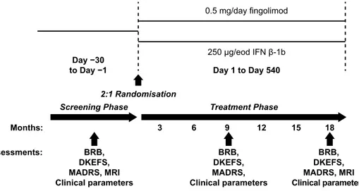
All patients provided written informed consent before enrolment. The study was conducted in accordance with the International Conference on Harmonisation Guidelines for Good Clinical Practice and the Declaration of Helsinki [26, 27]. The study was conducted at 36 study centres, 28 in Italy (22 recruiting) and 8 in Germany (4 recruit-ing), and the protocol was approved by the Independent Ethics Committee or Institutional Review Board at each centre. The study included a 1-month screening phase to determine eligibility. At baseline, eligible patients were randomised (2:1) to receive oral fingolimod (0.5 mg/day) or subcutaneous IFN β-1b (250 µg every other day; Fig. 1).
Study visits for patient clinical assessment were scheduled at screening, baseline and Months 3, 6, 9, 12, 15 and 18 of treatment.
Efficacy assessments
CI was assessed using the BRB of neuropsychological tests in MS [28], comprising five tests: Selective Reminding Test [SRT; includes long-term storage (SRT-LTS), consistent long-term retrieval CLTR) and delayed recall (SRT-d)]; 10/36 Spatial Recall Test (10/36 SPART) [total cor-rect responses (10/36 SPART-T) and delayed recall (10/36 SPART-DR)]; Symbol Digit Modalities Test (SDMT); PASAT and Word List Generation (WLG) test performed at screening, Month 9 and Month 18. All BRB tests have been validated in Italian and German and are available in two alternate forms (A and B), which were administered accord-ing to the scheme A–B–A to reduce the practice effect. Gore-tti et al. have suggested that, similar to other languages, the Italian B version of the BRB test may be easier than version A but also suggested that the application of the normative values provided in their study can overcome this issue [29]. We therefore considered that the BRB versions A and B are comparable only when the data are normalised. Moreover, normalisation of BRB in German is available only for ver-sion A.
Executive functions were assessed using the DKEFS-Sorting test [30], one of the nine tests presented in the DKEFS manual, at screening, Month 9 and Month 18. The DKEFS test consisted of two testing procedures: free sort-ing and sort recognition. In free sortsort-ing, six scores were obtained: confirmed correct sorts for card sets 1 and 2 (or 3 and 4 for version B), sum of confirmed correct sorts, free sorting description score for card sets 1 and 2 (or 3 and 4 for version B) and sum of free sorting description scores. In sort recognition, a description score for card sets 1 and 2 (or 3
Fig. 1 Study design. BRB
Rao’s brief repeatable battery,
DKEFS Delis–Kaplan executive
function system, eod every other day, MADRS Montgom-ery–Asberg Depression Rating Scale, MRI magnetic resonance imaging 0.5 mg/day fingolimod 250 µg/eod IFN β-1b Day 1 to Day 540 Treatment Phase 3 BRB, DKEFS, MADRS, MRI Clinical parameters Assessments: Months: Day −30 to Day −1 BRB, DKEFS, MADRS, MRI Clinical parameters BRB, DKEFS, MADRS, Clinical parameters 6 9 12 15 18 Screening Phase 2:1 Randomisation
and 4 for version B) as well as the sum of description scores of both sets were obtained.
The MADRS [14] was used to assess depression at
screening, Month 9 and Month 18.
MRI was performed locally at the participating centres on 1.5 T (or higher) scanners according to a prespecified protocol provided by the central reading facility (Neuroim-aging Research Unit, Institute of Experimental Neurology, San Raffaele Scientific Institute, Milan, Italy). MRI param-eters [T2-hyperintense, T1-hypointense and T1-enhancing lesions, normalised brain volume (NBV) and percentage brain volume change (PBVC) versus screening scan] were assessed (using central reading) at screening and Month 18. The identification of white matter lesions was performed by consensus of two experienced observers, and the volume of the identified lesions was measured using a semiautomated segmentation technique based on local thresholding (Jim 6.0; Xinapse System, UK). NBV at screening and PBVC during the follow up were measured on precontrast 3D T1-weighted images, using the SIENAx and SIENA software.
Patients were assessed for MS relapses during the course of the study.
EDSS scores were assessed by local raters, specifically trained to minimise variability, at screening, Month 9 and Month 18.
Safety assessments
Safety assessments included reporting of adverse events (AEs), serious AEs (SAEs), vital signs, physical/neurologi-cal examinations, skin examination, laboratory examina-tions, electrocardiogram (ECG) monitoring (as required) and ophthalmologic examinations.
AEs, SAEs and vital signs were assessed at each study visit. Physical examinations were performed at screening and Months 6, 12 and 18; ophthalmologic examinations were performed at screening and Months 3, 6 and 18; and skin examinations were performed at screening and Month 18.
Statistical analysis
For the efficacy data analysis, the full analysis set (FAS) population was considered instead of the per-protocol pop-ulation because of the high drop-out rate, particularly in the IFN β-1b group. The FAS population included all ran-domised patients who received at least one dose of the study drug and had at least one post-baseline assessment of both primary efficacy variables (i.e., non-missing information on BRB and DKEFS-Sorting test) without any major protocol deviations. The safety population included all randomised patients who received at least one dose of the study drug.
Continuous data were summarised by mean, standard deviation (SD), median, interquartile range, minimum and maximum, and 95% confidence limits (CLs), where applicable. Categorical data were presented by absolute and relative frequencies (n and %) or contingency tables. Homogeneity tests were run to assess differences between groups at screening/baseline. The normality of continuous variables was evaluated by means of a Shapiro–Wilk test to perform the t test (in case of normal data distribution) or the Wilcoxon–Mann–Whitney test (in case of non-normal data distribution).
Chi square tests were performed for categorical vari-ables, or Fisher’s exact test for cell frequencies < 5.
Differences between groups in each of the BRB tests were analysed by means of analysis of covariance (ANCOVA) on raw scores (no a priori data normalisation of raw scores based on age/gender and education), with propensity score as an independent variable. The following variables were considered in the propensity score adjust-ment: EDSS score at screening, disease duration, naïve or treated patients’ status, MADRS total score at screening, T2 lesion volume (LV), T1 hypointense LV and number of altered BRB tests at screening.
An ANCOVA model was applied to assess the differ-ences between groups in MRI variables, except for the number of T1-enhancing lesions and T2-hyperintense lesions, considering propensity score as an explanatory variable and screening value as the covariate. The pro-pensity score analysis considered the following factors: EDSS score, disease duration, naïve or treated patients, depression (MADRS score) and number of altered cogni-tive tests. Differences between groups in number of T1 gadolinium-enhancing (Gd+) lesions and new T2 lesions were assessed by means of a negative binomial regres-sion model, including the same propensity score vari-ables applied for the ANCOVA model on the other MRI variable.
Differences between groups in MADRS scores overtime were analysed by means of a mixed model for repeated measures (MMRM). The explanatory variables in the lon-gitudinal model included treatment, visit, treatment-by-visit interaction, missing data pattern (completers, miss-ing at Month 9, missmiss-ing at Month 18), propensity score and corresponding baseline. The propensity score analy-sis considered the following factors: EDSS score, disease duration, naïve or treated patients, number of altered tests, T2 LV and T1 hypointense LV.
The correlation between each BRB test and each MRI variable was performed using Pearson correlation in case of normal distribution of both the considered variables, or Spearman correlation otherwise.
Results
Patient disposition, demographic, and baseline characteristics
In total, 198 patients were enrolled and screened in the GOLDEN study, of whom 157 were randomised (2:1) to receive either fingolimod (n = 106) or IFN β-1b (n = 51). Overall, 30 of the randomised patients discontinued the study: 9 (8.49%) patients from the fingolimod group and 21 (41.18%) patients from the IFN β-1b group (p ≤ 0.0001) (Fig. 2).
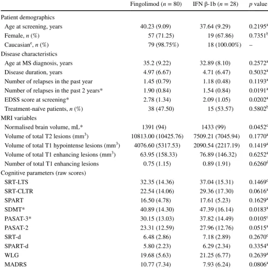
The safety population comprised 151 patients (fingoli-mod, 104; IFN β-1b, 47), whereas the FAS population com-prised 108 patients (fingolimod, 80; IFN β-1b, 28). Approxi-mately 50% of the patients in the FAS were treatment naïve (fingolimod, 47.5%; IFN β-1b, 53.57%; Table 1). The most recent previous treatments included glatiramer acetate, IFN β-1a, IFN β-1b, natalizumab, azathioprine and mitoxantrone.
Contrary to the safety population where baseline char-acteristics were similar across the two treatment groups, in the FAS population, patients randomised to receive fin-golimod had higher number of relapses in the past 2 years (p = 0.0191), higher EDSS scores (p = 0.0202), lower NBV (p = 0.0452) and worse cognitive test scores [PASAT-3 (p = 0.0105) and SDMT (p = 0.0183)] compared with the IFN β-1b group (Table 1).
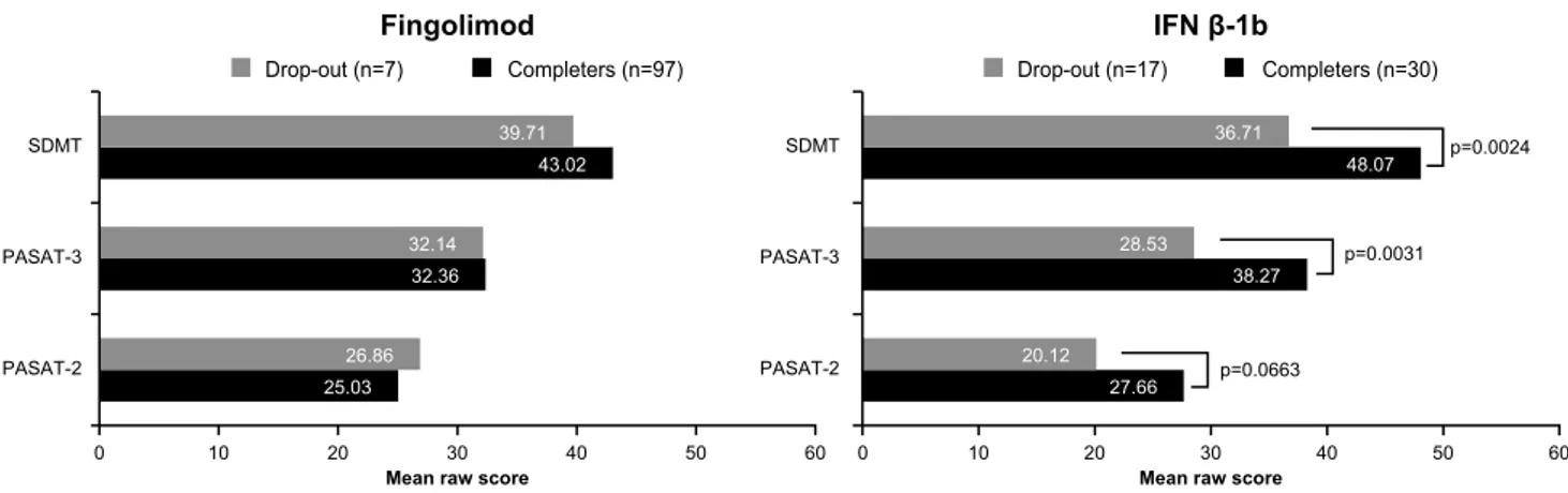
There were statistically significant differences between the baseline PASAT and SDMT (and WLG) scores of patients who completed the study versus those who dropped out of the study in the IFN β-1b group: in this group, patients who later dropped out had more severe baseline scores than the completers, whereas this effect was not observed for the
patients in the fingolimod group (only a non-statistically sig-nificant trend was noted for SRT; Fig. 3).
The total duration of treatment exposure was 537.00 ± 105.77 (range 4–650) days for fingolimod and 420.38 ± 183.37 (range 1–568) days for IFN β-1b (p ≤ 0.0001).
Efficacy results Cognitive function
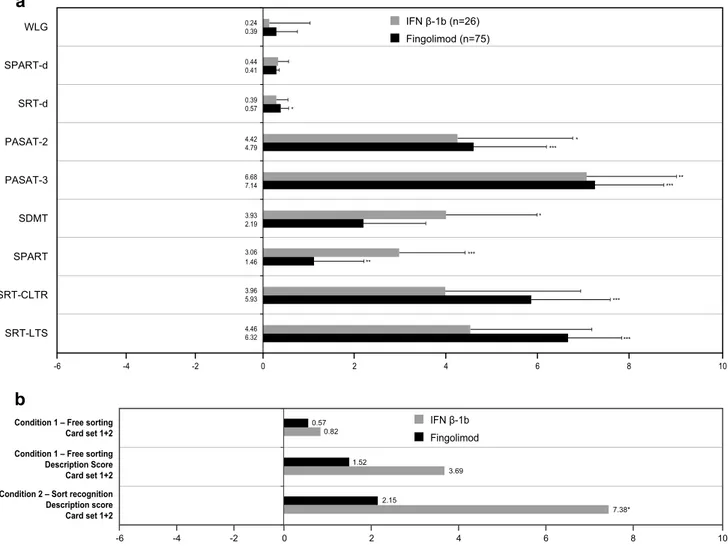
For the BRB test, both treatment groups showed improve-ments in mean changes of all parameters from screening to Month 18: SRT-d (p = 0.0163 for fingolimod; p = 0.3270 for IFN β-1b), PASAT-2 (p = 0.0002 for fingolimod; p = 0.0413 for IFN β-1b), PASAT-3 (p < 0.0001 for fin-golimod; p = 0.0022 for IFN β-1b), SDMT (p = 0.0540 for fingolimod; p = 0.0445 for IFN β-1b), SPART (p = 0.0058 for fingolimod; p = 0.0009 for IFN β-1b), SRT-CLTR (p = 0.0001 for fingolimod; p = 0.1246 for IFN β-1b), SRT-LTS (p < 0.0001 for fingolimod; p = 0.0534 for IFN β-1b), SPART-d (p = 0.0502 for fingolimod; p = 0.2210 for IFN β-1b) and WLG (p = 0.5017 for fingolimod; p = 0.8128 for IFN β-1b) (Fig. 4a). No significant differences were detected between the treatment groups in the mean changes in all parameters from screening to Month 18.
Executive function
Similar to the BRB test, both treatment groups showed improvements in the mean changes from screening to Month 18 for the components of the DKEFS Sorting test (Fig. 4b). No significant differences were detected between
Fig. 2 Patient disposition. AE
adverse event, IFN β-1b inter-feron beta-1b, RRMS relapsing– remitting multiple sclerosis
RRMS patients assessed for eligibility (n=198)
RRMS patients who underwent randomisation (n=157)
Randomised to fingolimod (n=106) Randomised to IFN β-1b (n=51)
97 fingolimod-treated patients
completed the study 30 IFN β-1b-treated patients completed the study
9 patients discontinued
3 (33.3%) discontinued due to AEs 2 (22.2%) had abnormal laboratory values 1 (11.1%) had unsatisfactory
therapeutic effect
3 (33.3%) subjects withdrew consent
21 patients discontinued
1 (4.8%) discontinued due to AEs 1 (4.8%) had abnormal laboratory values 7 (33.3%) had unsatisfactory therapeutic effect
the treatment groups in the mean changes from screening to Month 18 for components of DKEFS-Sorting test.
Depression
The MADRS total score was higher (though not signifi-cantly) in the fingolimod group at screening (10.77 ± 7.34
vs. 7.93 ± 6.24; p = 0.0806). At Month 18, fingolimod-treated patients still exhibited higher scores compared with IFN β-1b-treated patients; however, changes versus screening indicated slight improvement only in the fin-golimod group [−0.68 ± 7.57, 95% CL (− 2.45, 1.08) versus a change of 0.30 ± 5.63, 95% CL (− 1.93, 2.52)
Table 1 Baseline
characteristics of patients included in the FAS
Data represent mean (SD) unless specified otherwise
EDSS expanded disability status scale, FAS full analysis set, IFN β-1b interferon beta-1b, MADRS
Mont-gomery–Asberg rating scale, MRI magnetic resonance imaging, MS multiple sclerosis, PASAT paced audi-tory serial addition test, SD standard deviation, SDMT symbol digit modalities test, SPART 10/36 spatial recall test, SPART-d 10/36 spatial recall test-delayed recall, SRT-LTS selective reminding test-long-term storage, SRT-CLTR selective reminding consistent long-term retrieval, SRT-d selective reminding test-delayed recall, WLG word list generation
*Number of relapses in the past 2 years, EDSS score at baseline and cognitive test scores (PASAT and SDMT) were statistically significant, indicating that IFN β-1b group patients had more severe disease than fingolimod group patients
a Wilcoxon two-sample test b Chi-square
c t test
d Negative binomial regression model e For safety population
Fingolimod (n = 80) IFN β-1b (n = 28) p value
Patient demographics
Age at screening, years 40.23 (9.09) 37.64 (9.29) 0.2195a
Female, n (%) 57 (71.25) 19 (67.86) 0.7351b
Caucasiane, n (%) 79 (98.75%) 18 (100.00%) –
Disease characteristics
Age at MS diagnosis, years 35.2 (9.22) 32.89 (8.10) 0.2572a
Disease duration, years 4.97 (6.67) 4.71 (6.47) 0.5032a
Number of relapses in the past year 1.45 (0.79) 1.18 (0.48) 0.1193a
Number of relapses in the past 2 years* 1.90 (0.84) 1.54 (0.84) 0.0191a
EDSS score at screening* 2.78 (1.34) 2.09 (1.05) 0.0202a
Treatment-naïve patients, n (%) 38 (47.50) 15 (53.57) 0.5802b
MRI variables
Normalised brain volume, mL* 1391 (94) 1433 (99) 0.0452c
Volume of total T2 lesions (mm3) 10813.00 (10425.76) 7509.21 (7045.94) 0.1770a
Volume of total T1 hypointense lesions (mm3) 4076.60 (5317.53) 2090.54 (2217.19) 0.1419a
Volume of total T1 enhancing lesions (mm3) 63.95 (158.33) 76.89 (146.32) 0.6252a
Number of total T1 enhancing lesions 0.75 (1.15) 0.89 (1.91) 0.6260d
Cognitive parameters (raw scores)
SRT-LTS 32.35 (14.36) 37.04 (15.31) 0.1469c SRT-CLTR 22.54 (14.06) 29.36 (17.30) 0.0616a SPART 16.50 (4.78) 17.61 (5.23) 0.1629a SDMT* 40.89 (14.30) 47.39 (16.14) 0.0183a PASAT-3* 30.15 (13.03) 37.82 (14.49) 0.0105c PASAT-2 23.31 (12.59) 27.96 (12.76) 0.0515a SRT-d 6.48 (2.86) 7.18 (2.89) 0.2670c SPART-d 5.80 (2.23) 6.29 (2.34) 0.3354a WLG 19.68 (5.63) 21.25 (6.77) 0.2639a MADRS 10.77 (7.34) 7.93 (6.24) 0.0806a
in the IFN β-1b group], although the difference was not statistically significant (p = 0.3291).
MRI results
T2 LV at screening was higher in the fingolimod group versus the IFN β-1b group (p = 0.1770); although the mean T2 LV decreased in both groups from screening to Month 18, when taking the deltas into account, i.e. only patients who had both values, on average it increased from screening to Month 18 in both treatment groups (Fig. 5a). At Month 18, patients in the IFN β-1b group presented with more new T2 lesions (3.33 ± 4.44 vs. 1.25 ± 2.05) than those in the fingolimod group (p = 0.0276 between groups).
The number and volume of Gd+ lesions at screening were similar in the two groups; both number and vol-ume decreased in patients treated with fingolimod (sig-nificantly for the number of lesions, p = 0.0316) and increased in patients treated with IFN β-1b. The between-group difference was significant for the number of T1 Gd+ lesions (p = 0.0290; Fig. 5b, c).
NBV at screening was significantly lower in the fin-golimod group versus the IFN β-1b group (1391 vs. 1433 mL; p = 0.0452). During the study, the PBVC from screening to Month 18 in the IFN β-1b group (−0.96% ± 0.71%) was larger than that in the fingolimod group (−0.60% ± 0.83%; Fig. 5d), and the between-group difference was statistically significant in favour of fin-golimod (p = 0.0166).
No significant correlation was found between the effects of treatment on MRI parameters and on the vari-ous tests of the BRB or DKEFS.
Relapses and EDSS
The proportion of patients with at least one relapse dur-ing the study period was significantly higher in the IFN β-1b group than in the fingolimod group (31.91 vs. 15.38%, p = 0.0199; Table 2). Moreover, the annualised relapse rate was also higher in the IFN β-1b group than in the fingolimod group (0.39 vs. 0.12; Fig. 6).
EDSS scores remained virtually stable over the 18-month study period, with very small changes in scores at Month 18 versus screening, of 0.12 ± 0.84 [95% CL (− 0.07, 0.31)] in the fingolimod group and 0.19 ± 0.54 [95% CL (− 0.03, 0.40)] in the IFN β-1b group.
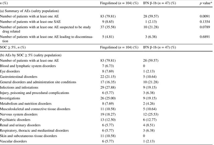
Safety results
Overall, AEs were reported in 79.81% of patients treated with fingolimod and 59.57% treated with IFN β-1b (Table 3a). No deaths were reported during the study.
The proportion of patients with SAEs was higher in the fingolimod group versus the IFN β-1b group (8.65 vs. 2.13%), with one SAE suspected of being related to the study treatment in the fingolimod group (second-degree atrioventricular block after first dose of the drug, in a patient who underwent overnight hospitalisation and then continued treatment with fingolimod without problems), while the proportion of patients discontinuing the study due to AEs was higher in the IFN β-1b group (6.38 vs. 4.81%). The most commonly reported (first five) system organ classes (SOCs) in both groups were ‘infections and infestations’ (primarily nasopharyngitis and influenza), ‘investigations’ (primarily alanine aminotransferase [ALT], blood cholesterol and transaminase increases for
b 1 -β N F I d o m il o g n i F
Drop-out (n=7) Completers (n=97) Drop-out (n=17) Completers (n=30)
p=0.0024 p=0.0031 p=0.0663 25.03 32.36 43.02 26.86 32.14 39.71 0 10 20 30 40 50 60 PASAT-2 PASAT-3 SDMT e r o c s w a r n a e M e r o c s w a r n a e M PASAT-2 PASAT-3 SDMT 27.66 38.27 48.07 20.12 28.53 36.71 0 10 20 30 40 50 60
Fig. 3 SDMT and PASAT scores at screening for patients who
completed the treatment versus those who dropped out of the study (safety population). IFN β-1b interferon beta-1b, PASAT paced audi-tory serial addition test, SDMT symbol digit modalities test. The
fig-ure represents mean values of PASAT and SDMT raw scores. The p value was calculated using the t test for SDMT and PASAT-2 in IFN group and PASAT-3 in fingolimod group; otherwise, the Wilcoxon two-sample test was used
fingolimod; transaminase, blood triglyceride and cho-lesterol increases for IFN β-1b), ‘nervous system disor-ders’ (mostly headache for fingolimod and MS relapse for IFN β-1b), ‘gastrointestinal disorders’(only one case of diarrhoea was thought to be related to treatment, in the fingolimod group), ‘general disorders and administra-tion site condiadministra-tions’ (fever, fatigue and influenza-like ill-nesses more frequent in the IFN β-1b group). Of the 11 cases of the SOC ‘Skin and subcutaneous tissue disorders’ that have been reported only among patients treated with fingolimod, two were considered possibly related to the
treatment: one case of psoriasis and one case of alopecia (Table 3b).
Discussion
Cognitive dysfunction is a common clinical problem in MS and is associated with functional impairment leading to dete-rioration in patients’ quality of life [4]. Thus, a number of studies have been performed on cognitive dysfunction in patients with MS; however, most of the evidence supporting the effect of DMTs on cognitive dysfunction comes from
SRT-LTS SRT-CLTR SPART SDMT PASAT-3 PASAT-2 SRT-d SPART-d WLG
a
IFN β-1b (n=26) Fingolimod (n=75) 0.39 0.24 0.41 0.44 0.57 0.39 * 6 4 2 0 -2 -4 8 10 -6 4.79 4.42 * *** 7.14 6.68 ** *** 2.19 3.93 * 1.46 3.06 *** ** 5.93 3.96 *** 6.32 4.46 *** -6 -4 -2 0Condition 2 – Sort recognition Description score Card set 1+2 Condition 1 – Free sorting Description Score Card set 1+2 Condition 1 – Free sorting
Card set 1+2 0.570.82 3.69 1.52 7.38* 2.15 6 4 2 8 10 IFN β-1b Fingolimod
b
Fig. 4 Cognitive impairment test results a Rao’s BRB test:
ANCOVA LS mean changes Month 18 versus screening (FAS popu-lation) *p < 0.05; **p < 0.01; ***p < 0.001. ANCOVA analysis of covariance, BRB brief repeatable battery, FAS full analysis set, IFN
β-1b interferon beta-1b, LS least squares, M month, PASAT paced
auditory serial addition test, SDMT symbol digit modalities test, SE standard error, SPART 10/36 spatial recall test, SPART-d 10/36 spatial recall test-delayed recall, SRT-LTS selective reminding test-long-term storage, SRT-CLTR selective reminding test-consistent long-term
retrieval, SRT-d selective reminding test-delayed recall, WLG Word List Generation, b DKEFS-sorting test: ANCOVA model estimated mean (LSmean) changes at Month 18 versus screening (FAS popula-tion). *p < 0.05; **p < 0.01. Note: Condition 1—free sorting: golimod 66, IFN β-1b 27. Condition 1—free sorting description: fin-golimod 65, IFN β-1b 27. Condition 2–sort recognition: finfin-golimod 50, IFN β-1b 21. ANCOVA analysis of covariance, DKEFS Delis– Kaplan executive function test, FAS full analysis set, IFN β-1b inter-feron beta-1b, LS least squares, M month
observational, often uncontrolled/non-randomised, studies using single tests that typically assess only one cognitive domain [5, 31–33]. Moreover, controlled trials investigat-ing the effect of DMTs in MS were primarily designed to evaluate clinical outcomes, and cognitive dysfunction was assessed as a secondary outcome or only in a subgroup of patients [5].
This is the first randomised, double-blind study to pro-spectively and comprehensively assess CI in MS patients treated with fingolimod versus an active control (IFN β-1b). A number of characteristics made our study design quite robust in assessing CI in MS patients over the given period of time. First, as described earlier, we have used the full BRB test, which is a sensitive and widely used tool in clini-cal practice, and also assesses executive functions with the DKEFS scale. Second, patients were included in the study only if they already presented CI at screening. This ‘enrich-ment design’ is important, since it is known that preserved cognitive function can remain stable over a long time in MS, whereas incipient cognitive decline seems to be widespread and progressive in nature [34]. Thus, trials in cognitively unselected populations have lower chances to detect the
1433.11 p=0.0452* 1390.48 IFN β-1b (n=28) Fingolimod (n=80) Normalised brain volume (mL) at screening Percentage change in brain volume -1 -0.5 0 0.5 1 1.5 1430 1420 1410 1400 1390 1380 1370 1360 -0.2 -0.4 -0.6 -0.8 -1 -1.2 0 1440 Screening 0.89 0.75 Screening 76.89 63.95 -0.6 -0.96 p=0.0166* 140 120 100 80 60 40 20 0 -20 -40 160 0 6 -10000 8000 6000 4000 2000 12000 0 Changes vs. Screening 711.81 176.25 p=0.1781 Screening 10813 p=0.1770 7509.21 Changes vs. Screening 63.33 p=0.0674 -38.35 Changes vs. Screening 0.59 p=0.0290* -0.64
a
c
b
d
IFN β-1b (n=28) Fingolimod (n=80)IFN β-1b (n=28) Fingolimod (n=80) IFN β-1b (n=28) Fingolimod (n=80)
Fig. 5 MRI measures. a Total T2 lesion volume (mm3), b
num-ber of T1 Gd+ lesions, c volume of T1 Gd+ lesions (mm3), and d
brain volume (mL). p values for the changes were calculated using an ANCOVA model, except for the number of T1 Gd+ lesions, which was computed by using a negative binomial regression model. At
baseline, the p values were calculated using the Wilcoxon two-sam-ple test, except for the brain volume, which was calculated using the
t test. *p < 0.05 within group. ANCOVA analysis of covariance, Gd+
gadolinium enhancing, IFN β-1b interferon beta-1b, M month, MRI magnetic resonance imaging
Table 2 MS relapse (safety population)
*Chi-square test
Fingolimod (n =
104) IFN β-1b (n = 47) p value* No. of patients
with at least one relapse
16 (15.38%) 15 (31.91%) 0.0199 Of above, patients with
1 relapse 13 (81.25%) 10 (66.67%) 2 relapses 2 (12.50%) 3 (20.00%) 3 relapses 1 (6.25%) 2 (13.33%) 0.12 0.39 0.0 0.1 0.2 0.3 0.4 0.5 Fingolimod IFN β-1b
Annualised relapse rate
Fig. 6 MS relapse rate (safety population). IFN β-1b interferon
differences in CI that will accumulate in such a population over the course of the study, particularly if the study dura-tion is short and does not run over many years, whereas these chances were maximised with our enriched population design. Third, in order to prevent the learning of specific test stimuli and thus potentially mitigate practice effects, all the tests of the BRB were provided at screening, Month 9 and Month 18 in two alternate forms (A and B, scheme of administration: A–B–A). Fourth, a standard MRI acquisition protocol was followed across all the sites to ensure uniform quality of the scans across the sites; scans were then ana-lysed at a central MRI evaluation centre by physicians who were unaware of the study-group assignments.
This notwithstanding, the following factors were taken into account when interpreting the results of our study, par-ticularly with regard to the less objective endpoints like cog-nitive scales, compared to the more objective endpoints like MRI. In addition, one has to remember that the study was neither powered nor designed to serve as a direct comparison between fingolimod and IFN β-1b on cognitive scales, but rather to provide side-by-side, pilot evidence. First of all
(and contrary to the safety population, where there were no differences between the treatment groups, which indicates that randomisation did work), in the FAS population patients in the fingolimod group had more severe disease character-istics at baseline compared with patients in the IFN β-1b group, significantly so in terms of number of relapses in the past 2 years, EDSS score, NBV and cognitive test scores (PASAT and SDMT). Second, as expected, a considerably higher percentage of patients treated with IFN β-1b versus fingolimod prematurely discontinued treatment (41.18 vs. 8.49%), with the main reasons for discontinuation being ‘unsatisfactory therapeutic effect’ and ‘withdrawal of con-sent’. One implication is that patients in the IFN β-1b group with more severe disease abandoned the trial, as confirmed by the difference between the randomised population and the FAS population. A second implication is that patients in the IFN β-1b group who completed the study and had Month 18 cognitive scores available were most probably those who were responding better to that treatment com-pared to the fingolimod group. This is supported by the sta-tistically significant differences between the baseline PASAT
Table 3 AEs and SAEs in the safety set
AE adverse event, IFN β-1b interferon beta-1b, SAE serious adverse event
*Chi-square test
n (%) Fingolimod (n = 104) (%) IFN β-1b (n = 47) (%) p value*
(a) Summary of AEs (safety population)
Number of patients with at least one AE 83 (79.81) 28 (59.57) 0.0091
Number of patients with at least one SAE 9 (8.65) 1 (2.13) 0.1354
Number of patients with at least one AE suspected to be study
drug related 37 (35.58) 10 (21.28) 0.0789
Number of patients with at least one AE leading to
discontinua-tion 5 (4.81) 3 (6.38) 0.6891
SOC ≥ 5%, n (%) Fingolimod (n = 104) (%) IFN β-1b (n = 47) (%)
(b) AEs by SOC ≥ 5% (safety population)
Number of patients with at least one AE 83 (79.81) 28 (59.57)
Blood and lymphatic system disorders 7 (6.73) 0
Eye disorders 8 (7.69) 1 (2.13)
Gastrointestinal disorders 22 (21.15) 5 (10.64)
General disorders and administration site conditions 17 (16.35) 10 (21.28)
Infections and infestations 29 (27.88) 9 (19.15)
Injury, poisoning and procedural complications 6 (5.77) 3 (6.38)
Investigations 26 (25.00) 9 (19.15)
Metabolism and nutrition disorders 8 (7.69) 2 (4.26)
Musculoskeletal and connective tissue disorders 11 (10.58) 5 (10.64)
Nervous system disorders 19 (18.27) 12 (25.53)
Psychiatric disorders 13 (12.50) 6 (12.77)
Renal and urinary disorders 6 (5.77) 4 (8.51)
Respiratory, thoracic and mediastinal disorders 6 (5.77) 3 (6.38)
Skin and subcutaneous tissue disorders 11 (10.58) 0
and SDMT (and WLG) scores of patients who completed the study versus those who dropped out of the study in the IFN β-1b group: in this group patients who later dropped out had more severe baseline scores than the completers, whereas this effect was not observed for the patients in the fingolimod group (only a non-statistically significant trend was noted for SRT; Fig. 3).
Our results show that both fingolimod and IFN β-1b improved all cognitive domains affected by MS, as evalu-ated through the various tests of the BRB (SRT-d, SRT-LTS, SRT-CLTR, SPART, SPART-d, SDMT, PASAT and WLG) with some differences in the improvement pattern. Fingoli-mod showed the best effect on PASAT and SRT scores, whereas IFN β-1b showed the best effect on SDMT scores. In this regard, it is worth noting that PASAT is a difficult and demanding test of the battery, relatively more difficult than the SDMT, and is sensitive even to early cognitive changes in patients where room for improvement is still limited [35, 36]. A longitudinal correlational research study suggested that PASAT is particularly sensitive to inflammatory activ-ity measured by Gd+ enhancement in otherwise physically stable patients with MS [37]. Hence, the observed effect of fingolimod on PASAT in this study is consistent with the effect observed on Gd+ lesions.
In general, our results are supported by previous studies evaluating the effect of IFN or fingolimod on CI in real-world setting using a non-randomised design. A 3-year, open-label, prospective, observational study [COGnitive Impairment in Multiple Sclerosis (COGIMUS)] showed that the proportion of cognitively impaired patients treated with IFN β-1a remained stable over the 3 years of treatment in at least three tests of the BRB and the Stroop Color-Word Task [33]. In another 1-year, open-label study, treatment with IFN β-1b in RRMS patients led to improved perfor-mance in the complex attention, concentration and visual learning and recall domains compared with patients with RRMS matched for neurological disability [31]. In a post hoc analysis of pooled data from a fingolimod phase III trial in patients with RRMS, fingolimod treatment resulted in early and sustained improvement in cognition, as measured by the change in PASAT-3 scores over 6, 12 and 24 months [23]. Results of a multicentre, examiner-blinded, prospec-tive trial also showed significant improvement in cogniprospec-tive function from the sixth month of initiation of fingolimod in patients with RRMS [24]. Additionally, real-world stud-ies in patients with RRMS have shown a positive impact of fingolimod treatment on cognitive parameters [22]. In a study comparing effectiveness of natalizumab and fingoli-mod treatment on cognitive functions by using the BRB tests in patients with RRMS from clinical practice, fingolimod was found to be more effective than natalizumab in improv-ing cognitive function [38]. However, our study provides novel information on the topic. Namely, we show that there
is not necessarily any direct correlation between the effects of a given treatment on MRI parameters (particularly on brain atrophy, which in turn has been shown to correlate with a beneficial effect on CI) and on the multiplicity of tests that compose the BRB or DKEFS. In general, studies have identified a significant correlation between MRI measures and CI [7, 39, 40] and, more specifically, studies suggest a robust correlation between cognitive deficits and irrevers-ible tissue loss in the brain, usually measured in terms of global and regional atrophy [41]. In our study, fingolimod showed better results than IFN β-1b on all MRI parameters, brain atrophy and clinical parameters (relapse rate and EDSS score). This is supported by previous studies in which fin-golimod was associated with early and consistent reduction in brain volume loss, compared with both placebo and intra-muscular IFN β-1a [42]. On the other hand, both fingolimod and IFN β-1b improved all cognitive domains affected by MS, as evaluated through the various tests of the BRB, with some differences in the improvement pattern. Apart from the confounding effects of the difference in baseline sever-ity and drop-out patterns across the two groups, which hold true both for the effects on cognitive scales and on MRI, this difference in effects on MRI versus BRB may also be due to other factors, including better sensitivity of MRI (the most sensitive tool to capture early tissue loss quantitatively and independent of any practice effects [43]) compared with cognitive scales for the detection of changes in endpoints supposed to be correlated (such as CI and atrophy), or to different sensitivity across the individual scales to changes in similar or different cognitive domains. Finally, the lim-ited duration of the trial (18 months) may have impacted on the possibility of observing the complete expression of dif-ferential effects between the treatment groups on cognitive impairment scales reflecting the between-groups differences on MRI. Trials of longer duration may therefore be required to assess this aspect.
This absence of a firm correlation (also due to the high number of confounding effects and biases in assessing CI) also shows that effects of interventions (pharmacologic or non-pharmacologic) on functional cognitive capacity should be shown directly and cannot be deduced from effects on brain volumetric measures.
With regard to the safety results of the study, although this was a pilot study of limited size and, therefore, not really comparable to larger studies reported in the literature for both drugs, the GOLDEN study supported the estab-lished safety and tolerability profile of both drugs. The only SAE suspected of being related to the study treatment was a self-limiting, second-degree atrioventricular block after the first-dose of fingolimod, which is consistent with the known first-dose cardiovascular effects of the drug. Apart from this, first-dose monitoring was relatively eventless, although some patients required extended monitoring for
precautionary measures due to lowered heart rate. AEs sus-pected of being related to treatment were in line with the tolerability and safety/tolerability profile of the two drugs as documented in the respective Summary of Product Char-acteristics or reported in the literature. No significant differ-ences between fingolimod and IFN β-1b groups with regards to safety parameters were evident except for the higher number of adverse events in the fingolimod group, and the greater proportion of patients experiencing an MS relapse in the IFN β-1b group.
In conclusion, the results of the GOLDEN study suggest that both fingolimod and IFN β-1b treatments were associ-ated with improvements in all cognitive parameters, with some differences in the improvement patterns. Despite a dis-advantage in terms of baseline characteristics and drop-out patterns, fingolimod treatment demonstrated significantly better effects than IFN β-1b on MRI parameters and relapse rate.
Also, although limited in size, the GOLDEN study confirms the favourable benefit–risk profile of fingolimod reported in previous studies.
Management of cognitive decline in MS, which substan-tially alters patients’ quality of life and is the leading cause of occupational disability in patients with MS [44], is an area that still needs further research. Among others, additional studies are warranted to better understand the effects of DMTs on cognitive function and its correlation with under-lying disease mechanisms.
Acknowledgements The authors would like to thank Richa Chhabra
and Avinash Thakur (Medical communications, Novartis Healthcare Pvt. Ltd) for medical writing assistance in developing the first draft of the manuscript, formatting, referencing, preparing tables and fig-ures, incorporating the authors’ revisions and submission, all under the direction of the authors. All authors have reviewed the manuscript for intellectual content, provided guidance during manuscript devel-opment and approved the final version submitted for publication. The final responsibility for the content lies with the authors. Members of the MRI analysis centre—Neuroimaging Research Unit, Institute of Exper-imental Neurology, Division of Neuroscience, San Raffaele Scientific Institute, Vita-Salute San Raffaele University, Ospedale San Raffaele, Milan, Italy: M. Filippi (Director), M.A. Rocca, L. Dall’Occhio, A. Meani, R. Messina, E. Pagani, M. Petrolini, P. Preziosa, M. Sibilia.
GOLDEN Study Principal Investigators (only centres who actively recruited patients are reported here, in order of recruitment): Italian
centres (1) Francesco Patti and Clara Grazia Chisari, Department of
Medical, Surgery Science and Advanced Technology “GF Ingrassia”, Section of Neurosciences—MS Centre, University Hospital, Catania, Italy, (2) Girolama Alessandra Marfia and Diego Centonze, Depart-ment of Neuroscience, University of Rome Tor Vergata, Rome, Italy, (3) Vincenzo Brescia Morra, MS Centre, Neurosciences, Reproductive and Odontostomatological Sciences Department, Federico II Univer-sity of Naples, Naples, Italy, (4) Ruggero Capra, Multiple Sclerosis Centre, Spedali Civili Hospital, Montichiari (Brescia), Italy, (5) Carlo Pozzilli and Valentina Bianchi, Department of Neurology and Psy-chiatry, Sapienza University of Rome, Rome, Italy, (6) Angelo Ghezzi and Marco Roscio, Neurology II, Multiple Sclerosis Centre, Gallarate Hospital, Gallarate, Italy, (7) Giancarlo Comi and Francesca Sangalli for Maria Emma Rodegher (co-PI at the time of the study), Department
of Neurology and Institute of Experimental Neurology, San Raffaele Hospital, Milan, Italy, (8) Erika Pietrolongo for Alessandra Lugaresi (PI at the time of the study), Department of Neuroscience Imaging and Clinical Science, University “G. d’Annunzio”, Chieti, Italy, (9) Ada Francia, Department of Neuroscience, Sapienza University of Rome, Rome, Italy, (10) Maura Chiara Danni, S.O.D. Neurological Clinic, University Hospital Umberto I, Ancona, Italy, (11) Ugo Nocentini, Neurology and Neurorehabilitation Unit, I.R.C.C.S. “Santa Lucia” Foundation; Department of Systems Medicine, University of Rome “Tor Vergata”, Rome, Italy, (12) Placido Bramanti, IRCCS Centro Neurolesi “Bonino-Pulejo”, Messina, Italy, (13) Gioacchino Tede-schi, Department of Medical, Surgical, Neurological, Metabolic and Ageing Sciences, University of Campania Luigi Vanvitelli, Naples, Italy, (14) Davide Maimone, Multiple Sclerosis Center, Neurology, Garibaldi Hospital, Catania, Italy, (15) Luigi Maria Edoardo Grimaldi, Neurology Unit, San Raffaele Institute “G.Giglio”, Cefalù (Palermo), Italy, (16) Elio Angelo Scarpini, Department of Pathophysiology and Transplantation, Neurology Unit, University of Milan, Fondazione Ca’ Granda, IRCCS Ospedale Policlinico, Milan, Italy, (17) Antonio Uccelli, Department of Neurosciences Ophthalmology and Genetics, University of Genoa, Genoa, Italy, (18) Maria Pia Amato, Department NEUROFARBA, University of Florence, Florence, Italy, (19) Maria-rosa Rottoli, Neurology and Autoimmune Diseases Unit, Hospital Papa Giovanni XXIII, Bergamo, Italy, (20) Stefano Ruggieri, Neurology Unit, Istituto Mediterraneo Neuromed, Pozzilli, Italy, (21) Maria Tro-jano, Department of Basic Medical Sciences, Neuroscience and Sense Organs, University of Bari, Bari, Italy, (22) Roberto Bergamaschi, IRCCS Neurological Institute “C. Mondino”, Pavia, Italy. German
centres (23) Florian Then Bergh, University of Leipzig, Department
of Neurology, Leipzig, Germany, (24) Mathias Buttmann, Department of Neurology, University of Würzburg, Würzburg, Germany, (25) Peter Rieckmann, Sozialstiftung Bamberg Hospital, Bamberg, Germany, (26) Ali Safavi, Praxis Burgstrasse 3, Alzenau, Germany.
Compliance with ethical standards
Conflicts of interest G Comi has received compensation for
consult-ing services and/or speakconsult-ing activities from Novartis, Teva, Sanofi, Genzyme, Merck, Biogen, Roche, Almirall, Celgene, Forward Phar-ma, Excemed. F. Patti received speaking honoraria and fee for advisory board activities by Amirall, Bayer, Biogen, Celgene, Merck, Novartis, Roche, Sanofi-Genzyme and TEVA; he also received research grant by MIUR and Fondazione Italiana Sclerosi Multipla. M. Rocca received speakers honoraria from Biogen Idec, Novartis, Genzyme, Sanofi-Aventis, Teva and Merk Serono and receives research support from the Italian Ministry of Health and Fondazione Italiana Sclerosi Multipla. M. Filippi is Editor-in-Chief of the Journal of Neurology; serves on a scientific advisory board for Teva Pharmaceutical Industries; has re-ceived compensation for consulting services and/or speaking activities from Biogen Idec, Merk-Serono, Novartis, and Teva Pharmaceutical Industries; and receives research support from Biogen Idec, Teva Phar-maceutical Industries, Novartis, Italian Ministry of Health, Fondazi-one Italiana Sclerosi Multipla, Cure PSP, Alzheimer’s Drug Discovery Foundation (ADDF), the Jacques and Gloria Gossweiler Foundation (Switzerland), and ARiSLA (Fondazione Italiana di Ricerca per la SLA). F.Mattioli received travel and conference fees and editorial con-sulence fees from Merck Serono, Biogen and Novartis. M. P. Amato received research grants and honoraria as a speaker and member of advisory boards by Bayer, Biogen Idec, Merck Serono, Novartis, Sa-nofi Genzyme, Teva, Almirall. P. Gallo has been a consultant for Bayer Schering, Biogen Idec, Genzyme, Merck Serono and Novartis, has re-ceived funding for travel and speaker honoraria from Merck-Serono, Biogen Idec, Sanofi-Aventis, Novartis Pharma and Bayer-Schering Pharma, Teva, has received research support from Bayer, Biogen Idec/ Elan, MerkSerono, Genzyme and Teva, and has received research grant
from the University of Padova, Veneto Region of Italy, the Italian As-sociation for Multiple Sclerosis, the Italian Ministry of Public Health. D. Centonze is an advisory board member of Almirall, Bayer Schering, Biogen, GW Pharmaceuticals, Merck-Serono, Novartis, Sanofi-Gen-zyme, Teva and received honoraria for speaking or consultation fees from Almirall, Bayer Schering, Biogen Idec, GW Pharmaceuticals, Merck Serono, Novartis, Sanofi-Genzyme, Teva. He is also the princi-pal investigator in clinical trials for Bayer Schering, Biogen Idec, Mer-ck Serono, Mitsubishi, Novartis, Roche, Sanofi-Genzyme, Teva. His preclinical and clinical research was supported by grants from Bayer, Biogen, Merck Serono, Novartis and Teva. C. Pozzilli has served on scientific advisory boards for Actelion, Biogen, Genzyme, Hoffmann-La Roche Ltd,Merck-Serono, Novartis, Sanofi, Teva, and has received consulting and/or speaking fees, research support and travel grants from Allergan, Almirall, Biogen, Genzyme, Hoffmann-La Roche Ltd, Merck-Serono, Novartis, Sanofi and Teva. F. Saccà received personal compensation from Novartis, Almirall, Genzyme, Biogen, Merck Se-rono Forward Pharma and TEVA for public speaking, editorial work and advisory boards. F. Then Bergh has received research support for investigator-initiated studies and support for local scientific symposia (to the University of Leipzig), travel support for scientific meetings, and personal compensation as a speaker or serving on advisory boards from Actelion, Bayer-Schering, Biogen-Idec, Genzyme, Merck-Sero-no, Novartis, Sanofi-Aventis, TEVA. He has received public research funding from the Deutsche Forschungsgemeinschaft, the German Min-istry of Education and Research and the Myelinprojekt. He has no di-rect financial interest in products or procedures described in this work or employed in the study reported. M. Bartezaghi and R. Turrini are employees of Novartis Farma.
Ethical standards All patients provided written informed consent
before enrolment. The study was conducted in accordance with the International Conference on Harmonisation Guidelines for Good Clini-cal Practice and the Declaration of Helsinki.
Funding The study was funded by Novartis Farma, Origgio, Varese,
Italy.
Open Access This article is distributed under the terms of the
Creative Commons Attribution 4.0 International License ( http://crea-tivecommons.org/licenses/by/4.0/), which permits unrestricted use, distribution, and reproduction in any medium, provided you give appro-priate credit to the original author(s) and the source, provide a link to the Creative Commons license, and indicate if changes were made.
References
1. Lublin FD, Reingold SC, Cohen JA, Cutter GR, Sorensen PS, Thompson AJ, Wolinsky JS, Balcer LJ, Banwell B, Barkhof F, Bebo B, Calabresi PA, Clanet M, Comi G, Fox RJ, Freedman MS, Goodman AD, Inglese M, Kappos L, Kieseier BC, Lincoln JA, Lubetzki C, Miller AE, Montalban X, O’Connor PW, Petkau J, Pozzilli C, Rudick RA, Sormani MP, Stuve O, Waubant E, Polman CH (2014) Defining the clinical course of multiple sclerosis The 2013 revisions. Neurology 83(3):278–286
2. Amato MP, Zipoli V, Portaccio E (2006) Multiple sclerosis-related cognitive changes: a review of cross-sectional and lon-gitudinal studies. J Neurol Sci 245(1–2):41–46. doi:10.1016/j. jns.2005.08.019
3. Chiaravalloti ND, DeLuca J (2008) Cognitive impairment in multiple sclerosis. Lancet Neurol 7(12):1139–1151. doi:10.1016/ S1474-4422(08)70259-X
4. Patti F (2009) Cognitive impairment in multiple sclerosis. Mult Scler 15(1):2–8. doi:10.1177/1352458508096684
5. Amato MP, Langdon D, Montalban X, Benedict RH, DeLuca J, Krupp LB, Thompson AJ, Comi G (2013) Treatment of cogni-tive impairment in multiple sclerosis: position paper. J Neurol 260(6):1452–1468. doi:10.1007/s00415-012-6678-0
6. Rosti-Otajarvi EM, Hamalainen PI (2014) Neuropsychological rehabilitation for multiple sclerosis. Cochrane Database Syst Rev (2):CD009131. doi:10.1002/14651858.CD009131.pub3
7. Cohen JA, Barkhof F, Comi G, Hartung HP, Khatri BO, Montal-ban X, Pelletier J, Capra R, Gallo P, Izquierdo G, Tiel-Wilck K, de Vera A, Jin J, Stites T, Wu S, Aradhye S, Kappos L, Group TS (2010) Oral fingolimod or intramuscular interferon for relapsing multiple sclerosis. N Engl J Med 362(5):402–415. doi:10.1056/ NEJMoa0907839
8. Patti F (2012) Treatment of cognitive impairment in patients with multiple sclerosis. Expert Opin Investig Drugs 21(11):1679–1699. doi:10.1517/13543784.2012.716036
9. Polman CH, O’Connor PW, Havrdova E, Hutchinson M, Kap-pos L, Miller DH, Phillips JT, Lublin FD, Giovannoni G, Wajgt A, Toal M, Lynn F, Panzara MA, Sandrock AW, Investigators A (2006) A randomized, placebo-controlled trial of natalizumab for relapsing multiple sclerosis. N Engl J Med 354(9):899–910. doi:10.1056/NEJMoa044397
10. Rao SM, The Cognitive Function Study Group of the National Multiple Sclerosis Society (1990) A manual for the brief repeat-able battery of neuropsychological tests in multiple sclerosis. Medical College of Wisconsin, Milwaukee
11. Langdon DW, Amato MP, Boringa J, Brochet B, Foley F, Fredrik-son S, Hamalainen P, Hartung HP, Krupp L, Penner IK, Reder AT, Benedict RH (2012) Recommendations for a brief international cognitive assessment for multiple sclerosis (BICAMS). Mult Scler 18(6):891–898. doi:10.1177/1352458511431076
12. Parmenter BA, Zivadinov R, Kerenyi L, Gavett R, Weinstock-Guttman B, Dwyer MG, Garg N, Munschauer F, Benedict RHB (2007) Validity of the Wisconsin card sorting and Delis– Kaplan executive function system (DKEFS) sorting tests in multiple sclerosis. J Clin Exp Neuropsychol 29(2):215–223. doi:10.1080/13803390600672163
13. Stroop JR (1935) Studies of interference in serial verbal reactions. J Exp Psychol 18:643–662
14. Montgomery SA, Asberg M (1979) New depression scale designed to be sensitive to change. Br J Psychiatr 134:382–389 15. Amato MP, Hakiki B, Goretti B, Rossi F, Stromillo ML, Giorgio
A, Roscio M, Ghezzi A, Guidi L, Bartolozzi ML, Portaccio E, De Stefano N, Italian RISMSSG (2012) Association of MRI metrics and cognitive impairment in radiologically isolated syndromes. Neurology 78(5):309–314. doi:10.1212/WNL.0b013e31824528c9
16. Calabrese M, Agosta F, Rinaldi F, Mattisi I, Grossi P, Favaretto A, Atzori M, Bernardi V, Barachino L, Rinaldi L, Perini P, Gallo P, Filippi M (2009) Cortical lesions and atrophy associated with cognitive impairment in relapsing-remitting multiple sclerosis. Arch Neurol 66(9):1144–1150. doi:10.1001/archneurol.2009.174
17. Rocca MA, Amato MP, De Stefano N, Enzinger C, Geurts JJ, Pen-ner IK, Rovira A, Sumowski JF, Valsasina P, Filippi M, Grp MS (2015) Clinical and imaging assessment of cognitive dysfunction in multiple sclerosis. Lancet Neurol 14(3):302–317. doi:10.1016/ S1474-4422(14)70250-9
18. Kappos L, Radue EW, O’Connor P, Polman C, Hohlfeld R, Cala-bresi P, Selmaj K, Agoropoulou C, Leyk M, Zhang-Auberson L, Burtin P, Grp FS (2010) A placebo-controlled trial of oral fingoli-mod in relapsing multiple sclerosis. N Engl J Med 362(5):387– 401. doi:10.1056/NEJMoa0909494
19. Hunter SF, Bowen JD, Reder AT (2016) The direct effects of fin-golimod in the central nervous system: implications for relaps-ing multiple sclerosis. CNS Drugs 30(2):135–147. doi:10.1007/ s40263-015-0297-0
20. Pelletier J, Karlsson G, Li BB, De Vera A, Francis G, Cohen J, Kappos L (2011) Effect of fingolimod on multiple sclerosis functional composite (MSFC) defined disability progression in patients with relapsing remitting MS (RRMS): results from two phase III studies. Neurology 76(9):A611
21. Barak Y, Magalashvili D, Paz T, Achiron A (2014) Fingolimod (Gilenya) confers cognitive stability in active relapsing–remitting multiple sclerosis patients. Mult Scler J 20:210
22. Fonseca J (2015) Fingolimod real world experience: effi-cacy and safety in clinical practice. Neurosci J 2015:389360. doi:10.1155/2015/389360
23. Langdon D, Penner I-K, Calabrese P, Cutter G, Häring D, Dahlke F, Tomic D, Kappos L (2016) Fingolimod effects on PASAT score and baseline determinants of PASAT in a large cohort of RRMS patients. Neurology 86(S16):P12.150
24. Ozakbas S, Cinar BP, Kosehasanogullari G, Yigit P (2016) Effects of fingolimod on cognitive status in patients with multiple sclero-sis: prospective, controlled trial. Neurology 86(S16):P13.071 25. Polman CH, Reingold SC, Edan G, Filippi M, Hartung HP,
Kap-pos L, Lublin FD, Metz LM, McFarland HF, O’Connor PW, Sandberg-Wollheim M, Thompson AJ, Weinshenker BG, Wolin-sky JS (2005) Diagnostic criteria for multiple sclerosis: 2005 revi-sions to the “McDonald criteria”. Ann Neurol 58(6):840–846. doi:10.1002/ana.20703
26. International conference on harmonisation of technical require-ments for registration of pharmaceuticals for human use (1996) ICH harmonised tripartite guideline. Guideline for good clinical practice. http://www.ich.org/fileadmin/Public_Web_Site/ICH_ Products/Guidelines/Efficacy/E6_R1/Step4/E6_R1__Guideline. pdf. Accessed 2 Aug 2017
27. World Medical Association Declaration of Helsinki (2017) Ethi-cal principles for mediEthi-cal research involving human subjects.
http://www.wma.net/en/30publications/10policies/b3/index.html. Accessed 2 Aug 2017
28. Rao SM, Leo GJ, Bernardin L, Unverzagt F (1991) Cognitive dysfunction in multiple sclerosis. I. Frequency, patterns, and pre-diction. Neurology 41(5):685–691
29. Goretti B, Patti F, Cilia S, Mattioli F, Stampatori C, Scarpazza C, Amato MP, Portaccio E (2014) The Rao’s brief repeatable bat-tery version B: normative values with age, education and gen-der corrections in an Italian population. Neurol Sci 35(1):79–82. doi:10.1007/s10072-013-1558-7
30. Delis DC, Kramer JH, Kaplan E, Holdnack J (2004) Reliabil-ity and validReliabil-ity of the Delis–Kaplan executive function system: an update. J Int Neuropsychol Soc 10(2):301–303. doi:10.1017/ S1355617704102191
31. Barak Y, Achiron A (2002) Effect of interferon-beta-1b on cog-nitive functions in multiple sclerosis. Eur Neurol 47(1):11–14. doi:10.1159/000047940
32. Flechter S, Vardi J, Finkelstein Y, Pollak L (2007) Cognitive dysfunction evaluation in multiple sclerosis patients treated with interferon beta-1b: an open-label prospective 1 year study. Isr Med Assoc J 9(6):457–459
33. Patti F, Amato MP, Bastianello S, Caniatti L, Di Monte E, Ferrazza P, Goretti B, Gallo P, Morra VB, Lo Fermo S, Pic-coni O, Tola MR, Trojano M, Grp CS (2010) Effects of immunomodulatory treatment with subcutaneous interferon beta-1a on cognitive decline in mildly disabled patients with relapsing-remitting multiple sclerosis. Mult Scler 16(1):68–77. doi:10.1177/1352458509350309
34. Kujala P, Portin R, Ruutiainen J (1997) The progress of cognitive decline in multiple sclerosis—a controlled 3-year follow-up. Brain 120:289–297. doi:10.1093/brain/120.2.289
35. Forn C, Belenguer A, Belloch V, Sanjuan A, Parcet MA, Avila C (2011) Anatomical and functional differences between the paced auditory serial addition test and the symbol digit modalities test. J Clin Exp Neuropsychol 33(1):42–50. doi:10.1080/13803395.2 010.481620
36. Karabudak R, Dahdaleh M, Aljumah M, Alroughani R, Alsha-roqi IA, AlTahan AM, Bohlega SA, Daif A, Deleu D, Amous A, Inshasi JS, Rieckmann P, Sahraian MA, Yamout BI (2015) Functional clinical outcomes in multiple sclerosis: current sta-tus and future prospects. Mult Scler Relat Disord 4(3):192–201. doi:10.1016/j.msard.2015.03.004
37. Bellmann-Strobl J, Wuerfel J, Aktas O, Dorr J, Wernecke KD, Zipp F, Paul F (2009) Poor PASAT performance correlates with MRI contrast enhancement in multiple sclerosis. Neurology 73(20):1624–1627. doi:10.1212/WNL.0b013e3181c1de4f
38. Iaffaldano P, Viterbo RG, Lucisano G, Trojano M (2016) Com-parative effectiveness of natalizumab and fingolimod treatment on cognitive functions in relapsing multiple sclerosis. Mult Scler J 22:687
39. De Stefano N, Airas L, Grigoriadis N, Mattle HP, O’Riordan J, Oreja-Guevara C, Sellebjerg F, Stankoff B, Walczak A, Wiendl H, Kieseier BC (2014) Clinical relevance of brain volume measures in multiple sclerosis. CNS Drugs 28(2):147–156. doi:10.1007/ s40263-014-0140-z
40. Kappos L, O’Connor P, Polman C, Hohlfeld R, Radue EW, Cala-bresi P, Selmaj K, Agoropoulou C, Leyk M, Zhang-Auberson L, Grp FS (2010) Oral fingolimod (FTY720) vs placebo in relaps-ing–remitting multiple sclerosis: 24-month clinical efficacy results from a randomized, double-blind, placebo-controlled, multicenter phase III study (FREEDOMS). Neurology 74(9):A194
41. Filippi M, Rocca MA, Benedict RH, DeLuca J, Geurts JJ, Rom-bouts SA, Ron M, Comi G (2010) The contribution of MRI in assessing cognitive impairment in multiple sclerosis. Neurology 75(23):2121–2128. doi:10.1212/WNL.0b013e318200d768
42. Radue EW, Barkhof F, Kappos L, Sprenger T, Haring DA, de Vera A, von Rosenstiel P, Bright JR, Francis G, Cohen JA (2015) Correlation between brain volume loss and clinical and MRI outcomes in multiple sclerosis. Neurology 84(8):784–793. doi:10.1212/Wnl.0000000000001281
43. Traboulsee AL, Li DK (2006) The role of MRI in the diagnosis of multiple sclerosis. Adv Neurol 98:125–146
44. Rahn K, Slusher B, Kaplin A (2012) Cognitive impairment in multiple sclerosis: a forgotten disability remembered. Cerebrum 2012:14