nasolacrimal duct obstruction, over the past decades, the popularity of EN-DCR has increased due to the obvious advantages already pointed out by the authors. The increased popularity of the EN-DCR is also supported by the better outcomes. In this respect, we chal-lenge the authors to agree that the different success rate in their 2 groups (91.0% versus 71.5%) is likely to be attributed more to the different surgical technique rather than the adjuvant use of MMC.
In fact, we note that the adjuvant MMC increases the success rate of revision EN-DCR,2 whereas the success rate of primary EN-DCR is already very high and might not benefit from the ad-junctive use of MMC.3Furthermore, the same findings of better wound healing at the osteotomy site in absence of increased success rate was previously reported.4
Because of the retrospective nature of the study, we would have welcomed a selection of more homogenous groups, including only patients with primary or revision DCR. We would have also ex-pected a comparative interventional study between patients treated with EN-DCR alone and EN-DCR with MMC to assess the effect of MMC, which was the purpose of this article. In contrast, the authors only used the adjunctive MMC in their patients undergoing EN-DCR and then compared the success rate of this group with a group of subjects undergoing external DCR without MMC.
With regard to the adjunctive use of silicone tube, we would like to cite a recent meta-analysis that concluded that silicone tubing after primary EN-DCR is not necessary.5This confirms the original findings of a previous prospective randomized interventional trial.6 We would like to stimulate the authors to provide more details regarding their study. In particular the following:
(a) Were the patients allocated to 1 of the 2 different groups ac-cording to particular preoperative characteristics?
(b) Were the patients operated on by the same surgeon?
(c) The size and location of the osteotomy were not reported. This has been found to be a common cause of failure.7Were size and location of osteotomy consistent throughout the study? (d) Could the authors provide a differentiated analysis of success
rates and postoperative complications for patients undergo-ing primary procedures and revisions among their 2 groups of patients?
(e) How the authors defined and assessed the success of their procedure? We would like to highlight that the patency on saline irrigation demonstrates the anatomic success, whereas the Jones fluorescein dye test, the height of the tear meniscus, and the freedom from epiphora are indicators of functional success.
Marco Carifi, MD A.O.R.N. ‘‘A. Cardarelli’’ Naples, Italy Bruno Zuberbuhler, PhD Manchester Academic Health Science Centre Manchester Gianluca Carifi, MD Moorfields Eye Hospital London, UK [email protected]
REFERENCES
1. Apuhan T, Yldrm YS, Eroglu F, et al. Effect of mitomycin C on endoscopic dacryocystorhinostomy. J Craniofac Surg
2011;22:2057Y2059
2. Penttila¨ E, Smirnov G, Seppa¨ J, et al. Mitomycin C in revision endoscopic dacryocystorhinostomy: a prospective randomized study. Am J Rhinol Allergy 2011;25:425Y428
3. Smirnov G, Tuomilehto H, Kokki H, et al. Symptom score questionnaire for nasolacrimal duct obstruction in adultsVa novel tool to assess the outcome after endoscopic dacryocystorhinostomy. Rhinology 2010;48:446Y451
4. Ugurbas SH, Zilelioglu G, Sargon MF, et al. Histopathologic effects of mitomycin C on endoscopic transnasal dacryocystorhinostomy. Ophthalmic Surg Lasers 1997;28:300Y304
5. Gu Z, Cao Z. Silicone intubation and endoscopic
dacryocystorhinostomy: a meta-analysis. J Otolaryngol Head Neck Surg 2010;39:710Y713
6. Smirnov G, Tuomilehto H, Tera¨svirta M, et al. Silicone tubing is not necessary after primary endoscopic dacryocystorhinostomy: a prospective randomized study. Am J Rhinol 2008;22:214Y217 7. Konuk O, Kurtulmusoglu M, Knatova Z, et al. Unsuccessful lacrimal
surgery: causative factors and results of surgical management in a tertiary referral center. Ophthalmologica 2010;224:361Y366
Management of a Needle
Breakage During Third Molar
Extraction With C-ARM
Digital Fluoroscope
To the Editor: Reviews of the scientific literature shows that needle breakage in the oral cavity is a potential complication of surgical procedures performed with local anesthesia with possible serious risks of injuries of vital structures, including blood vessels and nerves. Most cases occur during inferior alveolar nerve blocks be-cause of an inadequate technique or unexpected movement of the patient during anesthesia. Intraoperative localization comprises dif-ferent measures such as metal detector, magnets, x-rays, and C-arm fluoroscope.1Y3
For a correct surgery planning and an adequate removal of the needle, a detailed knowledge about the anatomic point of breakage is essential; most authors use image-guided technique to localize the fragment, either multiplane x-rays or fluoroscopy with at least 2 reference needles in place or three-dimensional computed tomo-graphic scans. Other authors suggest the use of a metal detector or a magnet.4,5
We describe a peculiar case of a needle breakage during a third molar extraction removed with the aid of a digital fluoroscope, en-riching the poor current scientific literature of image-guided removal of foreign bodies in the oral cavity. On July 2011, a 35-year-old woman was referred by her dentist to the Unit of Maxillo-Facial Surgery of
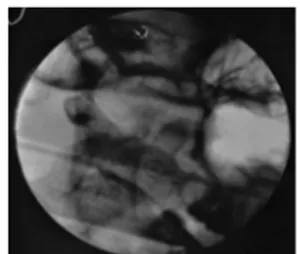
FIGURE 1. Needle broken in the pterygomandibular area; fluoroscopic intraoperative radiograph.
The Journal of Craniofacial Surgery
&
Volume 23, Number 5, September 2012 Correspondence* 2012 Mutaz B. Habal, MD
1583
the Novara University Hospital, Italy, for a broken-needle fragment after the inferior alveolar nerve block.
One month before, she had received local anesthesia for the extraction of the left inferior wisdom tooth; during the injection, the needle broke into the pterygomandibular space. The third molar was easily removed; however, after an unsuccessfully surgical search of the needle, the dentist referred his patient to our clinic. The patient complained of trismus and intraoral pain; nothing was locally objectified during the inspection. Orthopantomogram showed the location of the fragment of the dental needle in the left pterygo-mandibular space (Fig. 1).
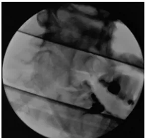
The patient was immediately taken to the operating theater for the removal of the needle via oro-endotracheal intubation under general anesthesia; the precise location of the foreign body was determined using the fluoroscope (C-arm Radius; Intermedical SRL, Grassobbio,
Italy). After a vertical 4-cm mucosal incision along the ascending ramus of the left mandible and blunt dissection, the needle was reached and removed (Fig. 2). Postoperation was uneventful, and the patient was discharged from our clinic after 2 days.
Matteo Brucoli, MD, Maura Deandreis, MD, Francesco Arcuri, MD, Arnaldo Benech, MD, PhD University of Piemonte Orientale ‘‘Amedeo Avogadro’’ Novara, Italy [email protected] REFERENCES
1. Augello M, von Jackowski J, Gra¨tz KW, et al. Needle breakage during local anesthesia in the oral cavityVa retrospective of the last 50 years with guidelines for treatment and prevention. Clin Oral Investig 2011;15:3Y8
2. Johansson B, Krekmanov L. Fragment of broken instrument removed from field of operation by an electromagnet. Br J Oral Maxillofac Surg 1987;25:265Y266
3. Moore UJ, Fanibunda K, Gross MJ. The use of a metal detector for localisation of a metallic foreign body in the floor of the mouth. Br J Oral Maxillofac Surg 1993;31:191Y192
4. Progrel MA. Broken local anesthetic needles. A case series of 16 patients, with recommendations. J Am Dent Assoc 2009; 140:1517Y1522
5. Ethunandan M, Tran AL, Anand R, et al. Needle breakage following inferior alveolar nerve block: implications and management. Br Dent J 2007;202:395Y397
FIGURE 2. Fluoroscopic intraoperative radiograph after the foreign body removal.
Correspondence The Journal of Craniofacial Surgery