molecules
ReviewResearch Progress in the Modification of Quercetin
Leading to Anticancer Agents
Alessandro Massi1 ID, Olga Bortolini1, Daniele Ragno1, Tatiana Bernardi1, Gianni Sacchetti2,
Massimo Tacchini2and Carmela De Risi1,* ID
1 Dipartimento di Scienze Chimiche e Farmaceutiche, Università di Ferrara, Via Luigi Borsari 46, I-44121 Ferrara, Italy; [email protected] (A.M.); [email protected] (O.B.);
[email protected] (D.R.); [email protected] (T.B.)
2 Dipartimento di Scienze della Vita e Biotecnologie, Sezione di Botanica Applicata, Piazzale Luciano Chiappini 3, I-44123 Ferrara, Italy; [email protected] (G.S.); [email protected] (M.T.) * Correspondence: [email protected]; Tel.: +39-0532-455287
Received: 26 June 2017; Accepted: 25 July 2017; Published: 29 July 2017
Abstract:The flavonoid quercetin (3,30,40,5,7-pentahydroxyflavone) is widely distributed in plants, foods, and beverages. This polyphenol compound exhibits varied biological actions such as antioxidant, radical-scavenging, anti-inflammatory, antibacterial, antiviral, gastroprotective, immune-modulator, and finds also application in the treatment of obesity, cardiovascular diseases and diabetes. Besides, quercetin can prevent neurological disorders and exerts protection against mitochondrial damages. Various in vitro studies have assessed the anticancer effects of quercetin, although there are no conclusive data regarding its mode of action. However, low bioavailability, poor aqueous solubility as well as rapid body clearance, fast metabolism and enzymatic degradation hamper the use of quercetin as therapeutic agent, so intense research efforts have been focused on the modification of the quercetin scaffold to obtain analogs with potentially improved properties for clinical applications. This review gives an overview of the developments in the synthesis and anticancer-related activities of quercetin derivatives reported from 2012 to 2016.
Keywords: quercetin; quercetin derivatives; methoxyflavones; anticancer; cell proliferation; cytotoxicity; multi-drug resistance (MDR)
1. Introduction
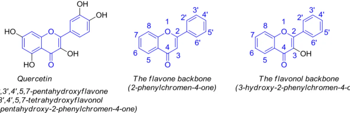
Quercetin, namely 3,30,40,5,7-pentahydroxyflavone (30,40,5,7-tetrahydroxyflavonol or 3,30,40 ,5,7-pentahydroxy-2-phenylchromen-4-one) (Figure1), belongs to the flavonol (3-hydroxyflavone) group of polyphenolic compounds known as flavonoids.
Molecules 2017, 22, 1270; doi:10.3390/molecules22081270 www.mdpi.com/journal/molecules
Review
Research Progress in the Modification of Quercetin
Leading to Anticancer Agents
Alessandro Massi 1, Olga Bortolini 1, Daniele Ragno 1, Tatiana Bernardi 1, Gianni Sacchetti 2,
Massimo Tacchini 2 and Carmela De Risi 1,*
1 Dipartimento di Scienze Chimiche e Farmaceutiche, Università di Ferrara, Via Luigi Borsari 46, I-44121 Ferrara, Italy; [email protected] (A.M.); [email protected] (O.B.); [email protected] (D.R.); [email protected] (T.B.)
2 Dipartimento di Scienze della Vita e Biotecnologie, Sezione di Botanica Applicata, Piazzale Luciano Chiappini 3, I-44123 Ferrara, Italy; [email protected] (G.S.); [email protected] (M.T.) * Correspondence: [email protected]; Tel.: +39-0532-455287
Received: 26 June 2017; Accepted: 25 July 2017; Published: 29 July 2017
Abstract: The flavonoid quercetin (3,3′,4′,5,7-pentahydroxyflavone) is widely distributed in plants, foods, and beverages. This polyphenol compound exhibits varied biological actions such as antioxidant, radical-scavenging, anti-inflammatory, antibacterial, antiviral, gastroprotective, immune-modulator, and finds also application in the treatment of obesity, cardiovascular diseases and diabetes. Besides, quercetin can prevent neurological disorders and exerts protection against mitochondrial damages. Various in vitro studies have assessed the anticancer effects of quercetin, although there are no conclusive data regarding its mode of action. However, low bioavailability, poor aqueous solubility as well as rapid body clearance, fast metabolism and enzymatic degradation hamper the use of quercetin as therapeutic agent, so intense research efforts have been focused on the modification of the quercetin scaffold to obtain analogs with potentially improved properties for clinical applications. This review gives an overview of the developments in the synthesis and anticancer-related activities of quercetin derivatives reported from 2012 to 2016.
Keywords: quercetin; quercetin derivatives; methoxyflavones; anticancer; cell proliferation; cytotoxicity; multi-drug resistance (MDR)
1. Introduction
Quercetin, namely pentahydroxyflavone (3′,4′,5,7-tetrahydroxyflavonol or 3,3′,4′,5,7-pentahydroxy-2-phenylchromen-4-one) (Figure 1), belongs to the flavonol (3-hydroxyflavone) group of polyphenolic compounds known as flavonoids.
Figure 1. Structure of quercetin and representation of flavone and flavonol backbones. Figure 1.Structure of quercetin and representation of flavone and flavonol backbones.
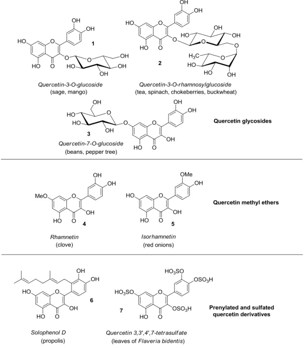
Quercetin is abundantly present in diverse plant materials (leaves, grains, fruits, and vegetables) as well as in common foods and drinks [1–3], with onions, apples, berries, broccoli, tea, and red wine serving as typical examples. In plants, quercetin can exist in either free (aglycone) or bounded form, mainly with carbohydrates (quercetin glycosides) and alcohols, mostly methanol (quercetin methyl ethers), while less frequently occurring are quercetin derivatives featuring prenyl and sulfate substituents [4]. Some representative quercetin conjugated compounds (1–7) are depicted in Figure2.
Quercetin is abundantly present in diverse plant materials (leaves, grains, fruits, and vegetables) as well as in common foods and drinks [1–3], with onions, apples, berries, broccoli, tea, and red wine serving as typical examples. In plants, quercetin can exist in either free (aglycone) or bounded form, mainly with carbohydrates (quercetin glycosides) and alcohols, mostly methanol (quercetin methyl ethers), while less frequently occurring are quercetin derivatives featuring prenyl and sulfate substituents [4]. Some representative quercetin conjugated compounds (1–7) are depicted in Figure 2.
Figure 2. Structures of quercetin derivatives 1–7 along with their occurrence in food and plants. With particular regard to quercetin glycosides, either monosaccharides or disaccharides are generally attached at the C-3 position of quercetin, however glycosylation of other hydroxyl groups may occur. For example, quercetin 3-O-glucoside 1 was found in sage and mango [5,6], with the latter containing quercetin 3-O-galactoside, rhamnoside, and xyloside too. In addition, quercetin 3-O-rhamnoside has been detected in spinach [7], hot pepper [8], and olives [9]. Quercetin
Figure 2.Structures of quercetin derivatives 1–7 along with their occurrence in food and plants.
With particular regard to quercetin glycosides, either monosaccharides or disaccharides are generally attached at the C-3 position of quercetin, however glycosylation of other hydroxyl groups may occur. For example, quercetin 3-O-glucoside 1 was found in sage and mango [5,6], with the latter containing quercetin 3-O-galactoside, rhamnoside, and xyloside too. In addition, quercetin 3-O-rhamnoside has been detected in spinach [7], hot pepper [8], and olives [9].
Molecules 2017, 22, 1270 3 of 27
Quercetin 3-O-rhamnosylglucoside (rutin, 2) is present in tea [10], spinach [7], chokeberries [11], and buckwheat [12]. Instead, quercetin 7-O-glucoside 3 occurs in beans and aerial parts of pepper tree [13,14], whereas quercetin 3-O-rhamnoside-7-O-glucoside is a typical component of pepper [8].
Once ingested, quercetin glycosides are hydrolyzed, and the released aglycone is adsorbed and metabolized giving rise to glucuronidated, methylated, and sulfated derivatives, i.e., quercetin-3-O-glucuronide, 30-O-methyl-quercetin (isorhamnetin, 5), isorhamnetin 3-O-glucuronide, and quercetin-30-O-sulfate, which enter the bloodstream [15]. Generally, neither free quercetin or its parent glycosides are detected in the plasma, wherein quercetin exists just in conjugate form.
Several decades ago quercetin attracted considerable attention as it was revealed to produce DNA mutations in bacteria. This result anticipated it as a cancer-causing agent, however inconclusive animal research as well as little evidence in humans did not seem to support this idea. On the contrary, recent years have evidenced several possible beneficial effects of quercetin, included its role in prevention and therapy of cancer [16].
In fact, quercetin functions as antioxidant, radical-scavenging, anti-inflammatory, antibacterial, antiviral, gastroprotective, immune-modulator, and is used in the treatment of obesity and cardiovascular diseases [15,17–22]. Moreover, quercetin may find application in anti-diabetic research [23], and is involved in the prevention of neurological disorders due to its neuroprotective effects [24]. Recently [25], it has been postulated that quercetin exerts protection against mitochondrial damages as a result of its ability to interact with several mitochondrial processes that are supposed to affect cells and tissues.
Due to its lipophilic nature, quercetin passes with ease through cell membranes and plays pleiotropic roles in triggering diverse intracellular routes implicated in chemoprevention (e.g., apoptosis, cell cycle, detoxification, antioxidant replication, cell invasion, angiogenesis) [26–28]. Nonetheless, there is no final proof regarding the anticancer mode of action of quercetin, with in vitro experiments showing that it could suppress multiple oncogenic signaling pathways [29,30]. On the contrary, it has been clearly demonstrated that anticancer effects of quercetin are site-specific [31].
Yet still, low bioavailability and poor solubility in water [32], together with rapid body clearance, fast metabolism, and enzymatic degradation, hamper to a great extent the clinical application of quercetin as an anticancer drug. Furthermore, quercetin has been shown to have in vitro toxic effects on normal human cell lines thereby setting limits to its possible in vivo use [33]. Speaking of which, intensive studies have been carried out on the pro-oxidant properties of quercetin and its metabolic conversion into potentially toxic quinones due to the presence of the catechol moiety [34]. In order to obtain quercetin analogs with improved properties for potential employment in cancer management, many synthetic efforts have been invested over the past ten years, and a water-soluble glycine carbamate ester quercetin prodrug (QC12) entered pre-clinical and clinical studies [35]. Regrettably, so far, no additional information about clinical development of this compound could be found in the literature.
The progresses in the synthesis and biological evaluation of quercetin derivatives as possible anticancer agents have been reviewed in 2009 by Hirpara et al. [36]. Since then, profuse research studies have been conducted on this topic, however no other literature survey has been hitherto reported, to the best of our knowledge. That being so, an overview of recent developments underlying the anticancer potential of synthetic quercetin derivatives is likely to be needed to provide an up-to-date picture of this research area.
In this review article, we gathered the diverse data published between 2012 and 2016, with attention being exclusively focused on compounds obtained using quercetin as the starting material. Among various anticancer-related effects of quercetin analogs, we selected activities against cancer cell lines as the exclusive focus of our manuscript. SciFinder database (Chemical Abstracts Service, Columbus, OH, USA) has been used as the literature source and papers in languages other than English have been excluded.
2. Synthesis and Anticancer-Related Activities of Quercetin Derivatives
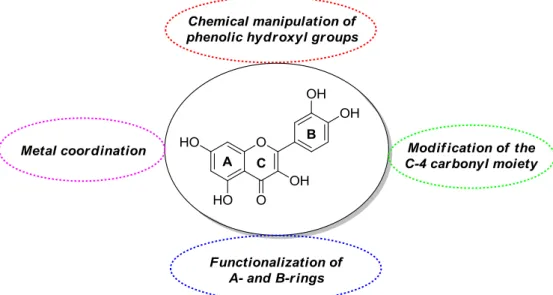
Quercetin derivatives with desirable properties for possible anticancer applications have been obtained through different synthetic routes. These include chemical manipulation of phenolic hydroxyl groups, possibly in combination with modifications at the C-4 carbonyl residue, functionalization of A- and B-rings, and metal coordination (Figure3).
Molecules 2017, 22, 1270 4 of 27
2. Synthesis and Anticancer-Related Activities of Quercetin Derivatives
Quercetin derivatives with desirable properties for possible anticancer applications have been obtained through different synthetic routes. These include chemical manipulation of phenolic hydroxyl groups, possibly in combination with modifications at the C-4 carbonyl residue, functionalization of A- and B-rings, and metal coordination (Figure 3).
Figure 3. General methodologies towards modified forms of quercetin.
The collected literature material has been organized in subsections according to the way the quercetin analogs have been synthesized. Thus, compounds obtained by elaboration of either the phenolic hydroxyl groups or the C-4 carbonyl moiety are discussed in Subsection 2.1, while Subsection 2.2 focuses on species arising from functionalization of A- and B-rings. Finally, Subsection 2.3 describes quercetin-based metal complexes. For the sake of clarity, in all cases we chose to provide just a brief description of the synthetic details to give as much information as possible about biological activities. Accordingly, Figures 4–9 and Figure 11 exclusively show the structures of the discussed quercetin derivatives, with the synthetic schemes depicting their preparation being omitted.
2.1. Chemical Modification of Phenolic Hydroxyl Groups and/or C-4 Carbonyl Moiety
Phenolic hydroxyl groups of quercetin have been mostly manipulated by etherification (O-alkylation) and esterification (O-acylation), with the O-alkylation strategy being possibly accompanied by conversion of the C-4 carbonyl group into the corresponding thiocarbonyl or selenocarbonyl functions. Also, interchange of catecholic hydroxyl groups with bioisosteric moieties has been developed.
2.1.1. O-Alkylation
It has been reported that insertion of methoxy groups into a flavone molecule results in metabolically more stable derivatives with increased solubility, bioavailability and cancer cell antiproliferative activity, as well as reduced toxic side-effects [37]. This information inspired several studies on the etherification of quercetin with either methyl or other alkyl groups in order to investigate their effect on either physico-chemical or anticancer-related properties.
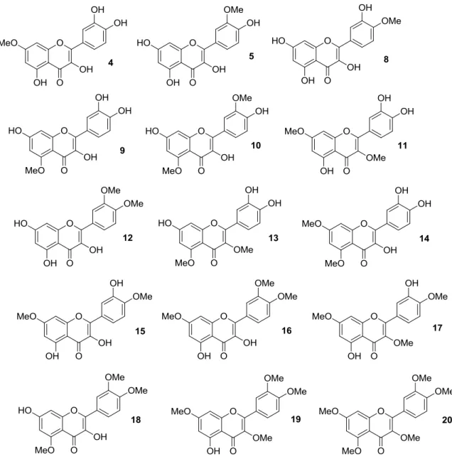
Thus, quercetin was converted into a series of monomethylated (4,5,8,9), dimethylated (10–15), trimethylated (16–18), tetramethylated (19), and pentamethylated (20) derivatives [38–42], which are shown in Figure 4.
Figure 3.General methodologies towards modified forms of quercetin.
The collected literature material has been organized in subsections according to the way the quercetin analogs have been synthesized. Thus, compounds obtained by elaboration of either the phenolic hydroxyl groups or the C-4 carbonyl moiety are discussed in Section2.1, while Section2.2 focuses on species arising from functionalization of A- and B-rings. Finally, Section2.3describes quercetin-based metal complexes. For the sake of clarity, in all cases we chose to provide just a brief description of the synthetic details to give as much information as possible about biological activities. Accordingly, Figures4–9and11exclusively show the structures of the discussed quercetin derivatives, with the synthetic schemes depicting their preparation being omitted.
2.1. Chemical Modification of Phenolic Hydroxyl Groups and/or C-4 Carbonyl Moiety
Phenolic hydroxyl groups of quercetin have been mostly manipulated by etherification (O-alkylation) and esterification (O-acylation), with the O-alkylation strategy being possibly accompanied by conversion of the C-4 carbonyl group into the corresponding thiocarbonyl or selenocarbonyl functions. Also, interchange of catecholic hydroxyl groups with bioisosteric moieties has been developed.
2.1.1. O-Alkylation
It has been reported that insertion of methoxy groups into a flavone molecule results in metabolically more stable derivatives with increased solubility, bioavailability and cancer cell antiproliferative activity, as well as reduced toxic side-effects [37]. This information inspired several studies on the etherification of quercetin with either methyl or other alkyl groups in order to investigate their effect on either physico-chemical or anticancer-related properties.
Thus, quercetin was converted into a series of monomethylated (4,5,8,9), dimethylated (10–15), trimethylated (16–18), tetramethylated (19), and pentamethylated (20) derivatives [38–42], which are shown in Figure4.
Molecules 2017, 22, 1270 5 of 27
Synthetically, the mono-protected compounds were prepared by suitable protection/deprotection steps of the phenolic hydroxyl groups in quercetin, with methyl iodide/K2CO3 system in N,N-dimethylformamide (DMF) being conveniently used at the time of installing the methyl ether moiety. Conversely, direct treatment of the flavonol starting material with methyl iodide and potassium carbonate in either DMF or acetone was carried out to yield the di-, tri- tetra- and penta-functionalized analogs.
Molecules 2017, 22, 1270 5 of 27 Synthetically, the mono-protected compounds were prepared by suitable protection/deprotection steps of the phenolic hydroxyl groups in quercetin, with methyl iodide/K2CO3 system in N,N-dimethylformamide (DMF) being conveniently used at the time of installing the methyl ether moiety. Conversely, direct treatment of the flavonol starting material with methyl iodide and potassium carbonate in either DMF or acetone was carried out to yield the di-, tri- tetra- and penta-functionalized analogs.
Figure 4. Structures of O-methylated quercetin derivatives (4), (5), and (8–20).
In early studies [38,39], it was found that 3,3′,4′,7-tetra-O-methylated quercetin 19 and 3,3′,4′,5,7-penta-O-methylated quercetin 20 could represent potential anti-multidrug resistance (MDR) agents due to their ability to influence the effects of breast cancer resistance protein (BCRP), which is known to determine resistance in cancer cells. Importantly, both 3′,4′-OMe substitution and the presence of 5-OH group were essential for optimum BCRP inhibition, whereas this activity decreased upon methylation of C-5 phenolic hydroxyl group.
In particular, investigations in Madin-Darby canine kidney (MDCK) BCRP cells evidenced that 19 and 20 were able to inhibit BCRP as a result of Hoechst 33342 and pheophorbide A accumulation, contrary to quercetin, which gave no inhibitory effect (Table 1) [39].
Figure 4.Structures of O-methylated quercetin derivatives (4), (5), and (8–20).
In early studies [38,39], it was found that 3,30,40,7-tetra-O-methylated quercetin 19 and 3,30,40,5,7-penta-O-methylated quercetin 20 could represent potential anti-multidrug resistance (MDR) agents due to their ability to influence the effects of breast cancer resistance protein (BCRP), which is known to determine resistance in cancer cells. Importantly, both 30,40-OMe substitution and the presence of 5-OH group were essential for optimum BCRP inhibition, whereas this activity decreased upon methylation of C-5 phenolic hydroxyl group.
In particular, investigations in Madin-Darby canine kidney (MDCK) BCRP cells evidenced that 19and 20 were able to inhibit BCRP as a result of Hoechst 33342 and pheophorbide A accumulation, contrary to quercetin, which gave no inhibitory effect (Table1) [39].
Table 1. Breast cancer resistance protein (BCRP) inhibition by compounds 19 and 20 compared to quercetin1. 1 19 20 Hoechst 33342 nd2 0.540±0.079 0.822±0.169 Pheophorbide A nd2 0.570±0.093 1.880±0.240 1IC
50±standard deviation (SD) values as µM.2No inhibitory effect was observed up to 10 µM.
Later, Shi et al. prepared the O-methylated compounds 4, 5 and 8–20, and evaluated their ability to inhibit cancer cell growth using a high-throughput screening (HTS) approach in an in vitro human disease-oriented cancer cell line, including melanoma (LOX-IMVI and M14), neck and head (M4E), cervical (HeLa), human breast cancer (SKBR) as well as human lung cancers (A549, H157, H460, 1792, 1944, H266, H522, Hop62, 1299, 292G, and Calu1) [40,41]. These investigations demonstrated that selective masking of the phenolic hydroxyl groups in quercetin is pivotal in determining antiproliferative activity. As a rule of thumb, it was possible to maintain inhibitory effects against all the cancer cell lines by methylation at the 40-OH and/or 7-OH positions, while the coexistence of 30- and 40-OMe groups improved activity. Also, additional introduction of a methoxy moiety may enhance the inhibition of cancer cell growth, with 30,40,7-trimethoxyquercetin (16) being more potent than 30,40-dimethoxyquercetin (12).
The antiproliferative action of 3,7-O-dimethylquercetin (11), 3,40,7-O-trimethylquercetin (17), and 3,30,40,7-O-tetramethylquercetin (19) against human androgen-refractory (DU-145 and PC-3) and androgen-sensitive (LNCaP) prostate cancer cell lines were examined as well [42], showing that methylation barely determined a weak enhancement of activity compared to parent quercetin.
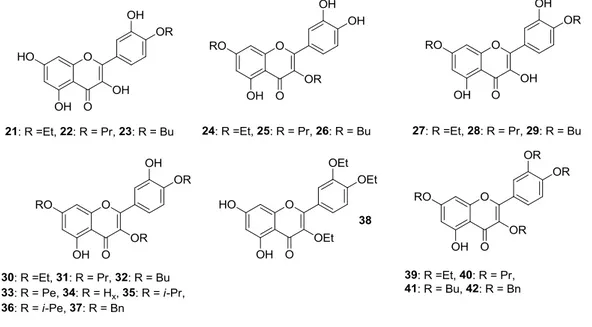
Besides, the preparation of 40-O-monoalkylated (21–23), 3,7-O-dialkylated (24–26), 40 ,7-O-dialkylated (27–29), 3,40,7-O-trialkylated (30–37), 3,30,40-O-trialkylated (38), and 3,30,40 ,7-O-tetraalkylated (39–42) derivatives of quercetin (Figure 5) was achieved in the same way as the quercetin methyl ether compounds [41,42]. Ensuing in vitro biological evaluation by the abovementioned HTS method lead Shi et al. to demonstrate that cancer cell growth inhibitory activities were retained when etherification of 3-OH and 40-OH was carried out using the long propyl chain or the short ethyl one, respectively [41]. On the contrary, introduction of two n-butyloxy moieties into the 3,7 or 40,7 sites enhanced the antiproliferative action.
Molecules 2017, 22, 1270 6 of 27
Table 1. Breast cancer resistance protein (BCRP) inhibition by compounds 19 and 20 compared to
quercetin 1.
1 19 20
Hoechst 33342 nd 2 0.540 ± 0.079 0.822 ± 0.169 Pheophorbide A nd 2 0.570 ± 0.093 1.880 ± 0.240
1 IC50 ± standard deviation (SD) values as M. 2 No inhibitory effect was observed up to 10 M. Later, Shi et al. prepared the O-methylated compounds 4, 5 and 8–20, and evaluated their ability to inhibit cancer cell growth using a high-throughput screening (HTS) approach in an in vitro human disease-oriented cancer cell line, including melanoma (LOX-IMVI and M14), neck and head (M4E), cervical (HeLa), human breast cancer (SKBR) as well as human lung cancers (A549, H157, H460, 1792, 1944, H266, H522, Hop62, 1299, 292G, and Calu1) [40,41]. These investigations demonstrated that selective masking of the phenolic hydroxyl groups in quercetin is pivotal in determining antiproliferative activity. As a rule of thumb, it was possible to maintain inhibitory effects against all the cancer cell lines by methylation at the 4′-OH and/or 7-OH positions, while the coexistence of 3′- and 4′-OMe groups improved activity. Also, additional introduction of a methoxy moiety may enhance the inhibition of cancer cell growth, with 3′,4′,7-trimethoxyquercetin (16) being more potent than 3′,4′-dimethoxyquercetin (12).
The antiproliferative action of 3,7-O-dimethylquercetin (11), 3,4′,7-O-trimethylquercetin (17), and 3,3′,4′,7-O-tetramethylquercetin (19) against human androgen-refractory (DU-145 and PC-3) and androgen-sensitive (LNCaP) prostate cancer cell lines were examined as well [42], showing that methylation barely determined a weak enhancement of activity compared to parent quercetin.
Besides, the preparation of 4′-O-monoalkylated (21–23), 3,7-O-dialkylated (24–26), 4′,7-O-dialkylated (27–29), 3,4′,7-O-trialkylated (30–37), 3,3′,4′-O-trialkylated (38), and 3,3′,4′,7-O-tetraalkylated (39–42) derivatives of quercetin (Figure 5) was achieved in the same way as the quercetin methyl ether compounds [41,42]. Ensuing in vitro biological evaluation by the abovementioned HTS method lead Shi et al. to demonstrate that cancer cell growth inhibitory activities were retained when etherification of 3-OH and 4′-OH was carried out using the long propyl chain or the short ethyl one, respectively [41]. On the contrary, introduction of two n-butyloxy moieties into the 3,7 or 4′,7 sites enhanced the antiproliferative action.
Figure 5. Cont. Figure 5. Cont.
Molecules 2017, 22, 1270 7 of 27
Molecules 2017, 22, 1270 7 of 27
Figure 5. Structures of O-alkylated quercetin derivatives (21–50). Abbreviations: Et: ethyl, Pr: propyl,
Bu: butyl, Pe: pentyl, Hx: hexyl, i-Pr: isopropyl, i-Pe: isopentyl, Bn: benzyl.
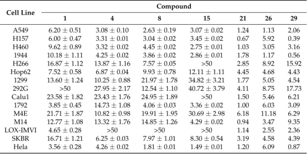
Speaking of these studies, cytotoxicity data of the most representative O-alkylated quercetin derivatives are listed in Table 2.
Table 2. Cytotoxicity data of the most representative O-alkylated quercetins compared to quercetin
by high-throughput screening (HTS) method 1.
Cell Line Compound
1 4 8 15 21 26 29 A549 6.20 ± 0.51 3.08 ± 0.10 2.63 ± 0.19 3.07 ± 0.02 1.24 1.13 2.06 H157 6.00 ± 0.47 3.31 ± 0.01 3.04 ± 0.02 3.45 ± 0.02 0.67 5.92 0.39 H460 9.62 ± 0.89 3.32 ± 0.02 4.45 ± 0.02 2.75 ± 0.01 1.03 3.05 3.16 1944 10.18 ± 1.11 4.25 ± 0.02 3.86 ± 0.02 2.86 ± 0.01 1.78 1.17 0.56 H266 16.87 ± 1.12 13.87 ± 1.16 7.57 ± 0.05 50 2.85 8.92 15.92 Hop62 7.52 ± 0.58 6.87 ± 0.04 9.93 ± 0.78 12.11 ± 1.11 4.45 4.68 4.43 1299 13.60 ± 1.24 10.25 ± 0.88 21.97 ± 1.78 34.82 ± 3.21 1.77 5.05 4.54 292G 50 27.95 ± 2.17 12.54 ± 1.10 40.72 ± 3.79 4.11 8.75 17.73 Calu1 23.58 ± 1.82 23.43 ± 1.76 24.95 ± 1.89 50 1.50 5.46 6.21 1792 3.85 ± 0.45 14.73 ± 1.08 4.06 ± 0.03 3.36 ± 0.02 1.00 6.03 3.09 M4E 21.71 ± 1.87 10.82 ± 0.98 19.91 ± 1.95 30.69 ± 2.98 6.18 11.18 6.29 M14 12.77 ± 1.08 13.32 ± 1.76 14.85 ± 1.26 4.29 ± 0.02 0.94 3.47 9.35 LOX-IMVI 4.65 ± 0.28 50 50 50 1.14 2.55 2.36 SKBR 16.71 ± 1.21 6.25 ± 0.03 7.97 ± 1.01 8.30 ± 0.54 3.19 4.58 4.39 Hela 3.56 ± 0.28 4.26 ± 0.02 1.81 ± 0.01 1.49 ± 0.01 1.20 6.09 0.87 1 IC50 ±SD or IC50 values as M.
Figure 5.Structures of O-alkylated quercetin derivatives (21–50). Abbreviations: Et: ethyl, Pr: propyl, Bu: butyl, Pe: pentyl, Hx: hexyl, i-Pr: isopropyl, i-Pe: isopentyl, Bn: benzyl.
Speaking of these studies, cytotoxicity data of the most representative O-alkylated quercetin derivatives are listed in Table2.
Table 2.Cytotoxicity data of the most representative O-alkylated quercetins compared to quercetin by high-throughput screening (HTS) method1.
Cell Line Compound
1 4 8 15 21 26 29 A549 6.20±0.51 3.08±0.10 2.63±0.19 3.07±0.02 1.24 1.13 2.06 H157 6.00±0.47 3.31±0.01 3.04±0.02 3.45±0.02 0.67 5.92 0.39 H460 9.62±0.89 3.32±0.02 4.45±0.02 2.75±0.01 1.03 3.05 3.16 1944 10.18±1.11 4.25±0.02 3.86±0.02 2.86±0.01 1.78 1.17 0.56 H266 16.87±1.12 13.87±1.16 7.57±0.05 >50 2.85 8.92 15.92 Hop62 7.52±0.58 6.87±0.04 9.93±0.78 12.11±1.11 4.45 4.68 4.43 1299 13.60±1.24 10.25±0.88 21.97±1.78 34.82±3.21 1.77 5.05 4.54 292G >50 27.95±2.17 12.54±1.10 40.72±3.79 4.11 8.75 17.73 Calu1 23.58±1.82 23.43±1.76 24.95±1.89 >50 1.50 5.46 6.21 1792 3.85±0.45 14.73±1.08 4.06±0.03 3.36±0.02 1.00 6.03 3.09 M4E 21.71±1.87 10.82±0.98 19.91±1.95 30.69±2.98 6.18 11.18 6.29 M14 12.77±1.08 13.32±1.76 14.85±1.26 4.29±0.02 0.94 3.47 9.35 LOX-IMVI 4.65±0.28 >50 >50 >50 1.14 2.55 2.36 SKBR 16.71±1.21 6.25±0.03 7.97±1.01 8.30±0.54 3.19 4.58 4.39 Hela 3.56±0.28 4.26±0.02 1.81±0.01 1.49±0.01 1.20 6.09 0.87 1IC 50±SD or IC50values as µM.
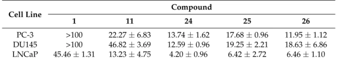
With particular regard to human prostate cancer cells, Al-Jabban et al. concluded that antiproliferative activity strongly depended on either length or steric hindrance of the introduced alkyl chain [42]. Indeed, cancer cell growth greatly dropped when linear long or bulky alkyl groups were simultaneously introduced into C-3, C-40and C-7 hydroxyl groups, as observed for compounds (31–34) and (35,36), respectively. On the other hand, the derivative (30) appended with the short, linear ethyl group showed a slightly increased activity, similarly to the corresponding methyl analog (17, Figure4). However, no significant change in activity was detected for 3,30,40-O-triethylquercetin (38). Importantly, the potency of 3,7-O-dialkylated derivatives (24–26) was 2–11 times higher than quercetin (Table3), with this behaviour being also observed for the corresponding dimethylated compound (11, Figure4).
Table 3. Antiproliferative activities of derivatives 11 and 24–26 compared to quercetin in human prostate cancer cells1,2.
Cell Line Compound
1 11 24 25 26
PC-3 >100 22.27±6.83 13.74±1.62 17.68±0.96 11.95±1.12 DU145 >100 46.82±3.69 12.59±0.96 19.25±2.21 18.63±6.86 LNCaP 45.46±1.31 13.23±4.75 4.20±0.96 6.42±2.72 6.46±1.10
1IC
50±SD values as µM.2After 72 h incubation.
It should be mentioned that the structures of the 40,7-O-dialkylquercetins reported by Shi et al. [41] have been found to be wrong by heteronuclear multiple bond correlation (HMBC) nuclear magnetic resonance (NMR) experiments, and were corrected as the corresponding 3,7-O-dialkylated isomers [42].
A recent work by Khan and coworkers evidenced that 3,40,7-O-triethylquercetin (30, Figure5) was able to inhibit cell proliferation in colon (HCT-116) cancer cells (IC50= 50 µM, 24 h incubation). Moreover, it behaved as apoptosis-inducer in the same cancer cell line without affecting normal cells growth [43]. It is worthwhile pointing out that 30 is supposed to take action through endoplasmic reticulum (ER) stress and a mitochondria-mediated pathway.
A three-step procedure involving peracetylation of quercetin followed by alkylation with a suitable alkyl chloride and base-mediated deacetylation gave access to 7-O-butylquercetin 43 and 7-O-geranylquercetin 44, which are shown in Figure5[44,45]. These compounds showed a moderate solubility (180 µM) in Dulbecco’s modified eagle medium (DMEM) [45], and exhibited much stronger antiproliferative effects than quercetin in estrogen receptor-positive human breast cancer cell line (MCF-7), likely due to their better accumulation capability [44]. More precisely, the proliferation inhibitory activity of 43 and 44 depended on their apoptosis-inducing effects which were anyhow higher than those of quercetin. In this regard, it was demonstrated that the apoptotic process of MCF-7 cells occurred through a caspase-independent Endonuclease G (Endo G)-induced mitochondrial route, unlike quercetin.
It is worthy of note that compounds 43 and 44 did not affect normal breast epithelial (MCF-10A) cells and were also effective in estrogen receptor-negative MDA-MB-231 breast cancer cells. Furthermore, 43 and 44 were proposed to possess reversal activities on MDR cancer cells, but no evidence in support of this hypothesis was furnished.
Further studies revealed that 44 had strong cytotoxicity on human colon (CaCo-2), human lung (NCI-H446 and A549) as well as human gastric (MGC-803 and SGC-7901) cancer cells thereby revealing potential antiproliferative properties [45]. In all cases, the observed activity proved to be higher as compared to quercetin. For the sake of clarity, the most relevant biological data regarding compounds 43and 44 are detailed in Table4.
Molecules 2017, 22, 1270 9 of 27
Table 4.Cytotoxicity data of compounds 43 and 44 compared to quercetin1,2.
Cell Line Compound
1 43 44 MCF-7 343 43.5±2.1 3 22.6±2.23 38.64 20.24 CaCo-2 340 66.8 43.7 NCI-H446 68.9 27.6 A549 77.2 29.5 MGC-803 80.6 25.4 SGC-7901 75.7 18.5 1IC
50±SD or IC50values as µM.2After 48 h incubation.3Data taken from [44].4Data taken from [45].
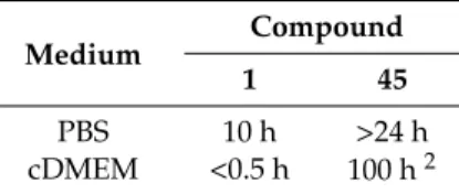
In order to extend previous results on quercetin conjugates bearing a pivaloxymethyl (POM) promoiety at the 3 or the 7 position [46], the 3,7-bis-O-pivaloxymethyl (POM) quercetin (45, Figure5) was prepared by sequential K2CO3-promoted alkylation of quercetin diphenylmethylketal with pivaloxymethyl iodide (POM-I) and deprotection [47]. In-depth studies evidenced that 45 had great stability in Dulbecco’s modified eagle medium complete (cDMEM) (Table5) and efficient uptake inside cells wherein it was selectively hydrolyzed to the corresponding 3-O-POM-quercetin, with no trace of other metabolites (i.e., 7-O-POM-quercetin or quercetin) being detected.
Table 5.Stability of compound 45 compared to quercetin in diverse media1.
Medium Compound
1 45
PBS 10 h >24 h
cDMEM <0.5 h 100 h2 1Half-time (t
1/2) values.2Compound 45 is converted exclusively to 3-O-POM-quercetin. PBS: phosphate-buffered saline; cDMEM: Dulbecco’s modified eagle medium complete.
Significant cytostatic activity of 45 was observed in MCF-7, HCT-116, and DU-145 cancer cell lines, as compared to quercetin which gave no inhibition of cell proliferation. Importantly, 45 displayed a cancer cell specific cytostatic effect, and no action was demonstrated on normal human diploid fibroblast (HS 27) cell line. Mechanistically, it has been proposed that the quercetin-POM conjugate 45 operates via a different pathway against quercetin, with cell cycle arrest taking place in the G0/G1 phase.
In a proof-of-concept study, Chong et al. demonstrated that 7-O-POM-quercetin (46, Figure5) was able to reverse MDR in drug-resistant MES-SA/Dx5 cells derived from the drug-sensitive human uterine sarcoma (MES-SA) cell line (Table6) [48,49].
Table 6. Multi-drug resistance (MDR)-reversing activity of compound 46 compared to quercetin and verapamil1.
Anticancer Drug Modulator
2
None (IC50) Verapamil (IC50/FR) 1 (IC50/FR) 46 (IC50/FR)
Doxorubicine 8.18±0.01 0.12±0.01/68.3 4.26±0.32/1.9 0.34±0.09/24.1 Actinomycin D 13.10±0.34 0.23±0.02/57.0 4.68±1.00/2.8 0.41±0.01/32.0 Vinblastine 12.25±0.19 0.24±0.01/51.0 4.90±0.13/2.5 0.43±0.06/28.5 Paclitaxel 10.53±0.21 0.22±0.04/47.9 4.66±0.11/2.3 0.41±0.04/25.7 1IC
50±SD values as µM (p < 0.01).2Used at 5 µM concentration. FR = fold-reversal (IC50of anticancer drug alone/IC50of anticancer drug combined with the modulator).
Mechanistically, it was evidenced competition of 46 with verapamil binding to the P-glycoprotein (P-gp), which is a major MDR target. Moreover, 46 proved to be considerably more potent than quercetin and as active as verapamil in inhibiting the drug efflux mediated by P-gp.
Importantly, 46 evidenced accumulation inside MES-SA/Dx5 cells wherein it persisted along with its hydrolyzed product quercetin and quercetin metabolites (glucuronide and sulfate) for more than 48 h. As a result, the intracellular levels of 46 were adequately high to elicit the increased MDR-reversal effect as compared to quercetin.
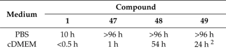
Suitably protected quercetin derivatives were reacted with iodomethyl isopropyl carbonate (POC-I)/K2CO3 system in either DMF or DMF/acetone mixture and the compounds obtained were then deprotected to afford the quercetin conjugates 47–49 (Figure 5) bearing an isopropyloxycarbonylmethoxy (POC) group at 3-OH and/or 7-OH [50]. These species were deeply studied with regard to solubility, stability, permeability and intracellular metabolism. Compounds (47) and (49) were poorly soluble in phosphate-buffered saline (PBS) differently to 48 which proved to dissolve well in the same medium even at high concentrations. Anyhow, complete dissolution of all derivatives was observed in cDMEM (up to 100 µM concentration). With regard to stability, it has been demonstrated that quercetin-POC conjugates were almost as stable as the quercetin-POM derivatives [46,47]. All compounds featured high stability in PBS (t1/2>96 h) (Table7), while either decomposition or hydrolysis occurred in cell-free culture medium. Thus, the 3,7-bis-O-POC derivative 49was hydrolyzed into 3-O-POC compound 48, whereas the 7-functionalized analog 47 gave rise to decomposition and/or hydrolysis to the mother quercetin. Among the series, 3-O-POC 48 showed the best stability profile in term of resistance to both decomposition and hydrolysis.
Table 7. Stability of quercetin-isopropyloxycarbonylmethoxy (POC) conjugates 47–49 compared to quercetin1. Medium Compound 1 47 48 49 PBS 10 h >96 h >96 h >96 h cDMEM <0.5 h 1 h 54 h 24 h2 1t
1/2values.2Compound 49 is hydrolyzed to 48.
Besides, membrane permeability assays assessed that the 7-conjugated derivative 47 behaved as quercetin, while 3-O-POC-quercetin 48 was the less permeable. In any case, the permeability of 48 is worthwhile noting as the corresponding 3-O-POM conjugate was totally impermeable [46]. Remarkably, no data could be obtained for 49 due to its low solubility in PBS at the concentration (25 µM) used for the membrane permeability test.
Cell-line-dependent hydrolytic and metabolic profiles were observed for quercetin derivatives 47–49. On the one hand, they were smoothly converted to quercetin and its metabolites in MCF-7 cell line, with quercetin glucuronide being predominantly formed in all cases, according to literature data [51]. It should be highlighted that these results were completely different from those observed for the quercetin-POM analogs, which have been shown to be less prone to both intracellular hydrolysis and metabolism [46,47]. In addition, 3-O-POC-quercetin 48 was easily hydrolyzed and metabolized contrary to 3-O-POM-quercetin and 3,7-bis-O-POM-quercetin 45 [46,47].
On the other hand, 47–49 underwent slow hydrolysis and low metabolism in HCT-116 cells. In particular, 7-O-POC-quercetin (47) hydrolyzed to quercetin but neither of its metabolites was detected, while 3-O-POC-quercetin (48) proved to be very stable (up to 12 h) giving no trace of quercetin. This metabolic profile was also typical of 3,7-bis-O-POC-quercetin (49), but its hydrolysis hastened (t1/2∼=3 h) in relation to cell-free medium (t1/2= 24 h, Table7). In this case, 3-O-POC-quercetin (48) was formed as the exclusive hydrolysis product.
Cytotoxic activities of 47–49 were strictly related to their stability properties. As a matter of fact, low antiproliferative effects against MCF-7 cells were observed for the POC-protected quercetins
Molecules 2017, 22, 1270 11 of 27
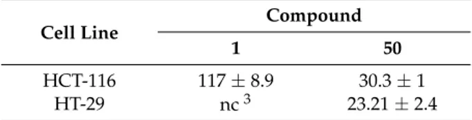
likely due to enhanced passive transport, intracellular hydrolysis, and metabolism. More precisely, compounds 47–49 were as active as quercetin. On the contrary, 47 and 49 displayed higher cytotoxicity than quercetin in HCT-116 cells, with 49 being more effective than 47. Given the slow hydrolysis and metabolism of 47 and 49 in HCT-116 cells, both these compounds and their hydrolyzed products were present at concentrations high enough to enhance cytotoxicity. Importantly, 48 was not cytotoxic at all. In addition to alkyl halides, 2,3-dichloro-1,4-naphtoquinone has been employed to alkylate quercetin using N,N-diisopropylethylamine as the base giving chloronaphtoquinone quercetin (50, CHNQ), which is depicted in Figure 5. This compound featured a 3-fold higher cytotoxicity than quercetin in colorectal (HCT-116 and HT-29) cancer cells (Table8), and strong apoptosis-inducing effects, too, have been observed [52].
Table 8.Anticancer activities of compound 50 compared to quercetin1,2.
Cell Line Compound
1 50
HCT-116 117±8.9 30.3±1
HT-29 nc3 23.21±2.4
1IC
50±SD values as µM.2After 24 h incubation.3Proliferation did not change.
Furthermore, likely due to the presence of the 1,4-napthoquinone framework, treatment of cells with 50 resulted in a potent generation of oxidative stress leading to reactive oxygen species (ROS)-induced autophagy in vitro. In particular, the authors highlighted that complete autophagy occurred in HCT-116 cells, while incomplete autophagy took place in the HT-29 ones. Herein, CHNQ promoted LC3 lipidation, with the formation of acidic vacuoles being not observed.
It should be pointed out that conversion of quercetin into an isomeric chloronaphtoquinone derivative has been previously reported by Danihelová et al. [53]. This structural modification led to lower the antioxidant properties of quercetin, but enhanced the cancer cell inhibitory activities. However, the chloronaphtoquinone derivative also featured cytolytic effects towards non-cancer murine fibroblast (NIH-3T3) at a concentration of 100 µM, but the total degeneration of cancer cells (HeLa) took place at lower concentrations (50 µM).
2.1.2. O-Alkylation and C-4 Carbonyl Group Modification
The O-alkylation of quercetin has been conveniently coupled with the bioisosteric conversion of the C-4 carbonyl group into the corresponding thiocarbonyl or selenocarbonyl moieties. So, synthetic routes involving suitable methylation of quercetin followed by either oxygen/sulfur or oxygen/selenium exchange and deprotection have been carried out to provide the sulfur- and seleno-compounds 51–54, which are shown in Figure6.
Molecules 2017, 22, 1270 11 of 27
CHNQ), which is depicted in Figure 5. This compound featured a 3-fold higher cytotoxicity than quercetin in colorectal (HCT-116 and HT-29) cancer cells (Table 8), and strong apoptosis-inducing effects, too, have been observed [52].
Table 8. Anticancer activities of compound 50 compared to quercetin 1,2. Cell Line Compound
1 50
HCT-116 117 ± 8.9 30.3 ± 1
HT-29 nc 3 23.21 ± 2.4
1 IC50 ±SD values as M. 2 After 24 h incubation. 3 Proliferation did not change.
Furthermore, likely due to the presence of the 1,4-napthoquinone framework, treatment of cells with 50 resulted in a potent generation of oxidative stress leading to reactive oxygen species (ROS)-induced autophagy in vitro. In particular, the authors highlighted that complete autophagy occurred in HCT-116 cells, while incomplete autophagy took place in the HT-29 ones. Herein, CHNQ promoted LC3 lipidation, with the formation of acidic vacuoles being not observed.
It should be pointed out that conversion of quercetin into an isomeric chloronaphtoquinone derivative has been previously reported by Danihelová et al. [53]. This structural modification led to lower the antioxidant properties of quercetin, but enhanced the cancer cell inhibitory activities. However, the chloronaphtoquinone derivative also featured cytolytic effects towards non-cancer murine fibroblast (NIH-3T3) at a concentration of 100 μM, but the total degeneration of cancer cells (HeLa) took place at lower concentrations (50 μM).
2.1.2. O-Alkylation and C-4 Carbonyl Group Modification
The O-alkylation of quercetin has been conveniently coupled with the bioisosteric conversion of the C-4 carbonyl group into the corresponding thiocarbonyl or selenocarbonyl moieties. So, synthetic routes involving suitable methylation of quercetin followed by either oxygen/sulfur or oxygen/selenium exchange and deprotection have been carried out to provide the sulfur- and seleno-compounds 51–54, which are shown in Figure 6.
Martins et al. reported the synthesis of analogs 51–53 via chalcogenation of the quercetin-derived 3,3′,4′,7-O-tetramethyl compound (19, Figure 4) [54]. All the compounds have been tested on nine human cancer cell lines, namely melanoma cells (A375), colorectal adenocarcinoma cells (HCT-15), pancreatic adenocarcinoma cells (BxPC3), MCF-7 cells and the multidrug-resistant variant MCF-7/ADR, cervical adenocarcinoma cells (A431) and the corresponding cisplatin-resistant one (A431/Pt), cisplatin-sensitive and cisplatin-resistant ovarian adenocarcinoma cells (2008 and C13*). For the purpose of comparison, the same cancer cell lines were used in parallel experiments with quercetin, 19, and cisplatin.
Figure 6. Structures of quercetin derivatives 51–54 obtained by manipulation of C-4 carbonyl moiety. Figure 6.Structures of quercetin derivatives 51–54 obtained by manipulation of C-4 carbonyl moiety.
Martins et al. reported the synthesis of analogs 51–53 via chalcogenation of the quercetin-derived 3,30,40,7-O-tetramethyl compound (19, Figure 4) [54]. All the compounds have been tested on nine human cancer cell lines, namely melanoma cells (A375), colorectal adenocarcinoma cells (HCT-15), pancreatic adenocarcinoma cells (BxPC3), MCF-7 cells and the multidrug-resistant variant MCF-7/ADR, cervical adenocarcinoma cells (A431) and the corresponding cisplatin-resistant one (A431/Pt), cisplatin-sensitive and cisplatin-resistant ovarian adenocarcinoma cells (2008 and C13*). For the purpose of comparison, the same cancer cell lines were used in parallel experiments with quercetin, 19, and cisplatin.
Thus, the selenium compound 51 showed a 9-fold and 3-fold higher cytotoxicity than quercetin and cisplatin, respectively, while 19 proved to be 3-fold less cytotoxic than quercetin (Table9).
Table 9.Cytotoxicity of compound 51 compared to quercetin, 19 and cisplatin.1,2,3.
Cell Line Compound
1 51 19 Cisplatin A375 25.13±2.62 2.21±1.07 69.72±3.32 3.12±1.13 HCT-15 16.35±2.29 2.23±1.01 68.11±2.35 12.31±1.26 BxPC3 24.12±1.85 2.42±1.19 >100 11.43±1.29 MCF-7 20.90±3.44 3.08±1.98 >100 7.6±2.49 MCF-7/ADR 22.15±2.14 (1.1) 3.35±1.58 (1.1) >100 8.41±1.22 (16) A431 23.04±1.07 3.11±1.23 57.54±2.28 1.62±1.25 A431/Pt 34.37±3.22 (1.5) 3.78±1.52 (1.2) 82.25±2.77 (1.4) 3.42±1.08 (2.1) 2008 21.18±1.84 2.09±1.27 >100 2.17±1.37 C13* 37.62±3.82 (1.8) 2.29±1.93 (1.1) >100 22.26±1.86 (10.3) 1IC
50±SD values as µM (p < 0.05).2After 72 h incubation.3Resistance factor (RF: IC50resistant cell/IC50parent cell line) values are reported in parentheses.
This result suggested that the selenocarbonyl moiety rather than the flavonoid core was responsible for the observed biological effects. On the contrary, unreproducible data were observed for the sulfur-containing derivatives 52 and 53 as a result of their low stability in solution. Furthermore, protected selenoquercetin 51 was able to overcome cisplatin-resistance due to comparable cytotoxic action against both the cisplatin-sensitive and cisplatin resistant cell lines. Remarkably, preliminary studies aimed at understanding the mechanism of cytotoxicity in MCF-7 cells evidenced that 51 hampered thioredoxin reductase (TrxR) activity, whilst it lacks efficacy to affect the glutathione peroxidase (GPx)/glutathione reductase (GR) redox system.
Exhaustive O-methylation of quercetin to the 3,30,40,5,7-O-pentamethyl derivative (20, Figure4) followed by thionation gave access to compound 54, which was eventually deprotected yielding 53[55,56]. It has been shown that both compounds possess in vitro antiangiogenic and antiproliferative properties [55]. As a matter of fact, they were able to inhibit the migration of human umbilical endothelial vascular cells (HUVECs) promoted by the vascular endothelial growth factor (VEGF). However, the antiangiogenic activity of thiocarbonyl compounds 53 and 54 was inferior to that of both quercetin and 20, with 54 being more active than 53. Besides, 53 and 54 had antiproliferative activity towards MCF-7 cancer cell line and the doxorubicin-resistant variant MCF-7/DX, but much higher concentrations (approximately a 10 to 100-fold increase) were required compared to those determining the antiangiogenic action. In particular, the observed antiproliferative effects were in the order quercetin > 53 > 20 > 54. Overall, derivative 20 was found to be the top of the line as it optimally balanced the high antiangiogenic activity with minimal toxicity. Further antiproliferative studies using breast cancer cell line MDA-MB-231 evidenced that the 4-thio compounds, and in particular 53, had greater effects than 20 and the parent quercetin [56]. However, 53 did not reach the IC50 at 10 µM. Moreover, 20, 53, and 54 proved to be less active than quercetin towards both MCF-7 and MCF-7/DX cell lines, with the lowest activity being observed in the latter. This result was supposed to depend on the fact that these compounds are likely to act as substrates of the P-gp efflux pump, which is known
Molecules 2017, 22, 1270 13 of 27
to be over expressed in the MCF-7/DX cell line. It is worthwhile noting that in all cases no stability problems of 53 were pointed out, in contrast with others [54].
2.1.3. Replacement of Catecholic Hydroxyl Groups
Along their studies aimed at separating the biological activities of quercetin from its antioxidant features, Cho et al. undertook the structural modification of quercetin by replacing the catechol hydroxyl groups with bioisosteric fluorine atoms [57]. Accordingly, a fragmentation-acylation-ring closure strategy was applied to convert quercetin into the corresponding 30,40-difluoro derivative 55 (Figure7).
Molecules 2017, 22, 1270 13 of 27
Figure 7. Structure of quercetin derivative 55 obtained via replacement of catecholic hydroxyl groups.
As anticipated, 55 proved to be stable against oxidative decomposition. Most likely, the non-radical-producing fluorine moieties prevent the production of o-quinone and p-quinone methide species that are typically formed upon oxidation of quercetin [58]. Nevertheless, 55 was as active as quercetin in the MCF-7 cell line, and a modest effect in hepatoma (Huh-7) cell line was also observed. Besides, it has been evidenced that the profile of early apoptotis of 55 resembles that of quercetin, albeit the latter induces cells in late apoptotic-necrotic stage more effectively.
Figure 8. Structures of O-acylated quercetin derivatives 56–63.
Figure 7.Structure of quercetin derivative 55 obtained via replacement of catecholic hydroxyl groups.
As anticipated, 55 proved to be stable against oxidative decomposition. Most likely, the non-radical-producing fluorine moieties prevent the production of o-quinone and p-quinone methide species that are typically formed upon oxidation of quercetin [58]. Nevertheless, 55 was as active as quercetin in the MCF-7 cell line, and a modest effect in hepatoma (Huh-7) cell line was also observed. Besides, it has been evidenced that the profile of early apoptotis of 55 resembles that of quercetin, albeit the latter induces cells in late apoptotic-necrotic stage more effectively.
2.1.4. O-Acylation
Introduction of ester and urethane moieties into the quercetin scaffold produced compounds with improved cytotoxic action. This is likely due to a better bioavailability as a possible result of lipophilization [53,59,60]. During the period 2012–2016, synthesis and anticancer activities of O-acylated quercetin derivatives 56–63 (Figure8) were reported.
Danihelová et al. prepared fifteen quercetin-derived compounds through condensation or selective protection reactions followed by acylation with acyl chlorides [53]. Among the series, pentaacetyl quercetin (56), di(tetraacetylquinoyl)quercetin (57), and tri(diacetylcaffeoyl)quercetin (58) exhibited the highest cytotoxicity towards HeLa cells and the non-cancerous cell line NIH-3T3. Notably, all these compounds were more effective than quercetin regardless of cell type.
Conversion of quercetin into the corresponding diphenylmethylketal followed by esterification with aspirin at the 3- and 7-OH gave access to quercetin aspirinates 59–61 showing higher cytotoxic effects against liver (HepG2) and promyelocytic leukemia (HL-60) cancer cells than quercetin [61].
Results of the cytotoxic activities of compounds 56–61 are reported in Table10.
It has been previously demonstrated that quercetin amino-acid conjugates possess properties (i.e., water solubility, hydrolytic stability, cell permeability) superior to those of quercetin [62]. Based on this result, Kim et al. prepared six quercetin derivatives appended with alanine or glutamic acid residues at 3-O and/or 7-O sites [63]. Key steps in the synthetic strategy concerned the selective protection/deprotection of quercetin hydroxyl groups and the coupling of the intermediates obtained with suitably protected alanine and glutamic acid compounds.
Molecules 2017, 22, 1270 14 of 27
Figure 7. Structure of quercetin derivative 55 obtained via replacement of catecholic hydroxyl groups.
As anticipated, 55 proved to be stable against oxidative decomposition. Most likely, the non-radical-producing fluorine moieties prevent the production of o-quinone and p-quinone methide species that are typically formed upon oxidation of quercetin [58]. Nevertheless, 55 was as active as quercetin in the MCF-7 cell line, and a modest effect in hepatoma (Huh-7) cell line was also observed. Besides, it has been evidenced that the profile of early apoptotis of 55 resembles that of quercetin, albeit the latter induces cells in late apoptotic-necrotic stage more effectively.
Figure 8. Structures of O-acylated quercetin derivatives 56–63. Figure 8.Structures of O-acylated quercetin derivatives 56–63.
Table 10.Cytotoxic data of analogs 56–61 compared to quercetin1.
Cell Line Compound
1 56 57 58 59 60 61 HeLa 35.5±1.12 29.6±1.92 19.5±0.82 16.5±1.52 NIH-3T3 20.9±0.92 15.5±0.72 16.1±0.42 10.6±0.12 HL-60 >100 68.713 69.293 >1003 HepG2 >100 54.223 38.493 55.803 1IC
50±SD or IC50values as µM.2After 72 h incubation.3After 48 h incubation.
The quercetin-amino acid analogs were tested for their cytotoxicity and ability to modulate MDR using the MES-SA cell line and the corresponding drug-resistant MES-SA/Dx5, that is known to overexpress P-gp [49]. At the concentration levels applied for MDR modulation (5 µM), the quercetin-amino acid conjugates showed no cytotoxic action in MES-SA cell line, likewise quercetin. Importantly, addition of the latter or the quercetin-amino acid derivatives did not affect the cytotoxic properties of a given anticancer agent, similarly to verapamil which has been used as the positive control. With regard to MES-SA/Dx5 cell line, MDR-reversal activity of the quercetin-amino acid
Molecules 2017, 22, 1270 15 of 27
compounds was strictly dependent on either the nature or position of the amino acid moieties. The 7-O-functionalized compounds in the series displayed the most potent effects, and the best results were evidenced for the quercetin-7-O-glutamic acid conjugate 62. As depicted in Table11, this compound proved to be 30.5-fold more active (fold-reversal, FR= 58.6) than quercetin (FR = 1.9) in reversing MDR towards doxorubicin, and was potent as much as the doxorubicin-resistance reversing drug verapamil (FR = 68.0). Additionally, 62 showed 13.8–14.8-times enhanced MDR-reversal effects against other anticancer drugs, including actinomycin D, vinblastine, and paclitaxel.
Table 11.MDR-reversing activity of compound 62 compared to quercetin and verapamil1.
Anticancer Drug Modulator
2 None Verapamil 1 62 Doxorubicine 8.20 0.12 4.26 0.14 Actinomycin D 0.13 0.23 4.68 0.34 Vinblastine 0.11 0.24 4.90 0.33 Paclitaxel 0.10 0.22 4.66 0.32 1IC
50values as µM.2Used at 5 µM concentration.
It is worthy of note that the MDR modulatory activity of 62 was not dictated by the stereochemistry of the amino acid promoiety. As a matter of fact, the synthetically prepared enantiomer of 62 was a potent MDR modulator (FR = 52.2), though less than 62 itself. Moreover, flow cytometric analysis and P-gp ATPase test evidenced that 62 inhibited the drug efflux by P-gp, and stimulated ATPase activity of P-gp by interaction with its drug-binding site, respectively.
Evaluation of the physico-chemical properties of compound 62 highlighted that the presence of the glutamic acid residue accounted for enhanced solubility, stability, and cellular uptake of quercetin. Indeed, 62 was markedly soluble in aqueous medium up to 400 µM, while quercetin solubility harshly dropped at concentration >100 µM. In comparison with quercetin, compound 62 possessed high stability both in PBS (t1/2> 72 h) and Roswell Park Memorial Institut (RPMI)-1640 complete culture medium (cRPMI), with decomposition occurring only after 9.3 h (Table12).
Table 12.Stability of quercetin-glutamic acid conjugate 62 compared to quercetin1.
Medium Compound
1 62
PBS 10.3 h >72 h
cRPMI 0.4 h 9.3 h
1t
1/2values. PBS: phosphate-buffered saline. cRPMI: Roswell Park Memorial Institut (RPMI)-1640 complete culture medium.
Importantly, the intracellular level of 62 remained adequately high for a prolonged period of time (6–24 h) due to slow metabolism to quercetin and quercetin metabolites (i.e., quercetin glucuronide and quercetin sulfate) that co-existed with 62. This result may profile the use of 62 as a safe MDR modulator given the riskless nature of quercetin. Quercetin was also reacted with n-butyl-isocyanate to obtain the 3,30,40,7-O-tetraacylated derivative 63. This compound proved to inhibit the proliferation of MCF-7 cells in vitro [64], with the IC50value obtained being 36 µM compared to 128 µM for quercetin.
2.2. Functionalization of A- and B-Rings
The quercetin framework has been suitably modified by the insertion of sulfonate, prenyl, aminomethyl, and phenylethenyl appendages into A- and B-rings to provide derivatives 64–71 which are listed in Figure9.
Molecules 2017, 22, 1270 16 of 27 It is worthy of note that the MDR modulatory activity of 62 was not dictated by the stereochemistry of the amino acid promoiety. As a matter of fact, the synthetically prepared enantiomer of 62 was a potent MDR modulator (FR = 52.2), though less than 62 itself. Moreover, flow cytometric analysis and P-gp ATPase test evidenced that 62 inhibited the drug efflux by P-gp, and stimulated ATPase activity of P-gp by interaction with its drug-binding site, respectively.
Evaluation of the physico-chemical properties of compound 62 highlighted that the presence of the glutamic acid residue accounted for enhanced solubility, stability, and cellular uptake of quercetin. Indeed, 62 was markedly soluble in aqueous medium up to 400 M, while quercetin solubility harshly dropped at concentration 100 M. In comparison with quercetin, compound 62 possessed high stability both in PBS (t1/2 72 h) and Roswell Park Memorial Institut (RPMI)-1640 complete culture medium (cRPMI), with decomposition occurring only after 9.3 h (Table 12).
Table 12. Stability of quercetin-glutamic acid conjugate 62 compared to quercetin 1.
Medium Compound
1 62
PBS 10.3 h 72 h
cRPMI 0.4 h 9.3 h
1 t1/2 values. PBS: phosphate-buffered saline. cRPMI: Roswell Park Memorial Institut (RPMI)-1640 complete culture medium.
Importantly, the intracellular level of 62 remained adequately high for a prolonged period of time (6–24 h) due to slow metabolism to quercetin and quercetin metabolites (i.e., quercetin glucuronide and quercetin sulfate) that co-existed with 62. This result may profile the use of 62 as a safe MDR modulator given the riskless nature of quercetin. Quercetin was also reacted with n-butyl-isocyanate to obtain the 3,3’,4’,7-O-tetraacylated derivative 63. This compound proved to inhibit the proliferation of MCF-7 cells in vitro [64], with the IC50 value obtained being 36 M compared to 128 M for quercetin.
Figure 9. A- and B-ring functionalized quercetin derivatives 64–71. Figure 9.A- and B-ring functionalized quercetin derivatives 64–71.
Zhang et al. reported the sulfonation of quercetin with concentrated sulfuric acid (H2SO4, 98%) to provide the water-soluble quercetin-50,8-disulfonate sodium (64) [65]. This compound had higher antiproliferative/cytotoxic activity than quercetin against human colon (LoVo) and breast (MCF-7) cancer cells (Table13). In particular, it has been found that 64 was less sensitive to LoVo cells as compared to the MCF-7 ones. As regards the cancer cell growth inhibition, it has been demonstrated that 64 acted like quercetin in both cancer cell types. Indeed, it decreased the cell cycle progression at G0/G1 phase and induced growth arrest at S-phase. In addition, 64 proved to be a more powerful pro-apoptotic agent than quercetin, with cell apoptosis taking place via a ROS-dependent pathway.
Recently, it has been shown that 64 could be a possible chemopreventive and chemotherapeutic agent for liver diseases, due to its potent hepatoprotective activity [66].
Table 13.Activity data for compounds 64 and 67–71 compared to quercetin1.
Cell Line Compound
1 642 673,4 683,4 693,4 705 715 LoVo 40.22 28.0 MCF-7 30.82 19.9 HL-60 16.65 ± 1.5 (11.45 ± 0.9) 14.22 ± 1.1 (11.45 ± 0.9) 7.56 ± 0.4 (11.45 ± 0.9) OVCAR-8 >50 (7.74 ± 0.5) >50 (7.74 ± 0.5) 16.69 ± 2.0 (7.74 ± 0.5) PC-3 42.74 ± 3.8 (9.49 ± 0.8) 32.28 ± 2.2 (9.49 ± 0.8) 15.45 ± 1.8 (9.49 ± 0.8) HepG2 22.16 ± 0.635,6 12.50 ± 0.9 (10.17 ± 0.6) 18.57 ± 1.1 (10.17 ± 0.6) 7.20 ± 0.5 (10.17 ± 0.6) >40 25.76 ± 1.12 SMMC-7721 >405 >40 >40 QGY-7703 18.90 ± 0.485 13.66 ± 0.90 17.70 ± 0.49 1IC
50±SD or IC50values as µM.2After 48 h incubation.3After 72 h incubation.4Data for HT-89 in parentheses.5 After 24 h incubation.6Data taken from [67] for comparison with activities of 70 and 71.
Molecules 2017, 22, 1270 17 of 27
Along their studies towards the synthesis of poinsettifolin A (65), a geranylated flavonol isolated from Dorstenia poinsettifolia var. angusta Engl. (Moraceae) [68], Escobar et al. condensed quercetin with citral under microwave irradiation to obtain the benzopyran intermediate 66 which could be possibly converted into the natural target by C-6 prenylation [69]. Unfortunately, this operation failed to provide C- and/or O-prenylated derivatives and gave degradation products of 66 depending on the reaction conditions. As a consequence, an alternative route to 65 was developed via prenylation of appropriately protected quercetin followed by condensation with citral at C-8.
Biological evaluation studies on both 65 and 66 evidenced that the latter had potent cytotoxic action against lung cancer (A549, IC50= 0.022 µM) and leukaemia (Jurkat, IC50= 0.005 µM) cells, with the adherent cancer epithelial A549 cells being less sensitive to 66 in comparison to Jurkat cells, which grow in suspension. On the contrary, 65 was totally inactive in the same cell lines. Aminomethyl residues were introduced on the C-6 site of quercetin by reaction with amines and formaldehyde providing the benzopyran derivatives 67–69, which were biologically essayed for in vitro cytotoxicity or RAC-alpha serine/threonine-protein kinase Akt1 inhibitory activity [70]. In detail, the cytotoxic action of 67–69 was tested against HL-60, OVCAR-8, PC-3, and HepG2 human cancer cell lines using the known Akt inhibitor HT-89 as a positive control [71]. These compounds displayed structure-dependent effects, with the N-methylbenzyl substituted analog 69 showing a better activity than the morpholine-and piperidine-containing ones (Table13). Furthermore, favored Akt1 inhibitory action was observed for compounds 67 and 69. Basing on these data, molecular dynamics simulation and molecular docking studies were carried out to have insights into the binding mode of the model substrate 69 to Akt1. The results obtained revealed that the Akt1 binding pocket enclosed 69, with π–π stacking interactions involving the benzyl residue, while the catechol and 5-OH groups participated in hydrogen bonding. Even so, a definitive opinion on the activity of compounds 65–69 cannot be given as the authors did not furnish any comparison with quercetin.
Acid-promoted reaction of quercetin with phenylacetaldehyde yielded the isomeric derivatives 70and 71 which featured moderate cytotoxic activity on the HepG2, SMMC-7721 and QGY-7703 liver cancer cell lines (Table13) [67].
2.3. Metal Coordination
Application of metal-based drugs for therapeutic purposes has attracted much attention since the introduction of cisplatin into clinical practice for anticancer therapy [72]. However, tolerance issues and resistance of tumors to cisplatin hardly hamper its clinical success [73]. So, intensive studies have been made to obtain new platinum-based alternatives as well as organometallic complexes containing a metal ion other than platinum [74], showing that the pharmacological effects depend on the metal ion, the organic scaffold, and DNA binding site [75]. In particular, the binding of transition metal complexes with DNA has drawn particular consideration [76], as they could find possible use in cancer therapy [77].
Due to the presence of hydroxyl and keto groups, quercetin is a very effective metal chelator. As depicted in Figure10, three coordination modes are possible using 3-hydroxy-4-keto (“maltol-like” coordination), 5-hydroxy-4-keto (“acetylacetone-like” coordination) and cathecol (“catechol-like” coordination) functionalities.
In the recent past, quercetin-metal complexes have been the subject of intensive research proving that chelation may produce biological activities depending on the coordinating metal. Importantly, quercetin-metal complexes show better pharmacokinetic properties in vitro due to a well-defined geometric spatial orientation in the active site resulting from incorporation of the metal ion [78].
Over the last five years, monometallic complexes of quercetin with Ge(IV), V(IV)O, and Sn(IV), as well as heterobimetallic quercetin-Cu(II)/Zn(II)-Sn2(IV) derivatives (Figure11) have been prepared and tested for their activity on diverse human cancer cell lines.
Molecules 2017, 22, 1270 18 of 27 participated in hydrogen bonding. Even so, a definitive opinion on the activity of compounds 65–69 cannot be given as the authors did not furnish any comparison with quercetin.
Acid-promoted reaction of quercetin with phenylacetaldehyde yielded the isomeric derivatives 70 and 71 which featured moderate cytotoxic activity on the HepG2, SMMC-7721 and QGY-7703 liver cancer cell lines (Table 13) [67].
2.3. Metal Coordination
Application of metal-based drugs for therapeutic purposes has attracted much attention since the introduction of cisplatin into clinical practice for anticancer therapy [72]. However, tolerance issues and resistance of tumors to cisplatin hardly hamper its clinical success [73]. So, intensive studies have been made to obtain new platinum-based alternatives as well as organometallic complexes containing a metal ion other than platinum [74], showing that the pharmacological effects depend on the metal ion, the organic scaffold, and DNA binding site [75]. In particular, the binding of transition metal complexes with DNA has drawn particular consideration [76], as they could find possible use in cancer therapy [77]. Due to the presence of hydroxyl and keto groups, quercetin is a very effective metal chelator. As depicted in Figure 10, three coordination modes are possible using 3-hydroxy-4-keto (“maltol-like” coordination), 5-hydroxy-4-keto (“acetylacetone-like” coordination) and cathecol (“catechol-like” coordination) functionalities.
Figure 10. Possible coordination modes of quercetin with metal ions (M).
In the recent past, quercetin-metal complexes have been the subject of intensive research proving that chelation may produce biological activities depending on the coordinating metal. Importantly, quercetin-metal complexes show better pharmacokinetic properties in vitro due to a well-defined geometric spatial orientation in the active site resulting from incorporation of the metal ion [78].
Over the last five years, monometallic complexes of quercetin with Ge(IV), V(IV)O, and Sn(IV), as well as heterobimetallic quercetin-Cu(II)/Zn(II)-Sn2(IV) derivatives (Figure 11) have been prepared and tested for their activity on diverse human cancer cell lines.
The quercetin-Ge(IV) complex 72, obtained by reacting quercetin with germanium dioxide in basic medium [79], proved to possess significant in vitro cell growth inhibitory activity in human lung (SPC-A-1), human esophageal (EC9706), HeLa, and PC-3 cancer cell lines (Table 14), but these data lack of a comparative relation to quercetin. Compound 72 showed apoptosis-inducing effects, too. Basing on previous results reported by Tan et al. [80], these activities were supposed to arise from the planarity of quercetin ligand which may assist the intercalation of the metal complex into DNA thereby inducing its oxidative damage.
Figure 10.Possible coordination modes of quercetin with metal ions (M).
Molecules 2017, 22, 1270 19 of 27
the electron rich phenyl moiety and the organotin apoptotic template, with the vacant coordination sites on Sn(IV) center being available to form relatively stable ligand-Sn bonds (e.g., Sn–N, Sn–O and Sn–C) that cause slow hydrolytic decomposition [86].
Figure 11. Structures of quercetin-metal complexes 72–77.
The heterobimetallic quercetin-Cu(II)/Sn2(IV) complex 76 and the corresponding Zn(II)/Sn2(IV)
analog 77 were respectively synthesized by treatment of tin tetrachloride with monometallic quercetin-Cu(II) and quercetin-Zn(II) complexes, in turn obtained upon reaction of quercetin with either copper or zinc nitrate, each in order [87]. Compounds 76 and 77 have been identified as potential metal-based anticancer drugs due to in vitro DNA binding and cleavage properties as well as topoisomerase I inhibitory activity.
In-depth studies revealed that both complexes featured a dual mode of binding to DNA. That is to say, the Sn(IV) ions coordinate the oxygen atoms of the phosphate backbone via electrostatic interactions, while Cu(II)/Zn(II) ions exhibit coordinate covalent binding to N-3/N-7 positions of the nucleobases. In addition, hydrogen bonding interactions occur between the functional groups of DNA nucleobases and quercetin hydroxyl moieties thereby providing possible site-specific molecular recognition in cells.
Figure 11.Structures of quercetin-metal complexes 72–77.
The quercetin-Ge(IV) complex 72, obtained by reacting quercetin with germanium dioxide in basic medium [79], proved to possess significant in vitro cell growth inhibitory activity in human lung (SPC-A-1), human esophageal (EC9706), HeLa, and PC-3 cancer cell lines (Table14), but these data lack of a comparative relation to quercetin. Compound 72 showed apoptosis-inducing effects, too.