A
A
l
l
m
m
a
a
M
M
a
a
t
t
e
e
r
r
S
S
t
t
u
u
d
d
i
i
o
o
r
r
u
u
m
m
–
–
U
U
n
n
i
i
v
v
e
e
r
r
s
s
i
i
t
t
à
à
d
d
i
i
B
B
o
o
l
l
o
o
g
g
n
n
a
a
DOTTORATO DI RICERCA IN
Ecologia Microbica e Patologia vegetale
Ciclo XXIV
Settore Concorsuale di afferenza: 07/F2- Microbiologia agraria Settore Scientifico disciplinare: AGR/16- Microbiologia agraria
TITOLO TESI
Therapeutic microbiology:
characterization of Bifidobacterium strains
for the treatment of enteric disorders in newborns
Presentata da: Irene Aloisio
Coordinatore Dottorato
Relatore
Paolo Bertolini
Diana Di Gioia
TABLE OF CONTENTS
PART 1: INTRODUCTION
1
Chapter 1. Intestinal microbiota in early infancy: composition
and development 1
1.1 Microbiota composition in early infancy 1
1.2 Influence of the mode of delivery on the infant microbiota composition 3 1.3 Effects of infant feeding on the gut microbiotic composition in infants 4 1.4 The intestinal bacterial colonization in preterm infants 5 1.5 Effects hospitalization on the microbiota composition in infants 6
Chapter 2. Interaction between gut microorganisms and
intestinal epithelial surface 7
2.1 Structure and fuctions of intestinal surface 7
2.2 Protective effects of the gut microbiota on the host 9 2.2.1 Competition for nutrients between indigenous microbiota and
enteric pathogens
9
2.2.2 Competition for intestinal adhesion sites 11
2.2.3 Stimulation of mucosal immune system 11
2.3 Experimental models of gut ecosystem 14
Chapter 3. Principal gastrointestinal diseases in infants and newborns
16
3.1 Necrotizing enterocolitis (NEC) in infants 16
3.2 Bacterial gastroenteritis 17
3.3 Infantile colics 18
3.4 Neonatal bacterial infections: group B Streptococcal infection 20
Chapter 4. Probiotics 22
4.1 History of Probiotics 22
4.2 Principal effects of probiotics on human gut 24
4.3 Use of probiotics in pedriatrics 25
4.4 In vitro selection of probiotic strains 26
4.4.1 Strain identification 28
4.4.2 Safety evaluation 29
4.4.3 Functional characterization 31
Chapter 5. The genus Bifidobacterium
5.1 Physiology and metabolism 32
5.2 Bifidobacterium spp. 33
5.3 Bifidobacterial population in the infant gut 34
5.4 Identification at the species level of the Bifidobacterium strains 34
Chapter 6. Prebiotics 37
6.1 The prebiotic capacity of different oligosaccharide compounds 37
6.2 FOS, fructooligosaccharides 38
6.3 Inulin 40
6.4 GOS, galactooligosaccharides 41
6.5 Human milk oligosaccharides: the prebiotic effect of human milk 43
Chapter 7. Molecular approaches to study the gut microbiota
46 7.1 Different molecular methods for studying the microbiota 46
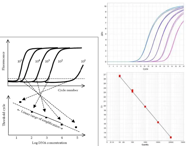
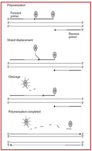
7. 2 Real-time PCR 47
PART 2: AIM OF THE WORK
52
PART 3: MATERIALS AND METHODS
53
Chapter 8 Study design 53
Chapter 9 Selection and characterization of Bifidobacterium strains 54
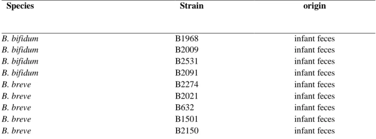
9.1 Bifidobacterium strains and culture conditions 54
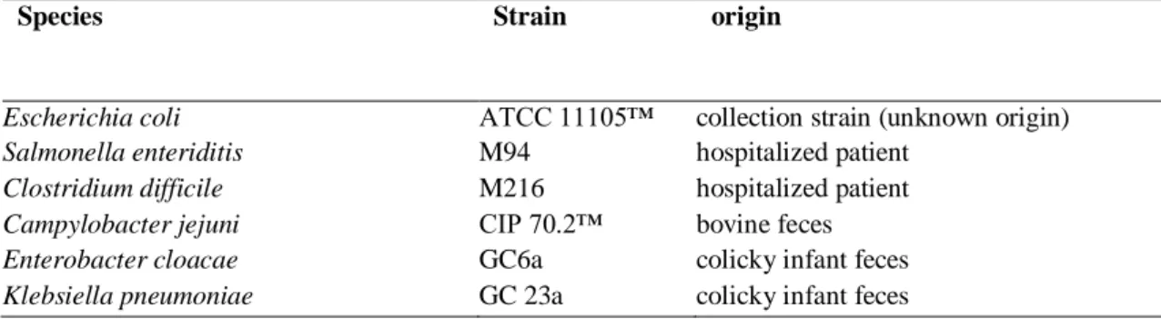
9.2 Antagonistic strains (potentially pathogenic) and culture conditions 57
9.3 In vitro inhibition of antagonistic strains 58
9.3.1 Agar spot test using living cells 58
9.3.2 Antimicrobial activity of Bifidobacterium spp. culture supernatants 58
9.4 Genetic typing of the strains 59
9.4.1 Enterobacterial Repetitive Intergenic Consensus PCR (ERIC-PCR) 59 9.4.2 PCR with genus-specific and specie-specific primers 59
9.5 Antibiotic resistance profiles 60
9.5.1 Minimal inhibitory concentration (MIC) 60
9.5.2 Screening of resistance genes 61
9.5.3 Plasmid detection 62
9.6 In vitro interaction between Bifidobacterium strains and human cells 65
9.6.1 Growth and maintenance of cell line 65
9.6.2 Cytotoxicity assays 66
9.6.3 Adhesion assay 66
9.6.4 Mitochondrial activity assay 66
9.6.5 Determination of Reactive Oxygen species (ROS): NO, H2O2 67
9.6.6 Dot-blot for interleukin 6 67
9.7 Experimental design, statistical analysis and strain selection criteria 68
Chapter 10 Evaluation of the most effective prebiotic fiber 69
10.1 Prebiotic activity assay 69
Chapter 11 Evaluation of the effects of intrapartum antibioticprophylaxis
on newborn microbiota 71
11.1 Newborn study design and sample collection 71
11.2 DNA extraction from faecal samples 71
11.3 Real-Time PCR assays 71
PART 4: RESULTS
74
Chapter 12 Selection and characterization of Bifidobacterium strains 74
12.1 Antimicrobial activity with the spot agar test 74
12.2 Antimicrobiobial activity of Bifidobacterium culture supernatants
against coliforms and S. enteriditis 77
12.3 Genotypic characterization of the Bifidobacterium strains 81
12.4 Antibiotic resistance profiles 84
12.4.1 Minimal inhibitory concentration (MIC) 84
12.4.2 Screening of resistance genes 86
12.4.3 Plamid detection 88
12.5 In-vitro interaction between Bifidobacterium strains and human cells 89
12.5.1 Cytotoxicity and adhesion 89
15.5.2 Stimulation of cell activity: mitochondrial activity, production of
reactive oxygen species and of interleukin 89
12.6 Selection of the best probiotic strains with the use of a synthetic index 94
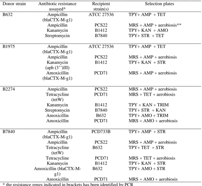
12.7 Transferability of antibiotic resistance traits 96
Chapter 13 Evaluation of the most effective prebiotic fiber 97
Chapter 14. Evaluation of the effects of intrapartum antibiotic
prophylaxis on newborn microbiota 103
14.1 Microbiological analysis of newborn fecal samples 103
PART 5: DISCUSSION
105
Chapter 15. Selection and characterization of Bifidobacterium strains 105
Chapter 16. Evaluation of the most effective prebiotic fiber 109
Chapter 17 Evaluation of the effects of intrapartum antibioticprophylaxis
on newborn microbiota 111
REFERENCES
113
PART 1: INTRODUCTION
Chapter 1. Intestinal microbiota in early infancy: composition
and development
1.1 Microbiota composition in early infancy
The intestinal microbiota of humans is a specific ecosystem made of a complex array of microorganisms (~ 1014 – 1015 CFU/g of lumen content) which forms an individual microbiota typical for each being . In particular, the human intestinal microbiota consists of more than 400 different species.
Birth brings about an immediate end to the sterility of the fetus enviroment: microbial colonization begins after birth, within a few hours bacteria start to appear in the feces. Studies of gnotobiotic mice have been particularly enlightening, illustrating the essential role of the gastrointestinal microbiota in normal gut development (Ley et al., 2006).Thaks to these studies, it is argued that the microbial diversity of the human gut is the result of coevolution between microbial communities and their hosts and that the peculiar structure of microbial diversity in the human gut resulted from natural selection operating at two different levels: the host level selection on the community which favours stable societies with a high degree of functional redundancy and a selection pressure driving microbial cells to become functionally specialized.
The first microbial population the newborn comes into contact with are the maternal intestinal and vaginal microbiota; successively, the newborn will be exposed to the microbes from the environment. Still, the microbial colonization of the infant gastrointestinal tract (GIT) is a remarkable episode in the human lifecycle.
A low amounts of bacteria is encountered a few hours after birth; the main bacteria genera isolated at these time are Staphylococcus, Streptococcus, Propionibacterium,
Corynebacterium. Following a rupture of the fecal membranes, bacteria of maternal
origin can be isolated.
The first bacteria encountered in the majority of healthy infants are facultative anaerobes, because the intestinal environmental of neonates shows a positive oxidation/reduction potential at birth. These bacteria remain predominant during the first few days of life, among them, Staphylococcus, Enterobacteriaceae and
Gradually the consumption of oxygen by these bacteria changes the intestinal environment into a more-reduced one, permitting the subsequent growth of strict anaerobes (Bezirtzoglou, 1997). Facultative anaerobic bacteria are followed by
Bifidobacterium spp., Bacteroides spp. and Clostridium spp. which are present within 2
days with an increased incidence in newborns delivered by a Caesarean section. In fact, in comparison with vaginal delivery, cesarean section resulted in lower colonization rates and counts of bifidobacteria and Bacteroides fragilis group species, whereas counts of Clostridium difficile and Escherichia coli are higher. The presence of C.
difficile is impoetant for the installation of other anaerobic putrefactive microorganisms
such as other bacteria belonging to the Clostridium genus.
As already mentioned before, the microbial population of the newborn changes in relation to many factors like diet (breast versus formula feeding), mode of delivery (natural delivery versus caesarean delivery), maternal diet, antibiotic use during the first few months of life and early enviromental surroundings (Table 1).
In recent year a first large epidemiologic study (KOALA study) on determinants of gut microbial composition in early infancy was carried out in the Netherlands (Penders et al., 2006). Within the KOALA project fecal samples of 1000 infants, 1 month of age, were analyzed in order to study the potential determinants in a multivariate manner and to distinguish their independent effects. Participants at 34 weeks of gestation with diverse lifestyles, i.e. pregnant women with a conventional lifestyle and pregnant women with an alternative lifestyle women that consume only organic food, follow Steiner principles and alternative medicines, were recruited.
In agreement with previous researches, the KOALA study confirm that term infants who were born vaginally at home and were exclusively breastfed seemed to have the most “beneficial” gut microbiota, with the highest numbers of bifidobacteria and lowest numbers of C. difficile and E. coli. Conversely, lifestyle appears not to influence gut microbial composition.
Table 1 Principal factors influencing intestinal microbiota development in newborns Factors
Place and mode of delivery
Maternal microbiota of intestine, vagina and epidermis Type of infant feeding
Antibiotic/antimycotic use Gestational age at birth Hospitalization after birth
1.2 Influence of the mode of delivery on the infant microbiota
composition
The environment is extremely important for intestinal colonization of infants born by cesarean section. Cesarean section new borns do not come in contact with the maternal vaginal and faecal microrganisms and may be separated from the mother for a long period after birth (Biasucci et al., 2010). In this situation the environment becames a crucial source of colonizing bacteria. These bacteria are mainly introduced from the environment of the hospital although it is known that bacteria introduced from the hospital environment have a low colonization ability during the first 7 days of life. Anaerobic colonization, especially by Bacteroides spp. is delayed but Bifidobacterium retrieval and E.coli presence was similar in vaginally and caesarean section delivered infants. Additionally, an increased incidence of Clostridium perfringens and C. difficile is reported in relation to the hospital environment (Penders et al., 2006).
Environmental contamination seems to be the main route for clostridial implantation in the newborn and the rapid implantation of C. perfringens in cesarean sectioned newborns seems to determine a decrease in redox potential which favors the subsequent colonization by anaerobic bacteria like other species of Clostridium and Bacteroides spp..
1.3 Effects of infant feeding on the gut microbiotic composition in
infants
Another important factor that can influence composition of the intestinal microbiota in the neonates is the type of feeding. Also in the KOALA study it has been demonstrated that diet can have an influence on the gut microbiota.
In both breast- and formula-fed infants, the GIT is initially colonized by streptococci and enterobacteria and these create anaerobic condition necessary for the establishment of the anaerobic Bacteroides spp. and Bifidobacterium spp. In full term breastfed neonates Bacteroides spp., bifidobacteria can appear 4 days after birth and after 1 week they dominate the faecal microbiota of breast-fed infants and their counts increase rapidly to constitute 80%-90% of the total flora. In contrast, the faecal microbiota of the formula-fed infants is more complex, with Bifidobacterium spp., enterobacteria and
Streptococcus spp. in similar proportion. Another notable difference is that formula fed
infants have much higher counts of Clostridium spp than breast fed infants (Penders et al., 2006).
An important difference is the relative buffering capacity of the two feeds. Breast milk has poor buffering capacity, compared with formula milk, and this leads to market differences in the colon pH of breast and formula fed infants : 5.1 and 6.5, respectively. This low pH promotes the growth of bifidobacteria and lattobacilli, but is inhibitory to many other bacteria (Tham et al., 2011). Moreover, a number of peptides capable of stimulating the growth of several bifidobacteria have recently been isolated from human milk. Another factor that could contribute to the dominance of bifidobacteriain the faeces of breast-fed infants is the presence in the human milk of glucoprotein, glycolipids, fucose, neuraminic acid, lactose, N-acetylglucosamine, and, a variety of oligosaccharides (Coppa and Gabrielli, 2008 ).
Both adults and neonates are regularly exposed to microorganisms via the diet, but with different effects. The microorganisms entering newborns throught breast milk are more likely to colonize than those entering in healthy adults with stable climax communities are. However, the results available to date on bifidogenic effects of milk molecules are still inconclusive and there is also a lack of information about the isolation and identification of commensal or potential probiotics bacteria, including bifidobacteria, from milk of healthy women. Even though authors are aware that human milk is difficult to sample and microbial contamination can never be totally discarded, some
studies have demostrated the presence of alive bifidobacteria in human milk ( Martin et al., 2003, Solis et al., 2010).
It has been hypothesized, within the KOALA study, that the maternal diet not only might be a determinant of the mother’s gut microbiota but also might influence her infant’s gut microbiotic composition. However, no association between maternal use of probiotics during pregnancy and the intestinal microbiotic composition at the age of 1 month was found (Penders et al., 2006).
Recent studies have been demonstrated that another additional anaerobic bacterial group is to be considered as dominant in breast-fed babies during the first days of life, i.e.
Ruminococcus (Morelli, 2008). It is also interesting to note that ruminococci seem to be
positively affected by oligosaccharides, at least in animal models. The complete role of ruminococci in protecting the health of babies is far from being understood, anyway
Ruminococcus is recognized to have an important protective effect on the host because
it produces ruminococcin A, a bacteriocin that can inhibit the development of many species of Clostridium.
1.4 The intestinal bacterial colonization in preterm infants
In contrast with full term neonates, little information concerning the composition of the microbiota in premature infants is available because only a few studies have determined the developmental aspects of the intestinal colonization in these subjects. It is difficult to draw firm conclusion on the fecal microbial community in preterm infants for several reasons: the inter-individual variability is very high and many parameters, such as antibiotic regiments and diet, may tend to increase study discrepancy. In particular, preterm often need parental feeding, due to the immaturity of their intestine and they often need respiratory support, they are vulnerable for infections and often require antibiotic treatment.
In addition, the limited number of patients analyzed usually do not allow to fully understand the microbiota composition. As this category of infants often require intensive care treatments due to an increased risk for serious infections, insight in the intestinal colonization is important.
At the first days of life, the preterm infants are predominantly colonized by facultative anaerobic bacteria, which remain at high levels, resembling the full term formula-fed
infants. However, the counts of enterobacteria and enterococci remain predominant until the 20th day of life and significantly higher than in full term breast-fed infants (Magne et al., 2005).
Moreover, one of the most significant differences between preterm and full term infants microbiota is the colonization of bifidobacteria that are not frequently identified in the first month of life of premature newborns (Westerbeek et al., 2006).
This alteration in the composition of the gut microbiota of preterm infants can be linked to the increased risk, for this subjects, of severe gastrointestinal disorders such as necrotizing enterocolitis (NEC) which affects predominantly premature and low weight newborns (Lin et al., 2008).
1.5 Effects hospitalization on the microbiota composition in infants
Prematurity is strongly associated with hospitalization. In addition, hospitalization itself is incriminated to changing the normal microbiota. Changes in the intestinal microbiota composition upon chemioterapic administration is observed, for example the oral use of antibiotics (mainly amoxicillin) by the infant during the first 1 month of life resulted in decreased numbers of bifidobacteria and B. fragilis-group species (Penders et al., 2006; Mangin et al., 2010).Moreover the simple impact of hospitalization, even without any antibiotic treatment produces changes in the normal microbiota. In hospitalized newborns intestinal colonization by Klebsiella, Proteus, Pseudomonas, as well as E.coli occurs more frequently (Penders et al., 2006).
Chapter 2. Interaction between gut microorganisms and
intestinal epithelial surface
The microbiota is in close conctact with the intestinal mucosa and epithelia surface which is, after the respiratory area, the largest surface of the body, occupying approximately 250-400 m2. Some anatomical and physiological aspect of the host organism are directly linked to the presence and activity of the intestinal microbiota such as formation of the intestinal walls, production of organic acids and vitamins, stimulation of immune system etc. The main fuctions of the microbiota on the host organism will be analysed in this chapter.
2.1 Structure and fuctions of intestinal surface
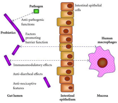
The intestinal mucosal surface is exposed to the lumen and the cells present in the external layer, along with their secretions, form a barrier between non sterile internal environment and the essentially sterility of the body (Duerr and Hornef, 2011) (Figure 1). As a result of these exposure the mucosal surfaces are the principal locus of attack by microorganisms.
Mucosa consists of three layers: the first is made up of the epithelial cells, which can be a single layer as in GI tract. The cells are attached to a basement membrane overlying the second layer, the lamina propria, which consists of subepithelial connective tissue and lymph nodes, underneath which is the third layer, a thin layer of smooth muscles called the muscolar mucosa.
The epithelial cells of the GI tract are squamous in the esophageal part but they became leaky and die before being shed into the lumen. This desquamation of the cells is an important mechanism of preventing microrganism invasion. In the intestinal tract the columnar epithelial mucus is secreted by goblet cells interspersed among the enterocytes. Enterocytes are polarized cells with a dinstinct apical and basolateral cytoplasmatic membrane. However the intestinal epithelium also contains M cells, which are present in Peyer’s patches and are part of the gut- associate lymphoid tissue. The M cells are specialized epithelial cells that transport antigens and microorganisms from their apical surface throught the cytoplasm to the basolateral surface by using
transcytosis. Immune cells such as macrophages and lymphocytes are located in their extracellular compartement underneath these cells, waiting for antigen presentation.
Figure 1 Anatomy of small intestine and colon immune system (Abreu, 2010)
The innate defense system consists of three components: mechanical, chemical, and cellular barriers.
The mechanical barrier is formed by the epithelial cells and the junctions between them (Yu and Yang, 2009). The chemical defence comes from antimicrobial proteins, peptides and cytokines that perform the immune response. The last component of the innate defence is the cellular defence enacted by M cells, dendritic cells, phagocytic cells, mast cells, lymphocytes and epithelial cells (Guarner, 2006).
The first defence that an invading pathogen would encounter is the preepithelial barrier, consisting of a secreted mucus gel. Mucus is therefore a unique physical gel that has both flow and rigidity properties. The secreted mucins are the principals viscous and gel-forming components of the mucus gel secretions. Mucins are high molecular weight glycoproteins.
Using in vitro and in vivo system (El Asmar et al., 2002; Cencič and Langerholc, 2010) it has been demonstrated that exposure to healthy commensal bacteria results in
establishment of the normal tight-junction barrier between epithelial cells, which represent the major determinant of gut permeability.
In particular the immaturity and the permeability of intestinal epithelial barrier may play a role in pathophysiology of intestinal complications in some neonates and mainly in preterm borns (Stratiki et al., 2007). Among the most severe gastrointestinal complications linked to the weakness of epithelial barrier, there are feeding intolerance, necrotizing enterocolitis (NEC), and gut associated sepsis. These intestinal complications that may occur mostly in the first weeks of life, will be further treated subsequently.
2.2 Protective effects of the gut microbiota on the host
The presence of an abundant commensal microbiota may provide some protection against incoming enteric pathogens and may activate the expression of virulence-related genes (Nataro, 2005).
In addition, experimental data suggest the existence of several complex interacting mechanism in the host defence such as competition with enteric pathogen bacteria for nutrients and adhesions site in the intestinal mucosa and stimulation of the mucosal immune system of the host by activating an appropriate inflammatory response or immune mechanisms against chronic infections (Figure 2).
2.2.1 Competition for nutrients between indigenous microbiota and enteric pathogens
The indigenous microbiota gains access to a nutrient enriched, stable environment, and thereby enters a symbiotic relation with the host’s intestinal tract. In vitro evidence supporting the nutrient-niche hypothesis has been reported by many researcher who used continous flow chemostat culture systems designed to mimic condition of the intestine (Laux et al., 2005). The use of these systems has demonstrated the importance of microbial association in the surfaces, the stability of the population, with respect of major genera, and the role of nutrient utilization in maintaining the population stable. If the analogy of a chemostat is applied to the intestinal tract, several hundred species of bacteria are in equilibrium, competing for resources from an extensive mixture of limiting nutrients, and the only way for a bacterial species to survive is to compete
gut is such a rich source of nutrients that it may seem unlike that this is the way in which the gut microbiota influences its own composition. However, it requires only one nutrient to be limiting for this mechanism to operate successfully. In vitro results suggest that probiotic microorganisms compete more efficiently than C. difficile for monomeric glucose, N-acetyl-glucosamine, and sialic acid found in the colonic contents (Fuller, 1991).
Furthermore, some polysaccharides which can occur naturally (e.g. in breast milk) or are used as food additives can enter in the colon indigested and they are able to stimulate the proliferation only of certain commensal bacteria like lactobacilli and bifidobacteria (Forchielli and Walker, 2005), this topic will be further treated below (see chapter 7).
Figure 2 Host defence against intestinal pathogenic bacteria (Britton and Versalovic, 2008).
2.2.2 Competition for intestinal adhesion sites
Adhesion to and colonization of the mucosal surfaces are possibly protective mechanisms against pathogens throught the competition of the binding sites. The ability of some potential probiotic strains belonging to the Bifidobacterium and Lactobacillus genera to strongly adhere to the intestinal mucosa has been widely studied in the last years (Collado et al., 2005, Del Re et al., 2000, Jankowska et al., 2008) . In particular, bacteria, that are able to adhere to mucus and unable to reach the epithelial cells, might be dislodged from the mucosal surface and washed away with the luminal contents. Indeed there are species of the normal human gut, often introduced in diary products like commercial strains, which should be carefully selected and characterized also for the adhesion to the mucosal surfaces. Many studies used enterocyte-like Caco-2 and HT29 cell lines to investigate the adherence of a large number of Lactobacillus and
Bifidobacterium strains (Del Re et al., 2000, Gopal et al., 2001, Candela et al.,
2008,Cencič and Langerholc, 2010).
However, a wide bibliography shows that the displacement activity exerted by probiotic bacteria towards enteropathogens is related to mechanisms other than mere competition for common adhesion sites. Lievin et al. (2000) have demonstrated that Bifidobacterium strains isolated from infants produce antibacterial lipophilic factor(s) effective in inhibiting S. enterica serovar Typhimurium invasion of Caco-2 cells and in killing intracellular enteropathogenic cells. Fujiwara et al. (2001) have purified a proteinaceous factor that inhibits in vitro adherence of an enterotoxigenic E. coli strain to gangliotetraosylceramide molecules, which are physiological constituents of the mammalian intestinal epithelium surface.
2.2.3 Stimulation of mucosal immune system
The communication between intestinal microorganisms and the GI epithelium has been extensively studied in the last decades using in vitro models and germfree animals. These studies showed that in the absence of the microorganisms, the intestinal immune system is underdeveloped and the morphology is disrupted (Wostmann, 1996), furthermore the germfree animals presented hypoplastic peyer’s patches and, a great reduction of immunoglobulin-A producing plasma cells (Macpherson and Harris, 2004) . They also exhibit an altered gene-expression profile of the intestinal epithelial cells.
Results of additional studies suggested that the indigenous intestinal microbiota in mammalians might contribute to the development of both humoral and cellular mucosal immune systems (Hooper, 2004). These interactions maintain a physiologically controlled inflammation or activation of gut-associated lymphoid tissue thoughout life (Neish et al., 2000).
The immune system is able to detect microorganisms by discriminating between self and nonself organisms. This discrimination is possible throught a sophisticated system of receptors that are called Toll-like receptors (TLRs), which provide considerable specificity for pathogen microorganisms. As soon as TLRs provide the alarm signal of infection, the host reacts with an immediate immune response system (Vinderola et al., 2005).TLRs are expressed by macrophages, dendritic cells, endothelial and epithelial cells and they are specialized in different classes like TLR4 that recognizes lipopolisaccarides (LPS) and gram-negative bacteria and TLR2 that recognizes a variety of microbial components such as peptidoglycan and lipoteichoic acids from gram-positive bacteria (Takeda and Akira, 2005).
Furthermore in vitro and in vivo finding allowed to analyze the secretion of interleukine-6 (IL-6) in responce to bacterial infection (Miller et al., 2002). IL-6 is a multifunctional cytokine involved in diverse biological processes, such as host response to enteric pathogens, acute-phase reaction , hematopoiesis, growth factor for normal or neoplasic cells, and terminal differentiation of B lymphocytes: IL-6 is condidered the product of proinflammatory cells (Montier et al., 2012). By now it is well known that the interaction between probiotics and intestinal cells could play an important role in the innate immune response induced by probiotics (Vinderola et al., 2005, Cencič and Langerholc, 2010).
Much has been learned during recent years about the capability of probiotic strains to induce IL_6 production from epithelial cells (Nissen et al., 2009) and it has been also demonstrated that LAB and bifidobacteria are able to use TLRs to send immune signals to the cells. It was reported that intestinal epithelial cells may be an important source of IL-1ß, IL-6 and IL-8 and that adherent population of Peyer’s patches was responsible for the production of gamma interferon (INF-γ) and tumor necrosis factor alpha (TNF-α) (Perdigon et al., 2002,Tanoue et al., 2008) (Figure 3).
Figure 3 Innate and cell-mediated immune response (Vanderpool et al., 2008).
In addition, reactive oxygen species (ROS) are classically thought of as cytotoxic and mutagenic molecules or as inducers of oxidative stress; recent evidence suggests that ROS play a role in signal transduction. ROS are implicated in stimulation or inhibition of cell proliferation, apoptosis, and cell senescence, moreover they can play an important role in host defence againts infections. Of particular interest, the production of NO and H2O2 by epithelial cells and macrophages mediates killing or
growth inhibition of bacteria, fungi and parasites (Park et al., 1999; Pipenbaher et al., 2009). The ROS compounds take part in the innate immune response (Keyaerts et al., 2004) and recent studies showed that some probiotic strains increase the production of ROS in small intestinal epithelial cells and in macrophages (Nissen et al., 2009, Pipenbaher et al., 2009, Maragkoudakis et al., 2010).
Moderate production of H2O2 and NO induced by probiotics used in food could have a
beneficial effect in manteining a balance and increasing resistence to infections. However, it should be noted that hight concentration of H2O2 and NO causes tissue
injury, disseminated intravascular coagulation and shock (Park et al., 1999).
Lastly, several studies showed that orally administration of lactic acid bacteria (LAB) stimulated IgA secretion and T-cells activation (Perdigon et al., 2001, Dogi et al., 2008), in particular LAB were able to increase IgA cells in a dose dependent manner.
Much of the research on interactions of LAB with epithelial cells has been conducted on tumoral cell lines such as HT-29 and CaCo-2,these studies allowed to better understand some of the complex mechanism of the interaction between microbiota and immune cells.
2.3 Experimental models of gut ecosystem
Human and animal gut is a complex system formed by a large community of microorganisms (intestinal microbiota) that interact with host in the development of intestinal epithelium, in nutrient acquisition and metabolism and in the development of host immune system; because of this complexity it is difficult to find an appropriate experimental model. Germ free animal models have been widely used till recent years but even if they are a good realistic model for such studies they presents major disadvantages like the disagreement with the bioethical spirit of reduce animal testing of EU. Their use is also not suitable in all laboratories because special facilities and special trained personnel are needed. They are also very expensive and ultimely it is not always possible to find a good human model for some of these kind of studies like, for example, pathogen studies (Cencič and Langerholc, 2010) .
As fully described by Cencič and Langerholc, (2010), in vitro cell models of the gut should functionally resemble the in vivo situation. Primary cells isolated from human or animal tissue conserve the majority of the in vivo ecosystem functionality, however the primary cells usually survive only a few days in in vitro culture. Primary cells derived from different individuals keep the diversity that is reflected on the results.
Anyway, in vitro cell models satisfy basic requirements: availability and easy handling and good human predictive power (Cencič and Langerholc, 2010). Moreover cell
models formed by a combination of epithelial and other cell lines responde to envimental factors like cytokines and inflammatory molecules.
As stated above, in most of the in vitro studies of the gut , human colon tumorigenic cell lines like Caco2, T84 and HT-29 have been widely used for mechanistic and functional studies of the gut. However, it is well assessed that the phenotype of tumorigenic cell lines traditionally used for this purpose distinguishes them profoundly from the normal gut epithelium (Tremblay and Slutsky, 2007), in fact adhenocancerogenic cell lines can be altereted in proliferation, glycosilation when compared to non tumorigenic ones. To study the interaction with probiotics and gut epithelium an interesting recent feature is to develop cell culture with non tumorigenic intestinal cells (Cencič and Langerholc, 2010). 3D intestinal epithelial models from various species were developed using both human and animal cell cultures. In particular these 3D models are built from intestinal epithelial cells in a microporous membrane by also adding an underlay of immune cells (macrophage and dendridic cells) that mime the mucosal lymphoid tissue. In the apical side of the membrane, intestinal bacteria can be added in order to makes these models close to in vivo situation.
Chapter 3. Principal gastrointestinal diseases in infants and
newborns
3.1 Necrotizing enterocolitis (NEC) in infants
NEC is the most common gastrointestinal emergency in the neonatal intensive care unit and a major cause of morbidity in preterm infants. It is characterized by gastrointestinal dysfunction progressing to pneumatosis intestinalis, systemic shock, and rapid death in severe cases. The most common risk factors cited are prematurity, enteral feeding and bacterial colonization, in particular intestinal injury in NEC may be the results of synergy of these three factors ( Claud and Walker, 2001).
However, there is a strong evidence that the initial bacterial colonization after birth plays a pivotal role in the developmentof NEC. As It has been already mentioned before, preterm newborns show a different colonization with respect to full term newborns where Bifidobacterium and Lactobacillus microrganisms are predominants. In preterm infants more pathogenic microrganisms such as enterobacteria and enterococci remain predominant until the 20th day of life, for this reason one it has been suggested that a major etiological factor for NEC is the abnormal microbiota, particularly as NEC usually occur after 8-9 days postpartum when anaerobic bacteria start to colonize the gut (Mai et al., 2011). It is also true that premature newborns have an immature and inappropriate intestinal ephitelial immunologic response to luminal bacterial stimuli. The observation that immature human enterocytes react with excessive pro-inflammatory cytokine production after pro-inflammatory stimulation can help in part to explain why prematures exposed to initial colonizing bacteria can develop NEC (Nanthakumar et al., 2000).
Several studies have shown that formula–fed infants have a higher incidence of NEC than breast-fed infants, this is due to the fact that breast milk contains passive immunity factors such as polymeric IgA that enhance intestinal maturation and antimicrobial factors providing protection to the newborn.
Moreover the fetal gut is exoposed to amniotic fluid containing hormones and peptides that may have a role in intestinal maturation and preparation for postnatal feeding ( Claud and Walker, 2001). The preterm infants may not have this maturation process when initially fed and for that reason they are unable to fully digest carbohydrates and
proteins, leading to the production of organic acids which may be harmful to the developing intestine.
However, despite these scientific evidences the exact etiology and pathogenesis of this disease have not been clearly delineated.
3.2 Bacterial gastroenteritis
Infectious gastroenteritis is one of the leading cause of morbidity especially in newborns and children under 5 years of age. Although gastroenteritis-associated mortality is rare in Western Europe, an increased incidence has been noted in some national registers over recent years (Wiegering et al., 2011). However, acute gastroenteritis vary from place to place depending on local socioeconomic conditions and geography.
Several studies have focused on the etiology of infectious diarrhea in hospitalized newborns and children. Rotavirus is the most common cause of infectious diarrhea in children worldwide, followed by adenovirus and norovirus. The clinical manifestations of viral gastroenteritis include diarrhea, vomiting, fever, anorexia, headache and abdominal cramps. None of these single symptoms clearly distinguishes viral gastroenteritis from diarrheal illness due to bacterial or parasitic organisms.
However, bacterial and viral gastroenteritis present with different clinical features. The differentiation of bacterial vs. non-bacterial and rotavirus vs. non-rotavirus diarrhea appears to be of particular clinical relevance. Rotavirus infections are known to be more severe and more often associated with a complicated course.
In the last few decades, several enteric bacteria (e.g., Salmonella spp., Shigella spp.,
Campylobacter spp., Clostridium difficile, Klebsiella pneumoniae, Enterobacter cloacae, E. coli ) and parasites (e.g., Cryptosporidium spp.) have been identified as
important causes of diarrhea in human, particularly in infants (Amisano et al., 2011). Diarrheagenic E.coli represents one of the most the bacterial cause of pediatric diarrhea in developing countries. E.coli is usually found in the commensal intestinal microbiota, but it can become a pathogen through acquisition of genetic determinants, which may enhance adhesiveness and toxicity. E.coli strains associated with diarrhea have been classified into six groups, based on clinical, epidemiological and molecular criteria: enteropathogenic E.coli (EPEC),enteroehaemorragic E.coli (EHEC), enteroinvasive
E.coli (EIEC), enterotoxigenic E.coli (ETEC), enteroaggregative E.coli (EAggEC) and
diffusely adherent E.coli (DAEC).
A further important etiological agent is Shigella spp., that is one of the most common pathogen in children over 1 year of age. Accordingly, Shigella spp. (particularly the S.
dysenteriae and S. flexneri serotypes) should also be regarded as a priority target for
vaccine development, especially since dysenteric illness is not treated primarily with oral rehydration salts, but usually requires antimicrobial therapy .
K. pneumoniae, E. clocae and C. difficile, on the other hands, are normal commensals
of the human intestine ubiquitously throughout most of the gut, and they can cause secondary bacteremia notably in the relatively vulnerable intestinal wall of young infants especially after mucosal damage due to rotavirus infection (Lowenthal et al., 2006).
In addition, Campylobacter emerged as a significant pathogen, mainly among under-6-month-olds.Campylobacter was associated with diarrhoea in some study sites, but mainly among 0-5-month-olds (Allen et al., 2010).
3.3 Infantile colics
Infantile colics are a common condition in the first months of life, about 10-30% of infants are effected by this clinical condition. The classic definition of infantile colic is based on the rule of three: fussy crying that last for > 3 hours per day; for > 3 days per week; and a minimum of 3 weeks. In fact the infant suffers from paroxysms of excessive, highpitched, inconsolable crying, frenquently accompanied by flushing of the face, meteorism, drawing-up of the legs and passing of gas. The crying episodes tend to increase at 6 weeks of age and are most frequent in the evening hours but fortunately this condition usually resolves spontaneously by the age of 3 months. Although infant colic is a common disturbance, the aetiology conditions remain obscure, however evidences suggest multiple independent causes.
The role of an aberrant intestinal microbiota has recently been reproposed to affect gut function and gas production that lead to colicky behaviour. According to Lehtonen et al.,1994, an anomalous microbial composition such as an inadequate bifidobacteria and lactobacilli level in the first months of life may affect the intestinal fatty acid profile thereby favouring the development of infantile colics.
Bifidobacteria and lactobacilli play an important role in the development of local and systemic immune responses in that way an inadequate balance of these microrganisms in colickly infants might underlie immaturity in the gut barrier and lead to aberrant immune responses and increase vulnerability. Furthermore recent studies (Savino et al., 2009) showed that colickly infants have higher counts of anaerobic Gram- negative bacteria than healthy infants and in particular of gas forming coliforms that are rod-shaped organisms that fermente lactose resulting in gas formation at 35-37°C. The most frequent faecal coliform genera are Escherichia, Enterobacter, Klebsiella and
Enterococcus.
Is is feasible that gas coliforms may contribute to colonic fermentation and consequently to excessive intra-intestinal air load, aerophagia and pain, which are the typical symptoms of infant crying, but many aspect of these relationships are still unknown and the contribution of coliforms colonization remains to be clarified (Savino et al., 2007).
Some recent evidences suggest that infantile colics might have many several independent causes, such as lactose intolerance. In this regard, infants, during the first period of life, may display malabsorption of carbohydrates present in breast milk or formula milk and recently, the hypothesis is that colic syntoms could be relieved by reducing the lactose content of the infant feed. According to other new theories, infant colics could be related to food allergy and sometimes could be manifestation of atipic deseases. According to Lindberg, 1999, infants with colic respond favourably to diet free of cow’s milk protein. Moreover, a recent trial suggested that a new formula with partially hydrolized proteins, a low amount of lactose and containig a mixture of galacto-oligosaccharides (GOS) and fructo-oligosaccharides (FOS) led a significant improvement of infantile gas colics and other gastrointestinal disorders (Savino et al., 2007). As indicated in a dedicated section (4.3), the possibly of reducing the synthoms of colics with the use of probiotics has been explored (Savino et al., 2010).
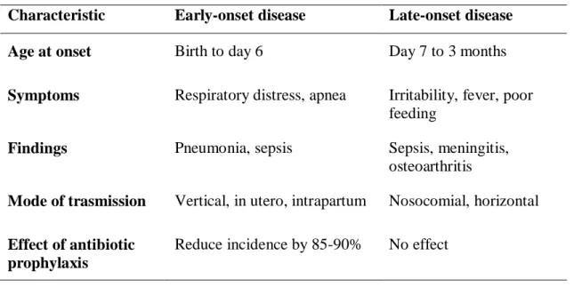
3.4 Neonatal bacterial infections: group B Streptococcal infection
Early-onset bacterial sepsis remain one of the major cause of neonatal morbidity and mortality although the sepsis-associated death rates have declined significantly in the last decade (2001-2011) (Ferrieri and Wallen, 2012). The reason of the reduction of mortality is due to the introduction of intrapartum antibiotic prophylaxis in pregnant women during labor and delivery. There are multiple says throught which bacteria can enter and infect newborns: the primary portal appear to be the respiratory tract, however acquisition via placenta is also possible. The leading cause of onset infection of fetus and newborn is group B Streptococcus (GBS). This gram-negative bacterium that resides in the cervix, vagine or rectum can reach the amniotic through intact or rupted membranes and lead to infection.
Identification of maternal colonization by GBS during pregnancy is very important for taking preventive measures, such as antibiotic prophylaxis, against neonatal disease. In 1996, the Centers for Disease Control and Prevention (CDC) published consensus guidelines for the prevention of neonatal GBS disease that approved the use of intrapartum antibiotic prophylaxis (IAP) for a maternal screening (Puopolo et al., 2005). Penicillin is recommended as the first-line agent for intrapartum antibiotic prophylaxis, while ampicillin is considered as an acceptable alternative. In penicillin-allergic women, who are not at high risk for anaphylaxis, clarithromycin and cefazolin are considered the agents of choice for intrapartum chemoprophylaxis because of its narrow spectrum of activity and ability to achieve high intraamniotic concentrations. In Table 2 the principal symptoms of the early-onset and late-onset infection have been reported . They are very different: in the first case the infection manifests with respiratory disturbance and apneic episodes while in the second case with fever and poor feeding.
As mentioned previously, over the past decade with the introduction of antibiotic maternal prophylaxis, there has been a significant decrease in the incidence of GBS to its current rate of approximately 0.32 per 1000 live births for early-onset disease, however there is no evidence that chemoprophylaxis prevents late-onset disease (Table 2). However, there are no information in the literature on the effect that the antibiotic treatment may have on the early colonization of bacteria in the newborn gut, which is known to be highly influenced by the microorganisms that derive from the mother.
Table 2 Manifestations of early-onset and late-onset group B streptococcal disease Characteristic Early-onset disease Late-onset disease
Age at onset Birth to day 6 Day 7 to 3 months
Symptoms Respiratory distress, apnea Irritability, fever, poor feeding
Findings Pneumonia, sepsis Sepsis, meningitis,
osteoarthritis
Mode of trasmission Vertical, in utero, intrapartum Nosocomial, horizontal
Effect of antibiotic prophylaxis
Chapter 4. Probiotics
4.1 History of Probiotics
The term probiotic, meaning “for life,” is derived from the Greek language and it is currently used to name bacteria associated with beneficial effects for humans and animals. The original observation of the positive role played by some selected bacteria is attributed to Eli Metchnikoff, the Russian Nobel Prize working at the Pasteur Institute at the beginning of the last century; Metchnikoff (1908) in his book “The Prolongation
of Life” was probably the first one to advocate, or rather postulate, the health benefits of
LAB associated with fermented milk products. He hinted that the longevity of the Caucasians could be related to the high intake of fermented milk products and that the intake of yogurt containing lactobacilli might result in a reduction of toxin-producing bacteria in the gut and that this could increase the longevity of the host. Tissier, a French paediatrician, recommended the administration of bifidobacteria to infants suffering from diarrhea, claiming that bifidobacteria supersede the putrefactive bacteria that cause the disease. The expression “probiotic” was probably first defined by Kollath in 1953 (Kollath, 1953), when he proposed the term to identify all organic and inorganic food complexes as “probiotics,” in contrast to harmful antibiotics in order to upgrade such food complexes as supplements.
Later, Lilly and Stillwell (1965) identified probiotics as “substances secreted by one microorganism which stimulates the growth of another”, against the concept of
antibiotic. It may be because of this positive and general claim of the definition that the
term probiotic was subsequently applied to other substances and gained a more general meaning. In 1971 Sperti (Sperti, 1971) applied the term to tissue extracts that stimulate microbial growth. Parker (1974) was the first to use the term probiotic in the sense that it is used today. He defined probiotics as “organisms and substances which contribute to intestinal microbial balance.” The use of the word substances in Parker’s definition of probiotics resulted in a wide connotation that included antibiotics. Although numerous definitions have been proposed since then, none has proved completely satisfactory because of the need for additional explanations, e.g., with regard to statements such as “beneficial balance,” “normal population,” or “stabilization of the gut flora.” In 1989, Fuller (Fuller, 1989) attempted to improve Parker’s definition of probiotic with the
following distinction: “A live microbial feed supplement which beneficially affects the host animal by improving its intestinal microbial balance.” This revised definition emphasized the requirement of viability for probiotics and introduced the feature of a beneficial effect on the host, which was, according to his definition, an animal. A similar definition was proposed by Havenaar and Huis in 't Veld (1992) “…mono- or mixed cultures of live microorganisms which, when applied to animal or man, beneficially affect the host by improving the properties of the indigenous microflora.” Probiotics are best known by the average consumer in relation to foods; in this contest the EU Expert Group on Functional Foods in Europe (FUFOSE) has defined them as “viable preparations in foods or dietary supplements to improve the health of humans and animals”. Salminen (1996) and Schaafsma (1996) broadened the definition of probiotics. According to Salminen, a probiotic is “a live microbial culture or cultured dairy product which beneficially influences the health and nutrition of the host.” According to Schaafsma, “Oral probiotics are living microorganisms which upon ingestion in certain numbers exert health effects beyond inherent basic nutrition.” , In 2001, Schrezenmeir and Michael de Vrese proposed the following definition: “A preparation of or a product containing viable, defined microorganisms in sufficient numbers, which alter the microflora (by implantation or colonization) in a compartment of the host and by that exert beneficial health effects in this host”. In 2002, FAO/WHO has adopted the definition of probiotics as “Live microorganisms which when administered in adequate amounts confer a health benefit on the host” (FAO/ WHO, 2002).
In the past decades studies in the area of probiotics have progressed considerably and significant advances have been made in the selection and characterisation of specific probiotic cultures and in the identification of the positive effects they have on health. Members of the genera Lactobacillus and Bifidobacterium are now mostly employed, but not exclusively, as probiotic microorganisms and a larger variety of probiotic foods are now available to the consumer.
The original assumption of Metchnikoff was that the dietary manipulation of the gut microbiota performed in order to increase the relative numbers of "beneficial bacteria" could contribute to the well being of the host. However he also stated that systematic investigations should be made on the relation of gut microbes to the age, and on the
influence of diets which prevent intestinal putrefaction in prolonging life and maintaining the forces of the body."
It is necessary to assess the efficacy and safety of probiotics and this constitutes an important part of their characterization for human use.
Microbes from many different genera are being used as probiotics. The most commonly used strains are members of the heterogeneous group of lactic acid bacteria; lactobacilli, enterococci and bifidobacteria.
4.2 Principal effects of probiotics on human gut
The mechanism of probiotic action is not totally known but different approaches could be developed. According to Fuller (1989) the probiotic effect of lactic acid bacteria and bifidobacteria may be expressed by three main mechanisms of action:
1. Suppression of pathogenic microorganisms in the intestinal tract by:
a) production of antibacterial substances including primary metabolites, such as lactic acid, acetic acid, carbon dioxide, diacetyl, acetaldehyde, hydrogen peroxide and bacteriocins; they are proteinaceous compounds with antimicrobial activities against other closely related bacteria;
b) competition for nutrients. In the large intestine, the competition is limited for some nutrients, in particular for specific carbohydrates and polysaccharides;
c) competition for adhesion receptors on the gut epithelium. Probiotic strains can adhere specifically or non-specifically. Specific adhesion takes place when a ligand on the bacterial cell binds to a receptor on the epithelial cell; this is commonly defined as a “ lock and key “ function. Non-specific adhesion is a more general phenomenon mediated by hydrophobic or electrostatic interaction and does not seem to have particular relevance in the colonisation of epithelia in vivo.
2. Alteration of microbial metabolism in intestinal tract:
a) increasing the activity of useful enzymes, e.g. β-galactosidase in the alleviation of lactose maldigestion;
b) decreasing the activity of some colonic enzymes such as nitroreductase and azoreductase known to have carcinogenic effects.
3. Stimulation of immunity: recent reports have shown that orally administered lactobacilli and bifidobacteria can improve immune status by increasing the circulating
and local antibody levels, the gamma interferon concentration, the macrophage activity and the number of natural killer cells (MacDonald and Monteleone, 2005). The inclusion of lactic acid bacteria and bifidobacteria as members of physiological indigenous microflora into the mucosa and the subsequent translocation to other organs is currently regarded as a crucial step for the development of the normal mucosal and systemic immunity.
4.3 Use of probiotics in pedriatrics
An increasing number of clinical trials have documented effects of ingestion of specific probiotics bacteria on the care of important infant diseases. The use of probiotics formula for infants older than 4 months of age has already been approved by the American Food and Drug Administration (FDA) and in particular B. lactis obtained the GRAS (generally regarded as safe) status. In addition some recent works have shown encouraging data about administration of Bifidobacterium breve strains in preterms and low birth weight infants (Li et al., 2004 and Wang et al., 2007) and a wide literature documentation reports clinical benefits with treatment of infant gastrointestinal disease with probiotics.
One of the best-studied clinical outcome with the use of probiotics bacteria has been acute diarrheal disease in infants. The majority of the studies have been included various species of lactobacilli and bifidobacteria, and by far, the most used have been
Lactobacillus rhamnosus (LGG), Lactobacillus reuteri and Bifidobacterium lactis
(Guandalini et al., 2000 and Weizman et al., 2005). The larger number of trials documents therapeutic use of probiotics as supplements early in the course of the disease and the most consistent effect reported is a reduction in duration of illness, while another part of literature examine the reduction in incidence of acute diarrheal disease after a preventive administration of probiotics and these studies documented reduction in incidence or severity of the illness (Saavedra and Tschernia, 2007). No study to date has documented an increase in diarrheal disease with any probiotic strain used. Moreover, several probiotics strains resulted effective in reducing the risk of antibiotic-associated diarrhea in newborns and children. A clinical trial, performed with 766 infants, indicated that concomitant treatment with probiotics, compared with placebo, reduced the risk of diarrhea from 28.5% to 11.9% (Szajewska et al., 2006).
Some recent works have described clinical trials conducted on preterm infants. The theoretical benefits of probiotics in preterm infants include the prevention of NEC. These initial studies are encouraging and demonstrate the efficacy of probiotics to re-establish the balance of the gut flora by increasing the number of bifidobacteria. The most used probiotics strains were Lactobacillus acidophilus, Lactobacillus rhamnosus,
Bifidobacterium longum subsp. infantis and Bifidobacterium bifidum. In all these works
the oral administration of probiotics showed a significant reduction in NEC incidence and NEC-associated mortality in respect with placebo group (Bin–Nun et al., 2005 and Lin et al., 2008).
A new aspect of the application of probiotics in the pediatric field is the treatment against gas colics. A published study (Savino et al., 2007) examinated, for the first time, the modulation of intestinal microbiota of colickly infants by administering a probiotic strain. A cohort of 90 breastfed colickly infants was randomly assigned to treatment with the probiotic Lactobacillus reuteri and simethicone. This study evidenced that infants treated with L. reuteri had a significant reduction in crying compared to infants treated with simethicone. The hypothesis, therefore, that probiotic supplementation can provided a reduction of gas colic symptoms and a modulation of intestinal microbiota was demonstrated (Savino et al., 2007, Savino et al., 2010).
To conclude, other clinical trials have shown a great improvement in infants affected by atopic dermatitis after administration with probiotics formula, in these cases, the severity of skin manifestation was strongly reduced (Viljanen et al., 2005). Lower counts of bifidobacteria have been reported in atopic vs non atopic children preceding allergen sensitization. Therefore, bifidobacteria are hypothesized to more effectively promote tolerance against antigen, stimulating GALT immune response.
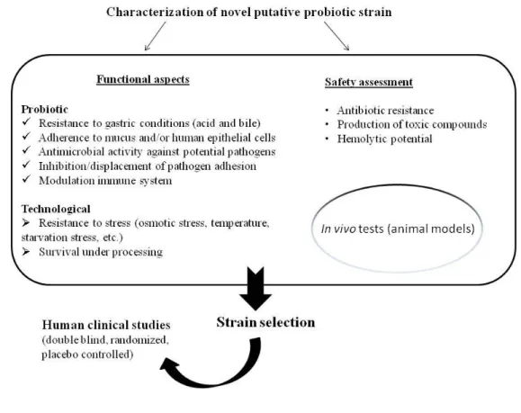
4.4 In vitro selection of probiotic strains
Although progress in probiotic research has been achieved over the past few years, not all of the available probiotic bacteria which are on the market have adequate scientific documentation. It should be desirable to understand the mechanisms that determine the nutritional and health benefits derived from products containing probiotic bacteria, and to use the most promising strains. The probiotic concept will only gain acceptance if these underlying mechanisms are elucidated. Consequently, it is necessary to establish
rational criteria for the screening and selection of candidate microorganisms and also to evaluate the efficacy of the selected strains or the food products in well-controlled human clinical trials.
Significant progress in legislation for the safety evaluation of probiotics has been made in USA, Canada, and Europe (EFSA, 2005a; HC, 2006; FAO/WHO, 2002); however, no unique standards are available. In the USA, microorganisms considered safe for human consumption are awarded the GRAS status by the FDA. In Europe, the European Food Safety Authority (EFSA) has introduced the concept of Qualified Presumption of Safety (QPS) similar in purpose to the GRAS approach. The QPS concept provides a generic assessment system for use within EFSA that in principle can be applied to all requests received for the safety assessments of microorganisms deliberately introduced into the food chain (EFSA, 2005b). EFSA has published a list of microorganism, which possess a known historical safety, proposed for QPS status (EFSA, 2007a). Although the FAO and WHO reports were mainly focused on foods enriched with probiotics, many of the recommendations, including the definition of probiotics, were approved at the Meeting of the International Scientific Association for Probiotics and Prebiotics in May 2002.
Figure 4 Procedure for the characterisation of novel strain with putative probiotic status.
The main steps for the selection of a novel probiotics strain are (Figure 4): 1. Strain identification;
2. Safety evaluation;
3. Fuctional characterization;
4.4.1 Strain identification
The first consideration is to identify and characterize the organism at the genus and species-level. Phenotypic tests may be useful to obtain a first tentative classification at the genus level but the identification results should in any case be confirmed by molecular methods. DNA-DNA reassociation is still considered as a reliable method for the delineation and description of a new bacterial species but it is impractical for the high cost and its complexity. Pattern- and sequence-based molecular methods provided actually a reproducible and easy methods thanks to the update of databases and data exchangeability. However, 16S rRNA does not allow a unequivocal separation of all the
taxa; for that reason it needs to be complemented by other molecular methods such as fingerprinting techniques: Amplified Fragment Length Polymorphism (AFLP), repetitive DNA element-PCR (rep-PCR) or Enterobacterial Repetitive Intergenic Consensus- PCR (ERIC-PCR). These techniques cold be used in association with sequencing of 23S rRNA , Internal Trascribed Spacer (ITS) elements and/or single copy genes (such as groEL, recA, tuf, atpD, dnaK and grpE).
Once the strain has been identified, a scientifically recognized name must be employed and the strains must be deposited in an internationally recognized culture collection.
4.4.2 Safety evaluation
As efficacy is inextricably linked to safety, any claims of health benefits for a probiotic require substantiation by scientific evidence.
The presence of antibiotic resistances and transferability of the antibiotic resistance genes are key factors in safety evaluation. In 2008, a decision of the FEEDAP Panel of EFSA updated the criteria used for the assessment of bacteria for resistance to antibiotics of human and veterinary importance (EFSA, 2008). The aim of this decision was to provide guidance for developing studies to show the potential of each bacteria strain to bear resistance and to transfer it. The basis of such evaluation starts with the determination in vitro of the minimal inhibitory concentration (MIC) for a relevant range of antibiotics of human and veterinary importance (Table 3). The detection of the MIC above the breakpoint levels for one or more antimicrobials required further investigations to make the distinction between acquired and intrinsic resistance; the microbiological breakpoints categorizing bacteria as resistant are expressed in table 3. According to the principle of FEEDAP, when a bacterial strain proves resistant to a specific antibiotic, while others species are normally susceptible to the same antibiotic, the applicant should evaluate the reason for such resistance. If an acquired resistance may be transferred or if known exogenous resistance genes are present, the probiotic strain is not considered suitable for use as food or feed additive
In addition, the determination of antibiotic resistance among probiotic microrganisms is affected by problems regarding the use of media, furthermore, MIC breakpoint values have been shown to be species specific and consequently they vary between species of the same genera.