Neurosurg. Rev. 20 (1997) 135-138
Intracerebral Aspergillus abscess: case report and review of
the literature
Marco Artico, Francesco S. Pastore, Maria Polosa, Shahram Sherkat, and Massi-
miliano Neroni
Institute of Neurosurgery, University of Rome "Tor Vergata", Italy
Abstract
Intracranial aspergillosis is a rare pathologic condition, dif- ficult to treat and often fatal, which generally affects irnmu- nodepressed patients. A case of brain abscess secondary to pulmonary localization in a patient with a non-Hodgkin lymphoma is described. The most significant clinico-path- ological findings of intracranial aspergillosis are examined in the light of the relevant literature.
Keywords:
Aspergillosis, brain abscess, immunosuppres- sion, mycoses.1 Introduction
Aspergillosis is one of the most common mycotic in- fections of the brain, although CNS localization is rare. The clinical picture of CNS aspergillosis may express with different conditions: meningitis, men- ingoencephalitis, granuloma, brain abscess, vascu- litis [11, 14, 17].
We report an instance of a 64-year-old man affected by a non-Hodgkin lymphoma, who developed a left fronto-parietal brain abscess secondary to a pulmo- nary infectious focus. The various clinical and path- ological features of this rare disease are also ana- lyzed in the light of the available literature.
2 Case report
This patient came to our attention in July 1990 for speech and gait disturbances. Neurologic examina- tion at admittance revealed a right hemiparesis and dysphasia. From November 1988 to December 1989
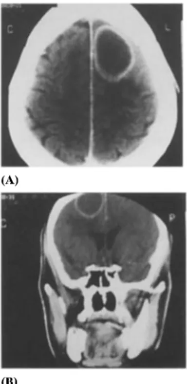
he had undergone chemotherapy (F-MACHOP protocol) following the diagnosis of non-Hodgkin Iymphoma. Eight weeks after the interruption of treatment he suffered a rapid spread of a pulmo- nary inflammatory condition and was, therefore, admitted to the Division of Hematology of our Uni- versity. Several sputum cultures disclosed the pres- ence of Aspergillus fumigatus, confirmed by sero- logic tests for Aspergillus antibodies. Chest plain X-ray and CT showed two globular lesions: one, 4 cm. in diameter was located in the superior field of the left lung and the other (2 cm. in diameter) in the median field of the right lung. The patient was con- sequently treated with 5-fluorocytosine (6 grams daily in divided doses). During this treatment he suddendly developed the above-mentioned neuro- logic symptoms. A C T of the brain was obtained which showed a left fronto-parietal hypodense le- sion with an enhancing ring, 4 cm. in diameter, interpreted as a "metastatic" abscess (Figure 1). Therefore, the patient was transferred to our Insti- tute and then operated on. A left parasagittal fron- tal craniotomy was performed and, after dural opening, the dome of a globular lesion appeared at the cortical surface and was punctured, drawing out necrotic fluid. A n anterior cleavage plane between the capsule and the surrounding cerebral tissue was hardly identified, but finally the lesion was totally removed. The hypothesis of the Aspergillus as the causative agent of the brain abscess was confirmed by the evidence of typical large septate hyphae o f Aspergillus fumigatus in the colture of abscess fluid. Postoperatively, amphotericin B (600 mg./ day) and 5-fluorocytosine (2 g./8 hours) therapy was established in the I.C.U. On 5th postoperative © 1997 by Walter de Gruyter & Co. Berlin- New York
136
Artico et al., Intracerebral aspergillus abscess
(A)
(U)
Figure L Cranio-cerebral contrast-enhanced CT scan. The
axial view (A) and the coronal (B) reconstruction show a
left fronto-parietal abscess with ring enhancement and
perilesional edema.
day an acute renal and cardiovascular failure oc-
curred, and the patient died.
3 Discussion
Candida, Aspergillus, Cryptococcus and few species
in the family of Mucoraceae are reported to be the
organisms most frequently responsible for mycotic
brain abscesses in immunocompromised patients [9,
11, 13, 16, 18, 20, 28, 29]. Aspergillus infection is rare
and usually fatal. Only few of the 350 classified types
of aspergillus show pathogenicity in humans: A. fu-
migatus, A. flavus, A. amstelodami, A. sjdowi, A.
candidus are those more often involved in CNS in-
fection [2, 19].
Brain aspergillosis is more fi-equently sustained by
A. fumigatus [15, 34]. The treatment with antibio-
tics, immunosuppressors, or steroids in patients with
debilitating illnesses greatly enhances the chances
of Aspergillus infection as will any process of weak-
ening of immune system [6, 8, 19, 21, 25]. Typically
these patients are immunodefective and have leuke-
mia, lymphoma, long standing diabetes, or renal dis-
eases. The lung is most frequently affected, followed
by the gastrointestinal tract and the brain [17].
Primitive brain aspergillosis is nevertheless very rare
[7, 11, 21], and commonly the infection is secondary
to lung diffusion [19, 20]. Aspergillosis typically gen-
erates large septate hyphae that are dichotomous,
branching, with evidence of vascular invasion, gra-
nuloma formation, and giant cell reaction [9, 11, 13,
18, 20]. Extension of fungal invasion to surrounding
neural tissue and blood vessels promotes hemor-
rhage, thrombosis, infarction, necrosis, meningitis,
and ventriculitis [18, 20] thus causing the various cli-
nico-pathological features of CNS aspergillosis.
Cerebral aspergillosis needs to be treated with both
chemotherapy and surgery. The drugs of choice are
free amphotericin B and liposomal amphotericin. 5-
fluorocytosine, fluconazole, miconazole, ketocona-
zole, itraconazole are also used, but their effective-
ness is questioned. A synergic action against asper-
gillus infection is reported for amphotericin B and 5-
fluor0cytosine [23]. However, the best results are
achieved combining surgery and chemotherapy [15].
Although cases of survival in CNS aspergillosis have
been reported [2,14, 21, 22, 25], this pathologic con-
dition carries an unfavourable prognosis, with a
mortality ranging in different reports from 80 % to
90 % [19, 23]. Data emerging from such studies indi-
cate that factors positively influencing the effective-
ness of therapy are:
1) evidence of single, isolated lesion without dissem-
ination [17]; 2) paucity of neurologic symptoms [17];
3) early diagnosis [7,10,11, 23, 28]; 4) preventive am-
photericin B administration in patients at risk for as-
pergillosis [10]. Literature data, moreover, underline
the difficulty of precisely determining the death
causes in such patients. A possible explanation is the
toxicity of the delivered drugs and particularly the
proximity of therapeutic and toxic dosage of these
drugs. Unexpectedly favourable outcome or sudden
worsening of conditions seem not related to the
patient's general condition or to the drug in use. In-
stead, diffusion pattern of the mycosis in the neu-
raxis and adequate immunologic response to infec-
tion of the patient are likely to be decisive for the fi-
nal outcome. In particular, an immunodeficiency
status appears responsible for the failure of various
organs and systems in the preterminal stages of this
mycosis.
Artico et al., Intracerebral aspergillus abscess 137
References
[1] ATKINSON GW, H L ISRAEL: 5-ftuorocytosine treat- ment of meningeal and pulmonary aspergillosis. A m l Med 55 (1973) 496-504
[2] BHALLA D, S KUMAR, DN PAL, V MALHOTRA, PL DHINGRA: Aspergilloma of the frontal lobe. Acta Neurochir 55 (1980) 135-139
[3] BORIANI G, A MIRRI, P IACOBITTI, G MAGNANI, R M FERRETTI, A GAMBA, F MAMPRIN, R FIOCCHI, P FER- RAZZI, G BINETTI, L PARENZAN, B MAGNANI: Asper- gillosi cerebrale e renale in un paziente con trapianto cardiaco ortotopico: diagnosi, trattamento e follow- up. Cardiologia 34 (1989) 807-811
[4] CAMARATA PJ, D L DUNN, AC FARNEY, RG PARKER, E L SELJESKOG: Continual intracavitary administra- tion of amphotericin B as an adjunct in the treat- ment of Aspergillus brain abscess: case report and review of the literature. Neurosurgery 31 (1992) 575-579
[5] COD~SH SD, JS TOBIAS, M HANN~GAN: Combined am- photericin B-5-fluorocytosine therapy in Aspergillus pneumonia. J A M A 241 (1979) 2418-2419
[6] COLEMAN JM, G G HOGG, JV ROSENFELD, KD WA- TERS: Invasive central nervous system aspergillosis: cure with liposomal amphotericin B, itraconazole and radical surgery - case report and review of the litera- ture. Neurosurgery 36 (1995) 858-863
[7] DAVID M, A CHARLIN, J MORICE, J NAUDASCHER: In- filtration mycosique h Aspergillus amstelodami du lobe temporal simultant un abcGs encapsul& Abla- tion en masse. Gu~rison operatoire. Rev Neurol 85 (1951) 121-124
[8] DENNING DW, DA STEVENS: Antifungal and surgical treatment of invasive aspergillosis: review of 2121 published cases. Rev Infect Dis 12 (1990) 1147-1201 [9] DENNING DW,, R M TUCKER, LH HANSON, DA STE-
VENS: Treatment of invasive aspergillosis with intra- conazole. A m J Med 86 (1989) 791-800
[10] ENZMANN DR, M BRaNT-ZAWADZKI, R H BRITT. CT of central nervous system infections in immunocom- promised patients. A J R 135 (1980) 263-267 [11] EPSTEIN NE, R HOLLINGSWORTH, K BLACK, P
FARMER: Fungal brain abscesses (aspergillosis/mu- cormycosis) in two immunosuppressed patients. Surg Neuro135 (1991) 286-289
[12] GOODMAN ML, RJ COFFEY: Stereotactic drainage of aspergillus brain abscess with longterm survival: case report and review. Neurosurgery 24 (1989) 96-99 [13] GRCEWC N, WF MATTEWS: Pathologic changes in
acute disseminated aspergillosis. A m J Clin Patho132 (1959) 536-551
[14] GUPTA R, A K SINGH, P BISHNU, V MALHOTRA: Intra- cranial aspergillus granuloma simulating menin- gioma on M R imaging. J Comput Assist Tomogr 14 (1990) 46%469
[15] HAMILL R, L A ONE'4 LR CRANE: Successful therapy for rhinocerebral mucomycosis with associated bilat-
era1 brain abscesses. Arch Intern Med 13 (1983) 581-583
[16] HEDGES TR, LSE LEUNG: Parasellar and orbital apex syndrome caused by aspergillosis. Neurology 26 (1976) 117-120
[17] HENZE G, P ALDENHOFF, U STEPHANI, G GROSSE, E KAZNER, F STAIB" Successful treatment of pul- monary and cerebral aspergillosis in an im- munosuppressed child. Eur J Pediatr 138 (1982) 263-265
[18] JINKINS JR, E
SIQUEIRA,
MZ AL-KAWI: Cranial man- ifestations of aspergillosis. Neuroradiology 29 (1987) 181-185[19] KARANDANIS D, JA SHULMAN: Factors associated with mortality in brain abscesses. Arch Intern Med 135 (1975) 1145-1150
[20] K~too TK, K SUGAr, TK LBONG: Disseminated asper- gillosis: case report and review of the world literature. A m J Clin Patho145 (1966) 697-703
[21] KLEIN HJ, H P RICHTER, W SCHACHENMAYR: Intra- cerebral aspergillus abscess: case report. Neurosur- gery 13 (1983) 306-309
[22] LOWE J, J BRADLEY: Cerebral and orbital aspergillus infection due to invasive aspergillosis of ethmoid si- nus. J Clin Patho139 (1986) 774-778
[23] LUCANTON~ D, R GALZIO, M ZENOBII, V MAGLIANI,
G SClARRA, C D'ARRIOO: Right occipital cerebral ab- scess caused by aspergitlus fumigatus. J Neurosurg Sci 31 (1987) 29-31
[24] MASUCCI EF, JA FABARA, N SAINI, JF KURTZKE: Ce- rebral mucormycosis (phycomycosis) in a heroin ad- dict. Arch Neuro139 (1982) 304-306
[25] MOHANDAS S, GH AHUJA, VP SOOD, V VIRMANI: As- pergillosis of the central nervous system. J Neurol Sci 38 (1978) 229-233
[26] PANKAJALAKSHMI VV, V V TARALAKSHMI, K TmRU- NEELAKANTAN: Cerebral aspergillosis: report of two cases. Sabonraudia 15 (1977) 225-230
[27] RAMOS-GABATIN A, R M JORDAN: Primary pituitary aspergillosis responding to transsphenoidal surgery and combined therapy with amphotericin B and 5-flu- orocytosine. Case report. J Nenrosurg 54 (1981) 839-841
[28] SALAKI JS, DB LOURIA, H CHMEL: Fungal and yeast infections of the central nervous system. A clinical re- view. Medicine 63 (1984) 108-132
[29] SERES JL, O t-[IROISA, EJ BENNER: Aspergillosis pre- senting as spinal cord compression: case report. J Neurosurg 36 (1972) 221-224
[30] SHUPER A, HI LEVITSKY, D R CORNBLATH: Early in- vasive CNS aspergillosis: an easily missed diagnosis. Neuroradiology 33 (1991) 183-185
[31] VANDEVELDEAG, AAMANCERI, JE JOIdNSON: 5-flu- orocytosine in the treatment of mycotic infections. A n n Intern Med 77 (1972) 43-51
[32] WALSH TJ, DB HIER, L R CAPLAN: Aspergillosis of Neurosurg. Rev. 20(1997)
138
the central nervous system: clinicopathological anal- ysis of 17 patients. Ann Neurol 18 (1985) 574-582 [33] WooDs KF, BJ HANNA: Brain stem mucormycosis in
a narcotic addict with eventual recovery. Am J Med 80 (1986) 126-128
[34] YOUNG RC, JE BENNETT, CL VOGEL, PP CARBONE, VT DE VIXA: Aspergillosis: the spectrum of the dis- ease in 98 patients. Medicine 49 (1970) 147-172
Artico et al., Intracerebral aspergillus abscess Submitted March 18,1996. Revised May 15,1996. Ac- cepted August 7, 1996.
Marco Artico, M. D., Pharm. D. Faculty of Pharmacy
Chair of Human Anatomy University of Rome "La Sapienza" Rle Aldo Moro 5
1-00185 Rome Italy