rstb.royalsocietypublishing.org
Review
Cite this article: Nistico` R, Mori F, Feligioni
M, Nicoletti F, Centonze D. 2014 Synaptic
plasticity in multiple sclerosis and in
experimental autoimmune encephalomyelitis.
Phil. Trans. R. Soc. B 369: 20130162.
http://dx.doi.org/10.1098/rstb.2013.0162
One contribution of 35 to a Discussion Meeting
Issue ‘Synaptic plasticity in health and disease’.
Subject Areas:
neuroscience
Keywords:
long-term potentiation, synaptic plasticity,
multiple sclerosis, experimental autoimmune
encephalomyelitis, hippocampus,
interleukin-1b
Author for correspondence:
Robert Nistico`
e-mail: [email protected]
Synaptic plasticity in multiple sclerosis
and in experimental autoimmune
encephalomyelitis
Robert Nistico`
1,2, Francesco Mori
3, Marco Feligioni
4, Ferdinando Nicoletti
1,5and Diego Centonze
1,31Department of Physiology and Pharmacology, Sapienza University of Rome, 00185 Rome, Italy 2IRCSS Santa Lucia Foundation, 00143 Rome, Italy
3Neurologic Clinic, Department of Systems Medicine, University of Rome ‘Tor Vergata’, 00133 Rome, Italy 4Pharmacology of Synaptic Plasticity Unit, EBRI—European Brain Research Institute, Rome, Italy 5IRCCS Neuromed, 86077 Pozzilli, Italy
Approximately half of all patients with multiple sclerosis (MS) experience cog-nitive dysfunction, including learning and memory impairment. Recent studies suggest that hippocampal pathology is involved, although the mech-anisms underlying these deficits remain poorly understood. Evidence obtained from a mouse model of MS, the experimental autoimmune enceph-alomyelitis (EAE), suggests that in the hippocampus of EAE mice long-term potentiation (LTP) is favoured over long-term depression in response to repeti-tive synaptic activation, through a mechanism dependent on enhanced IL-1b released from infiltrating lymphocytes or activated microglia. Facilitated LTP during an immune-mediated attack might underlie functional recovery, but also cognitive deficits and excitotoxic neurodegeneration. Having identified that pro-inflammatory cytokines such as IL-1b can influence synaptic function and integrity in early MS, it is hoped that new treatments targeted towards preventing synaptic pathology can be developed.
1. Introduction
Multiple sclerosis (MS) is a chronic inflammatory, autoimmune, demyelinating disease of the central nervous system (CNS). It is the most common cause of neurological disability in young adults, with disease onset peaking between 20 and 40 years. The disease takes three main forms: relapsing and remitting, where unpredictable acute attacks are interposed with periods of stability; primary-progressive, characterized by a gradual but steady progression of disability; and secondary-progressive, which begins with a relapsing –remitting course, and then becomes steadily progressive [1].
Clinical signs of MS are heterogeneous, reflecting the areas of the brain and spinal cord that are affected [2]. The neuropathological hallmarks of MS are demyelinating white matter lesions associated with inflammatory infiltrates, oxidative injury, excitotoxicity, astrogliosis and early axonal injury/neuronal damage, as well as disruption of the blood –brain barrier [3]. However, grey matter atrophy is also present early in the disease and worsens along with MS progression, correlating with motor, sensory, visual disability and cognitive deficits [4]. Accordingly, brain magnetic resonance imaging (MRI) studies in patients with MS have shown structural changes in both the cerebral cortex and hippocampus, with atrophy of the CA1 hippocampal subfield [5]. These data are in agreement with post-mortem studies showing demyelination and neuropathology in the hippocampus of MS patients [6], where cellular and mol-ecular alterations affecting synaptic plasticity, axonal transport and glutamate homeostasis occur [7]. Glutamate-mediated excitotoxicity is among the key fac-tors underlying neuronal damage in MS. Glutamate levels are significantly increased in the cerebrospinal fluid (CSF) [8] and brain of MS patients [9]. In addition, changes in the expression of glutamate transporters and receptors
have been found in MS patients [10–12] and in the experimen-tal autoimmune encephalitis (EAE) model of MS [13–15]. These results suggest that an increased excitatory neuro-transmission plays a role in the pathogenesis of MS [16]. We have recently found that synaptotoxicity might also be the result of the inflammatory damage of hippocampal inhibi-tory GABAergic interneurons, which shifts the inhibiinhibi-tory/ excitatory balance towards excessive excitation [17].
In this review, we will describe abnormal patterns of cortical plasticity in a sample of MS patients and in EAE, which models MS in mice. Our central hypothesis is that enhanced long-term potentiation (LTP) during immune attacks on the CNS might on the one hand facilitate functional recovery, and on the other trigger cognitive impairment and synaptic degeneration.
2. Abnormal neuroplasticity in multiple
sclerosis patients
LTP- and long-term depression (LTD)-like cortical plasticity can be explored safely and non-invasively in humans by a number of neurophysiological stimulation protocols, including intermittent (iTBS) and continuous theta-burst stimulation (cTBS), which have been developed in the attempt to mimic the physiological activity of hippocampal neurons during learning episodes [18,19].
Both forms of synaptic plasticity are altered in patients with MS, providing a plausible synaptic substrate for the cog-nitive deficits frequently associated with this disorder [20]. LTP induced by iTBS is absent in relapsing –remitting MS (RR-MS) patients during disease exacerbations (figure 1a) [21], probably because acute inflammation alters the metab-olism of amyloid-b (Ab) peptide, thereby restraining its effect on synaptic plasticity [22].
In MS, gadolinium (Gdþ
) lesions tend to be associated with an inflammatory response. In 42 MS patients stratified for the absence or the presence of acute inflammatory lesions (i.e. Gdþlesions at the MRI), we found that CSF levels of Ab1 – 42
were lower in Gdþ MS patients when compared with both
Gd– MS patients and non-MS controls. A striking correlation
between CSF Ab1 – 42 levels and LTP amplitude assessed by
iTBS was also found, indicating that Ab is a potent regulator of synaptic plasticity not only in animals [23] but also in the MS brain [22]. To examine whether cognitive impairment in MS patients was associated with reduced cortical LTP, we com-pared iTBS-induced LTP amplitude in MS patients with or
without cognitive impairment. We found that iTBS-induced LTP was lower in cognitively impaired MS patients [22], in agreement with the finding that the effects of repetitive tran-scranial magnetic stimulation (TMS) on synaptic plasticity are altered in patients with Alzheimer’s disease (AD) [24,25]. Our results indicate that acute inflammation in MS alters Ab metab-olism and cognitive abilities by interfering with LTP-like, activity-dependent synaptic plasticity [22].
Recent evidence suggests that certain mechanisms of neu-rodegeneration are shared between MS and AD. However, the relationship between inflammatory mediators and Ab metabolism in the two diseases is quite complex to define. In AD inflammatory changes, typified by activated microglia, are in close proximity to Ab-containing plaques, neurofibril-lary tangles and neuronal loss. It is still unclear, however, whether inflammation contributes to the pathogenesis of AD or is the consequence of the progressive neurodegenerative process [26]. A recent study showed that Ab-specific Th1 cells increase microglial activation and Ab deposition, chan-ges that are associated with impaired cognitive function in a transgenic mouse model of AD. Treatment with anti-IFN-g antibody attenuated the effects of Th1 cells, suggesting that release of IFN-g from infiltrating Th1 cells might induce the Ab misprocessing and behavioural deficits [27]. Moreover, BACE activity was found either increased in the CSF of AD patients [28] or decreased over time in the CSF of patients with RR-MS [29]. The reduction of CSF Ab in AD has been pos-tulated to reflect deposition of the aggregated insoluble fibrillar Ab peptides in senile plaque, with lower levels of diffusion into the CSF [30]. There is no evidence, however, of Ab tissue deposition in MS brains [31]. We cannot argue, therefore, that Ab CSF levels decrease as a consequence of tissue deposition in MS.
More recently, we also explored LTD-like plasticity by using the cTBS stimulation paradigm in MS. We found that cTBS caused the expected LTD-like effect in healthy individ-uals, while it resulted in no effect or even in LTP in MS subjects [32] (figure 1b). These findings differ from findings in AD patients where an impairment of LTP-like together with normal LTD-like cortical plasticity was recently reported [33]. In line with these findings, altered LTD in MS was associated with IL-1b and not with Ab CSF levels, consistent with the idea that specific inflammatory processes underlie synaptic plasticity alterations in different diseases.
Together with the finding that also iTBS-induced plasticity is altered in MS [22], these results indicate a profound
1.8 1.6 1.4 1.2 0.1 0.8 0.6 pre 0'
time after iTBS (min) 15'
* *
MEP amplitude (post/pre iTBS)
1.8 1.6 1.4 1.2 0.1 0.8 0.6 pre 0'
time after cTBS (min) 15'
*
MEP amplitude (post/pre cTBS)
(a) (b)
Figure 1. TBS-induced plasticity in MS and healthy controls. (a) iTBS induces the expected effects in MS Gd
2(grey line) patients and in healthy subjects (HS,
dotted line) but is altered in MS Gd
þ(black line) patients. Gd
2patients and the control group display the predictable LTP-like effect, while in MS Gd
þpatients no
plastic changes of cortical excitability are observed (adapted from [21]). (b) cTBS-induced effects diverge between MS (solid line) patients and healthy control
subjects (HS, dotted line). The control group displays the predictable LTD-like effect, while MS patients manifest LTP-like changes. *p
, 0.05, ANOVA.
rs
tb.r
oy
alsocietypublishing.org
Phil.
Trans.
R.
Soc.
B
369
:20130162
2subversion of plasticity rules and mechanisms in the MS brain, which is probably caused by the inflammatory milieu generated by infiltrating autoreactive T lymphocytes and by activated microglia in the CNS during immune-mediated attacks. Inter-estingly, a previous study reported the expected LTD-like plasticity in response to cTBS in a sample of 14 stable MS patients [34], while about half of our studied population (59 patients in total) was in the relapsing phase [32].
3. Animal models of multiple sclerosis
Two main animal model systems are used for the study of MS: EAE [35], and experimental viral infection models, exem-plified by Theiler’s murine encephalomyelitis virus (TMEV) [36]. Both models have significantly advanced our under-standing of MS and allowed preclinical testing of disease therapies. EAE is considered to be the best model of MS, because it recapitulates the main pathological features of the human disease. EAE can be induced by immunization of susceptible animals with defined myelin antigens, includ-ing myelin basic protein [37], proteolipid protein [38] and myelin oligodendrocyte glycoprotein (MOG) [39], which determine the different disease phenotypes and pattern of lesions. Chronic progressive EAE is usually induced in six-to eight-week-old C57BL/6 female mice by subcutaneous immunization with MOG 35 –55 amino acid peptide in incomplete Freund’s adjuvant supplemented with Mycobac-terium tuberculosis. Pertussis toxin is injected on the day of the immunization and again 2 days later to increase the per-meability of the blood–brain barrier [40,41]. In general, EAE symptoms appear 8–12 days post-immunization, peaking 10 days later. EAE clinical course usually begins with a wea-kened tail, gradually followed by hind limb paralysis and rarely by front limb paralysis. After the acute clinical phase, a gradual partial recovery is usually observed, resulting in a chronic phase characterized by less severe symptoms.
Like all animal models of human disease, also EAE presents several limitations. Different experimental variables including the species, strain, sex and age of the animals used; but also the specific induction method might account for the inconsistent results. Most EAE studies are performed in genetically identical groups of animals, which exclude an important source of vari-ation. Furthermore, genetically identical animals may differ in their susceptibility to EAE depending on environmental factors, which may not easily be controlled. Despite their intrinsic limit-ations, EAE models remain an important tool to understand the pathogenic cellular and molecular mechanisms of EAE and potentially of MS, as well as to assess the efficacy of novel disease-modifying therapies. Certainly, our understanding of EAE pathogenesis is still incomplete; thus caution is needed when translating the results of experimental research into clinical trials [42].
4. Long-term potentiation studies in
experimental multiple sclerosis
Animal models of human diseases represent excellent tools for the investigation of pathogenic mechanisms of diseases and the identification of potential targets for pharmacological interven-tion [43]. Cognitive funcinterven-tion in animal models is usually assessed by electrophysiological analysis of hippocampal
synaptic plasticity or behavioural analysis of learning and memory [44–46]. Of note, aberrant synaptic plasticity has been observed in experimental models of AD [47–49], Parkinson’s disease [50], autism spectrum disorders [51] and depression [52]. Few papers have investigated so far hippo-campal LTP in the EAE model. Ziehn et al. [53] found normal LTP at CA1 hippocampal synapses but impaired basal excit-atory synaptic transmission and paired-pulse facilitation (PPF) (a presynaptic form of synaptic plasticity) at 21–45 days after induction of EAE. By contrast, two subsequent studies showed that LTP was impaired at two different time points (14–19 days and 30–35 days) after EAE induction [54,55], while excitatory basal transmission and PPF were unal-tered in the acute phase of disease [55]. In the latter study, the LTP deficit was associated with a decreased expression of the NMDA receptor subunit NR2B and increased interleukin-1b (IL-1b) levels. Interestingly, a ketogenic diet could rescue motor disability, spatial learning and LTP impairment associ-ated with EAE by restraining pro-inflammatory cytokines and oxidative stress [54].
Our group has recently performed a comprehensive study of bidirectional synaptic plasticity at CA3–CA1 synapses from EAE (20–25 days after disease induction) compared to their respective complete Freund’s adjuvant (CFA) controls [17]. Animals were scored daily for clinical symptoms of EAE, as fol-lows: 0, no clinical signs; 1, flaccid tail; 2, hind limb weakness; 3, hind limb paresis; 4, complete bilateral hind limb paralysis; 5, death because of EAE; intermediate clinical signs were scored adding 0.5 value. Generally, only animals with a significant clinical disability score (at least 3–4) were selected for the experiments in the EAE group.
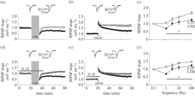
We showed that paired-pulse low-frequency stimulation (PP-LFS) at 1 Hz for 15 min, a protocol able to induce LTD in adult rodents [56], triggered the expected LTD in control slices, but slightly potentiated synaptic transmission in slices from EAE (figure 2a). This trend was also maintained at a stimulation frequency of 100 Hz (1 s), with LTP being sig-nificantly higher in slices from EAE mice (figure 2b). Figure 2c summarizes the frequency–response plots using different conditioning protocols. The rightward shift in the frequency–synaptic function response observed in EAE mice resembles the plasticity subversion observed in MS patients. As the amplitude of synaptic potentiation correlates with the concentration of the pro-inflammatory cytokine IL-1b in the CSF of MS patients [32], we tested whether the facilitated LTP in EAE mice was caused by IL-1b. Control hippocampal slices exposed to exogenous IL-1b showed responses similar to those seen in EAE, i.e. a moderate synaptic potentiation fol-lowing PP-LFS (figure 2d), and a more robust LTP folfol-lowing high-frequency stimulation (HFS; 100 Hz, 1 s) (figure 2e). These data suggest that IL-1b per se is capable of lowering the threshold for LTP induction (figure 2f), thus mimicking the abnormal frequency–response relationship observed in EAE. Differently from transgenic AD models where Ab-mediated LTD facilitation might promote loss of synapses, the contribution of LTD alterations to EAE pathogenesis still requires further investigation.
Contrasting electrophysiological data in EAE models are inherent to the high variability of these models in terms of mouse strains, methods used for EAE induction and housing conditions. It is not uncommon that data of synaptic plasticity are not homogeneous in animal models of neurological disorders [57].
rs
tb.r
oy
alsocietypublishing.org
Phil.
Trans.
R.
Soc.
B
369
:20130162
35. Role of inflammatory cytokines
In recent years, the canonical separation between inflammation and neurodegeneration has been challenged by compelling experimental evidence showing that both aspects are strictly interconnected either in neurodegenerative diseases, includ-ing AD and amyotrophic lateral sclerosis, or in traditional inflammatory disorders, such as MS [41]. Accordingly, inflammatory infiltrates with overt microglial and astroglial activation and specific neurodegenerative features have been found in discrete regions of the EAE brain, such as the cerebel-lum [58,59], striatum [40] and hippocampus [17,60]. In all brain regions, synaptic dysfunctions involving either glutamatergic or GABAergic neurotransmission often precede the onset of motor deficits. In general, glutamatergic transmission seems to be enhanced in EAE, while GABAergic transmission is reduced [17,40,59,61,62]. Notably, these synaptic alterations are likely to occur also in the MS brain and have been associ-ated with the activity of pro-inflammatory cytokines such as tumour necrosis factor-a (TNF-a) and IL-1b. We have recently demonstrated that abnormalities in both excitatory and inhibi-tory transmission found in the brain of EAE mice can be replicated in control brain tissue incubated with the CSF of MS patients [63,64]. Thus, these results indicate that in the course of MS, pro-inflammatory cytokines are released from infiltrating T cells and from activated microglial cells at concen-trations sufficient to diffuse into the CSF and cause widespread alteration of synaptic transmission. EAE-induced alterations of synaptic transmission occurring in the hippocampus are
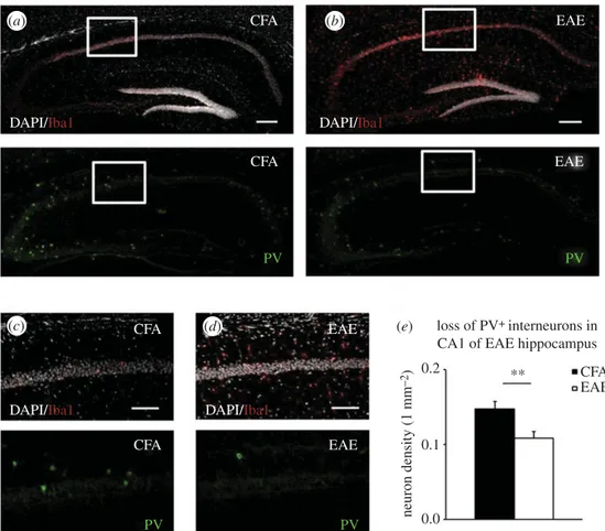
believed to play a crucial role in the cognitive deficits observed in EAE and also in MS patients [65]. In order to elucidate the cellular mechanisms underlying the subversion of synaptic plasticity observed in the EAE hippocampus [17], we per-formed immunohistochemistry and confocal imaging to analyse neuronal architecture in the CA1 region of EAE and CFA controls at 20 days after EAE induction. In line with pre-vious observations in the striatum [61] and cerebellum [62], we observed a lower density of parvalbumin-positive (PVþ) GABAergic interneurons associated with a strong microglia activation (Iba1þ cells), when compared with the hippo-campus of control mice (figure 3a–d). Specifically, the density of PVþ neurons was significantly reduced in the CA1 layer and stratum oriens of EAE mice (figure 3e). A strong Iba1þ labelling was also localized in proximity of the dentate gyrus, CA3 region and fimbria of the EAE hippo-campus. We also observed that IL-1b staining was clearly evident in the lesion sites endowed with activated microglia/ macrophages [17]. Besides microglia/macrophages, infiltrat-ing T lymphocytes may represent another potential source of IL-1b. Indeed, our immunofluorescence and confocal imaging analysis highlighted the presence of CD3þ lymphocytes in lesion sites of the EAE hippocampus [32]. Hippocampal slices incubated with preparations of either activated microglia or T lymphocytes taken from EAE mice displayed reduced GABAergic transmission, being prevented by blockade of IL-1b signalling with IL-1ra [17,32].
As local PVþ GABAergic interneurons strongly modu-late gamma oscillations in the hippocampus, regulating
2.0 0.5 mV 10 ms 0.5 mV 10 ms 0.5 mV 10 ms 0.5 mV 10 ms IL-1b 1.5 1.0 0.5 0 2.0 1.5 1.0 0.5 0 2.5 2.0 1.5 1.0 0.5 fEP S P sl ope (mV m s – 1) fEP S P sl ope (mV m s – 1) 2.5 2.0 1.5 1.0 0.5 fEP S P sl ope (mV m s – 1) 2.0 1.5 1.0 0.5 0 fEP S P sl ope fEP S P sl ope (mV m s – 1) 2.0 1.5 1.0 0.5 0 fEP S P sl ope 0 20 40 60 time (min) 80 0 20 40 60 time (min) 80 0.1 1 10 frequency (Hz) 100 LTP LTD LTP LTD * * IL-1b 1 Hz 1 Hz 100 Hz 100 Hz (a) (b) (c) (d) (e) ( f )
Figure 2. Shifted hippocampal frequency – response function in EAE mice and in response to IL-1b. (a) Superimposed pooled data showing the normalized changes
in field potential slope (+s.e.m.) (CFA, n ¼ 11; EAE, n ¼ 12) induced by paired-pulse low-frequency stimulation protocol (PP-LFS: 1 Hz, 15 min). Here and after,
insets show field EPSPs from representative experiments during a baseline interval and 60 min after delivery of conditioning train. (b) Superimposed pooled data
showing the normalized changes in field potential slope (+s.e.m.) (CFA, n ¼ 9; EAE, n ¼ 12) induced by high-frequency stimulation (HFS: 100 Hz; 1 s).
(c) Frequency – response function in EAE and CFA mice. The graph shows the percentage change in synaptic strength from baseline in EAE and CFA animals at
60 min in response to a variety of conditioning trains (at least seven slices were tested for each condition). Values are mean (+s.e.m.). *p , 0.05 (t-test)
at all frequencies tested. (d ) Superimposed pooled data showing the normalized changes in field potential slope (+s.e.m.) induced by PP-LFS in CFA animals
with (n ¼ 10) or without (n ¼ 11) IL-1b application (30 ng ml
21, duration of application indicated by bar). (e) Superimposed pooled data showing the
normalized changes in field potential slope (+s.e.m.) induced by HFS in CFA animals with (n ¼ 9) or without (n ¼ 9) IL-1b application. (f ) Shown is a
frequency – response graph of the fEPSP changes in response to a variety of conditioning trains in CFA animals with or without IL-1b (at least seven slices
were tested for each condition). Values are mean (+s.e.m.). *p , 0.05 (t-test) at all frequency tested (adapted from [17]). (a – c) Black circles, CFA; white circles,
EAE and (d – f ) grey circles, CFAþIL-1b; black circles, CFAþvehicle.
rs
tb.r
oy
alsocietypublishing.org
Phil.
Trans.
R.
Soc.
B
369
:20130162
4synchronization of pyramidal cell firing [59], we hypothesized that degeneration of PVþ neurons could also affect hippocam-pal gamma frequency, thereby exacerbating the functional consequences of abnormal synaptic plasticity in EAE mice. Accordingly, in addition to aberrant synaptic transmission and plasticity, slices from EAE mice were characterized by reduced hippocampal gamma oscillations [17].
Overall, our data suggest that the selective vulnerability of this neuronal population to the actions of soluble mediators might contribute to sustain synaptic hyperexcitability in EAE, and possibly cognitive impairment in MS. These findings are in line with previous studies conducted in EAE mice [60] and in the frontal cortex of MS patients [65,66].
6. Concluding remarks
Our results show that TBS-induced abnormal plasticity in MS patients and changes in synaptic function and network activity in the hippocampus of EAE mice largely rely on IL-1b-mediated suppression of GABAergic activity, at least before the degeneration of GABAergic interneurons is established. It is noteworthy that elevated IL-1b levels during focal inflammation in MS, as well as in the acute phase of EAE, can influence in opposite directions glutamatergic and GABAergic transmission (which is increased and inhibited by pro-inflammatory cyto-kines, respectively) [59,61,63,64]. Thus, neuroinflammation
and IL-1b signalling network might contribute to the high prevalence of cognitive impairment associated with MS [67] and to the spatial learning deficits in EAE [62,66]. On the other hand, we cannot discard the hypothesis that LTP-like phenomena occurring during immune attacks on the brain might represent a highly adaptive compensatory mechanism. This ‘plasticity reservoir’ may be crucial to counteract the clinical progression in MS by promoting recovery of function after a lesion. Likewise, potentiated excitatory synaptic transmission has been observed in areas surrounding a focal infarct, and this is associated with a better functional outcome [68]. Along this line, neurophysiological tools, such as repetitive TMS and transcranial direct current stimulation, have been recently intro-duced in the clinical setting to promote endogenous plasticity mechanisms aimed at improving functional outcome in drug-resistant neurological and psychiatric disorders [69].
Further research is required to understand the mediators and associated molecular pathways acting to preserve brain function and to limit the clinical consequences of neuronal injury in the progressive phases of MS and possibly in other acute and chronic neurological diseases.
Acknowledgements. We are indebted to Sonia Piccinin and Georgia Mandolesi for their major contribution to the work described.
Funding statement. This work was supported by Fondazione Italiana Sclerosi Multipla (Progetto Speciale FISM), by the Italian National Ministry of Health and the Italian National Ministry of University.
CFA EAE EAE CFA CFA CFA EAE ** CFA loss of PV+ interneurons in
CA1 of EAE hippocampus EAE ne u ron d en si ty (1 mm –2) 0.2 0.1 0.0 EAE PV PV PV PV DAPI/Iba1 DAPI/Iba1
DAPI/Iba1 DAPI/Iba1
(a) (b)
(c) (d) (e)
Figure 3. Loss of PVþ interneurons in the EAE hippocampus. (a– d) Immunostaining of Iba1þ microglia cells (upper panels), DAPI cell nuclei (grey, upper panels)
and PVþ neurons (lower panels), in hippocampal coronal sections derived from CFA (a) and EAE mice at 20 d.p.i. (b), respectively. A strong microglia activation and
loss of PVþ neurons characterize EAE hippocampus. (c) and (d ) are high magnifications of the white boxes in (a) and (b), respectively. The number of PVþ
interneurons (lower panels) in the CA1 layer and stratum oriens was reduced in EAE mice relative to CFA mice. (e) Histogram shows the mean percentage of
the PVþ density interneurons (1 mm
22) which was significantly reduced in EAE mice by about 27% relative to CFA in the acute phase of the disease.
**p
, 0.01, t-test. Scale bars: (a,b) 200 mm, (c,d) 100 mm. (Online version in colour.)
rs
tb.r
oy
alsocietypublishing.org
Phil.
Trans.
R.
Soc.
B
369
:20130162
5References
1. Vukusic S, Confavreux C. 2007 Natural history of multiple sclerosis: risk factors and prognostic indicators. Curr. Opin. Neurol. 20, 269 – 274. (doi:10.1097/WCO.0b013e32812583ad) 2. Noseworthy JH, Lucchinetti C, Rodriguez M,
Weinshenker BG. 2000 Multiple sclerosis. N. Engl J. Med. 343, 938 – 952. (doi:10.1056/ NEJM200009283431307)
3. Sorensen PS. 2005 Multiple sclerosis:
pathophysiology revisited. Lancet Neurol. 4, 9 – 10. (doi:10.1016/S1474-4422(04)00948-2)
4. Pirko I, Lucchinetti CF, Sriram S, Bakshi R. 2007 Gray matter involvement in multiple sclerosis. Neurology 68, 634 – 642. (doi:10.1212/01.wnl.0000250267. 85698.7a)
5. Sicotte NL, Kern KC, Giesser BS, Arshanapalli A, Schultz A, Montag M, Wang H, Bookheimer SY. 2008 Regional hippocampal atrophy in multiple sclerosis. Brain 131, 1134 – 1141. (doi:10.1093/ brain/awn030)
6. Geurts JJ, Bo¨ L, Roosendaal SD, Hazes T, Danie¨ls R, Barkhof F, Witter MP, Huitinga I, van der Valk P. 2007 Extensive hippocampal demyelination in multiple sclerosis. J. Neuropathol. Exp. Neurol. 66, 819 – 827. (doi:10.1097/nen.0b013e3181461f54) 7. Dutta R et al. 2011 Demyelination causes synaptic
alterations in hippocampi from multiple sclerosis patients. Ann. Neurol. 69, 445 – 454. (doi:10.1002/ ana.22337)
8. Sarchielli P, Greco L, Floridi A, Floridi A, Gallai V. 2003 Excitatory amino acids and multiple sclerosis: evidence from cerebrospinal fluid. Arch. Neurol. 60, 1082 – 1088. (doi:10.1001/archneur.60.8.1082) 9. Srinivasan R, Sailasuta N, Hurd R, Nelson S, Pelletier
D. 2005 Evidence of elevated glutamate in multiple sclerosis using magnetic resonance spectroscopy at 3 T. Brain 128, 1016 – 1025. (doi:10.1093/brain/ awh467)
10. Geurts JJ, Wolswijk G, Bo¨ L, van der Valk P, Polman CH, Troost D, Aronica E. 2003 Altered expression patterns of group I and II metabotropic glutamate receptors in multiple sclerosis. Brain 126, 1755 – 1766. (doi:10.1093/brain/awg179) 11. Geurts JJ, Wolswijk G, Bo¨ L, Redeker S, Ramkema
M, Troost D, Aronica E. 2005 Expression patterns of Group III metabotropic glutamate receptors mGluR4 and mGluR8 in multiple sclerosis lesions. J. Neuroimmunol. 158, 182 – 190. (doi:10.1016/j. jneuroim.2004.08.012)
12. Vallejo-Illarramendi A, Domercq M, Pe´rez-Cerda´ F, Ravid R, Matute C. 2006 Increased expression and function of glutamate transporters in multiple sclerosis. Neurobiol. Dis 21, 154 – 164. (doi:10.1016/ j.nbd.2005.06.017)
13. Pitt D, Werner P, Raine CS. 2000 Glutamate excitotoxicity in a model of multiple sclerosis. Nat. Med. 6, 67 – 70. (doi:10.1038/71555)
14. Groom AJ, Smith T, Turski L. 2003 Multiple sclerosis and glutamate. Ann. N. Y. Acad. Sci. 993, 229 – 275. (doi:10.1111/j.1749-6632.2003.tb07533.x)
15. Smith T, Groom A, Zhu B, Turski L. 2000 Autoimmune encephalomyelitis ameliorated by AMPA antagonists. Nat. Med. 6, 62 – 66. (doi:10.1038/71548)
16. Dong XX, Wang Y, Qin ZH. 2009 Molecular mechanisms of excitotoxicity and their relevance to pathogenesis of neurodegenerative diseases. Acta Pharmacol. Sin. 30, 379 – 387. (doi:10.1038/aps. 2009.24)
17. Nistico` R et al. 2013 Inflammation subverts hippocampal synaptic plasticity in experimental multiple sclerosis. PLoS ONE 8, e54666. (doi:10. 1371/journal.pone.0054666)
18. Huang YZ, Edwards MJ, Rounis E, Bhatia KP, Rothwell JC. 2005 Theta burst stimulation of the human motor cortex. Neuron 45, 201 – 206. (doi:10. 1016/j.neuron.2004.12.033)
19. Di Lazzaro V et al. 2008 The physiological basis of the effects of intermittent theta burst stimulation of the human motor cortex. J. Physiol. 586, 3871 – 3879. (doi:10.1113/jphysiol.2008.152736) 20. Amato MP, Zipoli V, Portaccio E. 2008 Cognitive
changes in multiple sclerosis. Expert Rev. Neurother. 8, 1585 – 1596. (doi:10.1586/14737175.8.10.1585) 21. Mori F et al. 2012 Early treatment with high-dose
interferon beta-1a reverses cognitive and cortical plasticity deficits in multiple sclerosis. Funct. Neurol. 27, 163 – 168.
22. Mori F et al. 2011 Cognitive and cortical plasticity deficits correlate with altered amyloid-b CSF levels in multiple sclerosis. Neuropsychopharmacology 36, 559 – 568. (doi:10.1038/npp.2010.187)
23. Shankar GM et al. 2008 Amyloid-beta protein dimers isolated directly from Alzheimer’s brains impair synaptic plasticity and memory. Nat. Med. 14, 837 – 842. (doi:10.1038/nm1782)
24. Inghilleri M, Conte A, Frasca V, Scaldaferri N, Gilio F, Santini M, Fabbrini G, Prencipe M, Berardelli A. 2006 Altered response to rTMS in patients with Alzheimer’s disease. Clin. Neurophysiol. 117, 103 – 109. (doi:10.1016/j.clinph.2005.09.016) 25. Battaglia F, Wang HY, Ghilardi MF, Gashi E,
Quartarone A, Friedman E, Nixon RA. 2007 Cortical plasticity in Alzheimer’s disease in humans and rodents. Biol. Psychiatry 62, 1405 – 1412. (doi:10. 1016/j.biopsych.2007.02.027)
26. Tuppo EE, Arias HR. 2005 The role of inflammation in Alzheimer’s disease. Int. J. Biochem. Cell Biol. 37, 289 – 305. (doi:10.1016/j.biocel.2004.07.009) 27. Browne TC, McQuillan K, McManus RM, O’Reilly JA,
Mills KH, Lynch MA. 2013 IFN-g production by amyloidb-specific Th1 cells promotes microglial activation and increases plaque burden in a mouse model of Alzheimer’s disease. J. Immunol. 190, 2241 – 2251. (doi:10.4049/jimmunol.1200947) 28. Mulder SD, van der Flier WM, Verheijen JH, Mulder
C, Scheltens P, Blankenstein MA, Hack CE, Veerhuis R. 2010 BACE1 activity in cerebrospinal fluid and its relation to markers of AD pathology. J. Alzheimers Dis. 20, 253 – 260.
29. Mattsson N et al. 2009 Reduced cerebrospinal fluid BACE1 activity in multiple sclerosis. Mult. Scler. 15, 448 – 454. (doi:10.1177/1352458508100031) 30. Englund H, Degerman Gunnarsson M, Brundin RM,
Hedlund M, Kilander L, Lannfelt L, Pettersson FE. 2009 Oligomerization partially explains the lowering of Ab42 in Alzheimer’s disease cerebrospinal fluid. Neurodegener. Dis. 6, 139 – 147. (doi:10.1159/ 000225376)
31. Dal Bianco A, Bradl M, Frischer J, Kutzelnigg A, Jellinger K, Lassmann H. 2008 Multiple sclerosis and Alzheimer’s disease. Ann. Neurol. 63, 174 – 183. (doi:10.1002/ana.21240)
32. Mori F et al. In press. Interleukin-1b promotes long-term potentiation in patients with multiple sclerosis. Neuromol. Med. (doi:10.1007/s12017-013-8249-7)
33. Koch G, Di Lorenzo F, Bonnı` S, Ponzo V, Caltagirone C, Martorana A. 2012 Impaired LTP- but not LTD-like cortical plasticity in Alzheimer’s disease patients. J. Alzheimers Dis. 31, 593 – 599.
34. Zeller D, Dang SY, Weise D, Rieckmann P, Toyka KV, Classen J. 2012 Excitability decreasing central motor plasticity is retained in multiple sclerosis patients. BMC Neurol. 12, 92. (doi:10.1186/1471-2377-12-92)
35. Mix E, Meyer-Rienecker H, Hartung HP, Zettl UK. 2010 Animal models of multiple sclerosis— potentials and limitations. Prog. Neurobiol. 92, 386 – 404. (doi:10.1016/j.pneurobio.2010.06.005) 36. Mecha M, Carrillo-Salinas FJ, Mestre L, Feliu´ A,
Guaza C. 2013 Viral models of multiple sclerosis: neurodegeneration and demyelination in mice infected with Theiler’s virus. Prog. Neurobiol. 101, 46 – 64. (doi:10.1016/j.pneurobio. 2012.11.003)
37. Zamvil SS, Mitchell DJ, Moore AC, Kitamura K, Steinman L, Rothbard JB. 1986 T-cell epitope of the autoantigen myelin basic protein that induces encephalomyelitis. Nature 324, 258 – 260. (doi:10. 1038/324258a0)
38. Tuohy VK, Lu Z, Sobel RA, Laursen RA, Lees MB. 1989 Identification of an encephalitogenic determinant of myelin proteolipid protein for SJL mice. J. Immunol. 142, 1523 – 1527.
39. Mendel I, Kerlero de Rosbo N, Ben-Nun A. 1995 A myelin oligodendrocyte glycoprotein peptide induces typical chronic experimental autoimmune encephalomyelitis in H-2b mice: fine specificity and T cell receptor Vb expression of encephalitogenic T cells. Eur. J. Immunol. 25, 1951 – 1959. (doi:10. 1002/eji.1830250723)
40. Centonze D et al. 2009 Inflammation triggers synaptic alteration and degeneration in experimental autoimmune encephalomyelitis. J. Neurosci. 29, 3442 – 3452. (doi:10.1523/ JNEUROSCI.5804-08.2009)
41. Centonze D, Muzio L, Rossi S, Furlan R, Bernardi G, Martino G. 2010 The link between inflammation, synaptic transmission and neurodegeneration in
rs
tb.r
oy
alsocietypublishing.org
Phil.
Trans.
R.
Soc.
B
369
:20130162
6multiple sclerosis. Cell Death Differ. 17, 1083 – 1091. (doi:10.1038/cdd.2009.179)
42. Friese MA, Montalban X, Willcox N, Bell JI, Martin R, Fugger L. 2006 The value of animal models for drug development in multiple sclerosis. Brain 129, 1940 – 1952. (doi:10.1093/brain/awl083) 43. Nistico` R, Pignatelli M, Piccinin S, Mercuri NB,
Collingridge G. 2012 Targeting synaptic dysfunction in Alzheimer’s disease therapy. Mol. Neurobiol. 46, 572 – 587. (doi:10.1007/s12035-012-8324-3) 44. Errico F et al. 2011 Persistent increase of
D-aspartate in D-aspartate oxidase mutant mice induces a precocious hippocampal age-dependent synaptic plasticity and spatial memory decay. Neurobiol. Aging 32, 2061 – 2074. (doi:10.1016/j. neurobiolaging.2009.12.007)
45. Errico F et al. 2011 Increased D-aspartate brain content rescues hippocampal age-related synaptic plasticity deterioration of mice. Neurobiol. Aging 32, 2229 – 2243. (doi:10.1016/j.neurobiolaging. 2010.01.002)
46. Molinaro P et al. 2011 Naþ-Ca2þexchanger (NCX3) knock-out mice display an impairment in hippocampal long-term potentiation and spatial learning and memory. J. Neurosci. 31, 7312 – 7321. (doi:10.1523/JNEUROSCI.6296-10.2011)
47. Balducci C et al. 2011 Theg-secretase modulator CHF5074 restores memory and hippocampal synaptic plasticity in plaque-free Tg2576 mice. J. Alzheimers Dis. 24, 799 – 816.
48. Middei S, Roberto A, Berretta N, Panico MB, Lista S, Bernardi G, Mercuri NB, Ammassari-Teule M, Nistico` R. 2010 Learning discloses abnormal structural and functional plasticity at hippocampal synapses in the APP23 mouse model of Alzheimer’s disease. Learn. Mem. 17, 236 – 240. (doi:10.1101/lm.1748310) 49. La Rosa LR et al. 2013 Age-related changes of
hippocampal synaptic plasticity in AbPP-null mice are restored by NGF through p75NTR. J. Alzheimers Dis. 33, 265 – 272.
50. Bonito-Oliva A et al. 2013 Cognitive Impairment and dentate gyrus synaptic dysfunction in experimental Parkinsonism. Biol. Psychiatry. (doi:10. 1016/j.biopsych.2013.02.015)
51. Pignatelli M, Feligioni M, Piccinin S, Molinaro G, Nicoletti F, Nistico` R. 2013 Synaptic plasticity as a therapeutic
target in the treatment of autism-related single-gene disorders. Curr. Pharm. Des. 19, 6480–6490. 52. Pignatelli M et al. 2013 Enhanced mGlu5-receptor
dependent long-term depression at the Schaffer collateral-CA1 synapse of congenitally learned helpless rats. Neuropharmacology 66, 339 – 347. (doi:10.1016/j.neuropharm. 2012.05.046)
53. Ziehn MO, Avedisian AA, Dervin SM, Umeda EA, O’Dell TJ, Voskuhl RR. 2012 Therapeutic testosterone administration preserves excitatory synaptic transmission in the hippocampus during autoimmune demyelinating disease. J. Neurosci. 32, 12 312 – 12 324. (doi:10.1523/JNEUROSCI.2796-12.2012)
54. Kim do Y, Hao J, Liu R, Turner G, Shi FD, Rho JM. 2012 Inflammation-mediated memory dysfunction and effects of a ketogenic diet in a murine model of multiple sclerosis. PLoS ONE 7, e35476. (doi:10. 1371/journal.pone.0035476)
55. Di Filippo M et al. 2013 Effects of central and peripheral inflammation on hippocampal synaptic plasticity. Neurobiol. Dis. 52, 229 – 236. (doi:10. 1016/j.nbd.2012.12.009)
56. Kemp N, McQueen J, Faulkes S, Bashir ZI. 2000 Different forms of LTD in the CA1 region of the hippocampus: role of age and stimulus protocol. Eur. J. Neurosci. 12, 360 – 366. (doi:10.1046/j.1460-9568.2000.00903.x)
57. Marchetti C, Marie H. 2011 Hippocampal synaptic plasticity in Alzheimer’s disease: what have we learned so far from transgenic models? Rev. Neurosci. 22, 373 – 402. (doi:10.1515/rns.2011.035) 58. Crocker SJ, Whitmire JK, Frausto RF,
Chertboonmuang P, Soloway PD, Whitton JL, Campbell IL. 2006 Persistent macrophage/microglial activation and myelin disruption after experimental autoimmune encephalomyelitis in tissue inhibitor of metalloproteinase-1-deficient mice. Am. J. Pathol. 169, 2104 – 2116. (doi:10.2353/ajpath.2006. 060626)
59. Mandolesi G et al. 2012 GABAergic signaling and connectivity on Purkinje cells are impaired in experimental autoimmune encephalomyelitis. Neurobiol. Dis. 46, 414 – 424. (doi:10.1016/j.nbd. 2012.02.005)
60. Ziehn MO, Avedisian AA, Tiwari-Woodruff S, Voskuhl RR. 2010 Hippocampal CA1 atrophy and synaptic loss during experimental autoimmune
encephalomyelitis, EAE. Lab. Invest. 90, 774 – 786. (doi:10.1038/labinvest.2010.6)
61. Rossi S et al. 2011 Impaired striatal GABA transmission in experimental autoimmune encephalomyelitis. Brain Behav. Immun. 25, 947 – 956. (doi:10.1016/j.bbi.2010.10.004) 62. Mandolesi G, Grasselli G, Musumeci G, Centonze D.
2010 Cognitive deficits in experimental autoimmune encephalomyelitis: neuroinflammation and synaptic degeneration. Neurol. Sci. 31, 255 – 259. (doi:10. 1007/s10072-010-0369-3)
63. Rossi S et al. 2012 Interleukin-1b causes synaptic hyperexcitability in multiple sclerosis. Ann. Neurol. 71, 76 – 83. (doi:10.1002/ana.22512)
64. Rossi S, Studer V, Motta C, De Chiara V, Barbieri F, Bernardi G, Centonze D. 2012 Inflammation inhibits GABA transmission in multiple sclerosis. Mult. Scler. 18, 1633 – 1635. (doi:10.1177/1352458512440207) 65. Clements RJ, McDonough J, Freeman EJ. 2008
Distribution of parvalbumin and calretinin immunoreactive interneurons in motor cortex from multiple sclerosis post-mortem tissue. Exp. Brain Res. 187, 459 – 465. (doi:10.1007/s00221-008-1317-9)
66. Dutta R et al. 2006 Mitochondrial dysfunction as a cause of axonal degeneration in multiple sclerosis patients. Ann. Neurol. 59, 478 – 489. (doi:10.1002/ ana.20736)
67. Chiaravalloti ND, De Luca J. 2008 Cognitive impairment in multiple sclerosis. Lancet Neurol. 7, 1139–1151. (doi:10.1016/S1474-4422(08) 70259-X) 68. Centonze D, Rossi S, Tortiglione A, Picconi B,
Prosperetti C, De Chiara V, Bernardi G, Calabresi P. 2007 Synaptic plasticity during recovery from permanent occlusion of the middle cerebral artery. Neurobiol. Dis. 27, 9 – 20. (doi:10.1016/j.nbd.2007. 03.012)
69. Pilato F, Profice P, Ranieri F, Capone F, Di Iorio R, Florio L, Di Lazzaro V. 2012 Synaptic plasticity in neurodegenerative diseases evaluated and modulated by in vivo neurophysiological techniques. Mol. Neurobiol. 46, 563 – 571. (doi:10.1007/s12035-012-8302-9)