MOLECULAR ANDCELLULARBIOLOGY, Mar. 2005, p. 2242–2259 Vol. 25, No. 6 0270-7306/05/$08.00⫹0 doi:10.1128/MCB.25.6.2242–2259.2005
Copyright © 2005, American Society for Microbiology. All Rights Reserved.
PC4 Coactivates MyoD by Relieving the Histone Deacetylase
4-Mediated Inhibition of Myocyte Enhancer Factor 2C
Laura Micheli,
1† Luca Leonardi,
1† Filippo Conti,
1Pasquale Buanne,
1‡ Nadia Canu,
1,2Maurizia Caruso,
1and Felice Tirone
1*
Istituto di Neurobiologia e Medicina Molecolare, Consiglio Nazionale delle Ricerche,1and Dipartimento
di Neuroscienze, Facolta` di Medicina e Chirurgia, Universita` di Tor Vergata,2Rome, Italy
Received 12 May 2004/Returned for modification 16 June 2004/Accepted 12 December 2004
Histone deacetylase 4 (HDAC4) negatively regulates skeletal myogenesis by associating with the myocyte enhancer factor 2 (MEF2) transcription factors. Our data indicate that the gene PC4 (interferon-related developmental regulator 1 [IFRD1], Tis7), which we have previously shown to be required for myoblast differentiation, is both induced by MyoD and potentiates the transcriptional activity of MyoD, thus revealing a positive regulatory loop between these molecules. Enhancement by PC4 of MyoD-dependent activation of muscle gene promoters occurs selectively through MEF2 binding sites. Furthermore, PC4 localizes in the nucleus of differentiating myoblasts, associates with MEF2C, and is able to counteract the HDAC4-mediated inhibition of MEF2C. This latter action can be explained by the observed ability of PC4 to dose dependently displace HDAC4 from MEF2C. Consistently, we have observed that (i) the region of PC4 that binds MEF2C is sufficient to counteract the inhibition by HDAC4; (ii) PC4, although able to bind HDAC4, does not inhibit the enzymatic activity of HDAC4; and (iii) PC4 overcomes the inhibition mediated by the amino-terminal domain of HDAC4, which associates with MEF2C but not with PC4. Together, our findings strongly suggest that PC4 acts as a coactivator of MyoD and MEF2C by removing the inhibitory effect of HDAC4, thus exerting a pivotal function during myogenesis.
Acquisition of myogenic identity, the initial step of myogen-esis leading to the generation of skeletal myoblasts and the ensuing differentiation into multinucleated myotubes are con-trolled by a family of myogenic basic helix-loop-helix (bHLH) transcription factors, including MyoD, Myf5, myogenin, and MRF4/Myf-6/Herculin (7, 8, 14, 18, 53, 65, 83). When ectopi-cally expressed in a number of cell types, the myogenic bHLH regulators are capable of initiating the skeletal muscle differ-entiation program (reviewed in reference 79). These transcrip-tion factors activate muscle gene transcriptranscrip-tion, forming het-erodimers with ubiquitously expressed bHLH proteins, termed E proteins, and hence binding to the consensus E-box se-quence (CANNTG) present in the promoters of many muscle-specific genes (4, 35). Targeted gene knockout and other ex-periments have revealed that each member of the myogenic bHLH family is expressed and plays a specific role at various stages of myogenesis (10, 36, 77). MyoD and Myf5 are essential for specifying and maintaining muscle cell identity (68), whereas myogenin is required for the differentiation of speci-fied precursors (31, 60) and MRF4 contributes to the later maturation steps (62).
Skeletal muscle differentiation involves the concerted action of myogenic bHLH factors and of the myocyte enhancer factor 2 (MEF2) transcription factor family. This family consists of four proteins (MEF2A, -B, -C, and -D) that share two highly conserved amino-terminal sequence motifs (referred to as the
MADS and MEF2 domains) responsible for DNA binding and dimerization, whereas the divergent carboxyl-terminal do-mains are important for gene activation and kinase responsive-ness. MEF2 homo- or heterodimers bind an A/T-rich DNA sequence [C/TTA(A/T)4TAG/A] within the regulatory regions of several muscle-specific genes (reviewed in reference 3). Re-markably, ablation of MEF2C in mice causes a phenotype of embryonic lethality with cardiac malformation (39), whereas ablation of the single MEF2 gene in Drosophila melanogaster leads to an absence of differentiated somatic, cardiac, and visceral muscle, indicating that MEF2 is essential for muscle differentiation (5, 38).
There is evidence to indicate that MEF2 and myogenic bHLH proteins are engaged in reciprocal regulatory circuits that generate a positive-feedback loop between the two fami-lies of regulators (reviewed in references 42 and 58). MEF2 proteins, although unable to activate myogenesis by them-selves, synergize with myogenic bHLH to regulate transcrip-tion through mechanisms involving their physical interactranscrip-tion and the transmission of a transcriptional activation signal (2, 56).
More recently, it has been shown that histone deacetylases (HDACs) play an important role in muscle differentiation. HDACs participate in the process of chromatin remodeling, by deacetylating histones and transcription factors, as corepres-sors in multiprotein complexes (for a review, see reference 81). Numerous data indicate that distinct HDAC families exert an inhibitory control on muscle-specific transcription. In fact, MyoD has been found to bind and be repressed by HDAC1, which belongs to class I of these enzymes (45, 63), whereas HDAC4, -5, and -7, members of class II, bind and repress MEF2, blocking the MyoD-induced muscle differentiation (16,
* Corresponding author. Mailing address: Istituto di Neurobiologia e Medicina Molecolare, Consiglio Nazionale delle Ricerche, Viale Marx 15, 00137, Rome, Italy. Phone: (06) 86090393. Fax: (06) 86090370. E-mail: [email protected].
† L.M. and L.L. contributed equally to this study. ‡ Present address: Dompe´ SpA, L’Aquila, Italy.
26, 37, 41, 54, 76). Class I HDACs appear to exert a negative control on the expression of late muscle genes (63), whereas class II HDACs inhibit the expression of both early and late muscle genes and their repression is removed by Ca-calmod-ulin kinase (CaMK), which triggers the nuclear export of HDAC4 and -5 (50, 89).
In this context the gene PC4, which has been previously shown by us to be required for muscle differentiation, plays its role. In fact, inhibition of PC4 expression in myoblasts, by antisense PC4 cDNA transfection or microinjection of anti-PC4 antibodies, prevents morphological and biochemical dif-ferentiation, impairing myogenin and myosin gene expression (27). Very recently, an important role for PC4 in muscle dif-ferentiation has been observed also in vivo. Mice lacking Tis7 (the murine homolog of PC4) display defective muscle regen-eration, characterized by reduced differentiation potential of muscle satellite cells and decreased levels of MyoD and myo-genin (73). PC4 was originally isolated as an immediate-early gene induced at the onset of the neuronal differentiation elic-ited by nerve growth factor in PC12 cells (72) or as a gene induced by tetradecanoyl phorbol acetate in NIH 3T3 cells (named Tis7) (74). The expression of PC4/Tis7, and of its human homolog (interferon-related developmental regulator 1 [IFRD1]) (9), is regulated during neuronal and muscle differ-entiation in cell lines and in vivo (27, 29, 33). PC4, as the gene is called here, is expressed in the mouse embryonic brain and skeletal muscle, attains an appreciable level in neural tissues at midgestation (embryonic day 10 [E10] to E12) and in back muscle at late gestation (E17), and presents maximal expres-sion in adult skeletal muscle and heart (9, 33). Taken together, these observations strongly suggest a role of PC4 in terminal differentiation of skeletal muscle cells. Interestingly, recent reports have revealed a role for PC4 as a regulator of tran-scription involved in tissue regeneration after ischemic stroke and in loss of epithelial cell polarity (67, 75, 80).
We show here that PC4 can potentiate the transcriptional activity of MyoD and MEF2C and reverse the HDAC4-depen-dent inhibition of muscle gene transcription. Our data lead to the conclusion that PC4 elicits these two effects due to its ability to antagonize the association of HDAC4 with MEF2C. Thus, PC4 may play an important regulatory role in the control of myogenesis.
MATERIALS AND METHODS
Cell culture, cell lines, and transfection.Clone 7 of the C2 line of mouse myoblasts (84) was obtained from M. Buckingham (Institut Pasteur, Paris, France) and was cultured in a humidified atmosphere of 5% CO2at 37°C in growth medium (GM), i.e., Dulbecco modified Eagle medium supplemented with 20% fetal bovine serum (HyClone, Logan, Utah). Clone S4 of C2C12 cells overexpressing PC4 was obtained by stable transfection of myoblasts with the construct pBAP-neo-PC4, as previously described (27). Myoblasts were passaged before reaching cell-cell contact to avoid selection. C3H10T1/2 fibroblasts were also grown in GM. The C3H-ER-MyoD cell line (a C3H10T1/2 cell line stably expressing an estradiol-inducible MyoD protein) has been previously produced and characterized (11). To induce differentiation, myoblasts were exposed for 2 days (or as indicated) to differentiation medium (DM; Dulbecco modified Eagle medium containing 2% fetal bovine serum). Differentiation of C3H-ER-MyoD cells was induced in DM in the presence of 10⫺7M estradiol.
Cell cultures were transfected by the liposome technique with Lipofectamine reagent (Invitrogen, Carlsbad, Calif.) according to the manufacturer’s instruc-tions.
Genomic clone isolation, sequencing, and primer extension.Approximately 500,000 plaques of a rat genomic library in an EMBL-3 vector were screened by
filter hybridization, using rat PC4 full-length cDNA32P labeled by random priming (19) as a probe, yielding eight clones with an average insert length of 20 kb. A further analysis of these genomic clones by digestion with restriction enzymes and Southern blotting, using the most 5⬘ region of PC4 cDNA (PstI-PstI fragment of 312 nucleotides [nt] excised from pCD-PC4 vector) (72) as a probe, indicated that four of them contained the region upstream to the 5⬘ untranslated region. The clone carrying the longest 5⬘ upstream sequence, named PC4G4, was cut into two fragments (5⬘XbaI-3⬘XbaI, 7.5 kb; 5⬘StuI-3⬘StuI, 3.6 kb), both hybridizing with32P-labeled oligonucleotides complementary to the 5⬘ region of PC4 cDNA, which were subcloned in pBluescript (constructs named PC4G4/3.1 and PC4G4/12, respectively). The transcription initiation site was mapped by primer extension using a 25mer oligodeoxynucleotide complementary to nt 31 to 55 of PC4 cDNA (5⬘-GGCTGAGAGGCGAGTCTCCGGCTAA-3⬘). The [␥-32
P]dATP-labeled oligonucleotide (by polynucleotide kinase) was annealed at 70°C with 20g of poly(A)⫹RNA obtained from PC12 cells treated or not with nerve growth factor (NGF; 100 ng/ml) for 1 h, and extended at 37°C by using Moloney murine leukemia virus reverse transcriptase (100 U; Promega). The extended products were digested with RNase A and separated by sodium dodecyl sulfate-polyacrylamide gel electrophoresis (SDS-PAGE) on a 6% polyacryl-amide gels with 7 M urea, in parallel with a dideoxy DNA sequencing reaction of clone PC4G4/3.1, performed by using the extension oligonucleotide as a primer (70).
Construction of the PC4 promoter reporter gene. Clone PC4G4/12 (i.e., 5⬘StuI-3⬘StuI fragment of PC4G4; 3⬘StuI is located at nt ⫺443) contained the PC4 sequence comprised between nt⫺778 and nt ⫺438, relative to the tran-scription start site. Instead clone PC4G4/3.1 (5⬘XbaI-3⬘XbaI fragment of PC4G4; 5⬘XbaI is located at nt ⫺560) started downstream to nt ⫺560. PC4 (⫺778⫹160)-CAT was obtained by (i) cloning the fragment 5⬘HindIII-3⬘XbaI excised from PC4G4/12 (3⬘XbaI being located at nt ⫺560) into the correspond-ing sites of the multiple cloncorrespond-ing region of vector pSV0t2CAT (44), (ii) addcorrespond-ing to the XbaI site of the obtained construct the insert 5⬘XbaI-3⬘SacI excised from clone PC4G4/3.1 (the 3⬘SacI site, located 35 nt before the ATG, was previously blunted and ligated to XbaI linkers), and (iii) removing from this construct the EMBL-3 lambda phage sequences (5⬘HindIII-3⬘SalI upstream fragment) and then blunting and religating the DNA ends. PC4 (⫺560⫹160)-CAT was obtained by subcloning the 5⬘XbaI-3⬘XbaI fragment excised from PC4 (⫺778⫹160)-CAT in vector pSV0t2CAT. PC4 (⫺133⫹160)-CAT was generated by subcloning the 5⬘BglII-3⬘XbaI fragment excised from PC4 (⫺560⫹160)-CAT and ligated to XbaI linkers in the XbaI site of vector pSV0t2CAT.
Plasmids, PC4 expression vectors, and mutants.The following expression vectors were kindly provided as indicated. pEMC11s (also named pEMSV-MyoD) and its empty vector pEMSV Scribe␣2 was provided by H. Weintraub (14). pcDNA1-MEF2C, pCDNA1-MEF2D and pVP16-MEF2C were provided by E. Olson (47, 57). pcDNA3.1-Myc-HDAC4 was provided by T. Kouzarides (54). Finally, activated SR␣-CamKI was provided by A. Means (30). The follow-ing constructs and reporter plasmids were generously provided as follows. pGEM7z-MEF2A (human) was provided by S. Ferrari. Glutathione S-trans-ferase (GST)–MyoD was provided by A. Lassar (34). pGEX-2T-MEF2C and pGEX-2T-MEF2C⌬MADS were provided by V. Sartorelli (71). pMyo84CAT, pMyo84(-E1)CAT, pMyo84(mutMEF2)CAT, pMyo84(mutMEF2/-E1)CAT were provided by E. Olson (17). pTK-MEF2x2CAT and the empty vector pBLCAT2 (86) were provided by S. Ferrari. 4RE-luciferase and 3x MEF2-luciferase were provided by E. Olson (41). Muscle creatine kinase MEF2-luciferase (MCK-LUC) was provided by V. Sartorelli (64). Finally, pt184RTK-CAT was provided by H. Weintraub (78). pEB-myogenin was obtained by subcloning the open reading frame (ORF) in the vector pEB (46).
The expression vector pSCT was from B. Scha¨fer (23), who obtained it by adding an artificial polylinker to the vector pSCT GAL 556X (69). pSCT-PC4 was constructed by cloning in 5⬘BamHI-3⬘HindIII the corresponding fragment excised from vector pGEM3z-PC4b, containing the complete coding region of PC4 cDNA (nucleotides 129 to 1570, the ORF being from nucleotides 146 to 1495). pGEM3z-PC4b (29) was obtained by cloning into the SalI site of pGEM3z vector (Promega) the fragment 5⬘BanI-3⬘HindIII excised from pCD-PC4, con-taining the full-length PC4 ORF (subclone OB83R) (72), previously blunted and ligated to SalI linkers.
Hemagglutinin (HA)-tagged pSCT-PC4 was generated by cloning into pSCT the fragment 5⬘HindIII-3⬘BamHI, excised from pGEM3z-HA-PC4, containing the whole PC4 ORF in frame with an upstream 2⫻ HA tag sequence (cloned in the 5⬘HindIII-3⬘SalI sites) preceded by a Kozak consensus sequence.
PC4 deletion mutants (identified by amino acid residues unless otherwise indicated) were obtained through an intermediate step. The PC4 cDNA region was PCR amplified and cloned in 5⬘SalI-3⬘KpnI of pGEM3Z-HA (containing a 2⫻ HA tag [see above]), and then the whole 5⬘HindIII-3⬘KpnI fragment
taining the HA-PC4 cDNA sequence) was excised and subcloned into the same sites of the pSCT polylinker. The following primers were used (SalI and KpnI sites are underlined in the forward and backward primer sequences, respec-tively): (i) HA-pSCT-PC4 1-295, corresponding to nt 149 to 1030 of PC4 cDNA (forward primer [5⬘-GCTGTCGACCCGAAGAACAAGAAGCGGAAC-3⬘] and backward primer [5 ⬘-AGGGTACCGGCCAATTCAAACAGAAGTGC-3⬘]), and (ii) HA-pSCT-PC4 290-449, corresponding to nt 1013 to 1495 of PC4 cDNA (forward primer [5⬘-GCTGTCGACCTTCTGTTTGAATTGGCCAGA-3⬘] and backward primer [5⬘-GTGGGTACCCTAGAAGAATTCTCCAACAT C-3⬘]).
pGEM3z-PC4 1-118 and pGEM3z-PC4 1-295 (used to generate in vitro-trans-lated proteins) were produced by excision of the fragments 5⬘XhoI-3⬘BamHI or 5⬘BalI-3⬘BamHI corresponding to amino acids (aa) 119 to 449 and aa 196 to 449, respectively, from the construct pGEM3z-PC4b; XhoI and BalI sites were blunted and ligated to BamHI linkers. pGEM3z-PC4 118-449 and pGEM3z-PC4 293-449 were obtained by subcloning the fragment 5⬘HindIII-3⬘BamHI amplified by PCR (nt 490 to 1570 and nt 1018 to 1570, respectively) into vector pGEM3z using as a template vector pGEM3z-PC4b, with the forward primers 5⬘-CAAA GCTTCCGCCAC CATGCTCGAGAGAAGAATGACT-3⬘ and 5⬘-CAAAGCT TCCGCCACCATGGAATT GGCCAGAGGAATG-3⬘ (the flanking 5⬘HindIII site is underlined), respectively, and with backward primer complementary to the T7 RNA polymerase promoter sequence present in the 3⬘ region of the pGEM3z-PC4b polylinker.
pcDNA3-HA-MEF2C was obtained by cloning the PCR-amplified MEF2C cDNA (using pcDNA1-MEF2C as a template) in frame in 5⬘SalI-3⬘XbaI of pGEM3Z-HA; the whole 5⬘HindIII-3⬘BamHI fragment containing the HA-MEF2C cDNA sequence was then subcloned into the same sites of pcDNA3 vector. Deletion mutants HDAC41-611 and HDAC4611-1084were obtained by subcloning the fragment 5⬘EcoRII-3⬘XbaI, amplified by PCR using pcDNA3.1-Myc-HDAC4 as a template vector into vector pcDNA6-Myc (in frame to the Myc tag downstream).
All of the constructs described above were checked by sequence analysis. mRNA analysis. The extraction of total mRNA from C3H-ER-MyoD, C3H10T1/2, and C2C12 cell cultures and the following Northern analyses were performed as previously described (11).
Constructs for two-hybrid assay in C3H10T1/2 cells.pMGAL4-PC4 was ob-tained by cloning the fragment 5⬘SalI-3⬘XbaI excised from pGEM 3Z-PC4ATG(⫺) (containing the PC4 ORF without ATG, preceded by a SalI site) into PM vector (Clontech), in frame with the GAL4 DNA-binding domain (DBD). pMGAL4-PC4 290-449 was constructed by cloning into 5⬘SalI-3⬘SalI sites of PM vector the PCR-amplified aa 290 to 449 region of PC4 in frame with the GAL4 DBD. The constructs were checked by sequence analysis.
Immunocytochemistry, confocal microscopy, and antibodies. Endogenous PC4 protein was detected by immunofluorescence staining in C2C7 cell cultures grown in 35-mm dishes fixed for 10 min at room temperature in phosphate-buffered saline (PBS) containing 3.75% paraformaldehyde. Cultures were then washed three times in PBS, permeabilized by a 5-min incubation with 0.2% Triton X-100 in PBS, washed again three times in PBS, and incubated for 60 min at room temperature with the rabbit polyclonal A451 primary antibody (de-scribed in reference 29; diluted 1:75 in PBS). After three washes in PBS, cells were incubated 30 min with the secondary antibody, either fluorescein isothio-cyanate (FITC)-conjugated (Jackson Immunoresearch) or TRITC (tetramethyl rhodamine isothiocyanate)-conjugated (Jackson Immunoresearch). Cells were finally washed in PBS and mounted with PBS-glycerol (3:1). Immunofluores-cence was observed by using an Olympus BX51 microscope with a Diagnostic Instruments digital camera (model 1.3.0). To detect nuclei, cells were incubated at the end of the immunofluorescence staining procedure for 2 min in Hoechst 33258 dye diluted in PBS at 1g/ml (Sigma), washed twice in PBS, and mounted as described above. Detection of endogenous PC4 and HDAC4 by confocal microscopy was performed on C2C7 or C2C12 cells grown on polylysine-coated coverslips layered onto a 35-mm dish. The immunofluorescence protocol de-scribed above was followed (using A451 and the anti-HDAC4 goat polyclonal L-19 [Santa Cruz]), except that 0.1% bovine serum albumin in PBS was used in a 30-min preincubation with 1% goat serum, in the incubation with the primary antibody, and also in the following washes. A final 30-min incubation with RNase followed (0.1 mg/ml diluted in PBS). Nuclei were visualized by incubating the cells 4 min in propidium iodide (0.1g/ml in PBS). Coverslips were then mounted on slides. Omission of the primary antibody demonstrated minimal background staining. Fluorescently labeled preparations were observed by a confocal laser scanning microscope LEICA TCS 4D (Leica Microsystems) sup-plemented with an argon-krypton laser. The excitation and emission wavelengths used were 488 nm and 510 nm for FITC labeling and 568 and 590 nm,
respec-tively, for TRITC labeling. The acquisitions were recorded by using pseudo-color representation.
Immunoprecipitations and immunoblots.Myoblasts (either C2C7 or C2C12 clone S4 [27] as indicated) and NIH 3T3 and HEK293 cell cultures, grown in 90-mm dishes, transfected or naı¨ve, were lysed by sonication in buffer containing 50 mM Tris-HCl (pH 7.4)–150 mM NaCl–1 mM EDTA–0.2% NP-40, with protease inhibitors, 1 mM Na3VO4, 10 mM-glycerophosphate, 10 mM NaF, 5 mM ATP, and 5 mM MgCl2. Then, 1.5 mg of myoblast or NIH 3T3 lysate was immunoprecipitated with anti-PC4 coupled to CH-Sepharose 4B or with an-ti-HA agarose-conjugated (Santa Cruz), as indicated. Lysates of HEK293 cells used in the experiments of displacement were obtained in PBS containing 0.5% Triton X-100 and 1 mM EDTA; 1.5 mg of lysate was then immunoprecipitated with anti-HA monoclonal antibody (clone 12CA5; a gift from O. Segatto).
In lysates from transfected cultures, the Myc-HDAC4 and HA-MEF2C pro-teins, or also the HA-PC4 mutants, were revealed by Western blots with anti-Myc (clone 9E10; Santa Cruz) and anti-HA monoclonal antibodies. Endogenous HDAC4 was revealed by Western blots with anti-HDAC4 rabbit polyclonal (Cell Signaling Technology). PC4, MyoD, myogenin, and-actin were revealed by Western blots with rabbit A451 antibody (29) and the mouse monoclonal anti-bodies 5.8A (15) (DakoCytomation), IF5D (82), and AC-15 (Sigma), respectively. Reporter gene assays.C3H10T1/2 cell cultures (35-mm dishes containing 105 cells seeded the day before transfection) were transfected with the indicated expression constructs by using the Lipofectamine reagent. Variations in the amounts of expression vectors were compensated by addition of the correspond-ing empty DNA plasmid vectors. Chloramphenicol acetyltransferase (CAT) as-says were performed as described previously (24): cells were harvested in TNE (40 mM Tris-Cl [pH 7.5], 1 mM EDTA [pH 8], 150 mM NaCl) and lysed in 250 mM Tris-HCl (pH 7.8) and 1 mM dithiothreitol by three freeze-thaw cycles. CAT levels were measured in cell extract aliquots containing equal amounts of pro-teins (determined by the procedure described in reference 6) incubated with acetyl coenzyme A (0.4 mg/ml) and [14
C]chloramphenicol (1.4Ci/ml). Lucif-erase assays were performed by the LucifLucif-erase assay system (Promega) according to the manufacturer’s instructions as previously described (28). The CAT and luciferase activity of each sample (Ai) was normalized for differences in trans-fection, measuring in each transfected cell extract the-galactosidase (-Gal) levels (Gi), as determined by a described procedure (66). The normalized activity of the reporter gene was thus equal to Ai⫻ Gm/Gi, where Gmis the average value for each experiment. The fold activity was then obtained by dividing each normalized reporter activity value by the average number of reporter activity units of the corresponding control culture.
GST fusion proteins.The construct pGEX-4T-PC4 was obtained by subclon-ing the codsubclon-ing region of PC4, amplified by PCR, in frame into 5⬘BamHI-3⬘SalI sites of the vector pGEX-4T3. The different GST fusion proteins (including GST-MEF2C and GST-MyoD) were purified through glutathione-Sepharose beads (Amersham-Pharmacia) and eluted as described by the manufacturer.
Pull-down assays were performed incubating 10l of GST proteins bound to glutathione-Sepharose resin beads with in vitro-programmed nuclease-treated rabbit reticulocyte lysates as described previously (28). For displacement assays of HDAC4-MEF2C complexes, equal amounts of lysates of transfected NIH 3T3 cells were incubated with either GST or increasing amounts of GST-PC4 over-night at 4°C. Afterward, lysates were immunoprecipitated with anti-HA agarose-conjugated antibody (Santa Cruz) for 2 h at 4°C. Bound proteins were collected by centrifugation and washed three times with lysis buffer. Immunoprecipitated proteins were analyzed by immunoblots with anti-Myc, anti-HA, and anti-GST antibodies.
Deacetylase assay. The deacetylase activity was assayed by measuring the release of [3
H]acetate from [3
H]acetyl histones with an HDAC assay kit (Upstate Biotechnology). HEK293 cells transfected with Myc-HDAC4 were lysed by son-ication in lysis buffer (50 mM Tris-HCl [pH 7.4], 150 mM NaCl, 1 mM EDTA, 0.2% NP-40, and 10% glycerol with inhibitors of proteases and phosphatases). Next, 2.4 mg of precleared cell lysates was immunoprecipitated by using 8g of anti-Myc antibody and protein G-Sepharose (Amersham) for 3 h at 4°C. Control samples were immunoprecipitated after preincubation with a synthetic peptide in 100-fold molar excess corresponding to the Myc epitope. Immunoprecipitated complexes were washed four times in lysis buffer (containing 0.5 M NaCl in the last two washes, as described in reference 22) and twice with HDAC buffer (10 mM Tris-HCl [pH 8.0], 10 mM NaCl, 10% glycerol). Equal aliquots of each purified protein sample were incubated with 75,000 cpm of [3
H]acetylated H4 peptide in 100l of assay buffer for 5 to 7 h at 37°C with or without 200 nM trichostatin A (Sigma). Another protein aliquot was used in Western blot to determine the amounts of immunoprecipitated protein, to which the deacetylase activities were normalized. Free [3
H]acetyl was measured with liquid scintillation counting.
RESULTS
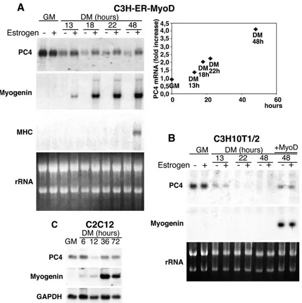
MyoD induces PC4 gene expression during myogenesis.We have previously shown that myoblasts in which the PC4 protein function has been inhibited, fail to express differentiation markers such as myogenin or myosin heavy chain, and do not undergo terminal differentiation (27).
To elucidate the role of PC4 during myogenesis, we sought in the first place to determine whether its expression is regu-lated by MyoD. To address this issue, we used a previously described cell line expressing a chimeric MyoD protein fused to the hormone-binding domain of the estrogen receptor (MyoD-ER), whose activity is inducible by treatment with
es-tradiol. The C3H10T1/2 fibroblast cell line stably expressing such a conditional MyoD protein (C3H-MyoD-ER) differen-tiates efficiently in the presence of estradiol and retains the undifferentiated phenotype in hormone-free conditions (11).
Figure 1A shows a Northern blot analysis of total RNA extracted from C3H-MyoD-ER cells cultured in GM or in differentiation medium (DM; with reduced serum concentra-tion), either in the presence or in the absence of estradiol. PC4 mRNA was expressed in GM both in the presence and in the absence of hormone but decreased significantly in hormone-free DM, indicating that expression of PC4 is downregulated after serum removal. Conversely, the hormone-dependent
in-FIG. 1. Induction of PC4 mRNA expression by MyoD. (A) C3H-ER-MyoD cells, expressing an estrogen-inducible MyoD protein, were cultured in GM or shifted to DM for increasing periods of time either in the presence or in the absence of estradiol (10⫺7M) as indicated. Afterward, total RNA was extracted and analyzed by Northern blotting. Identical filters were probed for myogenin, myosin heavy chain, and PC4 mRNAs. Ethidium bromide staining of rRNA, on one of the filters, was photographed under UV light. The graph on the right shows densitometric quantification of the MyoD-mediated induction of PC4 mRNA expression, expressed as the ratio between the levels of PC4 mRNA measured in the presence or in the absence of estradiol. (B) Expression of PC4 mRNA was analyzed by Northern blot in parental C3H10T1/2 cells cultured in the same conditions as C3H-ER-MyoD cells. The mRNA from C3H10T1/2 cells infected with a MyoD-encoding retrovirus was also analyzed (infected cells were kept 48 h in differentiation conditions; lanes⫹ MyoD). (C) Induction of PC4 mRNA expression during C2C12 myoblast differentiation. Cells were cultured in GM or shifted to DM for the times indicated. Total RNA was extracted and analyzed by Northern blotting, probing the same filter for PC4 and myogenin, as well as for GAPDH (glyceraldehyde-3-phosphate dehydrogenase) to control for RNA integrity and quantity.
duction of MyoD activity caused an increase of PC4 mRNA after 13 h in DM, concomitantly with the induction of myoge-nin expression. The PC4 mRNA levels increased up to fourfold during the following 48 h in DM (as indicated by the densito-metric analysis of the blots shown in Fig. 1A), when late mark-ers of differentiation (such as myosin heavy chain) were in-duced (Fig. 1A).
To verify that the estradiol treatment had no effect per se on the expression of PC4, the levels of PC4 mRNA were also measured in parental C3H10T1/2 fibroblasts under both grow-ing and differentiation conditions with or without estrogen (Fig. 1B). PC4 mRNA was completely downregulated within 22 h after serum withdrawal, irrespective of the presence of hormone; however, a retrovirus-mediated ectopic expression of MyoD caused an upregulation of PC4 mRNA in differen-tiation conditions that was comparable to that elicited by the estrogen-regulated form of MyoD (Fig. 1B).
Finally, expression of PC4 mRNA was also analyzed in C2C12 myoblasts undergoing differentiation. In these cells, similarly to the fibroblasts, PC4 mRNA was expressed in pro-liferating conditions but decreased soon after serum with-drawal, to regain the initial expression levels at the onset of the differentiation process, in concomitance with the maximal in-duction of myogenin (Fig. 1C).
Together, these results indicate that MyoD induces PC4 mRNA expression from the early stages of terminal differen-tiation.
To verify whether the increase of PC4 mRNA levels elicited by MyoD was the consequence of upregulation of PC4 tran-scription, we sought to analyze the effect of MyoD on the activity of the PC4 gene promoter. We therefore isolated the 5⬘-flanking region of the rat PC4 gene, extending from nt ⫺778 to nt⫹160 relative to the transcription initiation site (⫹1). This latter was determined by primer extension of poly(A)⫹ RNA isolated from rat PC12 cells induced for 2 h with NGF (given that PC4 is an NGF-inducible immediate-early gene; Fig. 2A to C). The same initiation site was mapped by using RNA from C2C7 myoblasts (data not shown). Inspection of the PC4 5⬘ flanking sequences (nt ⫺778 to nt ⫹160; Fig. 2B) did not show a TATA box but revealed the presence of a number of putative transcription factor consensus binding sites, including two E-boxes (nt ⫺684 to ⫺678; nt ⫺634 to ⫺628), the binding site of myogenic basic HLH factors (Fig. 2B) (4, 59).
The 5⬘ region of the PC4 gene was cloned upstream to a CAT reporter gene in the vector pSV0t2CAT, obtaining the plasmid PC4(⫺778⫹160)-CAT. This plasmid was then tran-siently transfected into C3H10T1/2 fibroblasts with either a MyoD expression plasmid or its empty vector. The results shown in Fig. 2E indicate that indeed MyoD was able to induce the activity of the PC4 promoter construct PC4(⫺778 ⫹160)-CAT up to sevenfold.
The most simple and testable mechanism by which MyoD could transactivate the PC4 promoter is by binding to the observed E-box consensus motifs. Thus, we generated a PC4 promoter 5⬘ deletion construct lacking the sequences that con-tain the E-boxes [PC4(⫺560⫹160)-CAT]. The activity of this PC4 promoter construct was still enhanced by MyoD (Fig. 2E), indicating that direct binding to the E-box motifs was not required for MyoD to mediate transcriptional induction of the
PC4 promoter. In contrast, a 5⬘ deletion extending up to nt ⫺133 caused a complete loss of promoter activation by MyoD (Fig. 2E). Thus, the sequences of the PC4 promoter targeted by MyoD appear to reside between nt ⫺560 and nt ⫺133. Further study will be required to identify them.
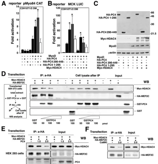
PC4 stimulates the MyoD-dependent activation of muscle-specific genes through MEF2. The observations presented above, indicating that MyoD upregulates the transcription of the PC4 gene, together with our previous observations showing a requirement for PC4 during myogenesis (27), led us to ques-tion whether PC4 could act as a regulator of MyoD activity. To verify this hypothesis, we assessed the influence of PC4 on the MyoD-dependent activation of the myogenin gene promoter. For our experiments, we used a reporter construct (pMyo84CAT) carrying 84 nucleotides of the myogenin pro-moter region, including an E-box and a MEF2 site (17, 25). These elements have been previously shown to mediate the responsiveness of the myogenin gene promoter to MyoD (17). Thus, the pMyo84CAT construct was transfected in C3H10T1/2 fibroblasts, together with MyoD and increasing amounts of PC4. We observed that PC4, in itself devoid of transcriptional activity, was able to potentiate the activation of the pMyo84 promoter by MyoD, dose dependently up to four-fold (Fig. 3A). We also tested whether PC4 could enhance the ability of MyoD to induce the endogenous myogenin gene. Cotransfection of PC4 with MyoD in C3H10T1/2 fibroblasts resulted in threefold higher levels of myogenin expression compared to those induced by MyoD alone (Fig. 3B).
Although MyoD can activate the myogenin promoter di-rectly through the E-box, the MEF2 site also mediates activa-tion by MyoD. In fact, the MEF2 site of the myogenin pro-moter has been shown to be required for myogenin transcription both in cultured cells and during embryonic de-velopment (17, 85), and a model has been proposed in which MEF2 factors, bound to DNA through the MEF2 site, can recruit MyoD, also to promoters devoid of E-boxes (56). We therefore sought to verify whether the potentiation of MyoD activity exerted by PC4 was mediated through the E-box or the MEF2 site. For this purpose we used either the report-er construct pMyo84(-E1)CAT, deleted in the E-box, or pMyo84(mutMEF2)CAT, mutated in the MEF2 site, or pMyo84(mutMEF2/-E1)CAT, lacking both of these sites. PC4 significantly potentiated the activation by MyoD of the pMyo84 promoter construct carrying only the MEF2 site but was without effect on the construct with the E-box only (Fig. 3C). This latter was stimulated by MyoD only weakly, in agree-ment with previous data (17) and with our observations indi-cating that optimal stimulation by MyoD occurs only in the presence of multiple E-boxes (see below, Fig. 3G). These data indicate that PC4 acts as a positive regulator of MyoD activity through MEF2.
To further investigate this point, we evaluated the functional interaction between PC4 and MEF2C, which is the MEF2 isoform with the highest ability to induce myogenic conversion when cotransfected with MyoD (56). In addition, MEF2C is specifically expressed in muscle and developing brain (43, 49, 61), where the expression of PC4 is high (9, 72). Therefore, we cotransfected an expression construct for MEF2C, together with MyoD, PC4, and the pMyo84 reporter. We observed that PC4 was able to further enhance the stimulation of pMyo84
promoter activity that resulted from the synergistic action of MyoD and MEF2C (Fig. 3D). MyoD and MEF2C protein levels in transfected cells were not influenced by PC4, as indi-cated by Western blot analysis (Fig. 3E).
To distinguish whether the target of PC4 was MyoD or MEF2C, we sought to analyze the effect of PC4 on the activity of each of these two factors independently. For this we used either an MEF2-responsive reporter construct, carrying two
MEF2 sites (pTK-MEF2x2 CAT; Fig. 3F), or a MyoD-respon-sive reporter construct, carrying four tandemly repeated MyoD binding sites (pt184RTK-CAT; Fig. 3G). It was found that PC4 potentiated the transactivation mediated by either MEF2C or MyoD of the corresponding reporter construct (Fig. 3F and G). It has to be noted that, while in the experiments with the MyoD-responsive reporter the transfected MyoD probably in-duced the endogenous MEF2C (13, 48), in the experiments
FIG. 2. Induction of PC4 promoter activity by MyoD. (A) Restriction map of the 5⬘ region of the rat PC4 gene. The transcribed region is represented in gray. (B) Nucleotide sequence of the 5⬘ region of the PC4 gene. Transcription factor consensus binding sequences are indicated. The transcription initiation site is indicated by the arrow. (C) Primer extension analysis of PC4 mRNA. RNA from rat PC12 cells either treated or untreated with NGF (lanes 1 and 2, respectively) was hybridized with the [␥-32P]dATP-labeled 31–55 PC4 oligonucleotide (see Materials and
Methods). The arrow indicates the extension product of 105 nt. The left lanes show the DNA sequence of the genomic clone PC4G4/3.1, obtained with the same primer, to correlate extension product size and sequence. (D) Scheme of the constructs carrying different PC4 promoter sequences (gray box) fused to the CAT reporter gene. A restriction map is shown: S, SalI; X, XbaI; ST, StuI; E, EcoRI; BG, BglII; SI, SacI. (E) MyoD stimulates PC4 promoter activity. A total of 1.0⫻ 105C3H10T1/2 cells, seeded onto 35-mm culture dishes the day before transfection, were
transfected with each of the PC4 promoter reporters shown in panel D (1g) and with either the pEMSV-MyoD expression construct (1 g) or the pEMVS Scribe␣2 empty plasmid (1 g). The cytomegalovirus (CMV)–-Gal expression construct was cotransfected in each sample as an internal control. At 24 h after transfection cells were placed in DM for 48 h, and then lysates were collected and assayed for CAT and-Gal activities. For each PC4 promoter construct the MyoD-dependent increase of CAT activity was calculated relative to the CAT activity in the absence of MyoD. Bars represent the average fold activity⫾ the standard error of the mean (SEM) of at least five independent experiments performed in duplicate. The CAT activities were measured as the percentage of the acetylated chloramphenicol/microgram of protein normalized to the-Gal activity.
FIG. 3. PC4 enhances the transcriptional activity of MyoD through MEF2 binding sites. (A) PC4 synergizes with MyoD in stimulating the myogenin promoter activity. C3H10T1/2 cells were cotransfected with the pMyo84 reporter plasmid carrying the myogenin promoter (0.8g) and the indicated amounts of the pEMSV-MyoD and pSCT-PC4 expression vectors. The fold increase in CAT activity was calculated relative to the activity of the control sample (transfected with the empty vectors). In panel A, as well as in panels B, C, D, E, and G, cells were placed in DM 24 h after transfection and harvested 48 h later. (B) PC4 cooperates with MyoD to induce the endogenous myogenin gene. C3H10T1/2 fibroblasts were transiently transfected with MyoD alone or in conjunction with PC4; the levels of PC4, MyoD and endogenous myogenin were determined by Western blotting. (C) The synergism between PC4 and MyoD requires the MEF2 site of the myogenin promoter. C3H10T1/2 cells were transfected with the indicated amounts of pEMSV-MyoD, pSCT-PC4, and either the pMyo84-CAT reporter plasmid, the pMyo84(-E1) CAT, the pMyo84(mutMEF2) CAT (mutated in the E-box or MEF2 sites, respectively), or the pMyo84(mutMEF2/-E1)CAT (lacking both sites). Samples were analyzed as for panel A. ❋, P⬍ 0.05; ❋❋, P ⬍ 0.02 (versus group with MyoD alone) (Student t test). (D) The synergism between MyoD and MEF2C is potentiated by PC4. C3H10T1/2 cells were transfected with the myogenin promoter construct pMyo84-CAT (0.8g) and with the indicated amounts of pEMSV-MyoD, pSCT-PC4, and pCDNA1-MEF2C. Samples were analyzed as for panel A. ❋, P⬍ 0.05; ❋❋, P ⬍ 0.02 (versus group with MyoD alone); @, P⬍ 0.03 (versus group transfected with pEMSV-MyoD and pCDNA1-MEF2C) (Student t test). (E) Western blot analysis of proteins expressed in C3H10T1/2 cells after a transfection experiment representative of those shown in panel D. (F) PC4 enhances the transcriptional activity of MEF2C. C3H10T1/2 cells were cotransfected with the indicated amounts of pSCT-PC4 or pCDNA1-MEF2C and with the reporter construct pTK-MEF2x2 CAT (carrying two MEF2C binding sites). Transfected cells were cultured in GM for 48 h before being harvested. Samples were analyzed as for panel A. ❋, P⬍ 0.05 (versus MEF2C group). (G) PC4 enhances the E-box-mediated MyoD transcriptional activity. C3H10T1/2 were transfected with the indicated amounts of pEMSV-MyoD, pSCT-PC4, and the reporter construct pt184RTK-CAT (carrying four E-boxes). Samples were analyzed as for panel A. ❋❋, P⬍ 0.02 (versus group with MyoD alone) (Student t test). In the assays in panels A, C, D, F, and G, CMV–-Gal was cotransfected, and the empty DNA plasmid vectors were used in place of the corresponding expression vectors to keep DNA amounts constant. Luciferase activities were measured as units/microgram of protein normalized to the-Gal activity. Bars represent the average fold activity⫾ the SEM of at least four independent experiments performed in duplicate.
with the MEF2x2 reporter the transfected MEF2C did not elicit expression of MyoD (56). Therefore, the ability of PC4 to stimulate the MEF2C-dependent activation of pTK-MEF2x2 CAT reporter (Fig. 3F) suggests that PC4 can interact func-tionally with MEF2C independently of MyoD.
As a whole, the experiments shown in Fig. 3 indicate that PC4 can stimulate the transcriptional activation exerted either by MyoD or by MEF2C. The observed synergism between PC4 and MyoD can occur either through MEF2 binding sites, or through a sufficient number of E-boxes, which are possibly necessary to allow the formation of myogenic complexes com-petent for cooperation with MEF2 factors (56).
Translocation of PC4 to the nucleus during muscle differ-entiation.The ability of PC4 to modulate the transcriptional activity of MyoD and MEF2C raised the question as to whether PC4, which is prevalently cytoplasmic (27), can trans-locate to the nucleus during differentiation. To assess this point, we evaluated the intracellular distribution of the endog-enous PC4 protein in differentiating C2C7 myoblasts by immu-nofluorescence (Fig. 4A to C). In undifferentiated C2C7
myo-blasts, growing in GM, PC4 was found mainly in the cytoplasm, although in a low percentage of cells (⬍15%) it was localized in the nucleus or both in the nucleus and in the cytoplasm (Fig. 4A and B). The number of cells showing exclusively nuclear or nuclear and cytoplasmic staining increased gradually during the first 36 h of differentiation up to ca. 70%, whereas, during the same time period, the percentage of cells displaying cyto-plasmic localization of PC4 decreased correspondingly. The cytoplasmic staining of PC4 increased again 72 h after the onset of differentiation in multinucleate myotubes, in which, however, PC4 remained detectable in a high percentage of nuclei (Fig. 4A and B). The localization of PC4 in the nucleus of terminally differentiated myocytes was confirmed by confo-cal microscopy, which also revealed a dot-like pattern of PC4 nuclear staining (Fig. 4C). These results suggest that the sub-cellular distribution of PC4 is dynamic and show that PC4 molecules enter the nucleus in significant numbers during mus-cle differentiation.
A deletion analysis carried out on the PC4 molecule indi-cated that the carboxyl-terminal region of PC4 (aa 290 to 445),
FIG. 4. Nuclear localization of endogenous PC4, analyzed by immunofluorescence and confocal microscopy. (A) A total of 7⫻ 104C2C7 cells
were seeded in 35-mm dishes and shifted 24 h later to DM to initiate differentiation. At the indicated times after the shift to DM, cells were fixed, permeabilized, and stained for immunofluorescence detection. Endogenous PC4 was visualized by using the anti-PC4 rabbit polyclonal antibody A451 (29), followed by incubation with goat anti-rabbit FITC-conjugated antibody. Nuclei were detected by Hoechst 33258 dye (corresponding photomicrographs on the right). Bar, 30m. (B) Percentage of cells with endogenous PC4 staining that was cytoplasmic only (black bars), nuclear and cytoplasmic (gray bars), or nuclear only (white bars). Values are calculated for each category as the percentages of the total number of cells scored at each time point (within three fields for each experiment). Means⫾ the SEM are from three independent experiments. The total number of cells counted for each time point is indicated at the top of the corresponding bar. (C) Confocal microscopy of endogenous PC4. A total of 7 ⫻ 104C2C7 cells were seeded onto circular coverslips placed in 35-mm dishes. Cultures were treated, and endogenous PC4 was detected as in panel
A. Nuclei are visualized in red by propidium iodide (Pr.i.). The panels on the left (merge) show the overlay of the green and red staining (in orange-green), which indicates the presence of endogenous PC4 protein in the nucleus. Bar, 10m.
isolated from the rest of the molecule, localizes to the nucleus (data not shown). Whether this region contains authentic nu-clear localization signals has yet to be determined. Further-more, we found that PC4 accumulates in the nucleus in the presence of leptomycin B, a specific inhibitor of the nuclear export receptor CRM1 (data not shown), indicating that PC4 is also subject to active nuclear export.
In vitro and in vivo interaction of PC4 with MEF2C. The experiments described above indicate that PC4 potentiates MyoD and MEF2C activity, which might imply its physical interaction with these molecules. This is also suggested by the observation that PC4 translocates to the nucleus during differ-entiation. Thus, we performed GST pull-down assays to
ana-lyze the ability of PC4 to bind MyoD and the MEF2 isoforms A, C, and D.
MEF2C, among the in vitro-translated isoforms of MEF2, was specifically able to associate with GST-PC4 and, similarly, in vitro-translated PC4 associated with GST-MEF2C (Fig. 5A, lane 8; Fig. 5B, lane 5). The efficiency of the binding of MEF2C to PC4 was comparable to that of MEF2C to MyoD (Fig. 5A). Indeed, MEF2C and bHLH factors (MyoD and myogenin) have been shown to physically interact and to synergistically regulate transcription (56). Furthermore, an MEF2C mutant deleted in the MADS domain (which is responsible, with the neighboring MEF2 domain, for DNA binding and dimeriza-tion) (57) was unable to bind PC4 (Fig. 5B lane 6). This
FIG. 5. PC4 interacts in vitro with MEF2C. (A) Binding of in vitro-translated35S-labeled MEF2A, MEF2C, and MEF2D to GST-MyoD and
GST-PC4. (B) Binding of in vitro-translated35S-labeled PC4 to GST-PC4, GST-MyoD, GST-MEF2C, and GST-MEF2C-⌬MADS. The amount
of GST fusion proteins bound to the glutathione-Sepharose resin beads used in the GST pull-down assay (10l), as detected by Coomassie blue staining, is shown below each lane. (C) Schematic representation of PC4 deletion mutants. The gray boxes identify the regions most conserved within the IFRD1 protein family (corresponding in the rat sequence to aa 65 to 157, aa 289 to 345, and aa 410 to 444) (9). (D and E) Binding to GST-MEF2C of PC4 mutants with deletions at the carboxyl terminus (D) or at the amino terminus (E). (A, B, D, and E) The indicated [35S]methionine-labeled products in vitro translated in rabbit reticulocyte lysates were incubated with the different GST-fused proteins bound to
glutathione-Sepharose 4B. Bound proteins were eluted and analyzed by SDS–8% PAGE, followed by autoradiography. The labeled input products loaded were about 30% (A and B) or 10% (D to E) of the amount used in the pull-down incubations.
pointed to the MADS domain as the requisite for the interac-tion of MEF2C with PC4.
In addition, a weak interaction occurred between PC4 and GST-MyoD (Fig. 5B, lane 4). Also, PC4 unexpectedly turned out to be able to strongly homodimerize (Fig. 5B, lane 3).
To determine the region(s) of PC4 that mediate the binding to MEF2C, we constructed four PC4 deletion mutants, carry-ing different domains of the PC4 protein, defined by the pres-ence of sequpres-ences conserved within the IFRD protein family (Fig. 5C) (9). The PC4 mutant proteins were translated in vitro and their binding to MEF2C was assessed by means of GST pull-down assays. The amino-terminal and central regions of PC4 (mutant proteins PC41-118 and PC41-295) showed null or
very weak binding to GST-MEF2C, whereas both mutants PC4118-449and PC4293-449bound strongly, thus indicating that
the carboxyl-terminal region of PC4 is required to bind MEF2C (Fig. 5D and E).
To define whether PC4 can interact with MEF2C also in vivo, we used a protein-protein-interaction assay system (20). PC4 was fused to the yeast GAL4 DBD to obtain the chimeric construct referred to as pMGAL4-PC4 (Fig. 6A). When intro-duced into C3H10T1/2 cells, pMGAL4-PC4 failed to activate the expression of the GAL4-dependent luciferase reporter gene pG5E1b-LUC (Fig. 6B, lane 5), indicating that PC4 does not harbor transcription activation domains. To test the inter-action between PC4 and MEF2C in this system, we used a truncated MEF2C protein encoding the MADS and MEF2 domains (aa 1 to 117) of MEF2C, fused to the VP16 activation domain (pVP16-MEF2C). This region of MEF2C was found to contain the sequences required for binding PC4 in vitro (as shown above in Fig. 5B). When pMGAL4-PC4 was expressed with pVP16-MEF2C, the activity of the reporter gene was induced⬃6-fold (Fig. 6B, lane 7). Furthermore, the mutant pMGAL4-PC4 290-449, encoding the GAL4 DBD fused to the carboxyl-terminal region of PC4, was seen to be even more efficient in this assay. In fact, when coexpressed with VP16-MEF2C, it was able to induce transcription more than eight-fold above the basal activity of the pG5E1b-LUC reporter (Fig. 6B, lane 10). These results indicate that PC4 and MEF2C can interact in mammalian cells and confirm that the MADS do-main of MEF2C is the determinant of the interaction with PC4 and that the carboxyl-terminal region of PC4 contains the MEF2C binding domain.
As a complementary approach to test the in vivo interaction between PC4 and MEF2C, we performed a coimmunoprecipi-tation assay. C2C12 myoblasts constitutively overexpressing PC4 (clone S4) (27) were transfected with HA-MEF2C; after-ward, the cells were either maintained in growing conditions (GM) or transferred to differentiation conditions (DM). The Western blot analysis of anti-PC4 immunoprecipitates indi-cated that MEF2C was able to associate with PC4, either in proliferating or in differentiating cells (Fig. 6C, panel IP: a-PC4).
Reversal by PC4 of the HDAC4-mediated inhibition of MEF2C and MyoD.The data reported above showed that PC4 stimulates the transcriptional activity of MyoD/MEF2C and can physically interact with MEF2C. A large body of evidence has recently highlighted that the activity of MEF2 transcription factors is regulated by class II deacetylases during the process of muscle differentiation (16, 41, 54, 76). HDAC4 has been
shown to repress MEF2C activity through association with the MADS domain (41, 76). Given that PC4 interacts with the same domain of MEF2C, one possibility was that PC4 could potentiate the activity of MEF2C by interfering with the inhib-itory action of HDAC4. To ascertain this, we tested whether PC4 could overcome the HDAC4-mediated inhibition of the muscle creatine kinase (MCK) promoter activity. The MCK
FIG. 6. In vivo interaction between PC4 and MEF2C. (A) Sche-matic representation of the mammalian two-hybrid assay, as applied to our molecular model. (B) Interaction of PC4 and PC4 290–449 with MEF2C as evaluated by the two-hybrid assay. C2C7 cells were trans-fected with pG5E1b-LUC, pMGAL4-PC4, pMGAL4-PC4 290–449, or pVP16-MEF2C or with the empty vectors as indicated. At 24 h after transfection the cells were placed in DM for 48 h before harvesting and luciferase assay measurements. ❋, P⬍ 0.05; ❋❋, P ⬍ 0.02 (versus the GAL4 control group [lane 11] and also versus the corresponding pVP16 control group [i.e., lanes 6 or 9, respectively]) (Student t test). (C) Coimmunoprecipitation of PC4 and MEF2C. C2C12 cells (clone S4 constitutively overexpressing PC4 [27]) were transfected with HA-MEF2C or with the empty vector, kept in GM or in DM for 48 h after transfection, and then lysed and immunoprecipitated with the poly-clonal A451 anti-PC4 antibody, covalently bound to Sepharose resin. The anti-HA or the anti-PC4 antibodies were used for Western blot analysis of the immunoprecipitated complexes (IP: a-PC4 panel) and of the input cell lysate (1/30 of the immunoprecipitated lysate).
regulatory region is known to contain both MyoD and MEF2 binding sites (1). As expected, HDAC4 inhibited the stimula-tion of the MCK LUC reporter gene mediated by MyoD and MEF2C (Fig. 7A). However, cotransfection of PC4 was able to rescue the MyoD activity, reaching a complete reversal of the HDAC4-imposed inhibition when MEF2C was also present (Fig. 7A).
We then monitored the influence of PC4 on the HDAC4-mediated inhibition of the MEF2C-responsive pTK-MEF2x2 CAT reporter construct in conditions promoting
differentia-tion. As shown in Fig. 7B, PC4 was indeed able to overcome the HDAC4-mediated repression of the MEF2C-dependent reporter. Western blot analysis of the protein extracts used in transcription assays indicated that PC4 did not alter the ex-pression levels of the MyoD, MEF2C, and HDAC4 transfected constructs (Fig. 7C).
PC4 can dissociate HDAC4 from MEF2C.Given that PC4 was able to overcome the HDAC4-dependent inhibition of MEF2C activity, we sought to determine whether such an effect of PC4 was dependent on its ability to interact with MEF2C. To address this issue, we analyzed the ability of the amino- or the carboxyl-terminal domains of PC4 to synergize with MyoD and to counteract HDAC4 (as shown in Fig. 5D and E, the carboxyl-terminal region of PC4 binds MEF2C). Thus, we coexpressed MyoD and either PC4, PC41-295, or
PC4290-449 in C3H10T1/2 cells with the reporter plasmid
pMyo84 CAT or MCK LUC. The carboxyl-terminal region of PC4, but not the amino-terminal region, enhanced the MyoD-dependent activation of both pMyo84 CAT and MCK LUC reporters as efficiently as full-length PC4 (Fig. 8A and B). Similarly, only the carboxyl-terminal region of PC4 was signif-icantly able to rescue the MCK promoter activity from HDAC4 inhibition (Fig. 8B). Western blot analysis of protein extracts from transfected cells indicated that PC4 full-length and the PC4 deletion mutants were expressed at similar levels (Fig. 8C). Thus, the ability of PC4 to synergize with MyoD and to counteract the inhibitory action of HDAC4 appears to cor-relate with the ability of PC4 to physically interact with MEF2C.
Because HDAC4 and PC4 bind to the same region of MEF2C (the MADS domain), we sought to determine whether PC4 could impair the association of HDAC4 with MEF2C. To test this hypothesis, we analyzed the formation of HDAC4-MEF2C complexes either in the presence or in the absence of recombinant PC4. Myc-HDAC4 and HA-MEF2C were ex-pressed in NIH 3T3 mouse fibroblasts, and their interaction was analyzed by anti-HA immunoprecipitation, followed by Western blot detection of the immunoprecipitated products (Fig. 8D). When the extracts were preincubated with increas-ing amounts of purified GST-PC4 protein, immunoprecipita-tion of extracts with anti-HA led to the recovery of HDAC4 in amounts inversely proportional to the quantity of immunopre-cipitated GST-PC4, as revealed by Western blotting with anti-Myc or anti-GST antibodies (Fig. 8D, see IP: a-HA). Consis-tently, in the supernatants after immunoprecipitation the amount of Myc-HDAC4 increased proportionally to the GST-PC4 added (Fig. 8D, see cell lysates after immunoprecipita-tion). Thus, the binding of PC4 and HDAC4 to MEF2C appear to be mutually exclusive.
As an alternative approach, we sought to evaluate the effects of transfection of PC4 on the MEF2C-HDAC4 complex in vivo. Therefore, we cotransfected HA-MEF2C and Myc-HDAC4 with increasing amounts of PC4. We found that the level of HDAC4 protein coimmunoprecipitating with MEF2C was lower in samples where PC4 was coexpressed, with a de-crease proportional to the amount of transfected PC4 (Fig. 8E, panel IP: a-HA). These results confirm the hypothesis that PC4 can displace HDAC4 from MEF2C.
It has been recently shown that CaMK enhances the tran-scriptional activity of MEF2C by negatively regulating the
in-FIG. 7. PC4 rescues the transcriptional activity of MEF2C and MyoD from the inhibition exerted by HDAC4. (A) The MCK LUC reporter was cotransfected in C3H10T1/2 cells with pEMSV-MyoD, pcDNA1-MEF2C, pcDNA-Myc-HDAC4, or PC4 in the indicated combinations. At 24 h after transfection, the cells were placed in DM and left for 48 h before luciferase assay determination. Luciferase activities were measured as units per microgram of protein normalized to-Gal activity and then calculated as the fold activation relative to the activity of control samples (transfected with empty vectors). Bars represent the average fold activity⫾ the SEM of at least three inde-pendent experiments performed in duplicate. ❋, P⬍ 0.05; ❋❋, P ⬍ 0.02 (versus the corresponding group without PC4) (Student t test). (B) The MEF2-responsive reporter was cotransfected in C3H10T1/2 cells with combinations of the expression vectors pEMSV-MyoD, pcDNA1-MEF2C, pcDNA-Myc-HDAC4, and pSCT-PC4, as indi-cated. Transfected cells were treated and analyzed as described in panel A. ❋, P⬍ 0.05 (versus the corresponding group without PC4) (Student t test). (C) Western blot analysis showing the expression levels of the constructs transfected in the experiments depicted in panels A and B.
teraction between HDAC4 and ME2C (40). Thus, we wanted to compare the effects of PC4 and CaMK. HDAC4 was almost completely displaced from MEF2C by an activated form of CaMK (Fig. 8F), similarly to what was observed with PC4 (Fig. 8E). Based on this observation, one would expect that the activated form of CaMK would favor the association of PC4 with MEF2. Indeed, we found that the ability of PC4 to bind to
MEF2C was somewhat increased by cotransfection of activated CaMK (about twofold; Fig. 9A). Finally, we observed that, although both PC4 and activated CaMK stimulated the tran-scriptional activity of MEF2 to a comparable extent (Fig. 9B), the simultaneous transfection of submaximal concentrations of PC4 and activated CaMK resulted in an additive effect, sug-gesting cooperativity between the two molecules (Fig. 9B).
FIG. 8. PC4 antagonizes HDAC4 and displaces it from MEF2C. (A and B) The carboxyl-terminal region of PC4 synergizes with MyoD and reverses the inhibition by HDAC4. (A) The pMyo84 CAT reporter was cotransfected in C3H10T1/2 cells with pEMSV-MyoD and with either HA-pSCT-PC4 or the deletion mutants HA-pSCT-PC4 290–449 and HA-pSCT-PC4 1–295, as indicated. (B) C3H10T1/2 cells were transfected with the MCK LUC reporter, with the expression constructs used for panel A, and with pcDNA-Myc-HDAC4, in the indicated combinations. In panels A and B, at 24 h after transfection cells were placed in DM for 48 h before luciferase assay determination. The luciferase activity was measured as units per microgram of protein normalized to the-Gal activity; the fold activations were calculated relative to the control sample (transfected with empty vectors). The average fold activity⫾ the SEM is shown for at least three independent experiments performed in duplicate. ❋, P⬍ 0.05 (versus the corresponding group without HA-pSCT-PC4 or without HA-pSCT-PC4 1–295 transfected) (Student t test). (C) Analysis by Western blotting of the expression levels of the constructs transfected, in one that is experiment representative of those shown in panels A and B. (D) PC4 displaces HDAC4 from MEF2C in an in vitro assay. Lysates of NIH 3T3 cells, cotransfected with Myc-HDAC4 and HA-MEF2C (or alternatively, with the empty vectors), were incubated with increasing amounts of purified GST-PC4 or GST protein and then immunoprecipitated with anti-HA antibody. The immunoprecipitated complexes (left panel, IP), as well as the cell lysates after immunoprecipitation (central panel) and the input cell lysates (1/30 of the total lysate), were analyzed by Western blotting with anti-Myc, anti-HA, or anti-GST antibodies. (E) PC4 displaces HDAC4 from MEF2C in an in vivo assay. Lysates of HEK293 cells, cotransfected with Myc-HDAC4 and HA-MEF2C (0.75g each) and with increasing amounts of PC4, were then immunoprecipitated with anti-HA antibody. The immunoprecipitated complexes (panel IP) and the input cell lysates (1/30 of the total lysate) were analyzed by Western blotting with the indicated antibodies. (F) Activated CaMKI dissociates HDAC4-MEF2C complexes. HEK293 cells were cotransfected with Myc-HDAC4 and HA-MEF2C (0.75g each) and with either CaMKI or its empty vector. Lysates of transfected cells were then immunoprecipitated with anti-HA. The immunoprecipitated complexes (panel IP) and the input cell lysates (1/30 of the total lysate) were analyzed by Western blotting with the indicated antibodies.
A functional requisite for the displacement of HDAC4 from MEF2C by PC4 is the colocalization of these molecules in the nucleus of differentiating myocytes. Since it is known that MEF2C is an essentially nuclear protein (49), whereas HDAC4 and PC4 translocate from the cytoplasm to the nu-cleus during differentiation (55, 89; the present study), we tested whether endogenous PC4 and HDAC4 indeed colocal-ize. As shown by confocal microscopy, PC4 and HDAC4 co-localized in the cytoplasm of proliferating C2C12 myoblasts. In differentiating myoblasts (after 24 h in DM) and, more
evi-dently, in mature myotubes (72 h in DM), PC4 and HDAC4 progressively accumulated and colocalized in the nucleus of the majority of cells, although a significant amount of these proteins was still detectable in the cytoplasm (Fig. 10). This indicated that a functional interaction between PC4, MEF2C, and HDAC4 can take place in the nucleus during differentia-tion.
Physical and functional interaction of PC4 and HDAC4.It has been recently reported that Tis7 (the mouse PC4 homolog) can interact with class I HDACs (75). To verify whether PC4 could also bind HDAC4, lysates of NIH 3T3 cells cotransfected with Myc-HDAC4 and PC4 were immunoprecipitated with the anti-PC4 (A451) antibody and subjected to Western blot anal-ysis with anti-Myc antibody to reveal the HDAC4 protein. As shown in Fig. 11A, HDAC4 could be detected in association with the immunoprecipitated PC4 protein. To verify the asso-ciation of PC4 with HDAC4 also in muscle cells, we immuno-precipitated PC4 from the C2C12 myoblast clone S4 (consti-tutively overexpressing PC4) transfected with Myc-HDAC4. As shown in Fig. 11B, PC4 associated with HDAC4, both in proliferation and in differentiation conditions (i.e., GM and DM). Moreover, the ability of endogenous PC4 and endoge-nous HDAC4 to coimmunoprecipitate in extracts from C2C7 cells indicated that their association occurs also in physiologi-cal conditions (Fig. 11C). A higher association of PC4 with HDAC4 was observed in differentiated myotubes.
To better understand the modalities of the interaction be-tween HDAC4 and PC4 observed in vivo, we sought to define whether they could associate in vitro and to identify the region
FIG. 9. Cooperativity between CaMK and PC4. (A) Activated CaMKI weakly increases the amount of PC4 associated with MEF2C. Clone S4 of C2C12 cells cultured in proliferating conditions were cotransfected with HA-MEF2C (6 g) and with either activated CaMKI (SR␣-CaMKI; 6 g) or the empty vector. MEF2C was then immunoprecipitated by the anti-HA monoclonal antibody, and the immunoprecipitated complexes were analyzed by Western blotting by using the anti-PC4 A451 or anti-HA antibody, as indicated. (B) PC4 and CaMK cooperate in stimulating MEF2 transcriptional activity. C3H10T1/2 cells were cotransfected with the reporter construct 3x MEF2 LUC (carrying three MEF2 binding sites [40]; 100 ng) with pCDNA1-MEF2C and with increasing amounts of either pSCT-PC4 or activated CaMKI, as indicated. Doses of PC4 and CaMKI above 1000 ng and 200 ng, respectively, did not induce further stimulation, indi-cating that these were maximally effective doses, within the linear range of the dose-response curve. Cotransfection of pSCT-PC4 and activated CaMKI (lane 9) produced additive effects. Transfected cells were cultured in GM for 48 h before being harvested. Luciferase activities were measured as units per microgram of protein normalized to-Gal activity and are expressed as the fold activation relative to the activity of the control sample (transfected with the empty vectors). Bars represent the average fold activity⫾ the SEM of three indepen-dent experiments performed in duplicate.
FIG. 10. Colocalization of endogenous PC4 and HDAC4 in prolif-erating and differentiating myoblasts. Endogenous HDAC4 and PC4 were visualized by confocal microscopy in C2C12 cells by using anti-HDAC4 and anti-PC4 rabbit polyclonal antibodies, followed by incu-bation with goat anti-rabbit TRITC- and FITC-conjugated antibodies, respectively. In proliferating myoblasts HDAC4 is prevalently cyto-plasmic, whereas in differentiating myotubes it is also nuclear, as shown in representative photomicrographs. The pattern of localization of HDAC4 overlaps with that of PC4, as indicated by the yellow pseudo-color produced by the overlay (merge) of the green and red staining. Bar, 10m.
of HDAC4 required to bind PC4. Thus, using GST pull-down assays, we tested the ability of GST-PC4 to bind in vitro-translated HDAC4 and its deletion mutants (HDAC41-611and
HDAC4611-1084). HDAC4 wild type and HDAC4611-1084were
both able to bind PC4 efficiently, whereas HDAC41-611was not
(Fig. 12A).
These data indicate that PC4 binds the carboxyl-terminal region of HDAC4, which harbors the deacetylase domain. Therefore, we wanted to analyze whether PC4 could inhibit the enzymatic activity of HDAC4. For this purpose, we measured the deacetylase activity associated with HDAC4 complexes immunoprecipitated from HEK293 cells transfected with HDAC4, either in the presence or in the absence of GST-PC4
(Fig. 12B). The results in Fig. 12B show that PC4 coimmuno-precipitated with HDAC4 and yet did not influence the HDAC4-associated deacetylase activity.
These results, taken together, indicate that PC4 can bind the carboxyl-terminal region of HDAC4 without inhibiting its deacetylase activity and also that HDAC4 binds PC4 and MEF2C through separate domains. Thus, we sought to test whether PC4 could counteract the inhibition exerted by the amino-terminal region of HDAC4 (HDAC41-611), which is
known to bind to and repress MEF2C as effectively as full-length HDAC4 (12). Therefore, we coexpressed the MCK LUC reporter, MyoD, and HDAC4 or HDAC41-611 in
C3H10T1/2 fibroblasts, with or without PC4. As expected, HDAC41-611 was able to inhibit the stimulation by MyoD of
the MCK promoter activity as efficiently as did full-length HDAC4, but its effect was completely reversed by PC4 (Fig. 12C). Thus, PC4 can counteract the repressor activity medi-ated by the amino-terminal region of HDAC4 which, notably, does not contain the PC4 binding site. This result clearly indi-cates that the ability of PC4 to overcome the HDAC4-depen-dent inhibition of MyoD activity does not rely on its ability to bind HDAC4.
DISCUSSION
In this study we have characterized the functional role of the PC4 gene in myoblast differentiation. Our results show that expression of PC4 mRNA is induced during the process of terminal differentiation, being upregulated in differentiating C2C12 myoblasts, as well as in a fibroblast cell line expressing a conditional MyoD protein. This MyoD-mediated induction of PC4 expression occurs at the transcriptional level, as indi-cated by the observation that MyoD stimulates the activity of the PC4 promoter.
As for the function of PC4, we found that the PC4 protein, which in itself is devoid of transcriptional activity, is able to significantly potentiate the MyoD-dependent transactivation of muscle gene promoters and that this effect of PC4 occurs selectively through MEF2 sites. Consistently, PC4 enhances the synergism of MyoD and MEF2C in activating muscle-specific promoters. A direct action of PC4 on MEF2C is sug-gested also by the ability of PC4 to increase the activation, elicited by MEF2C, of a synthetic reporter carrying only MEF2 sites. The observation that PC4 can override the repression imposed by HDAC4 on the muscle-inducing function of MyoD further supports the hypothesis that MEF2C is the target of PC4 action. Indeed, it has been well established that class II deacetylases suppress the myogenic function of MyoD by in-teracting with the MADS domain of MEF2 factors and re-pressing their transcriptional activity (16, 37, 41, 54, 76).
A key question raised by our findings is whether the two observed activities of PC4, i.e., the ability to potentiate the transcriptional activity of MyoD and to rescue MEF2C from the inhibition by HDAC4, rely on a common mechanistic basis. HDAC4 contains several domains active in transcriptional repression that can represent possible targets for regulation by PC4. The deacetylase catalytic domain of HDAC4, canonical within the class II HDACs family, resides in the carboxyl-terminal region of the protein (22, 26). It has been shown that this domain of HDAC4 acts by recruiting a corepressor
com-FIG. 11. PC4 associates with HDAC4. (A) NIH 3T3 cells were cotransfected with pcDNA-Myc-HDAC4 and pSCT-PC4 (or with the empty vector) as indicated, lysed 48 h after transfection, and immu-noprecipitated with the anti-PC4 antibody A451 covalently bound to Sepharose resin. Immunoprecipitated complexes (IP: a-PC4 panel) and input cell lysates were analyzed by Western blotting with anti-Myc or anti-PC4 antibodies. Asterisks in panels A and B indicate a back-ground protein (see reference 32). (B) C2C12 cells (clone S4 consti-tutively overexpressing PC4) were transfected with pcDNA-Myc-HDAC4 (or with the empty vector) as indicated, grown in GM or DM for 48 h after transfection, and then lysed and immunoprecipitated with the anti-PC4 antibody A451 covalently bound to Sepharose resin. The immunoprecipitated complexes (IP: a-PC4 panel), as well as the input cell lysates, were analyzed by Western blotting with anti-Myc or anti-PC4 antibodies. (C) C2C7 cells, cultured in GM or DM for 48 h, were lysed and immunoprecipitated with the anti-PC4 antibody co-valently bound to Sepharose resin. Immunoprecipitated complexes (IP: a-PC4 panel) and the input cell lysates were analyzed by Western blotting with anti-HDAC4 or anti-PC4 antibodies. As a control (mock IP panel), the immunoprecipitates obtained by using Sepharose resin coupled to preimmune serum were analyzed by Western blotting. For each experiment shown in panels A to C, 1.5 mg of protein lysate was immunoprecipitated and fractionated by SDS-PAGE. Western blot analysis of input lysates was carried out on 1/30 of the lysates used for immunoprecipitation.
plex composed of N-CoR, SMRT, and HDAC3 (21). The in-teraction of HDAC4 with MEF2C occurs through a separate region localized within the amino-terminal of HDAC4 (41, 76), which also effectively inhibits the MEF2 transcriptional activ-ity. In fact, the amino-terminal region of HDAC4 can interact with, and recruit to MEF2C, other transcriptional corepres-sors, such as HDAC1 (12), carboxyl-terminal-binding protein (87), and heterochromatin protein 1 (88).
We show that PC4 specifically binds MEF2C within the MADS domain (Fig. 5A and B), the same region through
which MEF2C interacts with HDAC4, and that PC4 can dis-sociate HDAC4 from MEF2C in a concentration-dependent manner (Fig. 8D and E). Such displacement of HDAC4 from MEF2C occurs concomitantly with an increased binding of PC4 to MEF2C, which indicates that PC4 and HDAC4 bind to MEF2C in a mutually exclusive fashion.
The observed dissociation of HDAC4 from MEF2C induced by PC4 can plausibly account for the ability of PC4 to coun-teract the HDAC4-dependent repression of the MyoD/ MEF2C transcriptional activity. This is also consistent with the
FIG. 12. Physical and functional interaction between HDAC4 and PC4. (A) The carboxyl-terminal region of HDAC4 binds PC4. GST pull-down assays were performed by incubating equal amounts of in vitro-translated [35S]methionine-labeled HDAC4 proteins, full-length or
truncated mutants, with equimolar amounts of either GST-PC4 or GST proteins bound to glutathione-Sepharose 4B resin. Bound proteins were eluted, separated by SDS–8% PAGE, and analyzed by using a phosphorimager. (B) PC4 does not influence the enzymatic activity of HDAC4. HEK293 cells, grown in 90-mm dishes, were transfected with 12g of either pcDNA-Myc-HDAC4 or the empty vector. The precleared lysates were incubated with GST or GST-PC4 and then immunoprecipitated with anti-Myc antibody. As a control, a synthetic peptide corresponding to the Myc epitope was also added in molar excess to samples before immunoprecipitation. Immunoprecipitated proteins, treated or not with trichostatin A (200 nM), were then incubated with preacetylated histones. Deacetylase activities are shown as percent values relative to the level of control samples (transfected with HDAC4 and without GST protein added) set to 100 (the absolute mean dpm values were about 1,400). The bars are the average⫾ the SEM of three independent experiments performed in duplicate, normalized to the amount of immunoprecipitated proteins by Western blotting (a representative blot is shown in the lower panel). (C) PC4 counteracts the inhibitory effect exerted by the HDAC4 amino-terminal region. The MCK LUC reporter was cotransfected in C3H10T1/2 cells with pEMSV-MyoD (50 ng) and the indicated combinations of HA-pSCT-PC4 (1g) and pcDNA-Myc-HDAC4 and pcDNA-Myc-HDAC4 1–611 (5 ng). At 24 h after transfection, cells were placed in DM for 48 h before luciferase assay determination. Luciferase activity was measured as units per microgram of protein normalized to the-Gal activity. The average fold activity⫾ the SEM shown (relative to control samples transfected with empty vectors) was calculated from three independent experiments performed in duplicate. ❋, P⬍ 0.05 (versus the corresponding group without PC4) (Student t test). (D) Working model for PC4 coactivation of MyoD/MEF2C. See the Discussion for more information.