Alma Mater Studiorum – Università di Bologna
DOTTORATO DI RICERCA IN SCIENZE BIOMEDICHE
MODULO 5: SCIENZE MORFOLOGICHE UMANE E MOLECOLARI
Ciclo XXVII
Settore concorsuale di afferenza: 05/H1
Settore Scientifico disciplinare: BIO/16
TITOLO TESI
Molecular basis of Osteoarthritis and aspects of
cellular senescence in disease
Esame finale anno 2015
Presentata da: Serena Guidotti
Coordinatore Dottorato Relatore
Prof. Lucio Cocco Prof. Lucio Cocco
Correlatore
INDEX
Abstract...1
1- INTRODUCTION
1.1 Osteoarthritis (OA)...3
General characteristics.1.2 Articular cartilage...6
Development of articular cartilage: from chondrogenesis to endochondral ossification.Structure of articular cartilage.
1.3 Role of senescence in OA...9
General features of senescence.The phenomenon of ‘‘intrinsic’’ or “replicative” senescence. The phenomenon of ‘‘extrinsic’’ or “stress-induced” senescence. Senescence in Aging and Osteoarthritis.
SASP: “Senescence associated Secretory phenotype".
1.4 Role of oxidative stress in OA...16
1.5 Involvement of oxidative stress in DNA damage...19
Double-strand breaks and γH2AX.
DNA mismatch repair (MMR) pathway.
Role of Gadd45β and p21 in DNA damage response and senescence.
1.6 Role of GSK3β in OA...26
1.7 NF-κB pathway: the role of IKKα...28
1.8 Nutraceuticals: a new way to counteract cell oxidative
stress and senescence in osteoarthritic chondrocytes...30
2- AIMS OF THE STUDY...33
3- MATERIALS AND METHODS
3.1 Effects of GSK3β pharmacological inactivation...36
Chondrocytes isolation.ROS production evaluation downstream GSK3β inactivation. Effects of GSK3β inhibition on proliferation and cell cycle. Real time PCR analysis.
Evaluation of senescence by mean of SA-β Gal activity and Glycogen content. Immunoblotting.
3.2 Cartilage explants studies...41
Immunohistochemistry and immunofluorescence.3.3 IKKα Knock Down cultures...42
Chondrocyte retroviral transduction.Effect of oxidative stress induction on IKKα KD and control cells. Mismatch repair gene expression analysis.
Flow-FISH determination of Telomere Length.
3.4 Nutraceuticals treatments...45
Hydroxytyrosol treatment of chondrocytes and protection from oxidative stress. Spermidine treatment of chondrocytes and protection from oxidative stress.3.5 Statistics...46
4- RESULTS
4.1 Effects of GSK3β inactivation in vitro...48
GSK3β inactivation in vitro determines ROS production and oxidative damage in chondrocytes.GSK3β inactivation affects cell proliferation with S-phase arrest of chondrocytes cultured in monolayer.
GSK3β inactivation in monolayer cultures induces senescence and hypertrophy in chondrocytes.
GSK3β inactivation in monolayer determines the activation of DNA damage response and increases expression of IKKα and MMP10.
4.2 GSK3β phosphorilation in cartilage explants...54
Evaluation of GSK3β inactivation in cartilage from OA patients.DNA damage response is associated with GSK3β inactivation in cartilage from OA patients.
4.3 Functional genomics analysis of IKKα effects on
oxidative DNA damage...57
IKKα impacts on double strand breaks (DSB) induction after an oxidative stress.IKKα impacts on telomere length after an oxidative stress.
IKKα impacts on DNA Mismatch Repair (MMR) system after an oxidative stress.
4.4 Nutraceutical treatments and anti-oxidant activity...63
Hydroxytyrosol attenuates γH2AX-foci formation and senescence after H2O2exposure in chondrocytes.
Spermidine pre-incubation reduces cell death and γH2AX-foci formation after H2O2 exposure in chondrocytes.
5- CONCLUSIONS AND DISCUSSION...69
6- BIBLIOGRAPHY...78
1
Abstract
The aim of this study is to investigate on some molecular mechanisms contributing to the pathogenesis of osteoarthritis (OA) and in particular to the senescence of articular chondrocytes. It is focused on understanding molecular events downstream GSK3β inactivation or dependent on the activity of IKKα, a kinase that does not belong to the phenotype of healthy articular chondrocytes. Moreover, the potential of some nutraceuticals on scavenging ROS thus reducing oxidative stress, DNA damage, and chondrocyte senescence has been evaluated in vitro.
The in vitro LiCl-mediated GSK3β inactivation resulted in increased mitochondrial ROS production, that impacted on cellular proliferation, with S-phase transient arrest, increased SA-β gal and PAS staining, cell size and granularity. ROS are also responsible for the of increased expression of two major oxidative lesions, i.e. 1) double strand breaks, tagged by γH2AX, that associates with activation of GADD45β and p21, and 2) 8-oxo-dG adducts, that associate with increased IKKα and MMP-10 expression. The pattern observed in vitro was confirmed on cartilage from OA patients.
IKKa dramatically affects the intensity of the DNA damage response induced by oxidative stress (H2O2 exposure) in chondrocytes, as evidenced by silencing strategies. At early time point an higher percentage of γH2AX positive cells and more foci in IKKa-KD cells are observed, but IKKa KD cells proved to almost completely recover after 24 hours respect to their controls. Telomere attrition is also reduced in IKKaKD. Finally MSH6 and MLH1 genes are up-regulated in IKKαKD cells but not in control cells.
2
Hydroxytyrosol and Spermidine have a great ROS scavenging capacity in vitro. Both treatments revert the H2O2 dependent increase of cell death and γH2AX-foci formation and senescence, suggesting the ability of increasing cell homeostasis. These data indicate that nutraceuticals represent a great challenge in OA management, for both therapeutical and preventive purposes.
3
1- INTRODUCTION
1.1 Osteoarthritis (OA)
General characteristics.
Osteoarthritis (OA), the most common arthritis in western population, is a degenerative joint disease characterized by progressive loss of cartilage, synovial inflammation, osteophyte formation, joint space narrowing and subchondral bone sclerosis (Castaneda, Roman-Blas et al. 2012); (Sharma, Jagga et al. 2013), and is responsible for structural and functional alterations of articular cartilage. This disease mostly affects knee, hip and hand and is one of the most common source of pain and disability in the elderly (Arden and Nevitt 2006) (Fig.1). OA affects one in six adults, and by 2030 it is estimated that 20% of people in Europe and United States will be affected by this pathology (De Bari, Kurth et al. 2010).
Fig.1 (A) Osteoarthritis at level of hand (deformations of the distal interphalangeal joints). (B) Plain radiograph of an osteoarthritic hip joint. (C) MRI of an osteoarthritic knee.
4
The aetiology of OA is multi-factorial and includes not only age, gender, weight but also joint injury and genetic factors. After the age of 50 years more women than men are affected (Bijlsma, Berenbaum et al. 2011). Osteoarthritis develops progressively over several years and chronic pain is frequently associated with significant functional impairment due to progressive degradation of articular cartilage, inflammation, stiffness and loss of mobility (Felson 2006). These symptoms diminish the patients’ quality of life.
On the biochemical level, OA is characterized by uncontrolled production of matrix-degrading enzymes, including aggrecanases (ADAMTSs) and matrix metalloproteinases (MMPs), which results in the destruction of cartilage extracellular matrix (Goldring and Goldring 2007). Other hallmarks of the disease are new bone formation at the joint margins (called osteophytes), limited inflammation of the synovial tissue (synovitis), and changes in the subchondral bone structure (sclerosis and cysts) (Fig. 2). This process is driven, in part, by signalling mechanisms induced by stressful or inflammatory mediators.
Histopathology of OA. Fig.2 Schematic drawing of an osteoarthritic joint Clinical and structural hallmarks of the disease are shown on the left. On the right the interplay between cartilage, bone and synovial tissue is displayed.
5
Osteoarthritis affects not only articular cartilage but also subchondral bone, synovial membrane and joint capsule (Goldring and Goldring 2007). The very early histopathological features are characterized by hypertrophic repair (Dreier 2010). In this context articular chondrocytes increase proliferation rate so they form “clusters”. Some anabolic activities are up-regulated such as an increased synthesis of proteoglycans and type II collagen. Besides this very early hypertrophic phase, the process is followed by a high catabolic activity, leading to an increased production of matrix metalloproteinases, aggrecanases, higher expression of regulatory proteins, increase of stress, senescence and apoptotic markers (Goldring and Marcu 2009). Expression of MMP-1, MMP-3, MMP-9, MMP-13 and MMP-14, as well as aggrecanases (ADAMTS-5, ADAMTS-4, and ADAMTS-9), drastically increases (van der Kraan, Blaney Davidson et al. 2010); (Madry, Luyten et al. 2012). The expression of MMP-13, known to be pivotal in OA pathology, is regulated by the interplay of two fundamental transcription factors (Runx-2 and Sox-9), contributing to the cartilage degeneration process (Orfanidou, Iliopoulos et al. 2009). These early alterations lead to a progressive loss of the cartilaginous structure, beginning with fibrillation of the superficial zone, followed by matrix loss, and appearance of simple and then more complex fissures (Goldring and Goldring 2007). Moreover, there is evidence of chondrocyte death and of chondrons, in which the cells are disoriented with lack of their normal spatial organization (Pritzker, Gay et al. 2006) . As a result, the thickness of the articular cartilage decreases.
In 2006, some OARSI (Osteoarthritis Research Society International) members proposed a grading and staging system for OA. This group has defined as “grade” a quantitative evaluation of the extent of OA progression into articular cartilage depth while the horizontal extent of the cartilage lesion is termed “stage”. The “score”, defined as the result of OA grade and stage, reflects OA severity and extent. The grade
6
value goes from 0.0 (articular cartilage surface is intact) to 6.0 (joint surface is deformed). On the other hand, with healthy cartilage being of stage 0, the stage value goes from 1 (less than 10% of joint involvement) to 4 (more than 50% of joint involvement) (Pritzker, Gay et al. 2006).
1.2 Articular Cartilage
Development of articular cartilage: from chondrogenesis to endochondral ossification. Chondrocytes are the only cellular component of articular cartilage and their primary function is to maintain cartilage homeostasis. During fetal development, the greater part of the skeleton is cartilaginous. At the level of the long bones, this “temporary” cartilage is gradually replaced by bone in a process called endochondral ossification, that ends at puberty, although the growth plates will complete their ossification only some years later. In contrast, the cartilage in the joints remains un-ossified during the whole life and is, therefore, “permanent”. The development of both temporary and permanent cartilage is called chondrogenesis and begins with the proliferation of chondroprogenitor from mesenchymal stem cells; at the completion of the process, in articular cartilage, the chondrocytes are kept in a state of maturational arrest and are prevented to complete the default route of their maturation process which ends with hypertrophy and terminal differentiation. Stage-specific transcription factors and signalling pathways play a key role in the process of chondrocyte maturation (Dreier 2010). As articular cartilage is formed to last for life, chondrocytes display only moderate metabolic activity under normal conditions to maintain their surrounding extracellular matrix (ECM) comprising collagens, proteoglycans and non-collagenous proteins. Under non-diseased conditions, the cells remain in a resting state and refrain from proliferation and terminal differentiation; this process is regulated by the
7
transcription factor Sox9. In a diseased OA state, however, some articular chondrocytes loose their differentiated phenotype and enter an endochondral ossification-like cascade of proliferation (Tallheden, Bengtsson et al. 2005) and hypertrophic differentiation followed by extracellular matrix calcification.
This process is associated with an increase of environmental stress, articular chondrocyte proliferation, expression of hypertrophy markers (for example, MMP-13 and collagen X), remodeling of the cartilage matrix by proteases, vascularization and focal calcification of joint cartilage with deposition of calcium hydroxyapatite crystals. This profound ECM perturbation and environmental stress can lead to chondrocyte programmed cell death. Hypertrophic differentiation and endochondral ossification are regulated by a transcription factor called Runx-2. Therefore some of the characteristics
Fig.3 Anatomy of articular cartilage and subchondral bone in normal and OA joints. Normal articular cartilage is divided into four zones. These zones consist of a small number of chondrocytes trapped in collagen matrix. Alteration in OA joint is represented by collagen matrix disruption in articular cartilage and thickening of subchondral bone. Fissuring and flanking in articular cartilage induces vascularization of cartilage, leading to exposure of subchondral bone to external surface.
8
of osteoarthritic cartilage resemble chondrocyte differentiation processes during skeletal development (Dreier 2010) and osteoarthritic chondrocytes undergo hypertrophy and terminal endochondral ossification, mimicking the pattern of differentiation of fetal skeletogenesis (Borzi, Olivotto et al. 2010); (Marcu, Otero et al. 2010)
Structure of articular cartilage.
In regard to its cellular composition, articular cartilage is less complex as compared to other tissues as it is neither vascularized nor innervated and does not contain tissue macrophages. Nevertheless it is a tightly regulated tissue, kept in a low homeostatic turnover by the chondrocytes that represent approximately 5% of the total tissue volume (Buckwalter, Mankin et al. 2005). Articular cartilage covers the end of bones of synovial or diarthrodial joints and acts bearing static and dynamic forces (shear, compression and tension). These mechanical loads are absorbed by the cartilage extracellular matrix (ECM) that is mainly constituted by collagens and proteoglycans. Type II collagen is composed of three polypeptide chains forming the fibrillar unit that polymerize outside the cells in a network with type XI and type IX collagen (Heinegard and Saxne 2011). Proteoglycans are mostly represented by large macromolecular aggregates of units called “aggrecan” anchored to a hyaluronan molecule. Aggrecan are macromolecular complexes with a core protein which establishes several interactions with glycosaminoglycans (GAG). The network of type II collagen fibrils is essential for maintaining the volume and shape, while proteoglycans ensure elasticity of the tissue. Collagens and proteoglycans are differently organized across the ECM thus it is possible to distinguished a territorial and an inter-territorial region as a function of the proximity to chondrocytes. In the inter-territorial area, the collagen component is highly prevalent. Conversely, the proteoglycan component is much more represented in the
9
territorial region (Madry, Grun et al. 2011). In articular cartilage four zones can be distinguished; the superficial, middle, deep and calcified zone. From a structural point of view, the superficial layer of articular cartilage is composed by a network of extracellular matrix proteins, proteoglycans, collagen, and non-collagenous proteins, and maintains a high water content. This zone includes a large percentage of immature and progenitor cells (Grogan, Miyaki et al. 2009). Middle layer contains randomly oriented collagen fibers with larger chondrocytes. Deep stratum is formed by vertical columns separated by collagenous fibrils. The last layer of articular cartilage is represented by calcified cartilage with partial mineralization and hypertrophic chondrocytes. This zone is separated from the deep zone by the so called tidemark of collagen X fibrils (Poole, Kojima et al. 2001). Subchondral bone plate is placed under the deepest calcified area of the articular cartilage (Burr 2004) (Fig. 3). The unique composition and organization of the matrix in articular cartilage are determined during embryonic and postnatal development. Articular cartilage is avascular, alymphatic and aneural, and oxygen and nutrients arrive to chondrocytes by diffusion from the synovial space. Oxygen gradient is found in the tissue and surface chondrocytes have an aerobic metabolism while the deeper-layer chondrocytes have an almost complete anaerobic metabolism.
1.3 Role of senescence in OA
General features of senescence.
The process of cell senescence is classically defined as the loss of the ability of mitotic cells to further divide in culture after a reproducible number of population doublings (generally from 30 to 40), the so called ‘‘Hayflick limit” (Hayflick 1984). This process refers to the irreversible growth arrest that occurs when dividing cells encounter many
10
different stimuli. The limited growth of human cells in culture is due to telomere erosion at each S phase of cellular cycle; besides the gradual loss of DNA at the ends of chromosomes, the eroded telomeres generate a persistent DNA damage response (DDR), which initiates and maintains the senescence growth arrest (d'Adda di Fagagna, Reaper et al. 2003); (Takai, Smogorzewska et al. 2003); (Rodier, Coppe et al. 2009). Genomic damage affects both telomeric and non-telomeric sites; for example DNA double strand breaks are especially potent senescence inducers that generate the persistent DDR signaling needed for the senescence growth arrest (Nakamura, Chiang et al. 2008). Another cause of senescence is the so-called “culture stress”, that includes not only inappropriate condition of culture (e.g., plastic, serum), but also an oxidative stress (Parrinello, Samper et al. 2003).
Senescence has been associated to several phenotypes that could be resumed in some points:
1- The senescence associated growth arrest is permanent and not reversible.
2- Senescent cells increase in size, enlarging more than two-fold relative to the size of non senescent cells (Hayflick 1965).
3- Senescent cells express a senescence-associated β-galactosidase (SA-βgal) (Dimri, Lee et al. 1995).
4- Most senescent cells express p16INK4a, that causes formation of senescence-associated heterochromatin foci (SAHF) (Narita, Nunez et al. 2003).
5- DDR-induced senescence is characterized by persistent nuclear foci that contain activated DDR proteins (d'Adda di Fagagna, Reaper et al. 2003).
6- Senescent cells with persistent DDR signaling secrete growth factors, proteases, cytokines, and other factors that have potent autocrine and paracrine activities
11
(Coppe, Patil et al. 2008) called senescence-associated secretory phenotype (SASP).
The phenomenon of ‘‘intrinsic’’ or “replicative” senescence.
According to its classical definition, senescence is a phenomenon affecting proliferating cells which loose their proliferative activity after a period of expansion in culture (‘‘intrinsic’’ or “replicative” senescence). It is accompanied by a reduction in proliferative potential and it is not only associated with an alteration of the morphology and cellular functions, but also with shortening of the total length of telomeres and the possibility of accumulating DNA damage (Lawless, Wang et al. 2010). Replicative senescence is characterized by a period of normal growth followed by cessation of cell divisions. The cells placed in long-term culture show a series of physiological, molecular and functional changes that lead to senescence (Bonab, Alimoghaddam et al. 2006). So this mechanism may have evolved in tissues to prevent cells with damaged DNA from being replicated and thus to prevent tumor formation (Loeser 2009). In vivo studies have demonstrated the relevance of replicative senescence for tissues with rapid turnover (such as skin), showing that fibroblasts isolated from older humans or animals reach replicative senescence sooner than cells isolated from younger individuals (Muller 2009). Replicative senescence is associated with changes in DNA structure and function including telomere shortening accompanied by telomere dysfunction (Itahana, Campisi et al. 2004); (Goyns 2002).
Telomeres are big nucleoprotein complex found at the ends of chromosomes (Blackburn 1994). In humans, the repetitive sequence of telomeric DNA is d(TTAGGG) (Meyne, Ratliff et al. 1989) and most of that DNA is organized in nucleosomes. Telomeric DNA is always oriented 5'-3' towards the terminal portion of the chromosome and has a
12
protrudent extreme of ~200 nucleotides as consequence of the problem of terminal replication. In humans, telomeres are bound by a six-protein complex called Shelterin,
comprised of TRF1 and TRF2 which in turn recruit RAP1, TIN2, TPP1 and POT1 which interacts with single-stranded and double-stranded telomeric DNA (Fig.4). This structure forms a large protective loop called T-loop which serves to sequester the chromosome terminus. At the very end of the T-loop, the single-stranded telomere DNA is held onto a region of double-stranded DNA creating a triple-stranded structure called D-loop (Gomez, Armando et al. 2012). These structures prevent chromosome ends from being recognised as DNA break (de Lange 2002). Telomeric DNA is gradually lost with each S phase because DNA polymerases are unidirectional and cannot initiate a new DNA strand, resulting in progressive shortening of DNA near the end of a chromosome; additionally, most normal differentiated cells do not express
Fig.4 Human telomeric structure. A schematic representation of telomeric DNA bound by the Shelterin complex consisting of six telomere binding proteins: TRF1, TRF2, Rap1, TIN2, TPP1 and POT1.
13
telomerase, the specialized enzyme that can restore telomeric DNA sequences de novo (Harley, Futcher et al. 1990); (Bodnar, Ouellette et al. 1998). Progressive telomere erosion with ongoing proliferation can therefore results in cell cycle arrest and replicative senescence. However, cell senescence appears to be much more complex than simple cell-cycle arrest occurring after a finite number of cell divisions. Replicative senescence has been associated with features of senescent phenotype such as enlarged flattened cells in culture, expression of the senescence-associated β-galactosidase (SA-βgal), and presence of markers of DNA damage (Rodier, Munoz et al. 2011).
The phenomenon of ‘‘extrinsic’’ or “stress-induced” senescence.
Progressive telomere shorting due to repeated cycles of cell division does not explain senescence in post-mitotic tissue like articular cartilage. It is known that cellular senescence might be provoked by various cellular events such as oxidative damage to DNA or other cell components. Oxidative damage, activated oncogenes, and inflammation can also damage telomeres and this is a much more likely mechanism for senescence in cartilage. This model of senescence may be called ‘‘extrinsic’’ or stress-induced and is relevant for post-mitotic tissues, in which damaged proteins, lipids and DNA accumulate over time. This type of senescence can occur following diverse stimuli including ultraviolet radiation, oxidative damage, activated oncogenes, and chronic inflammation (Itahana, Campisi et al. 2004); (Patil, Mian et al. 2005). It has been shown that post-mitotic cells are more vulnerable to accumulation of aberrant protein and metabolic waste (Vicencio, Galluzzi et al. 2008). Oxidative damage to DNA can directly contribute to stress-induced senescence and, because the ends of chromosomes are particularly sensitive to oxidative stress, this can result in telomere
14
shortening similar to that seen with replicative senescence (Itahana, Campisi et al. 2004); (Goyns 2002).
Senescence in Aging and Osteoarthritis.
Since the incidence of osteoarthritis increases with age, the aging of chondrocytes is accompanied by progressive cellular senescence and reduced ability to maintain and to restore articular cartilage. In fact chondrocytes from older adults share many of the changes exhibited by senescent cells, like telomere shortening, SA-β galactosidase staining, increase in p53 and p21 activity and in DNA damage (Mowla, Lam et al. 2014); (Martin 2001). Adult articular chondrocytes rarely, if ever, divide in normal tissue in vivo (Aigner, Zien et al. 2001) so it would seem unlikely that they could experience telomere shortening due to classical replicative senescence in vivo. However, chondrocyte proliferation is a feature seen in OA tissues and both telomere shortening and the presence of SA-β galactosidase have been observed in chondrocytes in OA lesions. Thus, it is much more likely that chondrocyte senescence is induced by chronic stress. This process, besides limiting replicative potential of the cells, is also associated to the so-called “Senescence associated Secretory phenotype” (SASP) sharing many features with the OA chondrocyte phenotype, such as increased production of IL-6 and IL-1, MMP-3 and MMP-13 and growth factors (Freund, Orjalo et al. 2010). During aging and osteoarthritis, articular cartilage extracellular matrix changes with respect to the total amount and composition, and undergoes proteolysis and other posttranslational modifications (Fig. 5).
15
In OA, increased proteolytic activity in cartilage and synovial fluid causes cartilage matrix changes, with an increase in degraded collagen molecules. This degradation is accompanied by the loss of cartilage in the superficial zone and loss of aggrecan (Lotz and Loeser 2012). Moreover, during aging and OA, chondrocytes are less responsive to anabolic growth factors like IGF-1 and TGF-β (Fortier, Barker et al. 2011) able to regulate their synthetic activity. With OA progression, an increased catabolic activity causes imbalance of cartilage homeostasis and cartilage matrix breakdown. These events are largely mediated not only by proinflammatory cytokines and matrix metalloproteinases (Sandell and Aigner 2001) but also by reactive oxygen species (ROS).
SASP: “Senescence associated Secretory phenotype".
Judith Campisi and collaborators demonstrated that genotoxic stress in cells induces senescence and is accompanied by secretion of a myriad of factors associated with inflammation (Coppe, Patil et al. 2008). They called this cellular state the senescence-associated secretory phenotype (SASP). Genotoxic stress, provoked by exhaustive replication, ionizing radiation or oxidative stress induced a similar type of SASP in
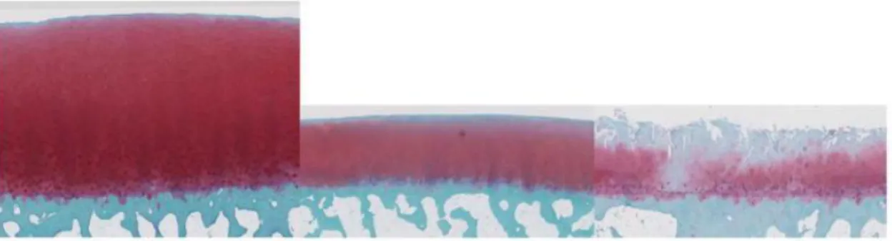
Fig. 5 Safranin O staining of human cartilage derived from young normal donor, old normal donor and OA patient.
16
normal human fibroblasts and epithelial cells. Secreted factors included interleukins and chemokines, e.g. IL-1α/β, IL-6, IL-8, MCP-2 and MIP-1α, growth factors, such as bFGF, EGF and VEGF, and several matrix metalloproteinases (Coppe, Desprez et al. 2010).
The signaling regulation of cellular senescence seems characterized by several positive, cell type specific feedback loops driven by secreted inflammatory mediators which can boost senescence via transcriptional regulation acting in an autocrine and/or paracrine manner. In fact many of the secreted compounds have effects not only in the tissue microenvironment but also at the systemic level. For example some of the released proinflammatory cytokines and chemokines can arrest cell growth (Salminen, Kauppinen et al. 2012). Moreover, inflammatory cytokines stimulate NF-κB signaling in senescent cells and can prevent apoptosis and thus maintain their senescent phenotype. Several studies have confirmed that apoptosis is reduced in senescent cells (Salminen, Ojala et al. 2011) . However, chronic inflammation is associated with many age-related diseases, e.g. metabolic disorders, cardiovascular and neurodegenerative diseases and osteoarthritis and the aging process itself also involves a low grade inflammation (Salminen, Huuskonen et al. 2008).
1.4 Role of oxidative stress in OA
Reactive Oxygen Species (ROS) are oxygen-containing molecules which are produced during normal metabolism. Oxidative stress results when the amount of ROS exceeds the anti-oxidant capacity of the cell. This process induces cell senescence not only in vitro but also in vivo, in fact there is evidence of age related oxidative stress in many tissues (Muller 2009). ROS are by-products of aerobic metabolism and include the superoxide anion (O2-), hydrogen peroxide (H2O2) and hydroxyl radicals (OH∙), all of
17
which have specific chemical properties for different biological targets (Schieber and Chandel 2014). ROS are also generated by enzymes such as NADPH oxidase and 5-lipoxygenase in response to specific cell signaling pathways. These ROS serve as secondary messengers that regulate signal transduction by activating redox-sensitive kinases and inhibiting redox-sensitive phosphatases (Finkel and Holbrook 2000).
Oxidative stress may play a major role in the link between the process of aging and the development of osteoarthritis: it has been reported that oxidative stress plays a role in chondrocyte senescence in vitro; exogenous addition of hydrogen peroxide or IL-1 (to mimic chronic inflammation) to cultured chondrocytes was found to induce markers of the senescent phenotype (Dai, Shan et al. 2006) and accelerates telomere shortening (Brandl, Hartmann et al. 2011). In this context ROS are often associated with the principles of oxidative stress contributing directly to cell senescence by causing damage to proteins, lipids, and DNA.
Increased levels of intracellular ROS were detected in cartilage derived from old rats when compared to young adults (Jallali, Ridha et al. 2005) and make human chondrocytes and rat chondrocytes more susceptible to cell death mediated by oxidants (Carlo and Loeser 2003) in an age-related manner. Not only aging and inflammation, but also other stressful factors contribute to production of ROS and induction of oxidative stress in chondrocytes of articular cartilage. In fact it has been reported that injurious mechanical loading may be a stimulus for excessive ROS production in cartilage (Green, Noble et al. 2006) and could thus contribute to stress-induced chondrocyte senescence and osteoarthritis.
There are many clues about the fact that oxidative stress may involve not only articular cartilage, but also subchondral bone and synovial membrane. Different clinical presentations of OA are associated with cellular senescence that might occur in the
18
different joint tissues: fibrosis and stiffness of joint capsule is one of these. It is documented that the cellular mechanism of fibrosis control is accompanied by cellular senescence (Krizhanovsky, Xue et al. 2008) and since fibrosis is a feature of OA, it is conceivable that intra-articular paracrine ROS messaging might promote senescence of fibroid cells to limit fibrosis (Fig.6). Moreover, joint inflammation is a characteristic of OA so it is likely that ROS induced senescence of synovial and cartilage cells can promote the inflammatory transition of the osteoarthritic joint (Ziskoven, Jager et al. 2010). Indeed senescent chondrocytes have paracrine effects on articular cartilage (Freund, Orjalo et al. 2010).
It has also been reported that mitochondrial respiratory chain is one of the major sites of ROS production and that inhibition of Complex III not only induces ROS production but also pro-inflammatory molecules (Turrens 2003). Moreover in aged and OA cartilage, a decrease of enzymes responsible of the cellular anti-oxidant defense (such as mitochondrial superoxide dismutase (SOD2)and glutathione peroxidase) has been documented.
Fig.6 Hypothetical relation between oxidative stress and osteoarthritis. Image modified by Ziskoven et al., 2010
19
1.5 Involvement of oxidative stress in DNA damage
When ROS production exceeds the detoxification and scavenging capacity of the cell, oxidative stress induces damage to DNA, proteins and lipids which consequently becomes implicated in the pathology of various diseases. Various forms of DNA damage were discovered such as those caused by oxidative damage that stimulate damage signals, activating checkpoint responses and in certain cases premature senescence (Colavitti and Finkel 2005). Regarding DNA damage, it is estimated that 80% of ROS attack bases while the remaining 20% act on sugar moieties generating multiple lesions that may lead to double strand breaks (Sedelnikova, Pilch et al. 2003). Another type of oxidative DNA lesion is the adduct 7,8-dihydro-8-oxo-2’-deoxyguanosine (8-oxo-G) that mostly affects purines (Brierley and Martin 2013) in genomic and mitochondrial DNA, as well as in RNA.
To deal with oxidative damage to DNA from various endogenous and exogenous sources, mammalian cells have evolved many mechanisms to first detect, and subsequently repair such damage.
Double-strand breaks and γH2AX.
DSBs are formed when both DNA strands are broken in sufficiently close proximity (<20 bp). They are one of the most critical lesions with respect to preservation of genomic integrity because affect the continuity of the genome. Cells undergo rapid and efficient error correction to preserve genomic stability by activating complex DNA repair pathways collectively known as the DNA damage response (DDR) (Khanna, Lavin et al. 2001 (Khanna, Lavin et al. 2001).
Phosphorylation of H2AX is one of the earlier cellular response to DSBs that accumulates in DNA foci and activates a complex molecular response (Gire, Roux et al.
20
2004). H2AX is a minor histone H2A variant which makes up between 2 to 25% of the H2A pool, depending on the cell type (Rogakou, Pilch et al. 1998). The H2AFX gene encodes H2AX; it is mapped onto position 23 on the q arm of chromosome 11 (Mah, El-Osta et al. 2010) and the sequence is highly conserved. In mammalians, Ser-139 at the C-terminus of the H2AX molecule is phosphorylated to produce γH2AX, which in recent years has emerged as one of the most well known markers of DNA damage and repair (Sedelnikova, Rogakou et al. 2002). This phosphorylation is mediated by the phosphatidylinositol 3-kinase-related kinase (PIKK) family of proteins. The three proteins potentially involved in the process are ataxia telangiectasia mutated (ATM), DNA-protein kinase catalytic subunit (DNA-PKcs) and ATM and RAD3-related (ATR) that act at the level of the highly conserved Serine-Glutamine (SQ) motif of H2AX following damage occurring after either UV exposure or metabolic stress or reactive oxygen species(Falck, Coates et al. 2005), but ATM seems the main kinase involved in H2AX phosphorylation (Fernandez-Capetillo, Chen et al. 2002). One aspect of H2AX phosphorylation is that it is not limited to the immediate vicinity, but spreads to a large chromatin region surrounding the 2Mbp region of chromatin around DSB and comprises about 2000 γH2AX molecules (Rogakou, Pilch et al. 1998).
Each γH2AX focus acts as a platform for the accumulation of DDR and repair factors and functions by altering chromatin structure to increase its accessibility (Paull and Lee 2005). Accumulating multiprotein complexes consist of other DNA repair and signaling molecules such as 53BP1, BRCA1, MDC1, NBS1, MRE11, RAD50 and RAD51 and form nuclear foci that have been also found to co-localize with γH2AX foci (Mah, El-Osta et al. 2010) (Fig.7). These proteins are recruited not only for their high affinity for γH2AX but also thanks to changes in chromatin conformation induced by the phosphorylation process (Andegeko, Moyal et al. 2001). Along with the accumulation
21
of DSB response and repair factors, a small fraction of ATM responsible for H2AX phosphorylation is retained in the nucleus and is found associated with γH2AX foci (Bonner, Redon et al. 2008).
H2AX phosphorylation is a crucial step in the DDR, because it induces DSB repair initiation by increasing DNA accessibility and facilitating the recruitment and accumulation of specific DDR proteins. The DDR is a cascade that senses DNA damage, induces cell cycle arrest and initiates DNA damage repair. Moreover, γH2AX modulates G2/M checkpoint responses and prevents cell cycle progression also inducing cellular senescence.This process avoids the duplication of damaged DNA into daughter cells to impede the propagation of corrupted genetic information (d'Adda di Fagagna 2008). Two types of γH2AX foci have been identified: one type is “transient” and DSBs are repaired and removed from the nucleus, the other type involves the formation of “persistent” foci where DSBs accumulate and remain unrepaired (Sedelnikova, Horikawa et al. 2004). Low levels of ROS are important for signal transduction and modulation of gene expression but high concentrations or prolonged exposures to ROS may have deleterious effects (Colavitti and Finkel 2005) including unrepairable DSBs likely due to persistent oxidative stress caused by defects in redox homeostasis (Marnett 2000).
The DNA damage response (DDR) is also responsible of the expression of a subset of SASP factors, including IL-6 and IL-8 (Rodier, Coppe et al. 2009). If the extent of DNA damage is broad, cells undergo either senescence or apoptosis, depending on the level of the DNA damage itself. In the case of senescence, cells arrest growth and maintain chronic low-level DDR signaling (d'Adda di Fagagna 2008).
22
This low-level DDR is persistent and necessary for a robust SASP. It is probable that the DDR stimulates the SASP by activating NF-κB that is a known target of ATM (Elkon, Rashi-Elkeles et al. 2005)
DNA mismatch repair (MMR) pathway.
DNA mismatch repair (MMR) is the main post-replicative correction pathway playing a key role in maintaining genomic stability and is therefore crucial for proliferating cells. The system corrects mispairs generated during replication, recombination and DNA damage and its efficiency can be monitored by assessing genomic instability at microsatellite loci (microsatellite instability or MSI) (Neri, Mariani et al. 2011). It has also been reported that it plays an important role in preventing mutations associated with the oxidative DNA lesion, 8-oxo-dG. The eukaryotic MMR pathway is formed by two components, both functioning as heterodimeric complexes, called MutS and MutL. The most abundant mismatch-binding heterodimer is MutSα, composed of MSH2 and MSH6 (Kolodner, Tytell et al. 1999). MutSβ is the other heterodimer composed of MSH2 and MSH3. Similarly, the MutL complexes are formed by different heterodimeric complexes, thus yielding MutLα (made up of MLH1 and PMS2 proteins), MutLβ (composed of MLH1 and PMS1 proteins) and MutLγ (containing MLH1 and MLH3) (Brierley and Martin 2013).
The initial recognition of mismatches is carried out by MSH2 bound to either MSH6 or MSH3. A second heterodimer of MLH1 bound to either PMS2 or PMS1 mediates the recruitment of additional proteins completing the repair process (Fig.8). Oxidative DNA damage can increase the frequency of MSI (Jackson, Chen et al. 1998) due to a functional inactivation of MMR by non-cytotoxic levels of H2O2 (Chang, Marra et al. 2002); (Lee, Chang et al. 2003).
23 Fig.7 Schematic representation of γH2AX-mediated DSB repair. DSBs induce the initial phosphorylation of H2AX mediated by ATM, or DNA-PK, that activates a nucleation reaction with the recruitment of the proteins of the complex.
This generates a feedback loop that leads to further phosphorylation of H2AX and the chromatin modifications required for the recruitment of 53BP1. The activation cascade culminates with the recruitment of RNF8 to phosphorylated MDC1 and the polyubiquitinylation of H2AX to recruit BRCA1/BARD1. γH2AX efficiently coordinates DDR signaling.
24 Fig.8 Schematic representation of MMR pathway in response to oxidative stress..
MutS detects the oxidative damaged DNA bases and recruits MutL. The MutS/MutL complex leaves the damaged site and slides along the DNA double helix and eventually encounters a single-strand gap on the daughter strand bound by accessory proteins (PCNA and RFC). This encounter displaces RFC and allows EXO1 to access the daughter-strand DNA to degrade DNA across the site of oxidative damage, before becoming inactivated by MutL. The oxidative damaged DNA is then excised, followed by the synthesis of new DNA by a DNA polymerase. Finally, the new DNA strand is ligated onto the existing daughter strand by a DNA ligase. EXO1, DNA exonuclease.
25
Role of Gadd45β and p21 in DNA damage response and senescence.
The gadd45 is a family of genes including gadd45α, gadd45β, and gadd45γ that encode for the Growth Arrest and DNA damage-inducible 45 proteins that are key players in cellular stress responses. These are small (18 kDa), evolutionarily conserved proteins that are highly homologous to each other (55– 57% overall identity at the aminoacid level), highly acidic, and localized within both the cell cytoplasm and nucleus (Liebermann, Tront et al. 2011). Expression of gadd45α, gadd45β, and gadd45γ is induced in response to environmental and physiological stresses (Gupta, Gupta et al. 2006) and it has been shown that GADD45 proteins participate in cell cycle arrest, DNA repair, cell survival and apoptosis in response to these stresses, as well as having a role in development.
In fact, GADD45β has been identified as an essential mediator of Col10a1 and Mmp13 gene expression in late-stage hypertrophic chondrocytes in the mouse embryo, acting as
a cell survival factor during terminal differentiation, driving chondrocyte hypertrophy
via p38 MAPK activation and preventing apoptosis of hypertrophic chondrocytes (Ijiri, Zerbini et al. 2005).
During cellular senescence cells show a variety of associated phenotypic changes and one of the most prominent initiator of senescence is the DNA damage response, with induction of cell cycle arrest through the activation of checkpoint proteins, notably p21 (Passos, Nelson et al. 2010). In the process of cellular senescence, a clear relationship has been shown between p21 and GADD45β that interact in a feedback loop mechanism. This process starts with mitochondrial dysfunction, production of reactive oxygen species (ROS) and p21 activation and results in the establishment of the senescent phenotype (Passos, Nelson et al. 2010). It has also been reported that p21 is a down-stream target of GADD45β suggesting the involvement of both proteins in the
26
molecular mechanism of chondrocyte senescence in mice (Shimada, Sakakima et al. 2011).
1.6 Role of GSK3β in OA
GSK3β (Glycogen Synthase kinase 3β) was initially identified as a key regulator of insulin-dependent glycogenesis, because of its role as a protein kinase able to phosphorylate and inhibit glycogen synthase (Embi, Rylatt et al. 1980). However, it is currently well known that it is a multi-functional kinase that performs a regulatory role in several cellular functions, including embryonic development, cell metabolism, proliferation and intracellular signaling (Cohen and Frame 2001). GSK3β in cells is present in a multiprotein complex with axin, APC (Adenomatosus Polposis Coli) and β-catenin. It is involved in the so called Wnt canonical signaling: in the absence of secreted Wnt glycoproteins, GSK3β is active and phosphorylates the other proteins of the complex at the level of cytoplasm. The principal function of this complex is to maintain β-catenin in an inactive state via its phosphorylation, thus preventing its nuclear translocation and transcriptional activation of TCF/LEF complex (Seidensticker and Behrens 2000).
Wnt binding to its receptors instead leads to stabilization of β-catenin via inhibition of GSK-3β (that occurs by phosphorylation of serine-9 that significantly decreases active site availability) (Fig. 9). Uncontrolled triggering of the Wnt pathway is linked to the development of a number of age-related pathologies such as osteoarthritis and an aberrant Wnt signaling can promote cell senescence (Maiese, Li et al. 2008). Disruption of the control of Wnt signaling has also been associated with altered joint formation, chondrogenesis, and OA (Dell'accio, De Bari et al. 2008); (Staines, Macrae et al. 2012).
27
For these reasons, in the context of skeletogenesis, GSK3β is considered among the major molecular constraints which keep chondrocytes in an “arrested state”, avoiding the process of endochondral ossification at the level of articular cartilage (Lories, Corr et al. 2013). It has also been reported that inhibition of GSK3β is a key event for chondrocyte maturation at the level of temporary cartilage during skeletogenesis. This process is under the control of regulatory kinases such as Akt (Rokutanda, Fujita et al. 2009) and cGMP dependent protein kinase II, that drive the process towards hypertrophy and terminal differentiation (Kawasaki, Kugimiya et al. 2008).
Moreover, OA-like changes occur in mice following both under- and over-activation of the Wnt pathway (Zhu, Tang et al. 2009). A fine regulation of GSK3β activity is therefore fundamental for the chondrogenesis and skeletal development because it guarantees the correct balance between cytoplasmatic and nuclear β-catenin (Lories, Corr et al. 2013).
In fact, evidence indicates that not only GSK3β inhibition induces OA changes in articular chondrocytes (Miclea, Siebelt et al. 2011); (Litherland, Hui et al. 2014), but also the ablation of β-catenin signaling pathway is associated with cartilage degeneration in transgenic mice (Zhu, Chen et al. 2008).
Collectively, functional genomics data about the effect of over- or under- activity of β-catenin suggest that the a fine tuning of β-β-catenin signaling via GSK3β regulation must be maintained to avoid osteoarthritis features at the level of human articular cartilage. Moreover it has been reported that GSK3 is also present at the level of mitochondria, and inactivation of “mitochondrial GSK3β” contributes to decreasing mitochondrial complex IV activity thus leading to subsequent ROS generation responsible for cell senescence in human diploid fibroblasts (Seo, Jung et al. 2008); (Byun, Jung et al. 2012).
28
1.7 NF-κB pathway: the role of IKKα
With regards to cell senescence, the secretory component of SASP (Senescence Associated Secretory Phenotype) involves cytokines, chemokines, growth factors and matrix metalloproteinases and NF-κB signaling is recognized as the master regulator of this kind of inflammatory responses. The NF-κB system is a conserved signaling pathway which is activated in response to a wide variety of insults and cellular stress to facilitate innate immunity responses and to establish cellular defense in order to maintain cell and tissue homeostasis (Vallabhapurapu and Karin 2009). NF‐κB transcription factors guide a wide range of inflammatory responses, regulating cell differentiation and development programs and ultimately control cell growth (Bonizzi and Karin 2004); (Karin and Greten 2005); (Basak, Kim et al. 2007).
NF-κB mediated transcriptional control is driven by the assembly of homodimers and heterodimers of 5 different NF-κB proteins (RelA/p65, RelB, c-Rel, NFκB1/p105 and
Fig.9 Schematic representation of Wnt canonic pathway. The presence of Wnt protein induces GSK3β inactivation and dissociation of the cytoplasmatic complex. This process is followed by nuclear translocation of β-catenin and transcription activation of specific target genes. Image modified by Cohen et al., 2001
29
NFκB2/p100). In the absence of any stimulus, NF-κB dimers are sequestered in the cytoplasm and their transcriptional activities blocked by one out of three small inhibitory NF-κB proteins (IκBα, IκBβ, IκBε). NF‐κB transcription factors are then
activated by N‐terminal phosphorylation of inhibitor IκBs, that releases the NF-κB dimers so they are free to migrate to the nucleus and activate target genes. The kinases responsible for this process are IKKα and IKKβ. IKKβ is essential for the nuclear translocation of NF‐κB while IKKα acts only occasionally as kinase of IκBα (Marcu, Otero et al. 2010). Interestingly, most of the signaling pathways acting in the cells in response to stressful conditions (genotoxic, environmental or inflammatory stress) target the IKK complex, which means that IKKα and IKKβ can even activate NF-κB independent targets, e.g. β-catenin, histone H3, TSC1, FOXO3a and several nuclear co-activators (Salminen, Kauppinen et al. 2012). In addition, the NF-κB system participates in crosstalk with several transcription factors which join together different signaling pathways, e.g. p53 and Wnt (Mengel, Hunziker et al. 2010).
Cellular senescence involves several morphological and energetic-metabolic changes during the generation of the senescent phenotype. It is known that NF-κB signaling can regulate energy homeostasis (Kawauchi, Araki et al. 2008) and the major housekeeping system, i.e. the autophagic cleansing system (Baldwin 2012). Cellular stress induces autophagocytosis, a self-eating process, which has many crucial functions in cellular survival. Moreover, the autophagic degradation capacity declines with aging in many tissues (Salminen and Kaarniranta 2009). In conclusion, it seems that the IKK complex, in particular IKKα, is an important player in the regulation of cellular senescence induction (Tilstra, Robinson et al. 2012).
It has also been reported that ablation of IKKα has peculiar effects on extracellular matrix remodeling (reducing the level of bioactive ECM degradation products), on
30
chondrocyte proliferative potential and cell cycle distribution, on hypertrophic differentiation and viability of the cells in maturing micromasses in conjunction with subcellular features of cell health (mitochondrial morphology and cell‐cell or cell‐ECM specialized junctions) thus suggesting that this kinase might contribute to the abnormal phenotype of osteoarthritic chondrocytes (Olivotto, Borzi et al. 2008).
1.8 Nutraceuticals: a new way to counteract cell oxidative stress and senescence in osteoarthritic chondrocytes
Osteoarthritis prevention is a major challenge for the research in this clinical field. Available pharmacological treatments are very expensive and almost not very effective; overall, there is currently no cure for OA, and there are no therapies which prevent, slow or arrest OA progression (Le Graverand-Gastineau 2010). Most treatments are focused on the control of the secondary symptoms of the disease, but fail to address the complex nature of OA, and have no beneficial effects on chondroprotection and therefore on OA prevention and modification. Furthermore, long-term use of available therapies is often associated with side effects at gastrointestinal, renal, and cardiovascular level (Cheng and Visco 2012).
In this scenario an alternative and safe opportunity is represented by nutraceuticals, defined as "Food, or parts of food, that provide medical or health benefits, including the prevention and/or treatment of disease". Indeed, it has been recognized that nutraceuticals may exert effects on molecular targeting of OA (Leong, Choudhury et al. 2013;(Henrotin, Lambert et al. 2011).
Recently, it has been reported that natural compounds found in fruits, teas, spices, wine, and vegetables such as phytoflavonoids, polyphenols, and bioflavonoids, have a great potential in modifying OA disease and symptoms thanks to their
anti-31
inflammatory and anti-catabolic actions, and their protective effects against oxidative stress (Shen, Smith et al. 2012). Another interesting compound with a protective effect on chondrocytes is sulforaphane, an isothiocyanate derived from cruciferous vegetables (Facchini, Stanic et al. 2011). An emerging role in reducing oxidative stress and DNA damage has been reported for hydroxytyrosol (HT), a phenolic compound found in the fruits of olive tree and in olive oil and highly present in Mediterranean diet, with an high anti-oxidant and cytoprotective activity (Facchini, Cetrullo et al. 2014). Spermidine (SPD), a natural dietary compound also found in high concentrations in Mediterranean diet (Soda 2010), belongs to the class of polyamines, naturally occurring polycations. It has been reported that supplementation with spermidine reduces oxidative stress and extends lifespan in yeast and flies by an autophagy-dependent mechanism (Eisenberg, Knauer et al. 2009; Guo, Harada et al. 2011; Minois, Carmona-Gutierrez et al. 2012).
These data open new perspectives for the study of possible ways to prevent, control or even revert osteoarthritis and for the development of a nutraceutical-based molecular targeting strategy for chondroprotection as an alternative to classical treatments.
33
2- AIMS OF THE STUDY
The main aim of this study is to investigate about the molecular bases of osteoarthritis with particular regards to aspects of cellular senescence in disease. This study will focus on the molecular mechanisms whereby two major enzymatic systems in chondrocytes may affect oxidative stress and DNA damage leading to chondrocyte senescence. More in detail we plan to investigate on the effects of pharmacological inhibition (LiCl) of GSK3β and of the activity of IKKα, by comparison with chondrocytes bearing a retroviral mediated IKKα Knock Down (KD). Moreover we will evaluate the ability of selected nutraceuticals of scavenging oxidative stress thus limiting senescence induction of osteoarthritic chondrocytes.
The first aim of the study will focus on the effect of GSK3β pharmacological inactivation on cellular senescence by evaluation of :
· ROS production by mitochondria;
· induction of "intrinsic" oxidative damage (assessed by increased 8-oxo-guanine adducts) and DNA damage response (evaluated by γH2AX foci formation) in osteoarthritic chondrocytes;
· induction of transient S phase arrest, reduced proliferation, and increased percentage of hypertrophic chondrocytes;
· induction of increased expression of the senescence marker GADD45β, p21, SA-β galactosidase and PAS staining.
The second aim of the project will focus on the DNA Damage Response in primary cultures of IKKα Wild Type or Knock Down osteoarthritic chondrocytes after induction of "extrinsic" oxidative stress using H2O2 and measuring:
34
· telomere length by Flow‐FISH; · γH2AX foci formation;
· expression of the DNA Mismatch repair system genes (Msh2‐Msh6‐Msh3‐Mlh1‐Pms1‐ Pms2).
The third aim of the project will focus on the ability of some nutraceuticals (i.e. hydroxytyrosol and spermidine) to attenuate oxidative stress inducted using H2O2 and measuring:
· effects of pre-treatment with hydroxytyrosol on γH2AX foci formation and cellular senescence;
· effects of pre-treatment with spermidine on cellular viability and γH2AX foci formation.
36
3- MATERIALS AND METHODS
3.1 Effects of GSK3β pharmacological inactivation
Chondrocytes isolation.
After Istituto Ortopedico Rizzoli Ethics Committee approval and patients’ informed consent, primary chondrocytes were obtained from 13 OA patients undergoing knee arthroplasty. Cartilage was cut from the subchondral bone using a sterile sharp scalpel blade and placed in serum free Dulbecco’s Modified Eagle’s Medium (DMEM) supplemented with antibiotics. Once all the cartilage was obtained, it was diced as finely as possible. In fact dicing the tissue finely improved the efficiency of the subsequent enzymatic digestion. All the cartilage obtained was put into a sterile Petri. Chondrocytes were isolated by mean of a sequential enzymatic digestion. The first step was a proteolytic digestion of ECM, my mean of cartilage incubation with Protease from Streptomyces griseus 26,5U/ml (Sigma-Aldrich) in serum free DMEM for 1hour at 37°C 5% CO2. At the end of this step supernatant was eliminated and cartilage was put into a sterile bottle containing Collagenase from Clostridium histolyticum 545U/ml (Sigma-Aldrich) in serum free DMEM and incubated for 1hour at 37°C in continuous stirring. Once digested, the cell suspension was strained through commercially available cell strainers (100µm pore size) and centrifuged (1800 rpm for 10 min) to obtain a cell pellet. This pellet was washed in DMEM containing 10% FBS and the cells counted. To obtain standard monolayer cultures, the cells were plated on culture plastic flasks at a density of 20,000 cells/cm2. Chondrocytes were expanded in culture up to passage 1 (p1) and then used as described below for monolayer culture.
37
ROS production evaluation downstream GSK3β inactivation.
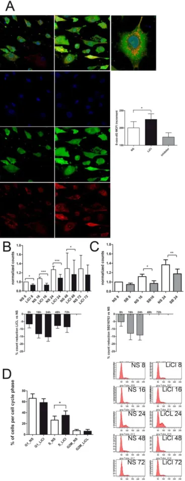
Reactive oxygen species (ROS) production at the level of mitochondria was assessed in live OA chondrocytes undergoing GSK3β inactivation. More in detail, the accumulation of ROS within mitochondria was assessed by mean of the overlap of the signals of selective fluorescent dyes for both mitochondria and ROS. Primary chondrocytes were plated in petri dishes with 0.17 mm thin glass well (Cell Culture Dish, World Precision Instruments Germany GmbH), suitable for signal detection at the confocal microscope. The cells were left to adhere for 72 hours, and then were either left unstimulated or treated with 5 or 10 mM LiCl or the highly selective GSK3β inhibitor SB216763 at 10 µM (Coghlan, Culbert et al. 2000) for 4 hours.
Analysis of the mitochondrial involvement was approached by “real time” and “time lapse” 30 µM DCHF-DA staining of increased ROS generation, overlapping with Mitotracker Orange CMTMRos mitochondrial staining (Molecular Probes ) along with Hoechst 33342 nuclear counterstaining.
Fluorescent signals were acquired by NIKON confocal microscope system A1 equipped with a Nikon Eclipse Ti microscope and an Argon Ion laser for 488 nm line, a DPSS laser for 561nm line and a diode laser for 640mn line. Emission signals were detected by a photomultiplier tube (DU4) preceded by emission filters BP 525/50 nm, BP 595/50 nm and BP 700/75 nm for Sybr green, Alexa Fluor555 or Dy Light 647, respectively. Laser scanning, image acquisition and processing were performed with Nikon Imaging Software NIS Elements AR-4 (Nikon Inc., USA). Fields of 210 µm x 210 µm (acquired with a Nikon plan apo 60x 1.40 oil objective) were acquired and analyzed.
38
Effects of GSK3β inhibition on proliferation and cell cycle.
Some experiments were carried out to investigate the effect of GSK3β inhibition on growth and cell cycle distribution of chondrocytes as well as downstream effects on cell senescence. At this purpose, experiments were established with p1 chondrocyte cultures obtained from 13 different patients. Cells were plated at low density (10,000-15,000 cells per cm2) in order to avoid culture conditions which could bias evaluation of senescence (Severino, Allen et al. 2000) and left for 72 hours. Then parallel cultures were kept either unstimulated or stimulated with 5 mM LiCl for 8, 16, 24, 48 and 72 hours. In some cases cultures were also stimulated with SB216763 (Coghlan, Culbert et al. 2000) which, used at 10µM, behaves as a rather selective inhibitor for GSK3. At the end, for each experimental conditions cells were trypsinized, counted and either fixed (10 min at RT with 2% PFA and stored at 4°C) for Flow Cytometry or senescence detection or pelleted for western blot analysis.
Cell cycle distribution was evaluated by flow cytometry by mean of DNA staining (Sytox green, Molecular Probes, at 5µM) of cells that had been previously fixed with 2% PFA and then post-fixed with 100µl methanol 90% on ice for 10minutes. A RNAse treatment was then applied by resuspending the cells with 100µl RNAse ONE buffer (Promega), preheating at 65°C 10 min and then addition of 1 µl (2.5 U) of RNAse One (Promega) and 1µL RNAse A (Sigma-Aldrich). Digestion was left for 30’ at 37 °C. Analyses were performed using a FACS Canto II flow cytometer (BD).
Light scattering properties of the cells were also analyzed by assessing both the forward scatter (FSC) of the cells which evaluate the cell size and therefore the hypertrophy promoting activity of LiCl as well as the side scatter (SSC), which correlates with granularity and has been reported to increase in cell senescence. The median values of several thousands of cells values were obtained and separated for each cell cycle phase
39
for both normal and lithium chloride treated cells, and normalized to the median size of control cells in the G1 phase.
Real time PCR analysis.
Cells from 4 patients were also dedicated for Real Time PCR analysis of gene expression. Total RNA was isolated from pellets obtained from cells either unstimulated or stimulated with 5 mM LiCl for 8, 16 and 24 hours. Total RNA was extracted from cells using TRIZOL (Invitrogen). RNA (0.5 μg) was reverse-transcribed using the Superscript VILO cDNA Synthesis kit (Invitrogen) and DNA treatment and removal was performed with DNA-free (Ambion). Each PCR reaction was carried out on 25 ng of cDNA sample using SYBR Premix EX Taq (Takara). The following conditions were used: 95 °C for 30 sec; 40 cycles at 95 °C for 5 s, 60 °C for 20 s and 65 °C for 15 s in a LightCycler 480 system (Roche diagnostics) and analyzed with the dedicated software. All values were normalized to GAPDH housekeeping gene and expressed as relative expression or fold change using the respective formulae 2− ΔCT or 2− ΔΔCt. Primers were as follows: GAPDH (NM_002046, forward 579–598 and reverse 701–683);
IKKα/CHUK (NM_001278, forward 1803-1826 and reverse 1865-1888); MMP10
(NM_002425.2, forward 1278–1298 and reverse 1472–1449).
Evaluation of senescence by mean of SA-β Gal activity and Glycogen content.
Senescence was evaluated by both assessment of SA-β Gal activity with the Senescent Cells Staining kit (SIGMA) and assessment of the increased glycogen content with the PAS staining (Sigma-Aldrich). Nearly 10,000 cells were cytospinned on a glass slide and further processed as recommended by the manufacturer. Development time was overnight for Senescent Cells Staining kit (Sigma-Aldrich). At the end, to facilitate their
40
automatic counting, the cells were treated with SyBr Green nuclear counterstaining. Then, an automatic analysis procedure was employed exploiting the NIS software which combined colorimetric and fluorescent staining. A threshold was set in order to take into account only cells above a given intensity of senescent staining. At least four fields were counted for each condition. The same cell suspension underwent evaluation of glycogen accumulation by mean of the PAS staining.
Immunoblotting.
Evaluation of the expression levels of proteins that are induced upon DNA damage was carried out by western blotting. Lysis buffer volumes were adjusted in order to load an equivalent of 150,000 cells. To achieve effective extraction of proteins, including those bound to DNA, radioimmunoprecipitation (RIPA) buffer with the addition of benzonase and protease inhibitor cocktail (PIC; Sigma-Aldrich) was used to extract proteins. The composition of the buffer was as follows: Tris-HCl 50mM pH 7.4, NaCl 150mM, Nonidet P-40 1%, SDS 0.1%, Na deoxicolate 0.5%, NaF 1mM, Na3VO4 1mM, PMSF 1mM, 1:200 PIC, and 100U/mL benzonase. Briefly, total cellular lysates in RIPA buffer were obtained from monolayers solubilized by keeping the pellet on ice. Samples were loaded in the wells of Nu-Page precast 4%–10% polyacrylamide gels (Invitrogen), which were subsequently transferred onto polyvinylidene fluoride membranes by a dry electroblotting method using I-Blot (Invitrogen) and then subjected to immunodetection exploiting the SNAP-ID device (Merck Millipore). Signals were detected with appropriate secondary antibodies and revealed with ECL Select kit (GE Healthcare), using the CCD camera acquisition system of Image Station 4000 MM and Carestream Molecular Imaging Software 5.0. (Carestream Health, Inc.). Immunoblot experiments were designed to kinetically assess the correlated protein expression of