Alma Mater Studiorum – Università di Bologna
DOTTORATO DI RICERCA IN
Biologia Cellulare e Molecolare
Ciclo XXVII
Settore Concorsuale di afferenza: 05/I1 Settore Scientifico disciplinare: BIO/19
TITOLO TESI
Study of the genes involved in Naphthenic acids (NAs)
degradation by Rhodococcus spp.
Presentata da: Alessandro Presentato
Coordinatore Dottorato Relatore
Prof. Davide Zannoni Prof. Davide Zannoni
Correlatore
Dott.ssa Martina Cappelletti
Esame finale: Bologna, Aprile 2015
Abstract
Naphthenic acids (NAs) are an important group of organic pollutants mainly found in hydrocarbon deposits. Although these compounds are toxic, recalcitrant, and persistent in the environment, we are just learning the diversity of microbial communities involved in NAs-degradation and the mechanisms by which NAs are biodegraded. Studies have shown that naphthenic acids are susceptible to biodegradation, which decreases their concentration and reduces toxicity. Nevertheless, little is still known about their biodegradability. The present PhD Thesis’s work is aimed to study the biodegradation of simple model NAs using bacteria strains belonging to the Rhodococcus genus. In particular, Rh. sp. BCP1 and Rh. opacus R7 were able to utilize NAs such as cyclohexane carboxylic acid and cyclopentane carboxylic acid as the sole carbon and energy sources, even at concentrations up to 1000 mg/L. The presence of either substituents or longer carboxylic acid chains attached to the cyclohexane ring negatively affected the growth by pure bacterial cultures. Moreover, BCP1 and R7 cells incubated in the presence of CHCA or CPCA show a general increase of saturated and methyl-substituted fatty acids in their membrane, while the cis-mono-unsaturated ones decrease, as compared to glucose-grown cells. The observed lipid molecules modification during the growth in the presence of NAs is suggested as a possible mechanism to decrease the fluidity of the cell membrane to counteract NAs toxicity. In order to further evaluate this toxic effect on cell features, the morphological changes of BCP1 and R7 cells were also assessed through Transmission Electron Microscopy (TEM), revealing interesting ultrastructural changes. The induction of putative genes, and the construction of a random transposon mutagenesis library were also carried out to reveal the mechanisms by which these
Index
1. Introduction ... 1
2 Naphthenic acids (NAs) ... 4
2.1 Origin and distribution of NAs ... 4
2.2 NAs chemical and physical properties ... 8
2.3 Toxicity of NAs ... 11
2.3.1 Aquatic environmental toxicity ... 12
2.3.2 Toxicity to Mammals ... 12
2.3.3 Toxicity to green plants ... 13
2.3.4 Mechanisms of toxicity in microorganisms ... 13
2.3.5 Lipid adaptation mechanisms of bacterial strains ... 15
3. Microbial biodegradation ... 18
4. The genus Rhodococcus ... 27
4.1. NAs degradation by Rhodococcus spp. ... 31
4.4. Rhodococcus opacus sp. R7 ... 38
Aims ... 40
5. General Materials and Methods ... 42
5.1. Bacterial strains, media and growth conditions ... 42
5.2. Extraction of genomic DNA from Rhodococcus spp. ... 44
5.3. RNAs isolation procedure ... 45
5.4. DNA manipulations and genetic techniques ... 46
5.5. Electroporation of Rhodococcus species. ... 47
6. Metabolic and physiological aspects of Rhodococcus single species growing on NAs ... 49
6.1. Materials and Methods ... 50
6.1.1. Preparation of Rhodococcus strains inocula ... 50
6.1.2. NAs stock solution preparation ... 50
6.1.3. Extraction and derivatization of model NAs ... 51
6.1.4. Gas Chromatography-Mass Spectrometry (GC-MS) analysis ... 51
6.1.5. Fatty acid methyl esters (FAMEs) analysis ... 52
6.1.6. Preparation of samples for TEM analysis ... 53
6.2. Rhodococcus spp.: Growth and degradation of model NAs ... 54
6.2.1. Cyclohexanecarboxylic acid (CHCA) and Cyclopentanecarboxylic acid (CHCA) ... 54
6.2.2. Cyclohexaneacetic acid (CHAA) and methyl-cyclohexanecarboxylic acid (mCHCA) ... 59
6.3. Fatty Acid Methyl Esters (FAMEs) analysis ... 62
6.4. Transmission Electron Microscopy (TEM) analysis ... 66
6.5. Discussion ... 70
7. Genetic analysis of model NAs (CHCA and CPCA) degradation by Rhodococcus sp. BCP1 and Rhodococcus opacus R7 ... 75
7.1. Materials and Methods ... 75
7.1.1. Semiquantitative RT-PCR ... 75
7.1.2. RT-qPCR ... 77
7.1.3. Transposon mutagenesis library ... 78
7.1.4. Two-step gene walking method ... 80
7.2. Genetic analysis about NAs degradation by Rh. sp. BCP1 ... 82
7.3. pTNR (TA)-mediated transposon mutagenesis of Rh. opacus R7 ... 88
7.4. Discussion ... 94
1. Introduction
Soil contamination is a global concern and can be considered a major barrier to sustainable development, since it ruins the balance of the ecosystem, causing economic loss and human health damage [Pimentel et al.; 1995]. These environmental issues derive from inadequate or irresponsible disposal measures, such as improper industrial discharge, mining tailings, waste disposal, and stockpiles [Wuana et al.; 2011]. Even Arctic soil contamination has various causes, such as blowouts and accidental oil leaks from tankers, trucks, pipelines, and storage tanks, in addition to various discharges from industrial sites, and/or military bases [Mohn et al.; 2000, Børresen et al.; 2007, Evdokimova et al.; 2012].
Over the last hundred of years, millions of tons of dangerous pollutants were generated, and a significant part of them consists of hydrophobic organic compounds that will persist in the environment for several decades [Head et al.; 2003].
Nowadays, there is a growing awareness concerning the toxic or even carcinogenic effects of a large fraction of the environmental pollutants [Triska et al.; 2004].The most common soil contaminants are heavy metals, organics, and radio-nuclides. Increased heavy metal levels in soil have been reported in many industrialized countries and areas. Metals and metalloids, such as chromium, cadmium, mercury, and lead, can threaten the ecosystem and human health through the food chains or direct exposure to the contaminated soil/water [Triska et al.;2004]. Organic pollutants such as volatile chlorinated solvents, polychlorinated biphenyls (PCBs) and petroleum products represent another pervasive concern due to their toxicity, mobility, and abundance. These organic pollutants remain stable in soil, being dangerous to animal and plant species [Arias-Estévez et al.; 2008]. In fact, the low volatility and water solubility of high molecular weight hydrocarbons makes them not only persistent in the environment, but also produce long-term adverse effects on the environment and human health including: prenatal toxicity, skin-related diseases, lung cancer, leukemia, and negative
effects on reproduction [Evans et al.; 2005]. While low-molecular weight aromatics volatize relatively rapidly, the heavier ones such as polycyclic aromatic hydrocarbons with three and more rings, remain in the environment for quite a long time. The biodegradation half-lives in natural habitats have been estimated for several PAHs including anthracene (three-rings, 170 days to 8 years), phenanthrene (three-rings, 2.5 days to 5.7 years), and benzo[α]pyrene (five-rings, 8.2 to 58 years) [Speight et al.; 2011]. Anthropogenic activities also generate radio-nuclides contamination [Elless et al.; 2012], deriving from atmospheric testing of nuclear weapons, the leakage of radioactive waste, and disasters such as those of Chernobyl and Fukushima nuclear-power plants. Apparently the high risk on human health and ecological security, derives from contaminated soils.
Efficient ways to dispose dangerous wastes are physical and chemical techniques that include combustion, photolysis, chemical degradation, and decomposition. Each chemical method can be successfully applied only within a certain range of concentration of organic compounds due to their solubility, toxicity, and persistence and even though these technologies are effective, they are in general too expensive and dangerous due to the use of hazardous chemicals resulting in high amount of wastes [Triska et al.; 2004]. Alternative methods for the decontamination are represented by biodegradation or phytoremediation procedures. Microbial biodegradation is considered the most cost-effective and ecological way to reduce or neutralize environmental contamination [Tandlich et al.; 2011]. In fact, microorganisms play a key role in the fundamental ecological processes such as biogeochemical cycling and organic contaminant degradation. Microorganisms are important degraders of both organic matter and xenobiotics, they represent an unexplored reservoir of genetic diversity and metabolic capability and provide several ecosystem services, most importantly the maintenance of soil and water quality, and products as nutrients to other organisms in the food chain [Megharaj et al.; 2011]. It is demonstrated that there are very few environments
where microbes havenot been able to survive, adapt and thrive. Microbes are able to utilize a wide combination of electron donors (pollutants) and electron acceptors (oxidants such as O2
and, in the case of anoxic respiration NO3-, Fe3+, Fe(OH)3, SO42-) to drive the metabolic
pathways required for their growth [Tiedje et al.; 1993]. In addition to these redox reactions, microbes have also developed a series of strategies enabling them to detoxify the environment, such as the so called cometabolism, allowing the use of a non-growth substrate (compound that is unable to support the cell growth) in the presence of a growth substrate, as it is observed in the case of chlorinated solvents biodegradation [Horvath et al.; 1972].
Bioremediation strategies are advantageous because they can be implemented in situ, which means directly in the contaminated site with no need to remove the contaminated material. Innovative in situ technologies allow the biological treatment of contaminated sites through biosurfactant molecules produced by microbes [Pacwa-Plociniczak et al.; 2011]. This provides a simpler, less intrusive, and cheaper method than conventional “pump and treat” systems that often employ hazardous chemicals that create and additional environmental risk, in the case of ex situ remediation. Microbes may be indigenous to a contaminated area or they may be isolated from elsewhere and brought to the contaminated site. In the latter case, this is referred to as bioaugmentation, whereas, if the naturally occurring population is encouraged to proliferate by the addition of exogenous electron donors or electron acceptors, this is called
biostimulation. The advantage of using microorganisms already living in the contaminated
area to be reclaimed, is that they are pre-adapted to a specific environmental condition so to lower the cost of the remediation procedure [Leung et al.; 2004].
2 Naphthenic acids (NAs)
2.1 Origin and distribution of NAs
The world’s largest accumulations of “oil sands” reserves occurs in the shallow reservoirs of North and South America [Head et al.; 2003]. One of these deposits, approximately 1200 billions barrels of bitumen, is located in Venezuela and it is slightly bigger than the Athabasca oil sands deposit in Alberta, Canada, with 900 billions of barrels (Fig. 2.1.1) [Head et al.; 2003, Zhou et al.; 2008].
Fig. 2.1.1 Canadian oil sands (National Geographic Photograph by Peter Essick)
Notably, the microbial biodegradation of crude oil leads to a decrease in hydrocarbon and aromatic hydrocarbon content, increasing viscosity, density, acidity, sulfur and metal content. Phenols, acyclic, cyclic, saturated and aromatic carboxylic acids, and heteroatom-bearing carboxlic acids are also produced [Larter et al.; 2008].
Several factors are required for the microbial petroleum biodegradation processes: low temperature of the reservoir (between 20 and 60 °C); presence of electron acceptors such as Fe3+, SO42-,NO3-; nutrients (e.g. nitrate and phosphate); an oil/water contact (the bounding
surface between oil and water in which petroleum is biodegraded by microorganisms) [Head et al.; 2003, Zhou et al.; 2008, Eschard et al.; 2008]. The Athabasca oil sands deposits are featured by having a low temperature (< 10 °C) which favors the biodegradation processes. In contrast, there are oil accumulation reservoirs characterized by a temperature around 80-90 °C, which causes inactivation of most of the hydrocarbon-degrading organisms, e.g. the oil reservoir of Peace River in Canada [Zhou et al.; 2008].
The most common approach for the production of crude oil from oil sands is called: “Clark Hot Water Extraction Process” [Clemente et al.; 2005, Frank et al.; 2009] developed by Dr. Karl Clark in 1920. The oil sand is characterized by loose sand or partially consolidated sandstone, which contains a mixture of inorganic materials such as sand, clay and silt, ranging between 80 and 87%, and water, saturated with a dense and viscous form of petroleum (between 6-16%), technically referred to as bitumen, that must be upgraded before it can be used by refineries to produce gasoline and diesel fuels (Fig. 2.1.2) [Liu et al.; 2005].
Fig. 2.1.2 Example of raw bitumen extracted from Alberta’s oil sands (Images courtesy of Syncrude Canada Ltd.)
During the“Clark Hot Water Extraction Process”, surface oil sands are mined and crushed to reduce size particles, which are mixed with hot water and caustic soda (NaOH) to allow the heavy bitumen to become less viscous and separate it from the sand. Because of this
treatment, the solubility of oil sands asphaltic acids, mainly aromatics containing oxygen functional group such as phenolic and carboxylic types, is increased due to the alkaline pH (around 8) that promotes their release [Allen et al.; 2008, Rogers et al.; 2002]. By increasing the pH, the superficial and interfacial tensions are reduced resulting in the disintegration of the oil sands ore structure and the recovery of the bitumen [Chalaturnyk et al.; 2002]. The process requires a large amount of fresh water, approximately 2 or 3 m3 per cubic meter of oil produced, generating 4 m3 of wastewater of which 80-88% is recycled back to the processing facility [Holowenko et al.; 2002]. The wastewater produced as a result of the bitumen extraction process contains sand particles, clay fines, silts, water, dissolved ions (mainly Na+ and SO42-), heavy metals, unrecoverable bitumen, inorganic and organic compounds. This
kind of wastewater is named Oil Sand Tailings Water (OSTW) or Oil Sand Process Water (OSPW), and its composition changes depending on the ore quality, source, extraction processes and age [Crowe et al.; 2001].
OSPWs produced during the extraction processes of crude oil are stored in settling tanks into the environment, known as tailing ponds (Fig. 2.1.3) [Del Rio et al.; 2006, MacKinnon et al.; 1989].
Fig. 2.1.3 Schematic representation of the tailing pond
These tailings ponds are open dumps with a high environmental impact and the existing ones (e.g. Athabasca region, Canada) cover an area of around 180 km2. Once in the pond, the sand quickly sinks to the bottom, and the water from the top three meters is recycled back to the process facility. However, tailings ponds present a number of challenges:
• The bottom layer, a mixture of clay and water called fine tailings, takes a long time to settle down and solidify, e.g. it can take up to 30 years to separate and dry out;
• Due to the extraction process, the remaining water contains chemicals that are toxic to living forms, e.g. fish;
• The risk of wastewater infiltration into the groundwater is significant;
• The small amount of residual oil that floats to the surface of the pond poses a risk for waterfowl.
OSPWs high toxicity is mainly due to the presence of compounds collectively known as naphthenic acids (NAs) [Allen et al.; 2008, Madill et al.; 2001, MacKinnon et al.; 1986]. NAs naturally occurring in petroleum and oil sands deposits [Fan et al.; 1991, Seifert et al.; 1969, Dzidic et al.; 1988] are believedto be generated by incomplete aerobic biodegradation of petroleum hydrocarbons. Indeed, earthquakes, erosions or tectonic movements can cause petroleum to be exposed to favorable conditions for the microbial aerobic biodegradation [Behar et al.; 1984, Meredith et al.; 2000, Zhou et al.; 2008]. NAs can also derive from insufficient catagenesis that is a physical process that occurs directly in oil deposits under high pressure and temperature conditions, leading to loss of carbonyl groups [Tissot et al.; 1978, Clemente et al.; 2005]. This phenomenon can alter biomolecules through isomerization, aromatization, cyclization and bond cleavage [Sinninghe et al.; 1997]. Finally, NAs can be produced by anaerobic hydrocarbon biodegradation processes in oil sands deposits where aerobic conditions are unlikely. The occurrence of this type of production is demonstrated by
the presence of anaerobic microorganisms and to the discovery of anaerobic hydrocarbon degradation pathways [Head et al.; 2003, Aitken et al.; 2004, Eschard et al.; 2008].
The continuous re-use of recycled water, causes a gradual accumulation of NAs in the tailing ponds at a concentration ranging between 40 and 120 mg/L [Allen et al.; 2008]. In the Athabasca region in Canada and other regions of Russia, Venezuela, Saudi Arabia, Iraq, Romania and West Africa, contamination from NAs were also detected in water resources at a concentration ranging from 0.1 to 0.9 mg/L [Schramm et al.; 2000] and groundwater aquifers (> 55 mg/L) [Conrad et al.; 1998], confirming once again the environmental importance of this issue.
Due to the overall toxicity of NAs in the tailings ponds, remediation procedures aimed at developing efficient treatment of areas holding OSPWs are needed [Quagraine et al.; 2005]. Nowadays, the big challenge for companies, industrial and research communities is to develop reclamation and remediation strategies to treat this wastewater and to release it back into the local environment [Herman et al.; 1994, Young et al.; 2008].
2.2 NAs chemical and physical properties
This class of pollutant compounds was first identified as cyclic carboxylic acids in petroleum or crude oil. Nowadays, the term “naphthenic acids”, refers to all acidic organic compounds found in crude oils including aromatic functionality [Hus et al.; 2000].
NAs are represented by the general formula CnH2+zO2, where n is the number of carbon atoms
and z indicates the hydrogen deficiency due to ring structure formation [Brient et al.; 1995, Clemente et al.; 2003].
The NAs mixture includes alkyl substituted alicyclic carboxylic acids and a smaller amounts of acyclic aliphatic acids [Brient et al.; 1995, Hus et al.; 2000]. This mixture also includes a low concentration of aromatic olefinic, hydroxy- and dibasic-acids that are described by the
5 [Barrow et al.; 2009]. These organic acids can be distinguished in acyclic, monocyclic, bicyclic, polycyclic groups, with a number of ring structures up to six or more (Fig. 2.2.1) [Herman et al.; 1993, Brient et al.; 1995].
Fig. 2.2.1 Aromatic and non-aromatic NAs structures. R represents an aliphatic group such as methyl; Z indicates hydrogen deficiency due to ring structure formation; m is the number of CH2 units [Modified
from Brient et al.; 1995]
Usually, the carboxylic group is bound to the cyclo-aliphatic ring as a side chain, and it is described by the chemical formula R-(CH2)m-COOH, where R represents the ring structure
(single or multi) that generally contains 5 or 6 carbon atoms, while the (CH2)m indicates the
carbonyl chain. NAs are also characterized by an aliphatic group with variable complex branching [Headley et al.; 2004, Headley et al.; 2007].
Due to their huge heterogeneity, the physical and chemical properties of NAs can significantly vary on the basis of their origin and composition. NAs general chemical composition of carboxylic acids makes them polar, non-volatile (Henry’s constant is 8,56 x 10⁻⁶ atm.m3/mole) and very stable compounds [Rogers et al.; 2002]. These physicochemical properties are strengthened as their molecular weight increases. NAs dissociation constant (pKa) is between 10-5 and 10-6 [Brient et al.; 1995, Headley et al.; 2005a], which is typical of
most carboxylic acids such as acetic acid, propionic acid, and palmitic acid. As they contain both a hydrophilic (COOH group) and hydrophobic (non-polar aliphatic) end, NAs are amphipathic compounds that act as surfactants and tend to accumulate in aqueous/non-aqueous interfaces [Armstrong et al.; 2008]. Generally, NAs solubility strongly depends on
the pH, their molecular weight and concentration of inorganic salts in aquatic environments [Quagraine et al.; 2005]. Indeed, at alkaline pH, NAs are generally soluble in water at a concentration up to 50 mg/L. Their solubility decreases with increasing chemical complexity and environmental salinity. They are soluble in organic solvents such as methanol, ethanol, acetone, vegetable and mineral oils. Their boiling points are ranging from 250 °C to 350 °C [Brient et al.; 1995].
NAs can precipitate as metal naphthenate salts, which can cause several problems because of deposit formations that can block pipelines, resulting in higher operating costs or expensive shutdowns. The mechanism through which calcium naphthenates deposition occurs is not fully understood, although it is known that involves a chemical reaction between NAs present in the oil and calcium ions in the water. Calcium naphthenate is the reaction product that is insoluble in both phases (oil and water), which tends to precipitate and accumulate in the oil/water interface [Baugh et al.; 2004, Lutnaes et al.; 2007]. The main fraction of calcium naphthenates deposited in Norwegian, China, West Africa, and UK offshore oilfields, was identified through a multimethod approach. This method consisted of an acidification of the acid mixture in order to collect it from the deposit followed by a fractionation procedure through an ion exchange resin method. The fractions were subsequently injected into a mass spectrometer. In this way, calcium naphthenates deposits were characterized as a family of C80 isoprenoid tetracids referred to as ARN acids with molecular weight between 1227 and 1237 Da [Brient et al.; 1995, Morii et al.; 1998, Clemente et al.; 2005, Smith et al.; 2007, Magnusson et al.; 2008]. Furthermore, NAs can act as oxidizing agents which are highly corrosive. Their corrosion property is due to the temperature of the environment, to the availability of carboxylic groups in their chemical composition to form metal complexes, and to their molecular size and structure [Piehl et al.; 1988, Clemente et al.; 2005]. It is known that the increase of the alkyl side chain length up to three methylene groups is strictly
correlated to the increase of their corrosive effect, while these effects are decreased when more than three methylene groups are present [Turnbull et al.; 1998, Kane et al.; 1999, Slavcheva et al.; 1999, Chen et al.; 2009]. For example, steel alloys that are normally resistant to corrosion, can be damaged by NAs corrosive power leading to safety and reliability problems (Fig. 2.2.2) [Kane et al.; 1999]. Conversely, NAs derivatives such as naphthenate esters can be used as corrosion inhibitors in oil well and petroleum refineries.
Fig. 2.2.2 NA-mediated pipeline corrosion (Image courtesy of Oil Plus Ltd).
2.3 Toxicity of NAs
NAs and naphthenates are considered the main toxic compounds found in OSPWs [Dorn et al.; 1992, Schramm et al.; 2000]. Their concentration can vary between 40 and 120 mg/L, reaching 130 mg/L in fresh oil sands tailings [Allen et al.; 2008]. These pollutant compounds exert their toxic effects towards a large number of organisms includingplants, fish, mammals, zooplanktons, phyto-planktons, and amphibians [Wort et al.; 1970, Dokholyan et al.; 1984, Rogers et. al.; 2002, Dorn et al.; 1992, Leung et al.; 2003, Pollet et al.; 2000].
2.3.1 Aquatic environmental toxicity
In aquatic environments, NAs exert their toxic effects on a large number of species of fish even at a low concentration (2.5-5 mg/L) [Dorn et al.; 1992]. NAs toxicity depends on several factors such as water temperature, water hardness, length of exposure, and dissolved oxygen concentration and fish species [Armstrong et al.; 2008, Dokholyan et al.; 1984]. Different studies have shown that OSPWs containing NAs are toxic to a variety of aquatic organisms. Dokholyan and coworkers (1984) studied the acute toxic effects of commercial NAs (used as sodium salts) supplied at a concentration ranging from 10 to 100 mg/L towards
Roach fish, for ten days. They found that the fish age may play a key role in NAs tolerance,
based on the evidence that 50% of two months old Roach died when they were exposed to 50 mg/L of commercial NAs, while a concentration of 75 mg/L was found to kill the same percentage of two years old Roach [Dokholyan et al.; 1984]. The most evident symptoms were related to changes to the gill and liver [Nero et al.; 2006], decreased in glucose blood level and leukocyte counts, increased muscle glycogen, and incidence deformity [Dokholyan et al.; 1984, Peters et al.; 2007].
2.3.2 Toxicity to Mammals
Information on the toxic effects of NAs on mammals are limited. It is known that commercial NAs can cause death by gastro-intestinal disturbances in rats, the oral LD50
evaluated (lethal dose of substance that results in 50% of mortality) was 3-5.2 g/Kg of body weight [Lewis et al.; 2000]. Garcia-Garcia and colleagues (2011) found that commercial NAs had no significant effects on mouse bone marrow derived macrophages (BMDM) viability when these type of cells were exposed to concentrations ranging from 6.25µg/mL to 50µg/mL. In vitro exposure of BMDM-derived macrophages to NAs caused down-regulation of the respiratory burst response of BMDM with a pronounced suppression of the reactive
oxygen species production, and down-regulation of genes coding for pro-inflammatory cytokines such as INFγ, IL-1β and CFS-1 [Garcia-Garcia et al.; 2011].
Other NAs toxic effects on mammalian cells are: central nervous system depression, convulsion, hepatoxicity, respiratory arrest [Pennisi et al.; 1977, Lai et al.; 1996, Conrad et al.; 1998, Rogers et al.; 2002].
2.3.3 Toxicity to green plants
NAs exert both stimulatory and inhibitory effects to land plants. Increased yield, dry mass of plants, higher ribonucleic acid proteins, and enzymes involved in nitrogen metabolism were found when Phaseolus vulgaris was exposed to commercial NAs [Wort et al.; 1973]. Increased photosynthesis was found when Typha latifolia was exposed to wetlands receiving oil sands effluent [Bendell-Young et al.; 2000]. Inhibitory effects regarding leaf growth, stomal conductance, and net photosynthesis were observed when Populos
tremuloides michx was exposed to a commercial mixture of NAs [Kamaluddin et al.; 2002].
Furthermore, NAs extracted from Athabasca OSPW caused more phytotoxic effects (growth plants reduction) because of their higher bioavailability to wetland plants such as Typha
latifolia, Phragmites australis and Scirpus acutus [Armstrong et al.; 2009].
2.3.4 Mechanisms of toxicity in microorganisms
NAs toxic effects on microorganisms are often related to their surfactant characteristics [MacKinnon et al.; 1986, Rogers et al.; 2002, Smith et al.; 2008], while the main factors that generally contribute to the NAs toxicity include pH, chemical structure, molecular size and hydrophobicity [Protic et al.; 1989, Nero et al.; 2006, Frank et al.; 2009]. As surfactants, NAs can easily come in contact with bacterial cell wall and penetrate into biological membranes [Quagraine et al.; 2005]. Because of (i) the lack of functional groups in
NAs that may target a specific receptor, (ii) the solubility of NAs in OSPW as ionic salts, and (iii) the surfactant properties of NAs, the probable mode of acute toxic action for NAs is narcosis [Frank et al.; 2008, 2009, Roberts et al.; 1991]. Narcosis is a phenomenon that correlates the disruption of the cellular cytoplasmatic membrane to the physical introduction of hydrophobic compounds in the lipid bi-layer, leading to the alteration of the membrane properties that include: increase of the fluidity, swelling, thickness, surface tension. These effects cause alterations of the membrane functionality and ultimately cause cell death [Schultz et al.; 1989, Frank et al.; 2008]. Basically, the narcotic effect of hydrophobic compounds is correlated to their size and hydrophobicity [Schultz et al.; 1989, Protic et al.; 1989]. NAs with molecular weight lower than 1000 Da are small and hydrophobic enough to enter into a cell membrane and elicit a greater cell response [Sanderson et al.; 2004, Frank et al.; 2009]. NAs with different molecular structures, in terms of carboxylic groups content and number of rings, have a different degree of hydrophobicity. Hydrophobicity decreases with the increase of the content of carboxylic groups and number of rings [Havre et al.; 2003]. Higher number of carboxylic groups and rings decreases the hydrophobicity of NAs by increasing the electrical charge of the molecule impairing its ability to enter into the cellular membrane. This reduces the toxic effect exerted through the narcotic mode of action [Frank et al.; 2010]. According to this, NAs of the Athabasca’s OSPW with a molecular weight around 500 Da were found to exert a more dramatic toxic effect compared to those with higher molecular weight [Seward et al.; 1999].
The toxicity of NAs is also affected by pH in aquatic environments. At pH > 6, NAs are in their ionized form and are less prone to penetrate the biological membranes resulting in lower toxicity. At lower pH, NAs are in their non-ionized form that is soluble in lipid and tends to be facilitated in entering the cytoplasmic membranes [Armstrong et al.; 2009]. NAs toxicity is therefore higher at acidic pH values compared to alkaline ones.
2.3.5 Lipid adaptation mechanisms of bacterial strains
The microbial cytoplasmic membrane represents the main target of pollutant compounds. Different kind of organic pollutants are able to penetrate into the cytoplasmic membrane resulting in the increase of the membrane fluidity. This increase leads to the loss of membrane functionality and to the damage of the bacterial cell. As a consequence, the toxicity of such pollutants can hamper the use of microorganisms for their removal [Heipieper et al.; 1994, Certık et al.; 2002]. Bacterial cells tend to counteract the disruptive effect of organic compounds by readjustment of the membrane fluidity. Most microorganisms have adopted mechanisms aimed to modulate the type of lipid content of the membrane in order to vary the membrane fluidity preventing solvent accumulation [Sikkema et al.; 1994, 1995].
A lipid bilayer can exist in either a ordered phase (gel) or a disordered phase (liquid-crystalline). The ordered phase of a biological membrane is characterized by lipid molecules organized in an highly packed and rigid arrangement, while in a disordered phase the lipid molecules are more fluid. This is due to the characteristic phase transition temperature that depends on the degree of saturation and length chain of lipid molecules. The phase transition temperature for long chain saturated fatty acid is very high compared to the unsaturated one. For example, palmitic acid (16:0) and palmitoleic acid (16:1cis) have phase transition temperature of 63 °C and 0 °C, respectively. This means that palmitic acid remains in an ordered phase below 63 °C in the lipid bilayer. [Weber et al.; 1996]. Consequently, microorganisms can alter the saturated/unsaturated fatty acids ratio in order to counteract the fluidizing effects of pollutant compounds. This mechanism was described for different gram positive and negative bacteria, such as Pseudomonas stutzeri grown in the presence of naphthalene, as well as three different Rhodococcus spp. grown in the presence of different aromatic compounds (phenol, 4-chloro-phenol, benzene, and cholo-benzene) [Zoradova et al.; 2011, Tsitko et al.; 1999, Gutierrez et al.; 1999]. Also, microorganisms can alter the content
of methyl-branched fatty acids in order to maintain their membrane rigidity when they grow under adverse conditions. Methyl-branched fatty acids are defined as iso and anteiso. iso
-Methyl branched fatty acids have the branch point on the penultimate carbon of the fatty acid, while
anteiso-methyl-branched fatty acids have the branch point on the ante-penultimate carbon
atom (Fig. 2.3.5.1).
Fig. 2.3.5.1 Example of iso- and anteiso-methyl-branched fatty acid.
These fatty acids are featured by different phase transition temperatures. In particular, it is higher for iso-methyl-branched fatty acid than anteiso configuration. This difference causes a remarkable change in the rigidity of the membrane when the iso/anteiso ratio is increased in a lipid-bilayer. According to the higher phase transition temperature and the more ordered packing of iso- than anteiso-methyl-branched fatty acid, microorganisms can alter the ratio
iso/antesio-methyl branched fatty acid ratio in order to make a more rigid cytoplasmic
membrane [Kaneda et al.; 1991]. Gram positive bacteria often contain methyl-branched fatty acids [Tsitko et al.; 1999], although they can be found also in Gram negative bacteria [Mrozik et al.; 2005, Zoradova et al.; 2011]. For example, Unell et al. (2007) reported that
Arthrobacter chlorophenolicus was able to increase the iso/anteiso ratio in response to the
presence of phenols in the growth medium. Methyl-branched fatty acids de novo synthesis depends on the energetic status of the cells as well as on de novo synthesis of their precursors (leucine, isoleucine, and valine) [Kaneda et al.; 1991]. In relation to this, the mechanism of adaptation cannot be supported by microorganisms that grow under inhibiting conditions. Another mechanism of adaptation that microorganisms can exploit is the cis-trans fatty acid isomerization [Heipieper et al.; 2004]. This mechanism is a short-term response triggered by
synthesis of fatty acids. The biological significance of this mechanism of adaptation is due to the steric structure of these fatty acids. In particular, the acyl chains of fatty acids in cis configuration have a non-movable bend of 30° that cause steric hindrance and disturbs the highly ordered fatty acid package. In contrast, the acyl chains of fatty acids in trans configuration with a non-movable bend of 6 °C does not hinder the tight fatty acid packing (Fig. 2.5.3.2) [Keweloh et al.; 1996].
Fig. 2.3.5.2 Elaidic acid in trans configuration (a), and oleic acid in cis configuration.
This mechanism allows the microorganisms to rapidly modify the lipid membrane when they grow under stress conditions. This, along with the increased saturated and methyl-branched fatty acids alterations, can help bacterial cells to survive during a long time period of adverse conditions.
3. Microbial biodegradation
Microbial biodegradation of NAs in aerobic conditions is the most cost-effective way of reducing their toxicity from wastewaters [Scott et al.; 2008]. However, the diversity of microbial communities involved in NAs degradation, and the mechanisms by which NAs are biodegraded in OSPWs, are poorly described [Holowenko et al.; 2002, Whitby et al.; 2010]. The degradation of NAs via microbial activity results in the production of CO2 and water.
Herman et al. (1994) showed that bacterial cultures isolated from oil sands tailings were able to utilize as sole carbon source both a commercial mixture of NAs and a mixture of organic acids derived from oil sands tailings ponds. During 24 days of incubation, the microbial activity converted into CO2 approximately 50% and 20% of organic carbon of the commercial
mixture of NAs and of the organic carbon of the oil sands tailings ponds NAs, respectively [Herman et al.; 1994]. Clemente et al. (2004) described the rate of commercial NAs mixture degradation performed by aerobic cultures isolated from oil sands process-affected waters, through high-performance liquid chromatography and gas chromatography-mass spectrometry (GC-MS). Within 10 days of incubation, the amount of NAs dropped from about 100 to <10 mg/L. This was accompanied by the release of about 60% of carbon from NAs as CO2 and the reduction of toxicity of the culture supernatant. This study also
demonstrated that biodegradation can change the composition of the NAs complex mixture and that low molecular weight NAs are degraded faster than high molecular weight ones [Clemente et al.; 2004].
The metabolic pathways putatively involved in the biodegradation of aliphatic and alicyclic carboxylic acids include β-oxidation, combined α- and β-oxidations [Rontani et al.; 1992] and aromatization [Taylor et al.; 1978]. β-oxidation represents the preferred route by which most microorganisms degrade aliphatic and alicyclic carboxylic acids [Quagraine et al.; 2005a,
Taylor et al.; 1978, Trudgill et al.; 1984]. Taylor and co-workers (1978) found that among 33 new microorganisms isolated from mud and soil samples from the Aberystwyth area in Wales (U.K.) 32 microbial strains were able to metabolize cyclohexanecarboxylic acid (CHCA) by β-oxidation of the coenzyme A ester. Cyclohexanecarboxylic acid (CHCA) was also metabolized through the β-oxidation pathway by several microorganisms that include
Acinetobacter anitratum, a strain named PRLW19 (reclassified as Alcaligenes fecalis), and Pseudomonas putida [Evans et al.; 1975, Blakley et al.; 1978, Blakley et al.; 1982]. For
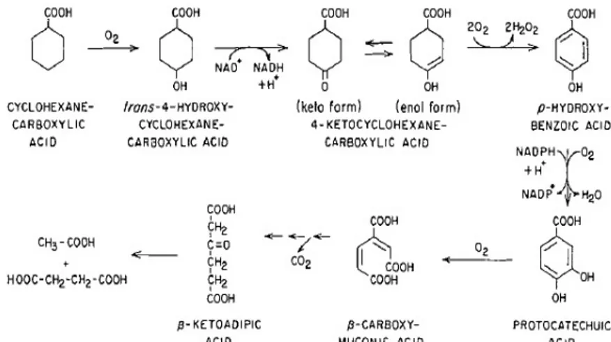
example, growth medium extracts of PRLW19 strain grown on CHCA were shown to contain 1-ene-CHCA and pimelic acid by GC-MS analysis. The presence of these two compounds in the growth medium and the high oxidative activity of cell suspensions with 1-ene-CHCA and 2-hydroxy-CHCA provided strong evidence for the metabolism of CHCA via β-oxidation pathway (Fig. 3.1). The activity related to the enzymes acyl-CoA synthetase, dehydrogenase, hydrolase and thiolase was found in crude extracts of PRLW19 cells grown on CHCA as sole carbon and energy source [Blakley et al.; 1978]. These two intermediates were also observed in the case of Rhodopseudomonas palustris, a purple non-sulfur phototrophic bacterium, during anaerobic degradation of benzoic acid that leads to CHCA formation as intermediate of reaction which is further degraded by a metabolic pathway similar to that found in PRLW19 strain [Pelletier et al.; 2000].
Fig. 3.1 β-oxidation pathway proposed for CHCA degradation by PRLW 19 [modified from Blakley et al.; 1978].
Other studies have provided results that support the aromatization pathway of CHCA [Blakley et al.; 1974, Kaneda et al.; 1974, Taylor et al.; 1978]. In Arthrobacter sp. culture, the microbial degradation of CHCA occurred through aromatization of the ring to form p-hydroxybenzoic acid before ring cleavage. In particular, cellular extracts of Arthrobacter sp. grown on CHCA were able to consume oxygen in the presence of CHCA, 4-hydroxy-CHCA, keto-CHCA, p-hydroxybenzoic acid, protocatechuic acid, quinic acid, and shikimic acid. 4-hydroxy-CHCA dehydrogenase and 4-keto-CHCA oxidase were found to be the key enzymes involved in the catabolism of CHCA, by analyzing the activity of the cellular extracts of CHCA-grown Arthrobacter sp. cells. The first enzyme requires Mg2+ while the second enzyme is very stable and does not require any cofactor for its activity [Blakley et al.; 1974] (Fig. 3.2).
Fig. 3.2 Aromatization pathway proposed for the catabolism of CHCA by Arthrobacter sp. [Blakley et al.; 1974].
The CHCA aromatization pathway shown in figure 3.1.3 was previously described in Gram positive bacteria such as Arthrobacter, Corynaebacterium, and Alcaligenes [Blakley et al.; 1974, Kaneda et al.; 1974, Taylor et al.; 1978]. Iwaki and co-workers (2005) described the function of the pobA gene coding for the enzyme responsible for the formation of protocatechuic acid from p-hydroxy-benzoic acid in the Gram positive Corynaebacterium
in the aromatic compound breakdown in Gram negative bacteria such as Pseudomonas
aeruginosa, Pseudomonas putida, and Azotobacter chroococcum [Entsch et al.; 1988, Quinn
et al.; 2001, Bertani et al.; 2001].
Rontani and colleagues (1992) reported that Alcaligenes sp. PHY12 when incubated in the presence of cyclohexaneacetic acid (CHAA) shows a combination of α- and β-oxidation to degrade this type NA. In particular, Alcaligenes sp. PH12 was able to decarboxylate CHAA to CHCA that was further degraded by β-oxidation [Rontani et al.; 1992]. Ougham et al.; (1982) isolated a strain of Arthrobacter (named Arthrobacter sp. CA1) from a soil contaminated with aviation fuel by enrichment on mineral salts medium containing cyclohexaneacetate (CHA) as unique carbon and energy source. By analyzing the cell extracts obtained from CHA-grown
Arthrobacter sp. CA1 cells, enzymes responsible for the formation of CHA-CoA were
detected. It was proposed that the degradation of CHA proceeded through the formation of a CoA ester followed by initiation of the β-oxidation cycle. Due to the formation of a tertiary alcohol, that is not amenable to dehydrogenation to form a ketogroup, the β-oxidation was blocked. The side chain of CHA was eliminated as acetyl-CoA by the action of a CoA lyase. Consequently, cyclohexanone was produced and it was degraded by a Baeyer-Villiger
oxygenase responsible for the insertion of an oxygen into the cyclohexanone ring forming a
lactone (6-capro-lactone) (Fig. 3.3).
Despite their overall toxicity and recalcitrance in wastewater, little information is available on the degradation of aromatic and highly branched alkanoic carboxylic acids that represent a small portion of NAs mixture contained in oil sands processed water (less than 10 %) [Hsu et al.; 2000]. Recently, Johnson and colleagues (2011) showed that increased alkyl side chain branching is responsible for NAs recalcitrance. In particular, they examined the biodegradation of four aromatic alkanoic acid isomers that differed in the length of the alkyl side chain branch: (4’-n-butylphenyl)-4-butanoic acid (n-BPBA); (4’-iso-butylphenyl)-4-butanoic acid (iso-BPBA); (4’-sec-butylphenyl)-4-(4’-iso-butylphenyl)-4-butanoic acid (sec-BPBA) and (4’-tert-butylphenyl)-4-butanoic acid (tert-BPBA) (chemically synthesized) (Fig. 3.1.5 a, b, c, and d) [Smith et al.; 2006]. A hydrocarbon degrading culture provided a pool of microbes putatively capable of BPBA degradation, as demonstrated through GC-MS analysis. By analyzing gas chromatograms after 35 days of incubation, the less branched n-BPBA was shown to be readily degraded, while the rate of degradation of the most branched n-BPBA was much slower. The major metabolic intermediate of butanoic acid biodegradation was the ethanoic acid (Fig. 3.4 e, f, and g), which, in turn, was further transformed to (4’-carboxybutylphenyl)ethanoic acid, the latter being a diacid formed by addition of a carboxylic acid group to the alkyl side chain. This confirmed that the initial degradation steps of the branched n-BPBA involve the removal of two carbons from the carboxyl side chain of the aromatic alkanoic acids, indicative of β-oxidation. Complete mineralization of n-BPEA and (4’-carboxybutylphenyl)ethanoic acid occurred in 49 days, while no further degradation of
iso-, sec- or tert-BPEA metabolites was detected. Through 454 Pyrosequencing and 16S
rRNA anlysis, Burkholderia, Pseudomonas and Sphingomonas spp. were identified as dominant microbial genera and key microorganisms involved in aromatic alkanoic acid transformations [Johnson et al.; 2011].
Fig. 3.4 Aromatic alkanoic acids: (4’-n-butylphenyl)-4-butanoic acid (n-BPBA) (a), (4’-iso-butylphenyl)-4-butanoic acid (iso-BPBA) (b), (4’-sec-butylphenyl)-4-(4’-iso-butylphenyl)-4-butanoic acid (sec-BPBA) (c), (4’-tert-butylphenyl)-4-butanoic acid (tert-BPBA) (d), (4’-n-butylphenyl)ethanoic acid (n-BPEA) (e), (4’-sec-butylphenyl)ethanoic acid (sec-BPEA) (f), and (4’-tert-butylphenyl)ethanoic acid (tert-BPEA) (g) [modified from Johnson et al.; 2011].
Nowadays, some information is available about alkyl phenyl alkanoic acids degradation by pure cultures. Johnson et al. (2012) attempted to isolate microbial species that could degrade BPBAs (Fig. 3.1.5 a, b, c, and d). A strain was isolated from hydrocarbon-contaminated sediment from Avonmouth (U.K.) that was used as inoculum of cultures supplied with n-BPBA as sole carbon and energy source. The strain isolated was designated as IS2.3 and it was phylogenetically related to Mycobacterium genus (16S rRNA 100% to Mycobacterium
aurum). IS2.3 was able to degrade n-BPBA or t-BPBA (Fig. 3.1.5 a, d) and to use them as
sole carbon and energy sources. During n-BPBA and t-BPBA degradation by IS2.3, (4’-n-butylphenyl)ethanoic acid and (4’-t-(4’-n-butylphenyl)ethanoic acid were produced as metabolic intermediates. This suggested that the degradation of BPBAs compounds proceeds via β-oxidation pathway. During t-BPBA degradation by the strain IS2.3, the diacids (4’-carboxy-t-butylphenyl)-4-butanoic acid and (4’-carboxy-t-butylphenyl)ethanoic acid were also formed. These two metabolites were quantified revealing that they were present in low abundance i.e. 4,9% for (4’-carboxy-t-butylphenyl)ethanoic acid and 1,5% for
(4’-carboxy-t-butylphenyl)-4-butanoic. The detection of these compounds during BPBA degradation could help to explain the presence of diacids, also named as O4 species in oil sands processed water. These type of
compounds are recalcitrant to microbial degradation and tend to concentrate in NAs-contaminated areas. Frank et al. (2009) and Headley et al. (2011) revealed these toxic O4 species in Athabasca contaminated sites through nuclear magnetic resonance spectroscopy analysis and electro-spray ionization mass spectrometry.
NAs biodegradation is principally affected by their chemical structure. Several studies showed how aerobic microbial activities are able to degrade low molecular weight NAs, while branched, cyclic and high molecular weight toxic compounds tend to accumulate in the contaminated areas as they are more persistent and recalcitrant to biodegradation [Holowenko et al.; 2002, Scott et al.; 2005, Clemente et al.; 2004, Biryukova et al.; 2007]. Smith et al. (2008) studied the rate of degradation of n-butyl-cyclohexyl-butanoic acid (BCHBA) and the
iso-, sec- and tert forms by the bacterial community found in sediments. They demonstrated
that the biodegradation efficiency decreases when alkyl side chain branching increases. They found that 97% of n-BCHBA with non-branched alkyl side chain was degraded, while 2,5% of tert-BCHBA with highly branched alkyl side chain was degraded in 30 days [Smith et al.; 2008]. Han et al. (2008) reported similar conclusions on the alkyl side chain branching. They also reported the negative effect of the cyclicity degree of the compound on the biodegradability of NAs.
Differences in biodegradation rates can be observed if we consider different isomers of the same model NAs. Holowenko et al. (2002) and Headley et al. (2002) studied the biodegradation of NAs from OSPW and/or commercial mixtures, respectively. They showed that cis-isomers are less biodegradable than trans-isomers. In particular, they investigated the biodegradation rates of methyl-1-cyclohexaneacetic acid (4-MACH) and trans-4-methyl-cyclohexanecarboxylic acid (4-MCCH) and their cis-isomers. The faster degradation
of trans-isomers compared to the cis ones was related to the lower bioavailability of the cis-isomers and the higher energy required to break their intra-molecular hydrogen bonds [Holowenko et al.; 2002, Tanapat et al.; 2002].
Other studies showed that methyl substitution can impair or reduce the rate of β-oxidation of both aliphatic- and cyclo-alkane carboxylic acids [Dias et al.; 1971, Hammon et al.; 1972, Beam et al.; 1974]. For example, Herman et al. (1994) showed that during 24 days of incubation, only 6-7% of methyl substituted pentyl-cyclohexanecarboxylic acid was mineralized to CO2, while 50% of CHCA was degraded. Apparently, although these data
about model NAs degradation are useful, we are far to fully understand the microbial degradation pathways required to OSPWs detoxification, as they contain thousands of different molecules.
Plants and microorganisms associated to plants have beneficial effects incontaminated soils. Many data show that plants-bacteria association support the degradation of contaminants in the rhizosphere [Davis et al.; 2002, Susarla et al.; 2002; Siciliano et al.; 2001]. The type of microorganisms and fungi that colonize the rhizosphere are influenced by root exudates that alter the chemistry of the soil in the area, and that can be used as selective growth substrates. In turn, microorganisms affect the composition and amount of root exudates by influencing root cell leakage, cell metabolism, and plant nutrition [Yang et al.; 2000]. Considering this, in phytoremediation studies, rhizobacterial populations have been studied for their ability to metabolize NAs and mitigate their toxicity. Biryukova et al. (2007) showed for the first time that microorganisms isolated from the rhizosphere in Athabasca contaminated soil can degrade commercially available NAs. After 17 days of growth of these root-associated microbial consortia on NAs, more than 90% of the initial concentration of the toxic compounds was metabolized
Few studies focused on the metabolic abilities of algae regarding NAs biodegradation, as well as the mechanisms by which algae can transform NAs. For example, algal communities have been characterized in the tailings ponds of the Athabasca Oil Sands [Leung et al.; 2001, 2003], while other studies described the ability of the algae to degrade model and tailings associated NAs [Headley et al.; 2008]. Among the 12 algal species that have been characterized by Headley et al. (2008), two Naviculla strains transformed the model NA
trans-4-methylycyclohexaneacetic acid (4-MCHAA). One of these two Naviculla strains was
also able to remove the cis-isomer of 4-MCHAA. NAs mixture derived from oil sands tailings water could not be degraded by any of the 12 algal species characterized. In a subsequent study, the green alga Chlorella pyrenoidosa was shown to metabolize the CHCA with the production of hydroxylated metabolites [Yoshizako et al.; 1991]. Two green algae (Dunaliella
tertiolecta and Chlorella vulgaris) and a cyanobacterium strain (Synechococcus leopoliensis)
were investigated by Quesnel et al. (2011) for their ability to degrade five model NAs at a concentration of 300 mg/L that exceeds that found in NAs-contaminated oil sands tailing waters (around 120 mg/mL). The NAs tested in this study were the CHCA, the cyclohexaneacetic acid (CHAA), the cyclohexanepropionic acid (CHPA), the cyclohexanebutyric acid (CHBA), along with a more complex NA such as 1,2,3,4-tetrahydro-2-naphthoic acid. As a result, Dunaliella tertiolecta was shown to degrade all the NAs under analysis except the 1,2,3,4-tetrahydro-2-naphthoic acid, while Synechococcus leopoliensis showed only tolerance towards all the tested NAs. In particular, Dunaliella tertiolecta showed the ability to grow on CHBA producing CHAA as metabolic intermediate. By GC-MS analysis of the growth medium derived from algal cultures treated with CHAA as sole carbon and energy source, cyclohexylidene acetic acid was found to be a degradation intermediate, as previously reported for the CHAA-degrading bacterium Arthrobacter sp. CA1 [Ougham et
exploiting a similar metabolic pathway to that described for Arthrobacter sp. CA1. D.
tertiolecta cultures incubated in the presence of CHPA revealed that CHCA was an
intermediate of degradation. CHCA was completely degraded via β-oxidation, similarly to that described for PRWl19 strain [Blakley et al.; 1978]. Conversely, algal cultures could not degrade the highly recalcitrant 1,2,3,4-tetrahydro-2-naphthoic acid, despite the fact it has the same molecular weight of CHBA. This suggested that NA persistence is influenced by the ring composition and not by the molecular weight of the molecule [Quesnel et al.; 2011]. In summary, all these data on the degradation of model NAs demonstrate the requirement for more research work in order to clarify the complexity of NAs degradation pathways.
4. The genus Rhodococcus
Rhodococci are Gram-positive, non-sporulating, aerobic bacteria. They are classified into the
suprageneric actinomycetes group known as mycolate-containing nocardioform, also including the genera Mycobacterium, Nocardia and Corynebacterium [Finnerty et al.; 1992].
Rhodococcus genus includes genetically and physiologically diverse bacteria, which are able
to colonize various habitats, from the sea level [Bell et al.; 1998] to Alpine soils [Margesin et al.; 2003], and from the deep sea [Heald et al.; 2001] to coastal sediments [Langdahl et al.; 1996] and Arctic and/or Antarctic environments [Whyte et al.; 2002, de Carvalho et al.; 2005]. Some Rhodococcus strains are also pathogens; for instance, R. fascians causes the formation of leaf gall in many plants while Rh. equi is an equine pathogen with the ability to infect other domestic animals [Gurtler et al.; 2004].
Rhodococcus genus members have many peculiar properties that can be summarized as
following:
- a peculiar cell wall, containing long aliphatic chains of mycolic acids, which facilitatesthe uptake of hydrophobic substrates into the cells. The presence of diverse organic compounds
in the growth medium may induce changes in the fatty acids composition of the membrane lipids and can alter the fluidity of the cell envelope. Notably, the ability to modulate the fatty acids composition of the cell envelope is important forthe resistance of Rhodococcus cells to many toxic compounds [de Carvalho et al.; 2005b].
- the capacity to produce surfactants improving the metabolism of hydrophobic substrates. Surfactants, such as trehalose-containing glycolipids, promote the adhesion of cells to hydrophobic phases in a two-phase system, decreasing the interfacial tension between phases and hydrophobic compounds [Finnerty et al.; 1992, de Carvalho et al.; 2005b];
- the ability to persist under stress conditions such as starvation [Martínková et al.; 2009] or after desiccation [LeBlanc et al.; 2008];
- the capacity to degrade pollutants though in the presence of more easily degradable carbon sources [Bell et al.;1998, Martínková et al.; 2009];
- the high frequency of recombination described in some Rhodococcus strains contributes to the plasticity of their genomes improving the ability to acquire new genes (by horizontal gene transfer) and consequently, new enzymatic activities [Larkin et al.; 2005].
- the large genomes of Rhodococcus strains, such as Rhodococcus jostii RHA1 with a genome around 9.7 Mb in size, provide a redundancy of catabolic pathways [McLeod et al.; 2006]. Moreover, Rhodococci also contain mega-plasmids carrying a large number of catabolic genes [McLeod et al.; 2006]. The wide range of Rhodococcus metabolic activities includes the degradation of alkanes and aromatic hydrocarbons, biotransformation of steroids and a significant set of xenobiotic compounds [Martínková et al.; 2009], antibiotics along with amino acids production [Wakisaka et al.; 1980, Yamada et al.; 1973], lignin degradation [Andreoni et al.; 1991], chemo-litho-autotrophic growth in the presence of hydrogen and carbon dioxide [Reh et al.; 1981], and production of biosurfactants [Bicca et al.; 1999, Philp et al.; 2002].
In summary, since Rhodococcus strains are characterized by numerous of enzymatic activities, unique and peculiar cell wall structure and suitable biotechnological properties, they are ideal organisms to be employed for bioconversion and biodegradation of many organic compounds in environmental remediation and in the pharmaceutical and chemical industries [Bell et al.; 1998, Larkin et al.; 2005, Van der Geize et al.; 2004]. Unfortunately, most of the genetic systems and regulatory mechanisms of genes and proteins required for these degradation/biosynthetic pathways in Rhodococcus, are still far from being understood. Indeed, the progress in Rhodococcus genetics and biochemistry is limited by the following features, namely:
- the high rhodococcal genetic diversity creates obstacles for finding molecular tools applicable to all Rhodococcus spp. [Shao et al.; 1995, Powell et al.; 1998].
- the recalcitrance of Rhodococcus strains cell walls to mechanical and chemical treatments hampers both the nucleic acids extraction and the introduction of exogenous DNA [Larkin et al.; 2005];
- the pleomorphism whereby many strains grow as short rods, cocci or branched multinucleated filaments [Williams et al.; 1976, Powell et al.; 1998] leads to problems in the segregation of mutants [Larkin et al.; 2005];
- the genomic instability may create problems with illegitimate integration upon electroporation of Rhodococcus cells with exogenous DNA [Larkin et al.; 2005];
- the high GC content genome creates problem in PCR amplification and DNA-DNA/ or RNA-DNA hybridization techniques;
- the presence of effective endogenous restriction systems that recognize unmethylated sites in exogenous DNA can cause the cleavage and the subsequent degradation of newly introduced
DNA with the consequence of low ‘fertility’ of some Rhodococcus strains in intergenic matings and/or transformation [Denome et al.; 1993, Schäfer et al.; 1990];
- the peculiar genetic codon usage by which the start codon of gene translation is often a GTG triplet instead of the typical ATG triplet and by which stop and start codons of consecutive genes clustered in operons often overlaps.
These genetic aspects can hamper the heterologous expression of rhodococcal proteins in E.
coli or Pseudomonas strains that are the typical host for protein functional experiments
because of the broad knowledge about their protein expression mechanisms and the significant amount of expression vectors that exist for these genera.
Due to the above reported limitations, and despite the importance of many potentially valuable Rhodococcus strains, the genetic analysis of this genus has hindered for a long time by the lack of efficient molecular tools [Sallam et al.; 2006]. During the last decade, several efforts have been towards the development of genetics strategies for the manipulation of
Rhodococcus members, namely: the construction of E. coli-Rhodococcus shuttle vectors
[Matsui et al.; 2007, Hirasawa et al.; 2001, Nakashima et al.; 2004], the development of transposon systems to create random transposon libraries [Sallam et al.; 2006, Fernandes et al.; 2001, Mangan et al.; 2001] and unmarked mutagenesis deletion systems with SacB as counter selection to generate mutants as a result of two consecutive DNA single crossover events [Jager et al.; 1995, Denis-Larose et al.; 1998, Van der Geize et al.; 2001]. Additionally, the following approaches have been recently applied in Rhodococcus strains: transcriptomic and proteomic techniques [Goncalves et al.; 2006, Hara et al.; 2007, Patrauchan et al.; 2005], analyses of regulator-operator interactions [Eulberg et al.; 1998, Nga et al.; 2004], studies of transcription using reporters [Nga et al.; 2004, Veselý et al.; 2007, Takeda et al.; 2004, Knoppová et al.; 2007], development of systems for the overexpression of genes involved in
key catabolic pathways and enzymes [Veselý et al.; 2003, Lessard et al.; 2004, Nakashima et al.; 2004, Na et al.; 2005].
The genomes of several Rhodococcus strains such as Rhodococcus jostii RHA1 [McLeod et al.; 2006], Rhodococcus opacus B-4 [Takarada unpublished] and Rhodococcus erythropolis PR4 [Sekine et al.; 2006] have recently become available on public databases. The genome sequences revealed very large and complex genomes, partly owing to the presence of (multiple) large (linear) plasmids along with catabolic complexity and diversity.
In conclusion, further efforts to improve Rhodococcus genomic knowledge and to develop new and more efficient tools for genetic engineering of this genus are needed. Breakthroughs in Rhodococcus genetics and molecular biology will support attempts to construct
Rhodococcus strains with suitable properties for environmental and biotechnological
applications [Van der Geize et al.; 2004].
4.1. NAs degradation by Rhodococcus spp.
Thanks to their metabolic capability and versatility, Rhodococcus genus, along with other closely related high-GC actinomycetes (Mycobacterium, Corynebacterium, Gordona, and
Nocardia), are widely recognized as ideal candidates for the biodegradation of pollutant compounds found in petroleum deposits [Yu et al.; 2006, Quek et al.; 2006].
Presently, only one experimental work is available on the Rhodococcus spp. ability to grow on NAs [Demeter et al.; 2014]. These authors isolated two Rhodococcus spp. from a microbial community derived from Athabasca OSPW, Rh. sp. MTF and Rh. sp. OSPW strains, which were shown to belong to Rh. erythropolis and Rh. fascians species, respectively. In addition to MTF and OSPW strains, other isolates were obtained belonging to
community was able to grow on a mixture of CHCA and CHAA, while none of the isolates could degrade these NAs. [Demeter et al.; 2014].
A few reports describe the ability of Rhodococcus spp. to degrade alicyclic aliphatic hydrocarbons that can be considered the chemical backbones of most of the NAs structures. Lee et al. (2007) characterized a microbial consortium able to utilize cyclo-hexane as sole carbon and energy source. Microorganisms isolated from the microbial consortium were tested for their ability to degrade hexane. The 16S rDNA of the most efficient cyclo-hexane-degrading strain shared 97% of similarity with the 16S rDNA of the nitrile-metabolizing bacterium Rhodococcus USA ANO 12 [Brandao et al.; 2002]. This new strain, named as Rh. sp EC1, was characterized by a versatile metabolism, being able to utilize structurally diverse compounds, including high carbon number diesel and lubricant oils [Lee et al.; 2007].
Rh. sp. EC1 cyclo-hexane degradation occurred via aromatization pathway. In particular,
2-cyclohexen-1-one, gamma-butyrolactone and phenol were identified as intermediates by GC-MS analysis of the growth medium of Rh. sp. EC1 culture incubated in presence of cyclo-hexane. It has been suggested that the cyclo-hexane can be degraded via aromatization pathway. Since that gamma-butyrolactone is considered as an intermediate of tetrahydrofuran (THF), and that THF was not directly detected as a metabolite of cyclo-hexane, the degradation pathway of THF was investigated to confirm the aromazation of cyclo-hexane as main removal mechanism of aromatic hydrocarbons by Rh. sp. EC1. The results showed that
Rh. sp. EC1 was able to degrade THF producing 2,3-hydrofuran and lactone as intermediates.
These evidences support the hypothesis that Rh. sp. EC1 could degrade cyclo-hexane via aromatization pathway (Fig. 4.1.1) [Yi et al.; 2011].
Fig. 4.1.1 Proposed cyclohexane degradation pathways by Rhodococcus sp. EC1; (1) cyclohexane (2) cyclohexanol (3) cyclohexanone (4) 2-cyclohexen-1-one (5) phenol (6) gamma-butyrolactone (7) tetrahydrofuran (8) 2, 3-hydrofuran (9) furan [Yi et al.; 2011].
Another mechanism described for the ring cleavage of alicyclic compounds is achieved through lactone formation. For example, Rh. NDKK48 was capable of cyclo-hexane and methyl-cyclo-hexane degradation when it was cultivated in the presence of hexadecane as co-carbon source. In particular, this Rhodococcus strain cleaved the ring of methyl-cyclo-hexane producing the following intermediates: 4-methylcyclohexanone, 4-methyl-2-oxepanone, and 3-methyladipic acid. The formation of 4-methyl-2-oxepanone indicated that the ring cleavage was performed by Baeyer-Villiger oxidation [Koma et al.; 2005]. Interestingly, a similar pathway was described regarding the degradation of cyclododecane by Rhodococcus ruber CD4 which was isolated from a mixed culture enriched with cyclododecane as unique carbon and energy source [Schumacher et al.; 1999]. On the basis of the metabolic intermediates detected, a putative pathway for the degradation of cyclododecane degradation by Rh. ruber CD4 was proposed. The substrate was initially hydroxylated to cyclododecanol and then dehydrogenated to the keto-cyclododecanone. This alicyclic ketone was then subjected to a Baeyer-Villiger oxidation, resulting in the formation of the lactone oxacyclo-tridecan-2-one, which was converted into 1-12-dodecandioic acid by two subsequent dehydrogenation steps (Fig. 4.1.2) [Schumacher et al.; 1999].
Fig. 4.1.2 Rhodococcus ruber CD4 proposed pathway for the degradation of cyclododecane [Schumacher et al.; 1999].
Baeyer-Villiger oxygenases are enzymes responsible for the homonym reaction that forms an ester from a keton or a lactone from a cyclic ketone. Kostichka and colleagues (2001) showed data about the characterization of a gene cluster involved in cyclododecanone degradation in
Rh. ruber strain SC1. This Rhodococcus strain was isolated from a mixed culture derived
from an industrial wastewater bioreactor by selecting for microbes that can utilize cyclododecanone as the sole carbon source. The characterization of Rh. ruber SC1 gene cluster revealed genes coding for cyclododecanone monooxygenase, an esterase, an alcohol dehydrogenase, and an aldehyde dehydrogenase. The four genes were cloned in suitable vectors for E. coli, and the enzyme activities were evaluated analyzing the intermediates produced during biotransformation experiments with cyclododecanol or cyclododecanone as substrates. The first two genes encode for the enzymes catalyzing the first two steps of the cyclododecanone oxidation. The last two genes encode for two different dehydrogenases responsible for the formation of docecanedioic acid from the oxidation of 12-hydroxylauric acid. On the basis of the intermediates produced during these experiments, a postulated pathway for Rh. ruber SC1 was proposed (Fig. 4.1.3) [Kostichka et al.; 2001].


![Fig. 4.1.2 Rhodococcus ruber CD4 proposed pathway for the degradation of cyclododecane [Schumacher et al.; 1999].](https://thumb-eu.123doks.com/thumbv2/123dokorg/8155447.126546/40.892.265.626.108.417/fig-rhodococcus-ruber-proposed-pathway-degradation-cyclododecane-schumacher.webp)