Case report
Κλινικό περιστατικό
J HELLENIC VET MED SOC 2019, 70(4): 1905-1910 ΠΕΚΕ 2019, 70(4): 1905-1910
ABSTRACT. Two mixed-breed bitches (18 and 19 months), that had been treated, one year before, with deslorelin acetate implant to postpone puberty, were hospitalized and monitored during their first heat. The heat was presumed by the owners, that observed vulvar swelling in both cases but no vulvar bloody discharge. The following diagnostic pro-cedures were employed: physical genital tract examination, vaginoscopy, vaginal cytology, endocrine assay, ultrasound and X-ray using vaginal infusion of iodum and pneumobladder as positive and negative contrasts. In bitch 1, vaginal cytology and progesterone levels confirmed the presence of an ovulatory “dry” oestrus, without cytological presence of red blood cells, progressing to dioestrus. Ultrasound showed preovulatory follicles and, in the following days, tran-sition to corpora lutea. The caudal abdomen presented a large ovoid cystic structure filled with echoic fluid, next to the bladder. Radiographic scans demonstrated a normal bladder profile, while the contrast medium failed to enter into the cranial vagina. On the basis of these findings, the bitch 1 was submitted to laparotomy 10 days after the end of oestrus. A vaginal dilatation (10x5 cm), from which brown fluid was aspirated, was found and resected together with uterus and ovaries. Bitch 2 had the same diagnostic route and findings, but she was laparotomized 3 months after the heat. During this period no spontaneous regression of the lesion was observed. At laparotomy, the vaginal dilatation (8x4 cm) was only aspirated and the bitch regularly neutered. In both cases, cytology of the fluid taken from the vaginal sac revealed superficial epithelial cells and abundant degenerate red blood cells. Histology (bitch 1) confirmed the vaginal origin of the dilatation and revealed an additional Gärtner duct cyst. The abnormality (hematocolpus) probably originated by an inadequate drainage of proestrous bloody discharge because of a severe vaginal stenosis. A congenital origin of the lesion was unlikely; it was strongly suspected that the treatment of the prepubertal bitches interfered, by an irreversible way, with the normal development of the vagina.
Keywords: Dog, hematocolpus, deslorelin, ultrasound, pathology
Complete vaginal stenosis and hematocolpus in two bitches with a history
of GnRH treatment to postpone puberty
G. Marino, M. Quartuccio, A. Sfacteria, S. Cristarella, A. Zanghì
Department of Veterinary Sciences, University of Messina, Messina, Italy
Corresponding Author:
Marino Gabriele, Department of Veterinary Sciences - University of Messina Via Palatucci, Polo Universitario Annunziata, 98168 Messina, Italy
Email address: [email protected]
Date of initial submission: 26-03-2019 Date of revised submission: 05-08-2019 Date of acceptance: 11-08-2019
CASE HISTORY
T
wo mixed-breed female dogs (bitch 1 and 2), not related to each other, treated at 6 months of age with 4.7 mg deslorelin acetate implant (Suprelorin®, Virbac), inserted into the interscapular region, were hospitalized and monitored during their first heat, ob-served at the age of 18 and 19 months, respectively.All procedures were approved for ethical implica-tions by the Center for Animal Reproduction and As-sisted Insemination of the University of Messina. The extra-label use of the drug, not designed for prepuber-tal female dogs, was approved by owners in agreement with literature (Trigg et al., 2006; Sirivaidyapong et al., 2012; Marino et al., 2014; Kaya et al., 2015). Even the diagnostic trials had the owners’ consent.
At presentation, bitch 1 (Europe) did not show any sign of disorders. The vulva was well developed, increased in volume, dry with wrinkled skin. Digital stimulation of the perivulvar skin evoked a lateral displacement of the tail, accompanied by a stiffening of the posterior limbs. No bloody discharge was ob-served. Abdominal palpation allowed appreciating an ovoid and firm structure consistent with an enlarged bladder for position and size. The cervix and the uter-us were not detectable by palpation. Digital vaginal exploration showed a normally shaped vestibulo-vag-inal channel, although slightly stenotic at the cin-gulus level. Vaginal cytology that was performed at two-day interval during proestrus-oestrus and weekly during dioestrus confirmed a normal transition from proestrus to dioestrus. Cornification during oestrus reached percentage of 80 % lasting approximately 9 days. Neutrophils were detected only at the dioestrus onset, in concomitance to the presence of interme-diate and parabasal cells. Red blood cells were not detected in any phase. Vaginoscopy was performed with a rigid endoscope (TCI-Endoscope, length 43 cm, Karl Storz) without sedation, at two-day inter-val during proestrus-oestrus until dioestrus onset. The introduction of the instrument was facilitated by a shunt system (Minitube), which also provided fixation to the vaginal wall allowing pumping air to facilitate the visualization of the vagina. Typical mu-cosal folds changed in appearance during the period of study, passing from oedematous, pink, and round (late proestrus) to pale and crenulated (oestrus) up to flattened and flaccid (dioestrus). No septa were ob-served at every level. The lumen was slightly stenotic and the bottom of the vagina and the cervix were not visualized, giving the appearance of a blinded vagina.
Blood sampling was performed every two days until dioestrus onset. The obtained serum was processed for the determination of progesterone, using an Enzyme Linked Fluorescent Assay (BioMeuriex, Minividas). Circulating progesterone progressively increased from values of 0.9 ng/ml at presentation to 24 ng/ml at dioestrus onset. Ultrasound examination of the abdo-men (Esaote, MyLab 40 Vet, 8 MHz convex probe, 12 MHz linear probe) was performed every two days un-til dioestrus onset. At presentation, multiple cavitary structures in the ovaries, with an anechoic content, 0.6-0.8 cm in diameter, compatible with preovulatory follicles were seen. At ovulation, an increase in echo-genicity of such structures was evident together with the finding of a small amount of liquid withheld in the ovarian bursa. The uterus showed normal appear-ance, a diameter of about 1.5 cm and the absence of intrauterine fluid. For the whole period of monitoring, a large cystic oval formation, at least 8-10 cm long and 4-5 cm wide, thick-walled, and with the presence of numerous and dense echoes within the lumen, was found (Fig. 1).
Figure 1. Bitch 1 (Europe). Caudal abdomen. Ultrasound
appear-ance of the large oval cystic structure (hematocolpus) with an echoic content.
This undefined cystic formation was found next to a second cystic oval anechoic structure, more at-tributable to the bladder. The ultrasound-guided cath-eterisation of the bladder confirmed the nature of the second structure. By contrast, the introduction of ul-trasound-guided catheter into the vagina, presented difficulties in the progression, stopping cranially, for the presence of a thin hyperechoic septum (Fig. 2). The abdominal X-ray examination (Univet, 300HS) was carried out on dorsoventral and lateral views. In order to enhance the contrast of the vaginal area, io-pamidol (Iopamiro 300, Bracco Imaging) was inocu-lated in vagina throughout a Foley catheter that was inserted and fixed in the caudal vagina.
Figure 2. Bitch 2 (Moon). Appearance of continuity between the
hematocolpus (H) and the vagina (V). Visible even a partial scan of the bladder (B).
The positive contrast medium showed a radiolog-ical stop in the vagina, emphasized by the presence of a substantial amount of iodum between the septum and the balloon of the Foley catheter. Then, pneumo-cystography was obtained by blowing filtered air into the bladder throughout a Foley catheter. The bladder, of normal appearance and content, had no connection with the undefined cystic formation. In light of these findings and the diagnostic suspicion of an abnormal collection in the vagina/uterus, the bitch was submit-ted to exploratory laparotomy and eventual ovari-ohysterectomy 10 days after the end of the oestrus. After premedication with intramuscular injections of tramadol hydrochloride (2 mg/kg b.w.; Contramal, Grunenthal Italia) and acepromazine maleate (0.05 mg/kg b.w.; Prequilan, Fatro), the patient was induced with diazepam (0.5 mg/kg b.w.; Diazepam, Intervet) and propofol (3 mg/kg b.w.; Propovet, Zoetis) ad-ministered intravenously. After intubation, anaesthe-sia was maintained with isoflurane in 100% oxygen. Laparotomy was performed on the linea alba. In prox-imity to the bladder, but in continuity with the uterus cranially and with the vagina caudally, an impressive fluid-filled dilatation was found (Fig. 3), including the cervix. About 200 ml of brown fluid was aspirat-ed from the dilatation (Fig. 4). Furthermore, a second cyst inside the main cystic structure was found, from which 10 ml of yellowish mucoid fluid was aspirat-ed (Fig. 5). The fluids were cytologically evaluataspirat-ed during laparotomy. The first fluid showed only epithe-lial vaginal cells, many of which were cornified and abundant degenerate red blood cells (Fig. 6).
Figure 3. Europe. Externalization of the formation caudal to the
body of the uterus.
Figure 4. Moon. Intraoperative aspiration of the brown fluid
con-tained in the hematocolpus.
Figure 5. Moon. Different aspect of the content of the second
Figure 6. Moon. Superficial epithelial cells and degenerate red
blood cells in the fluid aspirated from the hematocolpus. May - Grünwald Giemsa. Obv 20x.
The second fluid presented parabasal-like epithe-lial cells and mucus. In both fluids no bacteria and inflammatory cells were detected. A diagnosis of he-matocolpus was done. After ligating the vaginal ar-teries and branches of the internal pudendal arar-teries, the dilated vagina was amputated at 2/3 level and re-moved together with uterus and ovaries. The caudal wall of the dilated vagina was inspected to confirm the absence of communication with the caudal portion of the vagina (Fig. 7).
Figure 7. Europe. Visualization of the blinded caudal wall of the
hematocolpus.
The removed uterine horns were 10 cm length and 1.5 cm in diameter, with a spiral trend according to the phase of the cycle (dioestrus). The ovaries, respec-tively of 1.5 x 1.0 x 0.5 cm on the right and 2.5 x 1.0 x 0.6 cm on the left, presented both newly formed corpora lutea. The cervix was found inside the dilat-ed area, projecting in it, surrounddilat-ed by large haemor-rhagic areas (Fig. 8).
Figure 8. Europe. Gross appearance of the mucosa of the
hema-tocolpus with haemorrhagic areas. Following the dorsal median fold the cervix is visualized.
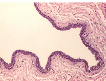
Specimens were processed for histopathological examination. The vaginal mucosa was thinned and partially degenerate, sometimes covered by a multi-layered epithelium. In the cranial part, the mucosal epithelium appeared monolayered with strong polar-ization, similar to endocervical epithelium. Inflamma-tory cells were not detectable in the mucosa and the underlying layers. In some areas the submucosa was characterized by haemorrhagic extravasation (Fig. 9). A large cavitary structure lined by a single layered ep-ithelium was found resembling a Gärtner duct epithe-lium (Fig. 10).
Figure 9. Europe. Hematocolpus. Vaginal mucosa close to the
cervix covered with a columnar epithelium. Note the absence of inflammatory infiltrates and the hemorrhagic extravasation. Hae-matoxylin and eosin. Magnification 20x.
Figure 10. Europe. Typical single layered epithelium lining the
Gärtner’s cyst. Haematoxylin and eosin. Magnification 20x.
Bitch 2 (Moon) had perfectly comparable findings. She was monitored until 3 months after the dry heat. During dioestrus, ultrasound monitoring of the vagi-nal dilatation was performed weekly, without evident changes in size and appearance. At laparotomy, the main cyst (hematocolpus) and the secondary smaller cyst (Gärtner duct cyst) were only aspirated and left in place; ligature was done at the uterine body level to remove uterus and ovaries. Moon had rectilinear uter-ine horns of 10.5 cm in length and 0.8 cm in diameter according to the phase of the cycle (anoestrus). The ovaries, sized respectively 1.8 x 0.9 x 0.7 cm on the right and 1.8 x 0.9 x 0.8 cm on the left, presented re-gressing corpora lutea. The uterine mucosa showed an initial picture of segmental hyperplasia. During fol-low-up, the stump was monitored by ultrasound and no changes were detected for a period of 1 year after the surgery.
DISCUSSION
Disorders of the development of the vagina and vestibulum are occasionally reported in the bitch and include vaginal septa, vaginovestibular stenosis, ves-tibulovulvar stenosis, segmental aplasia of the vagina (Gee et al., 1977; Wadsworth et al., 1978; Holt and Sayle, 1981; Wykes and Soderberg, 1983; Hawe and Loeb, 1984; Root et al., 1995; Archbald and Wolfs-dorf, 1996; Kyles et al., 1996). No breed and genetic predisposition for these abnormalities have been re-ported. Clinical signs reported in dogs with vaginal abnormalities include chronic vaginitis, mating diffi-culty, urinary incontinence, chronic urinary tract in-fections, dystocia and ambiguous external genitalia (Holt and Sayle, 1981; Wykes and Soderberg, 1983; Root et al., 1995; Archbald and Wolfsdorf, 1996;
Kyles et al., 1996), despite some bitches with vagi-nal septa or circumferential stenosis are asymptom-atic (Wykes and Soderberg, 1983; Root et al., 1995; Kyles et al., 1996). The terms hydrocolpus, mucocol-pus, hematocolmucocol-pus, pyocolpus refer to the abnormal distension of the vagina for the accumulation of fluid, mucus, blood and pus, respectively. These conditions may be related to a difficult drainage of the vagina and the few reported cases in the bitch were generally secondary to developmental disorder of the vestibu-lo-vaginal tract (Gee et al., 1977; Wadsworth et al., 1978; Hawe and Loeb, 1984; Tsumagari et al., 2001; Viehoff and Sjollema, 2003; McIntyre et al., 2010; Marinho et al., 2013; Alonge et al., 2015).
GnRH agonists are used as anticonceptionals in the canine species. Deslorelin acetate is a GnRH ago-nist developed as an implant, whose main indication is transitory contraception in adult male dogs. Recent studies have proved the efficacy of the treatment in prepubertal dogs to postpone puberty (Trigg et al., 2006; Sirivaidyapong et al., 2012; Marino et al., 2014; Kaya et al., 2015). There are few data about the side effects of GnRH agonists in veterinary medicine, and they are generally considered safe and totally re-versible. Persistent oestrus related or not to ovarian cysts, uterine disorders, urinary incontinence and hair abnormalities have occasionally been described (Arlt et al., 2011; Fontaine and Fontbonne, 2011; Palm and Reichler, 2012). In previous studies on prepubertal bitches, delayed epiphyseal closure, hip dysplasia, transient juvenile vaginitis and marked atrophy of the internal genital tract have been reported with minimal or no clinical impact (Marino et al., 2014; Kaya et al., 2015).
In this paper, two bitches with an abnormal dis-tension of the vagina (hematocolpus) were studied. It was remarkable that both bitches received an im-plant of deslorelin acetate in a prepubertal time. The presence of an obstruction in the genital tract caused an obstacle to normal drainage and an accumulation of fluid cranially to the stenotic point. When cycling female dogs have such obstructions the collection of fluid mixes with the typical bloody discharge of proestrus; these animals show the so-called dry heats (without bloody discharge from vulva) (Viehoff e Sjollema, 2003). Specific genes involved and herita-bility of developmental disorders of the genital tract have not yet been fully defined in bitches (McIntyre et al., 2010). Developmental disorder of the gonad and kidney may be associated finding, as consequence of
failure of gene expression or lack of bloody supply in the genital segment (McIntyre et al., 2010). Some causes may even be acquired after birth, since the development of the internal and external genitalia is completed by puberty. There was a reasonable suspi-cion that the treatment received by prepubertal bitch-es contributed to the determinism of the lbitch-esion. The treatment applied at prepubertal time is able to arrest
the development of the genital tract throughout the period of treatment (Marino et al., 2014). In some fe-male dogs, these changes are probably not completely reversible at vaginal level.
CONFLICT OF INTEREST
The authors declare no conflict of interest.
REFERENCES
Alonge S, Romussi S, Grieco V, Luvoni GC (2015) Congenital abnor-mality of the vagina complicated by haemato-pyocolpos in a 1-year Labrador Retriever. Reprod Dom Anim 50:514-516.
Archbald LF, Wolfsdorf K (1996) Theriogenology question of the month. Vaginal constriction, probably a congenital malformation. J Am Vet Med Assoc 208:1651-1652.
Arlt SP, Spankowsky S, Heuwieser W (2011) Follicular cysts and pro-longed oestrus in a female dog after administration of a deslorelin implant. N Z Vet J 59:87-91.
Fontaine E, Fontbonne A (2011) Clinical use of GnRH agonists in canine and feline species. Reprod Dom Anim 46:344-353.
Gee BR, Pharr JW, Furneaux RW (1977) Segmental aplasia of the Mülle-rian duct system of a dog. Can Vet J 18:281-286.
Hawe RS, Loeb WF (1984) Caudal vaginal agenesis and progressive renal disease in a Shi tzu. J Am Anim Hosp Assoc 20:123-130.
Holt PE, Sayle B (1981) Congenital vestibulo-vaginal stenosis in the bitch. J Small Anim Pract 22:67-75.
Kaya D, Schäfer-Somi S, Kurt B, Kuru M, Kaya S, Kaçar C, Aksoy Ö, Aslan S (2015) Clinical use of deslorelin implants for the long-term contraception in prepubertal bitches: effects on epiphyseal closure, body development, and time to puberty. Theriogenology 83:1147-1153.
Kyles AE, Vaden S, Hardie EM, Stone EA (1996) Vestibulovaginal ste-nosis in dogs: 18 cases (1987-1995). J Am Vet Med Assoc 209:1889-1893.
Marinho GC, De Jesus VLT, Palhano HB, Abidu-Figueire M (2013) Pyo-colpos in a Pinscher bitch: a case report. J Morphol Sci 30:206-208.
Marino G, Rizzo S, Quartuccio M, Macrì F, Pagano G, Taormina A, Cris-tarella S, Zanghì A (2014) Deslorelin implants in pre-pubertal female dogs: short- and long-term effects on the genital tract. Reprod Dom Anim 49:297-301.
McIntyre RL, Levy JK, Roberts JF, Reep RL (2010) Developmental uter-ine anomalies in cats and dogs undergoing elective ovariohysterecto-my. J Am Vet Med Assoc. 237:542-546.
Palm J, Reichler IM (2012) The use of deslorelin acetate (Suprelorin®)
in companion animal medicine. Schweiz Arch Tierheilkd 154:7-12. Root MV, Johnston SD, Johnston GR (1995) Vaginal septa in dogs: 15
cases (1983-1992). J Am Vet Med Assoc 206:56-58.
Sirivaidyapong S, Mehl N, Trigg T (2012) Delay of puberty and reproduc-tive performance in male dogs following the implantation of 4.7 and 9.4 mg GnRH-agonist deslorelin at an early pre-pubertal age. Reprod Dom Anim 47:400-402.
Trigg T, Doyle A, Walsh J, Swangchan-uthai T (2006) A review of advanc-es in the use of the GnRH agonist dadvanc-eslorelin in control of reproduc-tion. Theriogenology 66:1507-1512.
Tsumagari S, Takagi K, Takeishi M, Memon MA (2001) A case of a bitch with imperforate hymen and hydrocolpos. J Vet Med Sci 63:475-477. Viehoff FW, Sjollema BE (2003) Hydrocolpus in dogs: surgical treatment
in two cases. J Small Anim Pract 44:404-407.
Wadsworth PF, Hall JC, Prentice DE (1978) Segmental aplasia of the va-gina in the beagle bitch. Lab Anim 12:165-166.
Wykes PM, Soderberg SF (1983) Congenital abnormalities of the canine vaginal and vulva. J Am Anim Hosp Assoc 19:995-1000.