UNIVERSITÀ POLITECNICA DELLE MARCHE
FACOLTÀ DI MEDICINA E CHIRURGIA
Corso di Dottorato di Ricerca in Salute dell’Uomo
XXXI Ciclo
The ciliary neurotrophic factor. A novel role as leptin
sensitizer acting at the central nervous system.
Dottorando:
Docente tutor:
Wiebe Venema
Prof. Antonio Giordano
Correlatrice:
Ilenia Severi
Table of Contents
List of abbreviations 4
Abstract 6
INTRODUCTION 8
General introduction 8
Hypothalamic control of metabolic homeostasis 10 Brainstem control of metabolic homeostasis 13 Peripheral signals influencing satiety and appetite 15
The Cilliary Neurotrophic Factor (CNTF) 19
The distribution of CNTF and its receptor 20
The effects of CNTF on energy balance 22
Aims 24
MATERIALS & METHODS 25
Animals and experimental conditions 25
Experiment A 25
Experiment B 25
Tissue processing 26
Immunohistochemistry and light microscopy 27 Immunofluorescence and confocal microscopy 27
Western blotting 28
Statistical Analysis 29
Antibody table 29
RESULTS 31
Ob/ob and fasted mice do not show STAT3 activation in the hypothalamic arcuate nucleus
upon CNTF administration 31
ERK signaling is activated in hypothalamic β-tanycytes upon CNTF administration 32 STAT3 activation in the mouse tuberal hypothalamus is uprgeulated by co-administration
of CNTF and leptin 34
CNTF administration activates ERK signaling in the dorsal vagal complex of the mouse
CNTF co-administration with leptin promotes P-STAT3 activation in the nucleus of the
solitary tract 37
db/db mice are insensitive to the leptin sensitizing effect of CNTF 38
DISCUSSION 41
List of abbreviations
α-MSH - Alpha-melanocyte stimulating hormone AgRP - Agouti related protein
AMPK - Adenosine monophosphate-activated protein kinase
AP - Area postrema
APX - Area postrema lesioned ARC - Arcuate nucleus
ALS - Amyotophic lateral sclerosis BBB - Blood brain barrier
BDNF - Brain-derived neurotrophic factor BMI - Body mass index
CART - Cocaine and amphetamine- regulated transcript CCK - Cholecystokinin
CNS - Central nervous system CLC - Cardiotrophin-like cytokine CNTF - Cilliary neurotrophic factor CNTFRα - CNTF receptor α
CSF - Cerebrospinal fluid
CRH - Corticotrophin-releasing hormone CT-1 - Cardiotrophin-1
CVO - Circumventricular organ DIO - Diet induced obesity
DMV - Dorsal motor nucleus of the vagus DVC - Dorsal vagal complex
EGF - Endothelial growth factor
ERK - Extracellular signal–regulated kinases GABA - Gamma-aminobutyric acid
GHSR - Growth hormone segretagogue receptor
GI - Gastro-intestinal
GLP1 - Glucagon like peptide HFD - High fat diet
IHC - Immunohistochemistry IL-6 - Interleukin 6
IL-11 - Interleukin 11
IRS - Insulin receptor substrate
JAK-SAT - Janus kinase/signal transducer and activator of transcription LEPRb - Long isoform leptin receptor B
LHA - Lateral hypothalamus LIF - Leukaemia inhibitory factor LIFRb - LIF receptor-b
MAPK - Mitogen-activated protein kinase MBH - Mediobasal hypothalamus MC3R - Melanocortin receptor 3 MC4R - Melanocortin receptor 4
MCH - Melanin concentrating hormone
ME - Median eminence
mTOR - Mammalian target of rapamycin NPY - Neuropeptide Y
OSM - Oncostatin M PFA - Perifornical area POMC - Proopiomelanocortin
PI3K - Phosphatidylinositol-3-OH kinase PVN - Paraventricular nucleus
PYY - Hormone YY
SF1 - Steroidogenic factor-1
TH - Tyrosine Hydroxylase
TRH - Thyrotropin-releasing hormone UCP1 - Uncoupling protein 1
VMH - Ventromedial hypothalamus
VOLT - Vascular organ of the lamina terminalis Y1 - Neuropeptide Y receptor type 1
Abstract
The ciliary neurotrophic factor (CNTF) induces satiety and increases energy expenditure in rodents and humans through a leptin-like activation of the JAK-STAT3 signaling pathway. Hypothalamic glial cells constitutively express CNTF, and its expression is up-regulated in obese mice. We found that in mice with a low leptin level CNTF treatment did not induce activation of JAK-STAT in any of the hypothalamic feeding centers besides the median eminence. These results prompted us to study alternative pathways in which CNTF could play a role on food intake. In vitro studies have shown that CNTF can activate the ERK pathway. A recent report showed that activation of ERK in tanycytes of the median eminence is involved in the regulation of the access of leptin to the ARC. We studied if this pathway is activated by CNTF in the median eminence, and the dorsal vagal complex of the mouse brainstem. We treated 4 groups of mice with respectively vehicle, leptin, CNTF or CNTF and leptin and analysed activation of ERK and STAT3 in the hypothalamus and brainstem via western blotting and immunohistochemistry (IHC). Our findings indeed indicated that CNTF treatment was able to activate ERK signalling in the median eminence of the hypothalamus, further analysis showed that these cells were tanycytes. Interestingly, by western blotting we saw that co-treatment induced an increase in P-STAT3 expression compared to CNTF treatment alone significantly higher than leptin treatment compared to control. IHC showed that the P-STAT3 positive cells were located in all hypothalamic-feeding centers responsive to leptin, and the activation in these centers was significantly higher compared to leptin treatment alone. Furthermore our results in the brainstem showed activation of ERK in glial cells in all nuclei of the dorsal vagal complex. In the nucleus of the solitary tract (NTS), co-treatment with leptin and CNTF was able to significantly increase the activation of STAT3 signaling in this area targeted by leptin. Finally we show that these effects of the co-treatment with CNTF and leptin are not seen in leptin insensitive db/db mice. In conclusion, our findings show that CNTF does not directly target neurons in the arcuate, ventromedial and dorsomedial nuclei of the hypothalamus and the NTS of the brainstem. It primarily targets multiple signaling pathways in the circumventricular organs of the hypothalamus and brainstem. In particular the ERK pathway has been shown to play an essential role in the entrance of leptin in the mediobasal hypothalamus where it exerts its anorectic effects. Our
findings confirmed a role for CNTF in this mechanism both in the hypothalamus as in the brainstem, possibly through glio-vascular modifications resulting in greater leptin permeability.
INTRODUCTION
General introduction
The ability to properly adapt metabolism to changes in nutritional and environmental conditions can be described as metabolic flexibility [1]. An impaired metabolic flexibility, i.e. an inability to match food intake with energy expenditure can lead to various health problems. When food intake exceeds energy expenditure, excess energy gets stored in the form of triglycerides leading to an increase in the amount and size of adipocytes in adipose tissue [2, 3]. If this condition continues for an extended period of time, obesity and related disorders such as insulin resistance and type 2 diabetes mellitus, some types of cancer, coronary heart disease, muscoskeletal disorders, mood disorders and other serious health problems can develop [4, 5]. The incidence of obesity worldwide (Body Mass Index (BMI) ≥30kg/m²) is increasing and is predicted to increase even further in the next years, in the United States 38,2 percent of the population was obese in 2015, and it is predicted that by 2030 more than 45 percent of the population will be obese [6].
Research into the control of energy homeostasis has increased the understanding of the role that different factors play in the development of obesity. However, up until now this has not led to many opportunities in the treatment of this disease, and further research is necessary to discover potential targets to increase energy expenditure or decrease food intake.
Food intake is influenced by appetite and satiety, and is regulated at three levels in the human body; at the cellular level, sensors detect changes in energy status inside cells and start multiple processes in response. At the peripheral level signals are generated and conveyed to the central nervous system (CNS) were they influence appetite and satiety. The hypothalamus is one of two main regions in the brain responsible for the control of many physiological aspects critical for homeostasis, including energy regulation.
First evidence of the role of the hypothalamus in energy regulation came from lesion studies by Hetherington and Ranson [7] and by Anand and Brobeck [8], which showed that lesions in different areas of the hypothalamus caused either hyperphagia or obesity in the case of ventrally located ventromedial hypothalamus (VMH) and arcuate nucleus (ARC) or aphagia or even death by starvation in the case of the lateral hypothalamus (LHA) and the dorsally located paraventricular nucleus (PVN). These
findings suggested that the hypothalamus contains two distinct centers controlling food intake, one inducing satiety and the other inducing hunger, called the “dual center” model. Although this model has been useful, the control of hunger and satiety in the CNS has proven to be more complex. Recently specific neuronal pathways that control food intake via the integration of various neural and humeral inputs have been described [9]. First, hormonal and neurochemical signals reflecting the metabolic status of the individual reach the hypothalamus where they activate two neurochemically distinct and antagonistic sets of neurons in the ARC [10], increasing or activating neurons in one group results in higher food intake and lower energy expenditure, increasing or activating neurons in the other group has the opposite effect. Next to the hypothalamus, the second main region involved in processing information regarding energy status and controlling food intake is the brainstem. In this area, information from the alimentary organs enters the brain via, primarily, the vagus nerve. Afferent signals from the vagus nerve excite neurons in the nucleus of the solitary tract (NTS) in the brainstem and reduce meal size and duration [10-12]. Furthermore, projections from both the brainstem and the hypothalamus activate higher order neurons in the brain to initiate and regulate food intake.
Hypothalamic control of metabolic homeostasis
The hypothalamus, a small structure in the brain is the main regulator of many physiological processes such as, temperature regulation, biological cycles, hormonal balance, reproduction and energy regulation. It consists of different nuclei namely the arcuate nucleus (ARC), the paraventricular nucleus (PVN), the dorsomedial nucleus (DMH), the lateral hypothalamic area (LH) and the ventromedial hypothalamus (VMH), all playing an important role in energy balance [9].
Figure 1 - Hypothalamic nuclei involved in energy balance. 3V, third ventricle; ARC, arcuate nucleus; DMH, dorsomedial hypothalamic nucleus; LH, lateral hypothalamus; PVN, paraventricular nucleus [13].
The arcuate nucleus is the best-studied region for hypothalamic neuronal control of energy balance. It is positioned in the mediobasal hypothalamus, adjacent to the floor of the third ventricle and near the median eminence. The arc is, like many other nuclei involved in energy homeostasis, in direct contact with the third ventricle. The third ventricle is lined with layers of specialized ependymal cells called tanycytes; tanycytes form a long single basal projection into the hypothalamic parenchyma [14]. The dorsally located tanycytes near the DMH and VMH are often referred to as type α1 and α2, while the ventrally located tanycytes near the ARC and the median
eminence are type β1 and β2. The β2 tanycytes are unique in that they extend from the ependymal wall of the third ventricle to permeable fenestrated vessels at the pial surface of the brain, and form a barrier between blood and the cerebrospinal fluid in the third ventricle [15, 16]. By regulating the access of peripheral signals to the ARC and other hypothalamic sites, β2 tanycytes play a crucial role in brain homeostasis [17].
Distinct and antagonistic neuronal populations in the ARC respond to central and peripheral signals, such as neuropeptides, neurotransmitters and hormones controlling food intake [9]. Within this brain region two main neuronal populations are found that control satiety and energy expenditure, the an-orexigenic neurons, expressing proopiomelanocortin and cocaine and amphetamine- regulated transcript (POMC/CART) and the orexigenic neurons expressing neuropeptide Y and agouti related protein (NPY/AgRP) [18]. POMC/CART expression is up regulated in response to feeding by the release of leptin and insulin, and downregulated in response to fasting. Conversely NPY/AGRP neurons are up regulated during fasting, when leptin and insulin levels are low. These so called first-order neurons of the ARC project to second-order neurons in the LHA, PVN, VMH, the perifornical area (PFA) adjacent to the fornix, and to neurons of the NTS in the brainstem [9].
Upon activation, POMC neurons produce and secrete the anorectic hormone alpha-melanocyte stimulating hormone (α-MSH) which activates melanocortin receptor 3 (MC3R) and 4 (MC4R) on second order neurons, leading to suppressed food intake and increased energy expenditure [19, 20] Moreover, the activation of these neurons is affected by the activation of the neughbouring NPY/AgRP neurons, which through the production and secretion of AgRP inhibit the activity of α-MSH on MC4R. Furthermore, central administration of NPY activates Y1 and Y5 receptors in the ARC, VMH and the PVN, which increases food intake. Modifications in the NPY/AgRP and POMC/CART genes have been studied extensively and are linked to anorexia and weight loss or obesity and metabolic syndrome [21-23].
The paraventricular nucleus is located directly above the third ventricle in the anterior hypothalamus. It integrates information from the ARC and other extra-hypothalamic areas such as the NTS of the brainstem. The PVN expresses high levels of MC3R and MC4R and reacts with high sensitivity to the change in neuropeptides regulating food intake including AgRP, NPY and α-MSH [24, 25]. Two subsets of neurons expressing either thyrotropin-releasing hormone (TRH) or
corticotrophin-releasing hormone (CRH) mediate the effects of endogenous neuropeptides in the PVN, which are directly and indirectly involved in the control of energy balance [26, 27].
The dorsomedial nucleus (DMH) integrates information from most of the hypothalamic nuclei, but mainly the ARC, and sends projections to the LHA and the PVN. Neuropeptides such as NPY and CRH and many receptors for other peptides involved in food intake are found within the DMH. Furthermore, overexpression of NPY in the DMH has been linked to diet induced obesity (DIO), as has been reported in several rodent models of obesity [28-30]. Moreover, the DMH plays a role in different physiological processes such as stress, thermoregulation and circadian rhythms.
The lateral hypothalamic area, classically also known as the feeding center in the hypothalamus, plays an important role in the stimulation of appetite. This is mainly attributable to the presence of melanin concentrating hormone (MCH) and orexin-producing neurons. Upon fasting, orexin neurons produce orexin A and orexin B that via activation of orexin 1 receptor stimulates refeeding [31, 32]. Orexin neurons have widespread projections within the brain, influencing other physiological processes such as sleep/wake cycle and body temperature. Similarly, an increase in MCH is seen during fasting and an overexpression of MCH has been linked to obesity [33]. The ventromedial hypothalamus has been described as the satiety center in the hypothalamus, it receives AgRP and POMC projections from the ARC, and sends projections throughout the hypothalamus and the NTS in the brainstem. VMH neurons respond to leptin and express the leptin receptor B (LEPRb). For this reason, this nucleus is important in regulating the effect of leptin in energy homeostasis [34, 35]. Other neurons playing a role in preventing body weight gain are steroidogenic factor-1 (SF1)-neurons, which play a role in the development of the VMH and have been found exclusively in the VMH [36], and neurons expressing brain-derived neurotrophic factor (BDNF). Selective deletion of BDNF in the VMH of adult mice led to increased food intake and body weight [37].
Figure 2 - First order NPY/AgRP and POMC/CART neurons in the ARC, regulated by blood borne molecules project to other brain regions, mainly the hypothalamus and brainstem [38].
Brainstem control of metabolic homeostasis
First evidence of the involvement of the caudal brainstem in controlling energy regulation came from studies performed by Grill and colleagues in 1978 [39, 40]. Experiments on decerebrated rats demonstrated the ability of this brain area to receive and respond to signals arising from the mouth and from the gastrointestinal tract, which are critical for determining meal size [39, 40]. Furthermore, it has been demonstrated that the caudal brainstem was able to respond to hormonal signals regulating food intake such as cholecystokinin (CCK) and insulin [41, 42]. Similar to the hypothalamus, the brainstem consists of heterogeneous neuronal populations expressing neuropeptides important in the modulation of appetite, such as, proglucagon, tyrosine hydroxylase (TH), gamma-aminobutyric acid (GABA), CART, BDNF, NPY and POMC. Moreover, these neurons express receptors for a variety of the aforementioned neuropeptides, suggesting local circuits involved in the control of food intake [43]. Furthermore, the brainstem contains a number of receptors for circulating hormones such as CCK, insulin, leptin and glucagon like peptide (GLP1) [41, 42, 44, 45] illustrating the importance of the brainstem in metabolic homeostasis.
The majority of published data showed that the transmission of meal related visceral afferent information from the tubural region of the gastrointestinal tract is mediated by the afferent vagus nerve. A number of experiments manipulating the activity of the vagus nerve have been performed, showing a reduction in food intake and body weight in rats in which the vagus nerve was chronically or acutely stimulated [46, 47]. In particular in the brainstem, food intake is regulated primarily by the dorsal vagal complex (DVC), which is formed by three distinct regions, the nucleus of the solitary tract (NTS), the area postrema (AP) and the dorsal motor nucleus of the vagus (DMV). The anatomical and functional integration of these structures makes the DVC capable of providing behavioural, autonomic and endocrine reactions to energy related peripheral signals [48].
The nucleus of the solitary tract is the first site in the CNS to receive sensory afferent information from organs of the gastrointestinal tract and abdominal organs, as well as gustatory information from the oral cavity. The transmission of these gastrointestinal and gustatory information to the NTS is mediated by the vagus nerve and sympathetic fibers, and integration with hypothalamic input in the NTS is essential in the control of food intake [9, 49, 50]. POMC neurons from the hypothalamic ARC project into the NTS, where high amounts of MC4R’s have been found [51]. Furthermore, it receives orexigenic information from the LHA and satiety signals from the PVN and disruption of these signaling leads to both obesity and hyperphagia or weight loss [52, 53]. Circulating adiposity signals such as insulin and leptin potentiate the effect of satiety signals originating from the gastrointestinal tract, such as CCK on the NTS [54], and the presence of leptin receptors and POMC neurons in the NTS underlines its involvement in energy homeostasis [9, 55].
The area postrema is located dorsomedially to the NTS and it is one of the circumventricular organs (CVO). The lack of a complete blood brain barrier (BBB) allows CVOs to convey circulating substances that normally do not cross the BBB to the brain. Furthermore, the direct contact with the cerebrospinal fluid (CSF) in the fourth ventricle allows the AP to transport neurochemicals in and out of the CSF. The AP has connections to the NTS and similar to the NTS, the AP receives neuronal information directly from various thoracic and abdominal viscera via several cranial nerves, and projections from PVN in the hypothalamus [56]. Studies on the ablation of the AP demonstrated its involvement in feeding behaviour. AP-lesioned (APX) rats showed a loss of hypoglycaemic feeding responses induced by insulin indicating a
role in CNS glucose sensing [57]. Furthermore, although over consuming highly palatable food, APX rats were hypophagic and showed lower body weight compared to sham lesioned rats [58].
The dorsal motor nucleus of the vagus is located medially and ventrally to the NTS, directly adjacent to the central canal; it is the main source of vagal efferent innervation of the gastrointestinal (GI) tract via the subdiaphragmatic vagal branches [59, 60]. It plays an important role in the control of gut secretion and motility, and mediates pancreatic secretory function [61].
In conclusion, the brainstem processes energy status information at different levels, hereby contributing to the control of energy balance. It senses metabolites and hormones released by peripheral organs in the bloodstream; it innervates and receives input from the GI tract via the vagal nerve; it receives and projects from and into local brainstem circuits and other brain regions to integrate information important in the control of energy balance [43].
Peripheral signals influencing satiety and appetite
Peripheral signals involved in food intake regulation, glucose homeostasis and energy expenditure can be divided into episodic and tonic signals. Episodic signals arise usually after eating and are mainly anorexigenic, but can be orexigenic, whereas tonic signals are generated by tissue stores and exert a tonic anorexigenic pressure on the expression of appetite [62]. Tonic signals generated by the adipose tissue can be called adiposity signals if they fulfil certain criteria; they circulate in direct proportion to the total amount of adipose tissue; they should be able to cross the blood brain barrier and directly act on receptors within the CNS; and changes in their levels or activity should produce predictable changes in food intake and energy expenditure [63]. The most studied adiposity signal is leptin, the hormone discovered in 1994 that changed the understanding of the central regulation of energy balance [64].
Leptin is an adipose tissue-derived satiety signal circulating in proportion to fat mass [65], it is the product of the ob gene and it conveys information regarding energy status to the CNS by crossing the BBB via a saturable transport system [66]. Multiple leptin receptors have been identified, with the long isoform leptin receptor (LEPRb) being essential for the effects of leptin. Binding of leptin to the LEPRb leads to homodimerization of the receptor and to activation preferentially of the janus
kinase/signal transducer and activator of transcription (JAK/STAT) pathway, a mechanism of action in common with other cytokines [67]. Moreover, leptin also acts via another important pathway regulating food intake and glucose homeostasis, the phosphatidylinositol-3-OH kinase (PI3K) pathway [68].
The LEPRb is highly expressed in the ARC and in other hypothalamic regions involved in energy balance. POMC and AgRP neurons in the ARC are the main targets of leptin, on which it exhibits opposing effects: inhibiting AgRP neurons and stimulating POMC neurons [19]. Furthermore, LEPRb has been found on SF1 positive neurons in the VMH, where it plays an important role in reducing body weight [69].
More recent studies have shown the presence of LEPRb on a subpopulation of glial cells on the border of the NTS and the AP, indicating the role of leptin also on the level of the DVC in the brainstem [70].
Deletion of LEPRb or the lack of leptin production lead to a phenotype characterized by reduced energy expenditure and hyperphagia leading to severe obesity [71-73]. Most obese patients are characterized by a state of leptin resistance, in which high circulating levels of leptin are unable to exert anorexigenic actions in the CNS, this renders the use of leptin as a therapeutical anti-obesity agent unlikely [74].
The mechanisms underlying leptin resistance are poorly understood, but defects in leptin receptor signaling, suboptimal leptin transport through the BBB into the hypothalamus and blockades in downstream neuronal circuitries may be one factor underlying the state of leptin resistance in obese patients [75-77].
Indeed, recent studies have shown promising results, triggering extracellular signal– regulated kinases (ERK) signaling in tanycytes with endothelial growth factor (EGF) re-establishes leptin transport through the BBB into the hypothalamus. Activation of ERK signaling leads to mediobasal hypothalamic (MBH) neuronal activation and increased energy expenditure in obese mice, and accelerates the restoration of leptin sensitivity upon the return to a normal-fat diet [78].
Insulin is a hormone produced by beta (β)-cells of the pancreas; besides from its role in glucose metabolism it plays an important role in the CNS as an anorectic signal. Similarly to leptin, insulin secretion induced by glucose into the bloodstream is in proportion to fat mass [79], and its receptor is expressed in hypothalamic areas important in the control of food intake [80]. Binding of insulin to its receptor leads to autophosphorylation of the receptor and recruitment of insulin receptor substrate
(IRS) proteins, which coincides with the PI3K pathway activated by leptin [81]. Activation of the insulin receptor in the hypothalamus leads to an increase in POMC expression and a decrease in NPY and AgRP expression in neurons of the ARC resulting in an anorexigenic effect [80, 82].
Furthermore, leptin and insulin regulate activation of adenosine monophosphate-activated protein kinase (AMPK) a cellular and organismal energy sensor, regulating energy homeostasis in the in ARC and VMH [83]. A recent study has shown that AMPK inhibition by leptin in the ARC, VMH, DMH and PVN through phosphorylation on serine491 by mammalian target of rapamycin (mTOR/p70S6K), is
essential for the effects of leptin on food intake [84].
Ghrelin is a hormone that can be classified as an episodic signal; its production by the stomach is regulated by the ingestion of nutrients [85]. During fasting, circulating ghrelin levels are high, while they decrease after re-feeding. It has been described as a hunger signal since it stimulates food intake [85, 86]. Within the ARC, ghrelin activates NPY/AGrP neurons and decreases the activity of POMC neurons by increasing the release of the neurotransmitter GABA [87, 88]. The biological action of ghrelin on energy balance is through the growth hormone segretagogue receptor (GHSR), which is present in hypothalamic nuclei, but also in the area postrema. In the AP, two different subpopulations of ghrelin responsive neurons exist; the hormone hyperpolarizes one subpopulation, and depolarizes the other subpopulation. Further studies are necessary to identify the projections of these subpopulations [89].
Amylin is a hormone co-secreted with insulin by β-cells of the pancreas; it has an anorexigenic effect in the response to food intake, and it plays an important role in meal termination [90, 91]. Since AP-lesioned (APX) rats did not reduce food intake in response to amylin treatment [92, 93], the suppression of food intake by amylin seems to depend on a direct effect on neurons in the brainstems area postrema. Furthermore direct AP application of low doses of amylin inhibits food intake, whereas antagonism of AP receptors increases food intake and overrule the anorexigenic effects of exogenous peripheral amylin [94].
Cholecystokinin (CCK) is secreted by the intestinal mucosa in response to meals, its delivery into the bloodstream has been shown to reduce food intake in animal models, but also in human [95, 96]. CCK receptors have been found in the hypothalamus and in the brainstem, however, the main anorectic effects of CCK are mediated by sensory neurons of the vagus nerve, which project into the NTS of the brainstem [97]. CCK
activates POMC neurons in the NTS, and MC4R signaling within the brainstem is essential for the effects of CCK [98, 99]. Furthermore, it has been shown that leptin potentiates and ghrelin attenuates the effect of CCK on appetite [100, 101].
Glucagon-like peptide-1 (GLP-1) is another anorectic peripheral signal, it is postprandially secreted by intestinal mucosa and, next to its peripheral anorectic effects, it acts directly on the CNS to control food intake. Its receptor is expressed in key areas of the hypothalamus and the brainstem that play a role in the control of energy balance [102].
Hormone YY (PYY), similarly to GLP-1 and CCK, is secreted by the mucosa from the intestine in response to nutrient ingestion [103, 104]. Its effects are anorexigenic and it increases neuronal activity in the ARC [105], furthermore effects on the brainstem and the vagal–brainstem circuit have also been described [106, 107].
The Cilliary Neurotrophic Factor (CNTF)
Cilliary neurotrophic factor (CNTF) is a 23 kDa molecule of 200-amino acid first discovered by the group of Adler in 1979, which described its ability to support survival of parasympathetic neurons from chick ciliary ganglion [108]. CNTF belongs to the alpha-helical cytokine family including interleukin 6 (IL-6), leukaemia inhibitory factor (LIF), interleukin 11 (IL-11), oncostatin M (OSM), cardiotrophin-1 (CT-1) and cardiotrophin-like cytokine (CLC) and neuropoietin [109]. Cytokines of the IL-6-type can play important roles in regulating complex cellular processes such as gene activation, proliferation and differentiation [110]. These molecules can act on many different target cells, and are able to influence other cytokines in an additive, synergistic or antagonistic way. Besides these effects, several different cytokines of this family can achieve similar biological effects [110]. Cellular responses to these cytokines are elecited by different multiunit receptor complexes that always include the membrane spanning 130 kD glycoprotein (gp130) [111]. In 1994 it was shown that cytokines of the IL-6 family use tyrosine kinases of the JAK family and transcription factors of the STAT family to mediate signal transduction, however they can also activate a cascade involving PI3K or activate the mitogen-activated protein kinase MAPK (ERK) pathway [111].
CNTF binds to a receptor consisting of three subunits, the specific binding subunit, receptor α (CNTFRα) that is attached to the cell membrane by a glycosylphosphatidylinositol linkage and the signal transducing subunits LIF receptor-b (LIFRb) and gp130. Binding of CNTF to CNTFRα leads to heterodimerization of gp130 and LIFRb activating the receptor complex [112]. This in turn activates the JAK family, particularly JAK1 and JAK2, leading to tyrosine phosphorylation, dimerization and nuclear translocation of STATs, mainly STAT3 [113]. Phosphorylated STAT3 (P-STAT3) dimers translocate to the nucleus where they bind to specific response elements in DNA promoter regions to activate the transcription of target genes. Besides STAT3, the STAT family is comprised of 6 other members; STAT1, STAT2, STAT$, STAT5a, STAT5b and STAT6 [67]. The JAK/STAT pathway plays an important role in different physiological processes such as cell proliferation, differentiation, migration and it inhibits apoptosis [114]. The signal transducer subunits of the CNTF receptor, gp130 and LIFRb share sequence similarity and signaling capabilities with the leptin receptor and as described earlier,
leptin activates the same JAK/STAT signaling pathway [115]. While the Jak-STAT pathway is considered the principal CNTF signal transduction pathway, CNTF has also been demonstrated to act via other intracellular signaling pathways such as, MAPK (ERK) and PI3K [116-118]. CNTF can activate the MAPK (ERK) pathway via SH2 domain-containing proteins, including SHP2 and Shc, which are bound to the gp130 and LIFR subunits of the CNTF receptor complex [119, 120]. The resulting signaling cascade leads to translocation of MAPK (ERK) to the nucleus, where ERK activates transcription factors that result in gene expression [121]. The MAPK (ERK) pathway is important for cell growth, proliferation, and survival [122] and it has been shown to play a role in the activities of leptin and insulin in the hypothalamus [78, 123, 124].
Figure 3 - Various combinations of receptor subunits are the target of different members of the neuropoietic IL-6 cytokine family and lead to activation of Jak-STAT and MAPK pathways [125].
The distribution of CNTF and its receptor
CNTF was first described as being present in high amounts in chick ciliary ganglion neurons [126]; later studies, described the presence of CNTF and its bioactivity in sciatic nerves of the adult rat [127]. Subsequent studies, obtaining the final purification of CNTF from rabbit and rats peripheral nerves, showed high levels of CNTF mRNA in peripheral nerves, but not in skeletal muscles or skin [128, 129]. During embryonic development, CNTF mRNA was detectable in rodents only after postnatal day 4. In adult rodents CNTF is expressed in glial cells of the peripheral and central nervous systems, mainly in white matter astrocytes and peripheral Schwann cells [130-133]. Furthermore, higher expression of CNTF has been described in the
olfactory bulb and optic nerve, and the nervous system has been identified as the sole source of CNTF production [132, 133]. In grey areas of the CNS constitutively expression of CNTF is low [134], but in response to mechanical or ischemic lesions it is significantly upregulated at the lesion site, but also at the target area of the lesioned neurons [135, 136], indicating the involvement of CNTF in the protection of injured neurons and of axonal projections [132, 134].
CNTF receptor (CNTFRα) has a more extensive expression in respect to its ligand; within the CNS it is found predominantly, but not exclusively, in the brainstem, cerebral cortex, olfactory bulb, thalamus and hypothalamus [137, 138]. In both the developing as the adult nervous system, CNTFRα is mainly expressed in neurons [139-141], but also in cultured astrocytes, suggesting that glial cells can form a target for CNTF [142, 143]. After intraperitoneal or intravenous administration of CNTF, CNTF is shown to diffuse through the CVOs into distinctive areas of the brains parenchyma. In the hypothalamus of mice and rats the activation of the JAK-STAT pathway upon systemic injection of recombinant CNTF has been described predominantly in neurons of the ARC, in the ependymal wall of the ventricles and in CVOs such as the median eminence (ME) and the vascular organ of the lamina terminalis (VOLT), but also in the brainstems area postrema [144-146].
Besides its expression in the CNS, the CNTFRα has a widespread expression in peripheral organs, such as skeletal muscle, lungs, heart, adrenal gland, kidney, liver skin and testis [141].
Since the JAK-STAT pathway is the main pathway upon which CNTF acts, detection of Phospho-STAT3 positive neurons bearing the CNTFRα after CNTF treatment has been described as a reliable tool for the characterization of CNTF responsive cells [141].
Tanycytes and ependymal cells lining the third ventricle of the hypothalamus have recently been shown to both produce CNTF and bear its functional receptor [147]. These results support the possibility that the ependyma of the third ventricle contain dependent autocrine and/or paracrine signaling loops involved in the CNTF-mediated responses to physiologic or pathological stimuli [147].
The effects of CNTF on energy balance
The role of CNTF in energy balance regulation was discovered coincidentally during clinical trials as a possible therapeutic agent to treat amyotrophic lateral sclerosis (ALS). It was demonstrated that CNTF promotes survival of motor neurons [131], but disease progression was not affected; however, interesting side effects such as weight loss and anorexia were discovered [148]. During these clinical trials most of the patients developed neutralizing antibodies for the drug, but it was the start of many studies investigating the effects of CNTF on energy balance. Clinical trials with Axokine (CNTFAx15), a variant of CNTF with improved stability and potency, were started a couple of years later. The results seemed promising; treatment with CNTFAx15 lead to significant weight loss in obese patients. However, after approximately three months, 70 percent of the patients developed antibodies against CNTFAx15, and the clinical trials were stopped [149]. Efforts to understand the mechanisms of CNTF’s anorectic effects were done and it was described as a cytokine sharing several anatomic and structural properties with leptin, and activating similar pathways in the CNS [145, 150]. Next to weights loss, administration of Axokine, ameliorates obesity-associated conditions such as hyperinsulinemia, hyperglycemia, and hyperlipidemia [115, 144, 151]. Further studies have shown that the effects of exogenously administrated CNTF or Axokine could be the result of leptin-like actions in the hypothalamus via activation of the JAK-STAT pathway in the ARC [144]. However, a critical difference between CNTF and leptin is the unique ability of exogenous CNTF to elicit weight loss in diet-induced obese mice, in ob/ob and db/db mice and in MC4R- deficient mice, in which leptin is ineffective [115, 144, 152]. Interestingly, the deletion of CNTFRα in neurons co-expressing leptin receptor in the hypothalamus did not impede the anorectic effects of CNTFAx15; however, it decreased phosphorylation of STAT3 in the hypothalamus significantly, suggesting that CNTF can act on different targets in the hypothalamus compared to leptin [153]. Another unique ability of CNTF administration is to remain effective after therapy has ended; it was shown that mice maintained a decreased body weight after cessation of CNTF treatment for three to four weeks [144]. The ability of CNTF to promote cell proliferation in the adult mouse hypothalamus could be one of the potential explanations for this phenomenon. In fact, after CNTF treatment in mice, newly formed cells expressing neuronal markers were found in the hypothalamus and some
of these cells were described as leptin-responsive neurons. Eliminating the proliferation of these neural cells blocked the long-term, but not the short-term effects of CNTF on energy balance, suggesting that neurogenesis in the hypothalamus may play a role in energy balance [154].
CNTF reacts to dietary interventions in a distinct way within the hypothalamus; mice rendered obese by a high fat diet (HFD) showed an increase of CNTF in the ependymal layer and in tanycytes of the tuberal and mammillary regions, while calorie restriction causes a significant decrease in CNTF expression in these regions [147]. Interestingly these changes in CNTF expression corresponded to changes in CNTFRα, suggesting that CNTF signaling is increased in mice fed a HFD is and decreased in mice on calorie restriction. This data supports the notion that CNTF signaling in the hypothalamus of HFD mice can be a compensatory mechanism to prevent weight gain during periods of a positive energy balance [155]. Interestingly, ob/ob mice, although being obese, showed no evidence of increased CNTF signaling in the hypothalamus, making it necessary to elucidate the interplay of leptin and CNTF signaling in energy balance homeostasis [155].
Moreover, CNTF exerts its effects on the energy balance in peripheral tissues, such as the liver, muscle and adipose tissue. In the liver, it reduces hepatic steatosis in db/db mice [156], whereas in skeletal muscle CNTF has been shown to enhance oxidation of fatty acids and decrease the deposition of lipids and metabolites, consequently improving insulin sensitivity [151]. Furthermore, CNTFRα is expressed in adipose tissue and an upregulation of uncoupling protein 1 (UCP1) in brown adipose tissue of db/db mice treated with recombinant CNTF has been demonstrated [157, 158]. In vitro studies showing phosphorylation of STAT3 and p42/44 MAP kinase in brown adipocytes treated with CNTF supported this role of CNTF in brown adipose tissue[159].
Collectively, CNTF plays a role in energy balance regulation at CNS level, reducing appetite via actions on the hypothalamus and the brainstem, and it has peripheral effects, stimulating thermogenesis in brown adipose tissue and increasing insulin sensitivity in muscle and liver [160]. Elucidating its mechanisms of action may reveal novel targets for the pharmacological treatment of obesity.
Aims
Preliminary results from ob/ob mice and fasted mice treated with CNTF showed no activation of JAK-STAT signaling in neurons of the ARC raised questions on the direct action of CNTF on neurons of the ARC via the JAK-STAT pathway. This experimental evidence prompted us to study alternative pathways in which CNTF could influence the energy balance via the hypothalamus. CNTF is known to activate the MAPK/ERK pathway in vitro [121], demonstrating that the MAPK-ERK pathway mediates CNTF-induced neuronal survival in hypothalamic organotypic cultures [121]. A recent report showed that ERK signaling is involved in the regulation of the access of leptin to the ARC through β-tanycytes of the median eminence [17], showing a crucial role for this signaling in brain metabolic homeostasis [161]. Our aim is to study the effects of CNTF on ERK signaling in the median eminence, as an alternative mechanism by which CNTF exert a satiating effect. As a second objective, we will analyze if the same signaling pathway can be activated in the dorsal vagal complex by CNTF injections.
MATERIALS & METHODS
Animals and experimental conditions
Experiment A
Male C57BL/6 wild type, C57BL/6-Lepob ob/+ and C57BL/6-Lepob ob/ob mice
purchased from Charles River at 4 weeks of age were housed individually and kept under constant environmental conditions with free access to standard chow diet and water. They were used for experimental procedures at 12–14 weeks of age. All animals were sacrificed in a fed state between 11.00 and 12.00 am. At twelve to fourteen weeks of age mice were given a single intraperitoneal injection of either; recombinant rat CNTF (R&D Systems, Minneapolis, MN, USA; 0.3 mg/kg of body weight) or vehicle (pyrogen-free saline). The injected volumes of CNTF, or vehicle were kept equal according to body weight, and ranged from 180 μl to 360 μl, injections were made using a Hamilton syringe. Forty minutes after treatment mice were intraperitoneally anaesthetized with 2,2,2-tribromoethanol (Avertin) (Sigma-Aldrich, Saint Louis, MO, USA) killed and processed for either morphological or molecular analyses.
Experiment B
Male C57BL/6 wild type and C57BL/6-Lepdb db/db mice purchased from Charles
River at 4 weeks of age were housed individually and kept under constant environmental conditions with free access to standard chow diet and water. They were used for experimental procedures at 12–14 weeks of age. All animals were sacrificed in a fed state between 11.00 and 12.00 am, except for a subgroup of mice which was fasted for 48 hours before CNTF treatment and sacrifice. Mice were given a single intraperitoneal injection of either; recombinant rat CNTF (R&D Systems, Minneapolis, MN, USA; 0.3 mg/kg of body weight) plus vehicle, recombinant mouse leptin (Sigma-Aldrich, Saint Louis, MO, USA; 3 mg/kg of body weight) plus vehicle, recombinant rat CNTF plus recombinant mouse leptin; in the same concentrations as before, or vehicle (pyrogen-free saline). The injected volumes of CNTF plus vehicle, leptin plus vehicle, CNTF and leptin or vehicle were kept equal according to body weight, and ranged from 240 μl to μl, injections were made using a Hamilton syringe.
Forty minutes after treatment mice were intraperitoneally anaesthetized with 2,2,2-tribromoethanol (Avertin) (Sigma-Aldrich, Saint Louis, MO, USA) killed and processed for either morphological or molecular analyses.
All efforts were made to minimize animal suffering and to reduce the number of animals used. Experiments were carried out in accordance with EC Council Directive 86/609/EEC of 24 November 1986.
Tissue processing
For morphological analyses, after anaesthetising mice were transcardially perfused with 4% paraformaldehyde in 0.1 M phosphate buffer (PB), pH 7.4. After perfusion brains were removed and post-fixed in 4% paraformaldehyde for 24 hours. Brains were washed briefly in PB and divided in two parts using a brain matrix. Free-floating coronal sections (40-μm-thick) of the hypothalamus and the brain stem were cut using a Leica VT1200S vibratome (Leica Microsystems, Vienna, Austria) and kept in phosphate buffered saline (PBS), pH 7.4, at 4 °C until use. Nissl staining of adjacent brain sections was performed to localize the exact position of individual hypothalamic nuclei and areas [162].
For molecular analyses mice were decapitated after anaesthetisation and brains were rapidly removed from the skull and placed ventral side up in a pre-cooled adult mouse coronal brain matrix (ASI Instruments, Warren, MI, USA). From each brain a 2mm-thick midsaggital slice was collected, from which the area postrema and the bottom portion of the tuberal hypothalamus, containing the median eminence and the arcuate nucleus, were micropunched with a size 1.0mm Harris Uni-Core device (Electron Microscopy Sciences, Hatfield, PA, USA). The samples were snap-frozen in liquid nitrogen and stored at -80°C. To assess whether the proper brain areas were micropunched, the remaining part of the midsaggital slice was fixed, cut and stained according to standard procedures. If the mediobasal hypothalamus or the area postrema were not precisely dissected the samples were discarded.
Immunohistochemistry and light microscopy
Standard peroxidase immunohistochemical (IHC) staining methods were used for detection of pERK; Briefly; free-floating sections were treated with H2O2 (0,3% in PBS; 30 min) to block endogenous peroxidase, rinsed with PBS and treated with normal serum blocking solution (3% in PBS; 60 min) to block non-specific binding of the secondary antibody. Next primary antibody was applied to the sections (diluted in PBS; overnight at 4°C). Sections were rinsed in PBS and biotinylated secondary antibody was added (1:200 in PBS; 30 min), rinsed again in PBS and and incubated in avidin-biotin peroxidase complex (in PBS; 60 min) (ABC elite PK6100, Vector, Burlingame, CA, USA). Sections were rinsed in PBS several times and finally incubated in 3,3’ diaminobenzidine tetrahydrochloride (0.05% in 0.05 M Tris with 0.03% H2O2; 5 min) After immunohistochemical staining, sections were mounted on slides, air-dried, cleared with xylene and covered with Eukitt. For each staining procedure a negative control, in which the primary antbody was omitted, was added to check non-specific staining. Unmasking procedures were used for immunohistochemical detection of P-STAT3 [163]. Free-floating sections were reacted with 1% NaOH and 1% H2O2 (20 min), 0.3% glycine (10 min) and 0.03% sodium dodecyl sulphate (10 min). After rinsing in PBS, they were blocked with 3% normal serum (in 0.2% Triton X-100; 1 h) and primary antibody dilution was applied (diluted in PBS; overnight at 4°C). The next day the staining procedure was continued as described above. The IHC staining reactions were analysed using a Nikon Eclipse E600 microscope (Nikon; Sesto Fiorentino, Florence, Italy), a video camera was fitted to the microscope to obtain images.
Immunofluorescence and confocal microscopy
For double labeling studies, if P-STAT3 expression was being studied free-floating sections were treated according to the P-STAT protocol as described earlier until the incubation with the primary antibodies. In all other double labelling experiments the standard IHC protocol, was used until the incubation with the primary antibodies. After the pre-treatment of the sections with normal donkey serum, sections were incubated overnight at 4°C with a mixture of primary antibodies raised in different
species (for dilutions see antibody table. The next day sections were washed in PBS, treated with normal donkey serum (4% in PBS; 30 min) and incubated in a cocktail of fluorophore-linked secondary antibodies (1:400 in PBS; 1 h). The secondary antibodies were Alexa Fluor 488 donkey goat IgG, Alexa Fluor 488 donkey mouse IgG, Alexa Fluor 555 donkey mouse IgG, Alexa Fluor 647 donkey anti-mouse IgG and Alexa Fluor 555 donkey anti-rabbit IgG (Invitrogen, Carlsbad, CA, USA). If necessary, nuclei were stained with TOTO3-iodide 642/660 (Thermo Fisher, Waltham, MA, USA), sections were subsequently washed twice with PBS, mounted on standard glass slides, air-dried and coverslipped using Vectashield mounting medium (Vector, Burlingame, CA, USA). Sections were viewed under a motorized Leica DM6000 microscope at different magnifications. Fluorescence was detected with a Leica TCS-SL spectral confocal microscope (Leica Microsystems, Buccinasco, MI, Italy) equipped with an Argon and He/Ne mixed gas laser. Fluorophores were excited with the 488 nm, 543 nm and 649 nm lines and imaged separately. Images (1024 x 1024 pixels) were obtained sequentially from two channels using a confocal pinhole of 1.1200 and stored as TIFF files. Brightness and contrast of the final images were adjusted using Photoshop 6 (Adobe Systems, Mountain View, CA, USA).
Western blotting
Proteins were isolated from the phenol-ethanol supernatant obtained using homemade RIPA buffer. Soluble protein was quantified using a Bradford protein assay (Bio-Rad, Richmond, CA, USA) and equal amounts of proteins were loaded onto homemade polyacrylamide gels, proteins were seperated using sodium dodecyl sulfate polyacrylamide gel electrophoresis (SDS-PAGE) and transferred onto nitrocellulose membranes using a trans-blot turbo transfer system (Bio-Rad) Membranes are incubated in non-fat dry milk (Bio-Rad) (5% in Tris-buffered saline with 0.1% Tween 20 (TBS-T); 1 h) to block non specific binding of the antibodies, and incubated with the primary antibody (see table) (in TBS-T; overnight). Membranes were washed thoroughly with TBS-T and incubated with the secondary antibody (see table) (in TBS-T, 2 h) Membranes were developed using clarity ECL substrate (Bio-Rad) Protein levels were assessed in at least three separate experiments per molecule by densitometric analysis using a Chemidoc imaging system and ImageLab software
(Bio-Rad). Quantities were plotted as phosphorylated protein/non phosphorylated protein. Results were expressed as fold changes in relative protein expression compared with the control group.
Statistical Analysis
Data were analysed using GraphPad PRISM (V7) using Student’s t-tests in case of two groups and 1-way ANOVA in case of more then 2 groups. ANOVA was followed by Tukey multiple-comparison tests, to test for differences between groups. Data are presented as mean + SD, and the threshold for significance was p < 0.05.
Antibody table
The different primary antibodies used in this study are shown in table 1.
Description Marker Host/isotype IHC IF WB Manufacturer
Signaling anti-phospho- specific- (Tyr705)-STAT3 Rabbit/IgG Goat/IgG 1:900 1:600 1:700 1:1000 9145S, Cell Signaling Technology Inc. (Beverly, MA, USA)
Sc-7993, Santa Cruz Biotech. (Santa Cruz,
CA, USA)
anti-phospho-specific- (Thr202/Tyr204
)-ERK1/2
Rabbit/IgG 1:4000 1:2000 1:1000 4370, Cell Signaling Technology Inc.
anti-STAT3 Rabbit/IgG 1:2000 9132L, Cell Signaling Technology Inc. anti-ERK1/2 Rabbit/IgG 1:100 Sc-514302, Santa Cruz
Cell marker Glial Fibrillary Acidic Protein
(GFAP)
Mouse/IgG 1:1000 G3893, Sigma-Aldrich (St Louis, MO, USA)
Human Neuronal
Protein (HuC/D) Mouse/IgG 1:50 technologies (Carlsbad, A21271, Life CA, USA) Nestin Mouse/IgG 1:300 MAB353, Merk
Millipore Vimentin Goat/IgG 1:300 sc-7557, Santa Cruz
Biotech. Table 1 – Primary antibodies used for immunohistochemistry (IHC), immunofluorescence (IF) and western blotting (WB) experiments.
RESULTS
Ob/ob and fasted mice do not show STAT3 activation in the hypothalamic
arcuate nucleus upon CNTF administration
In the mouse hypothalamus, CNTF administration causes an activation of STAT3 in arcuate nucleus neurons, in scattered ME cells and diffusely in the ependyma of the third ventricle in respect to no treatment, as previously described [147] (Fig. 4A and B). In ob/ob mice (Fig. 4C) and in mice fasted for 48 hours (Fig. 4D), conditions in which circulating leptin was absent or vey low, we found that CNTF administration caused an activation of STAT3 signaling only in the ependymal and median eminence cells, but not, or to a very low extent, in the arcuate nucleus neurons.
Figure 4 – P-STAT3 immunohistochemistry in coronal sections of mouse brain.
In control mice (A) weak P-STAT3 staining was detected in neurons of the ARC and ME. In CNTF treated mice, P-STAT3 positive cells were found in the ependymal layer of the third ventricle, and in the ARC and ME (B). In CNTF treated ob/ob and fasted mice, P-STAT3 staining was found in the ME, and the ependyma, but not in neurons of the ARC (C and D).
ERK signaling is activated in hypothalamic β-tanycytes upon CNTF administration
To study the possible activation of alternative signaling by CNTF administration, we first analyzed MAPK/ERK signaling by western blotting and no differences were found between control and treated mice (Fig. 5A). Next immunohistochemical analyses were performed to evaluate p-ERK in hypothalamic coronal sections from control (Fig. 5B) and CNTF treated mice (Fig. 5C). This technique revealed a strong activation of the ERK signaling pathway in ependymal cells and β-tanycytes lining the floor of the median eminence (Fig. 5C) and this was not present in leptin treated mice (Fig 5D). Further immunofluorescence analysis showed that these p-ERK positive cells were nestin and vimentin positive (Fig. 5E and 5F).
Figure 5 – P-ERK in hypothalamus.
No differences were found in P-ERK expression between treatment groups in western blotting (A). Immunohistochemistry showed that in control mice P-ERK staining was absent in the ependyma, ARC and ME (B). In CNTF treated mice, P-ERK positive cells were found in ependymal cells of the third ventricle, and the ARC and ME (C). In leptin treated mice, P-ERK staining was absent in the ependyma, ARC and ME (D). Double staining showed that the P-ERK positive cells in CNTF treated mice are also positive for nestin (E; on the left panel P-ERK staining is showed in red, on the middle panel nestin in green and on the right the merge. on the lower panels an enlargement of the above panels) and vimentin (F; on the left panel P-ERK staining is showed in red, on the middle panel vimentin in green and on the right the merge).
STAT3 activation in the mouse tuberal hypothalamus is uprgeulated by co-administration of CNTF and leptin
To study if activation of ERK signaling in the -tanycytes was able to increase leptin delivery to hypothalamic satiety centers via CSF, we analyzed the expression and distribution of P-STAT3 in the hypothalamus of control mice, mice treated with leptin, mice treated with CNTF and mice co-treated with leptin and CNTF. Western blotting analysis (Fig, 6A) showed that leptin induced a small non-significant increase of STAT3 phosphorylation in the whole hypothalamus; the CNTF treatment strongly induced STAT3 activation and co-administration of leptin and CNTF induced a phosphorylation of STAT3 significantly higher even more than CNTF alone. This difference was bigger than the difference between leptin and control mice.
Immunohistochemical assessment of P-STAT3 activity confirmed that in the control mice only few neuronal cells of the arcuate nucleus were positive (Fig. 6B). Leptin treatment caused a low activation in several neuronal cells in the arcuate, ventromedial and paraventricular nuclei (Fig. 6C), and CNTF treatment strongly induced P-STAT3 activity in ependymal and median eminence cells (Fig. 6D). Interestingly, analysis of corresponding sections from leptin-treated and leptin plus CNTF treated mice (Fig. 6E) revealed that in the hypothalamic parenchyma co-treatment with leptin and CNTF was able to significantly increase the activation of STAT3 signaling in the hypothalamic centers targeted by leptin.
Figure 6 – P-STAT3 in hypothalamus.
No differences were found in P-STAT3 expression between control and leptin treated mice, CNTF treatment significantly increased P-STAT3 expression over control, and co-treatment with leptin and CNTF significantly increased P-STAT3 expression over control and over CNTF treatment alone in western blotting (A). Immunohistochemistry showed that in control mice weak P-STAT3 staining was evident in neurons of the ARC, VMH, DMH and ME. In CNTF treated mice, P-STAT3 positive staining was found in ependymal cells of the third ventricle, and in the ARC and ME (C). In leptin treated mice P-STAT3 staining was absent in the ependyma, and weak in the DMH, VMH, ARC and ME (D) Co-treatment with leptin and CNTF (E) showed a strong P-STAT3 staining in ependymal cells, the ARC, VMH, DMH and ME.
CNTF administration activates ERK signaling in the dorsal vagal complex of the mouse brainstem
There were no significant differences in protein expression between control and treated mice (Fig. 7A) However, clear ERK activation was seen in cells of the area postrema via immunohistochemical analysis (Fig. 7B and C). Immunofluorescent studies showed that these p-ERK positive cells were nestin and vimentin positive (Fig. 7D and 7E).
Figure 7 – P-ERK in the brainstem.
No differences were found in P-ERK expression between treatment groups by Western Blotting (A). Immunohistochemistry showed that in control mice P-ERK staining was absent in the AP and NTS and DMV (B). In CNTF treated mice, P-ERK positive staining was found in AP, NTS and DMV (C). Double staining showed that P-ERK positive cells in CNTF treated mice are also positive for nestin (D; on the left panel P-ERK staining is showed in red, on the middle panel nestin in green and on the right the merge) and vimentin (F; on the left panel P-ERK staining is showed in red, on the middle panel vimentin in green and on the right the merge).
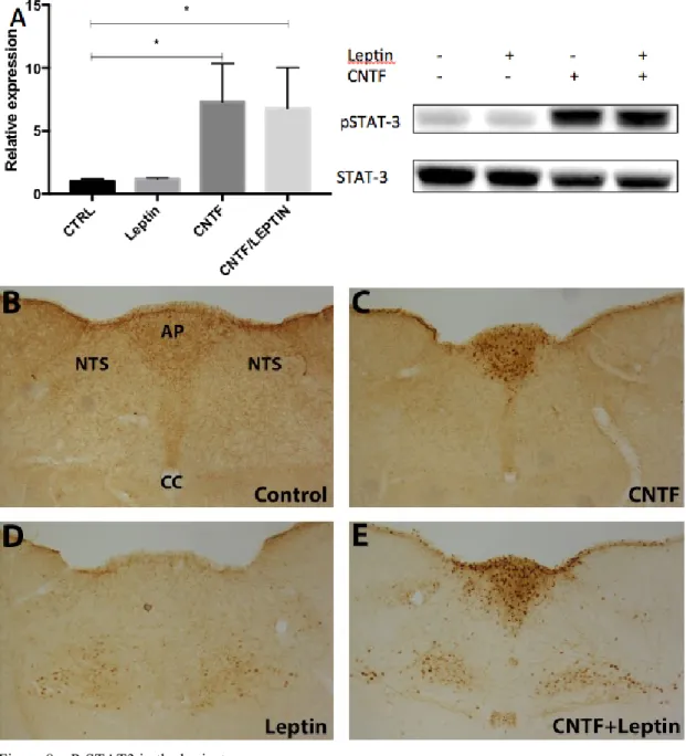
CNTF co-administration with leptin promotes P-STAT3 activation in the nucleus of the solitary tract
To analyze if activation of ERK signaling in the glial cells of the area postrema was able to influence leptin delivery to the nucleus of the solitary tract, we analyzed the expression and distribution of P-STAT3 in the brainstem of control mice, mice treated with leptin, mice treated with CNTF and mice co-treated with leptin and CNTF. Western blotting analysis, showed that leptin induced a small non-significant increase of STAT3 phosphorylation in the whole brainstem; the CNTF treatment strongly induced STAT3 activation and co-administration of leptin and CNTF induced a phosphorylation of STAT3 to CNTF alone (Fig, 8A).
Immunohistochemical assessment of P-STAT3 activity confirmed that in the control mice only few neuronal cells of the NTS were positive (Fig. 8B). Leptin treatment caused a wide activation in several neuronal cells in the NTS (Fig. 8C), and CNTF treatment strongly induced P-STAT3 activity in the AP (Fig. 8D) in nestin- and vimentin-positive glial cells, as previously described (data not shown) [146]. Analysis of corresponding sections from leptin-treated and leptin plus CNTF treated mice (Fig. 8E) revealed that in the NTS, co-treatment with leptin and CNTF was able to significantly increase the activation of STAT3 signaling in this area targeted by leptin.
Figure 8 – P-STAT3 in the brainstem.
No differences were found in P-STAT3 expression between control and leptin treated mice, CNTF treatment and co-treatment with leptin and CNTF significantly increased P-STAT3 expression over control in western blotting (A). Immunohistochemistry showed that in control mice no P-STAT3 staining was present. In CNTF treated mice, P-STAT3 positive staining was found in the AP (C). In leptin treated mice, weak P-STAT3 staining was present in the NTS and DMV(D) Co-treatment with leptin and CNTF showed a strong P-STAT3 staining in the AP and the DMV and some staining in the NTS (E).
db/db mice are insensitive to the leptin sensitizing effect of CNTF
To ascertain whether the increased phosphorylation of STAT3 observed in the tuberal hypothalamic neurons and neurons of the nucleus of the solitary tract was indeed dependent on leptin signaling, we treated leptin receptor lacking obese db/db mice with CNTF or with CNTF plus leptin. By western blotting, coadministration of leptin did not result in any significant increase of STAT3 phosphorylation in the
hypothalamus (Fig. 9A) nor in the brainstem (Fig. 9B). By immunohistochemistry analysis of P-STAT3 expression in db/db mice treated with CNTF (Fig. 9C) or treated with CNTF and leptin (Fig. 9D), only cells in the median eminence exhibited specific staining, whereas tuberal hypothalamic nuclei were not stained. In the area postrema of db/db mice treated with CNTF (Fig. 9E) or treated with CNTF and leptin (Fig. 9F) P-STAT3 staining was only evident in the AP and not in the NTS or DMV. Collectively, these data showed that the increased STAT3 phosphorylation seen in CNTF plus leptin treated mice is dependent on leptin action on neuronal hypothalamic and brainstem neurons respectively through the long form of the leptin receptor.
Figure 9 – P-STAT3 in the brainstem and hypothalamus of db/db mice.
No differences were found in P-STAT3 expression between CNTF and co-treatment with leptin and CNTF in the hypothalamus (A) or brainstem (B) via western blotting. Immunohistochemistry showed that in CNTF treated (C and E) and co-treated mice (D and F) P-STAT3 staining was only present in the ME and the ependymal of the third ventricle of the hypothalamus and in the AP of the brainstem.
DISCUSSION
Obesity and its related disorders such as type 2 diabetes mellitus, coronary heart disease, some types of cancer and other serious health problems can be due to an impaired metabolic flexibility, i.e. an inability to match food intake with energy expenditure [1]. Food intake is influenced at different levels, at the peripheral level signals are generated, which are conveyed to the central nervous system, through specific transporters in the blood brain barrier (BBB) and/or through sensory circumventricular organs. The importance of the transport of signals through the sensory CVO’s in maintaining metabolic homeostasis is exemplified by leptin, the adipose tissue derived satiety signal that conveys information about body lipid stores. The inability of leptin to reduce food intake and increase energy expenditure is known as leptin resistance and is an almost universal aspect of obesity in humans and mice [164]. Ineffective diffusion of leptin in the hypothalamic arcuate nucleus seems to be the limiting step in the regulation of its central effects [165, 166]. Circulating factors such as leptin enter the brain through the median eminence of the hypothalamus. β-2 tanycytes in the ME, lining the third ventricle, are able to take up leptin from the blood and mediate the entrance of leptin into the CSF [78]. Once in the CSF, leptin and possibly other circulating signals can enter the ARC via β-1 tanycytes, which differ from β-2 tanycytes in polarity and barrier properties [16].
The ability of CNTF to overcome leptin resistance in mouse models of obesity and in obese patients highlights its role as a potential anti-obesity drug. Further insights into its anorectic effects may reveal new mechanisms, pathways and brain circuits that are involved in metabolic homeostasis. The satiating effects of CNTF were first attributed to its ability to act on NPY- and POMC-containing neurons of the ARC, activating Jak-STAT3 signaling [144, 145]. Later, its ability to induce neurogenesis in hypothalamic regions of adult mice involved in energy balance regulation was shown to make the animals more sensitive to leptin [154]. Interestingly, mice with a conditional knock out of the CNTFRα in leptin receptor containing neurons did not show changes in the anorectic response to CNTF [153]. This study suggests that CNTF might act on different neuronal populations within the hypothalamus than leptin to influence feeding behavior. They found a significant decrease in P-STAT3 reactive hypothalamic neurons, indicating that leptin responsive neurons in the hypothalamus are not necessary for the effect of CNTF on food intake and suggesting
![Figure 1 - Hypothalamic nuclei involved in energy balance. 3V, third ventricle; ARC, arcuate nucleus; DMH, dorsomedial hypothalamic nucleus; LH, lateral hypothalamus; PVN, paraventricular nucleus [13]](https://thumb-eu.123doks.com/thumbv2/123dokorg/2967820.27057/10.892.142.738.361.800/hypothalamic-involved-ventricle-nucleus-dorsomedial-hypothalamic-hypothalamus-paraventricular.webp)
![Figure 2 - First order NPY/AgRP and POMC/CART neurons in the ARC, regulated by blood borne molecules project to other brain regions, mainly the hypothalamus and brainstem [38]](https://thumb-eu.123doks.com/thumbv2/123dokorg/2967820.27057/13.892.139.750.104.561/figure-neurons-regulated-molecules-project-regions-hypothalamus-brainstem.webp)
![Figure 3 - Various combinations of receptor subunits are the target of different members of the neuropoietic IL-6 cytokine family and lead to activation of Jak-STAT and MAPK pathways [125]](https://thumb-eu.123doks.com/thumbv2/123dokorg/2967820.27057/20.892.131.720.473.774/various-combinations-receptor-subunits-different-neuropoietic-cytokine-activation.webp)