A C-terminally truncated form of
β-catenin
acts as a novel regulator of Wnt/β-catenin
signaling in planarians
Hanxia Su1☯, Miquel Sureda-Gomez2☯, Neus Rabaneda-Lombarte2, Maria Gelabert2, Jianlei Xie1, Wei Wu1, Teresa Adell2*
1 MOE Key Laboratory of Protein Science, School of Life Sciences, Tsinghua University, Beijing, China, 2 Departament de Genètica, Microbiologia i Estadı´stica,Facultat de Biologia, Universitat de Barcelona and Institut de Biomedicina de la Universitat de Barcelona (IBUB), Universitat de Barcelona, Barcelona, Catalunya, Spain
☯These authors contributed equally to this work. *[email protected]
Abstract
β-Catenin, the core element of the Wnt/β-catenin pathway, is a multifunctional and evolu-tionarily conserved protein which performs essential roles in a variety of developmental and homeostatic processes. Despite its crucial roles, the mechanisms that control its context-specific functions in time and space remain largely unknown. The Wnt/β-catenin pathway has been extensively studied in planarians, flatworms with the ability to regenerate and remodel the whole body, providing a ‘whole animal’ developmental framework to approach this question. Here we identify a C-terminally truncatedβ-catenin (β-catenin4), generated by
gene duplication, that is required for planarian photoreceptor cell specification. Our results indicate that the role ofβ-catenin4 is to modulate the activity ofβ-catenin1, the planarianβ -catenin involved in Wnt signal transduction in the nucleus, mediated by the transcription fac-tor TCF-2. This inhibifac-tory form ofβ-catenin, expressed in specific cell types, would provide a novel mechanism to modulate nuclearβ-catenin signaling levels. Genomic searches and in
vitro analysis suggest that the existence of a C-terminally truncated form ofβ-catenin could be an evolutionarily conserved mechanism to achieve a fine-tuned regulation of Wnt/β -cate-nin signaling in specific cellular contexts.
Author summary
The Wnt signaling pathway is essential for proper intercellular communication in every developmental process since it controls basic cellular events as cell fate or proliferation. The key element of the Wnt signaling isβ-catenin, which controls the transcription of multiple genes in the Wnt receiving cell. A main level of regulation of the Wnt/β-catenin signaling occurs in the cytoplasm, whereβ-catenin protein levels depend on the activity of theβ-catenin destruction complex. However, once it reaches the nucleus,β-catenin tran-scriptional activity requires a fine-tuned regulation to enable the multiple context-specific responses that it performs. These nuclear mechanisms that regulate the Wnt/β-catenin
a1111111111 a1111111111 a1111111111 a1111111111 a1111111111 OPEN ACCESS
Citation: Su H, Sureda-Gomez M, Rabaneda-Lombarte N, Gelabert M, Xie J, Wu W, et al. (2017) A C-terminally truncated form ofβ-catenin acts as a novel regulator of Wnt/β-catenin signaling in planarians. PLoS Genet 13(10): e1007030.https:// doi.org/10.1371/journal.pgen.1007030
Editor: Ken M. Cadigan, University of Michigan, UNITED STATES
Received: May 9, 2017 Accepted: September 17, 2017 Published: October 4, 2017
Copyright:© 2017 Su et al. This is an open access article distributed under the terms of theCreative Commons Attribution License, which permits unrestricted use, distribution, and reproduction in any medium, provided the original author and source are credited.
Data Availability Statement: The sequence of 5 genes are available from NCBI: Smed-βcatenin3, KY196224; Smed-βcatenin-4, KY196225; TCF-1, KY196226; TCF-2, KY196227; Smed-TCF-3, KY196228. All other data are within the paper and its Supporting Information files.
Funding: This work was supported by grants BFU2008-01544 and BFU2014-56055-P (Ministerio de Educacio´n y Ciencia, Spain) to TA, grant 2009SGR1018 (Agència de Gestio´ d’Ajuts Universitaris i de Recerca, AGAUR, from La
signaling remain poorly understood. Here we report the existence of C-terminal truncated forms ofβ-catenin in planarians (β-cat3 and 4), which, in vitro, do not show transactiva-tion activity and compete with the canonical planarianβ-catenin (β-cat1), thus acting as competitor inhibitors. Functional analyses in planarians indicate thatβ-cat4 acts as a neg-ative regulator ofβ-cat1 during planarian eye photoreceptor specification. We provide evidence to suggest that this novel mechanism for the regulation of nuclearβ-catenin activity could be conserved across animal evolution.
Introduction
The Wnt/β-catenin pathway is an evolutionarily conserved intercellular signaling pathway with essential roles in virtually every developmental process [1–3] and links to a wide range of human diseases [3–7]. Given its multiple, context-dependent roles, the pathway must be exten-sively regulated. A key element of this pathway isβ-catenin, a bi-functional protein first dis-covered as a component of adherens junctions [8,9].β-catenin transduces the Wnt signal to the nucleus [5,10] and is primarily regulated at the level of nuclearization. Binding of Wnts to their receptors (Frizzleds and LRP5/6) uncouples theβ-catenin destruction complex (mainly composed of APC [adenomatous polyposis coli], Axin, CK-1 and GSK-3) and promotes β-catenin stabilization and its nuclear translocation [5,11–14]. Inhibition of the ligand-receptor interaction through secreted inhibitory molecules (WIF, sFRP, DKK) also represents a com-mon level of Wnt/β-catenin signal regulation [15–18]. Sinceβ-catenin does not have a DNA binding domain, once it reaches the nucleus it must interact with a member of the DNA-bind-ing T cell factor/lymphoid enhancer factor (TCF/LEF) family to regulate its downstream tar-gets [19,20]. Severalβ-catenin/TCF partners have been identified, which mainly target the C-terminal part ofβ-catenin, and which confer master regulatory properties to β-catenin since they are mainly involved in regulating chromatin structure and RNA polymerase II [21,22]. Thus, the final activity ofβ-catenin relies not only on its nuclearization but also on its ability to bind to TCFs and their nuclear co-factors. Although an increasing number of factors have been reported to modulate the transcriptional activity of theβ-catenin/TCF complex (e.g. ICAT, Groucho and Chibby) [20,22–27], the regulation ofβ-catenin activity once it reaches the nucleus remains poorly understood.
Planarians, flatworms with an almost unlimited ability to regenerate and remodel their tis-sues during their whole life span [28–30], have become a robust model to study the function of Wnt/β-catenin signaling in different developmental contexts [31–39]. Although most organ-isms have a single bi-functionalβ-catenin protein, gene duplication and functional specializa-tion have led to the generaspecializa-tion of twoβ-catenins in planarians: Smed-β-catenin1 (β-cat1) and Smed-β-catenin2 (β-cat2) [40].β-cat1 is the intracellular effector of Wnt/β-catenin signaling but exerts no role in cell adhesion, whereasβ-cat2 is exclusively found in cell-cell junctions [40]. This functional diversification provides an ideal scenario in which to study the Wnt sig-naling properties ofβ-catenin. A functional specialization of β -catenins has also been found in the nematodeC. elegans. However, their specific role in nuclear signaling appears extremely complex and, apparently, divergent [41,42]. Thus, although genetic tools to generate cells that exclusively lack canonical Wnt pathway activity have been reported in mouse [43], planarians represent an excellent scenario in which to study the signaling properties ofβ-catenin in vivo without interference of the cell adhesion properties.
Functional analysis ofβ-cat1 and the main elements of the Wnt/β-catenin signaling path-way demonstrate an essential role for this pathpath-way in the specification of the antero-posterior
Generalitat de Catalunya) to TA, as well as grant 30911130362 to WW from the National Natural Science Foundation of China. The funders had no role in study design, data collection and analysis, decision to publish, or preparation of the manuscript.
Competing interests: The authors have declared that no competing interests exist.
(A-P) axis during planarian regeneration and homeostatic cell turnover [31–34,44].β-cat1 silencing generates a range of anteriorized phenotypes, from “tailless” to “radial-like hyperce-phalized” planarians [32,45]. Recently, novel functions forβ-cat1 have been reported in pla-narian brain and eye regeneration, and in gonad development [34,36,46]. Importantly, analysis ofβ-cat1 protein localization reveals that it is present in the nucleus of posterior cells, according to its role in A-P axial identity specification, and also in the main planarian tissues [45]. Thus, given that it has both activity- and context-dependent effects, nuclearization alone cannot account for its regulation. This makes planarians an excellent model to further under-stand how the transcriptional activity ofβ-catenin might be regulated once it is in the nucleus.
Here we report the existence of two new planarianβ-catenins,β-catenin3 (β-cat3) and β-catenin4 (β-cat4), which have a truncated C-terminal transactivation domain and are expressed primarily in the nervous system.β-cat3 and β-cat4 can bind to TCF but do not activate the Wnt signal in vitro. Functional analysis in planarians indicates thatβ-cat4 acts as a negative regulator of nuclearβ-cat1 in planarian eye photoreceptors probably by competing for binding to TCF-2, a new TCF found in planarian photoreceptor cells. We provide evidence to suggest that this novel mechanism for the regulation of nuclearβ-catenin activity could be conserved across animal evolution.
Results
Smed-β-catenin3 and 4 are two new
β-catenin homologs
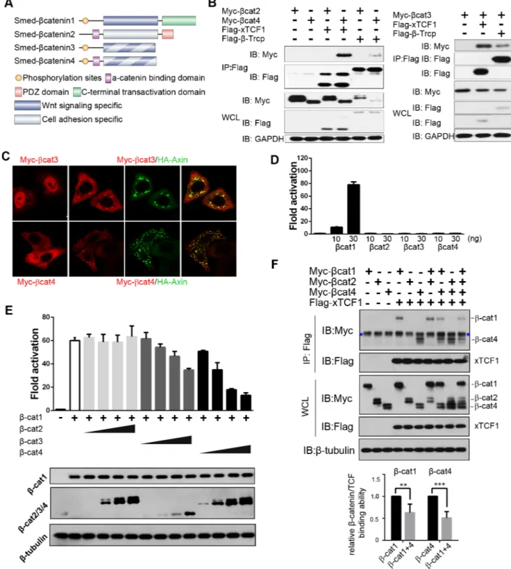
A search forβ-catenin family members in the Schmidtea mediterranea transcriptomes revealed two new genes with protein sequences indicating that they wereβ-catenin homologs (S1 Fig). We named themSmed-β-catenin3 (β-cat3) and Smed-β-catenin4 (β-cat4), since two β-catenin paralogs had been already reported in this species [31–33,40].β-cat3 and 4 proteins conserve the GSK3 phosphorylation sites in the N-terminal region, and their armadillo repeats contain the interacting amino acids for multipleβ-catenin-binding proteins, including APC, Axin, TCF and E-cadherin (Fig 1AandS1 Fig). Theα-catenin binding sites are conserved in β-cat4 but not inβ-cat3 (Fig 1AandS1 Fig). Importantly, the C-terminal transactivation domain, which interacts with crucial chromatin-dependent factors, is lost in bothβ-catenins (Fig 1A andS1 Fig). The finding that the Wnt signaling domains but not the transactivation domain are conserved suggests thatβ-cat3 and 4 could function as dominant-negative forms of β-cat1, which is theβ-catenin homolog involved in signaling to the nucleus in planarians [37,40].
β-cat3 and 4 inhibit
β-cat-dependent Wnt signaling in vitro
Sinceβ-cat3 and 4 contain several conserved domains or residues involved in Wnt signaling and cell adhesion, we further tested the interaction ofβ-cat3 and 4 with the main components involved in these two processes in mammalian cell lines. Co-immunoprecipitation experi-ments indicated thatβ-cat4 strongly interacts with the cell-cell adhesion elements E-cadherin andα-catenin (S2A Fig), whereasβ-cat3 showed interaction with E-cadherin but not α-catenin (S2B Fig). This observation is consistent with the conservation of their functional protein domains (Fig 1AandS1 Fig). In this experiment,β-cat1 was used as a control, since it retains the essential residues for the interaction with E-cadherin, but not the conservedα-catenin binding domain [40]. Interestingly, bothβ-cat3 and 4 were able to interact with the elements involved in Wnt signaling,β-Trcp and TCF (Fig 1B). In this experiment, planarianβ-cat2 was used as a control and, according to its reported role in cell adhesion but not in the Wnt cascade [40], it showed no binding toβ-Trcp or TCF. Moreover, immunofluorescence assays revealed co-localization ofβ-cat3 and 4 with Axin, the core element of the β-catenin destruction com-plex, which is also consistent with their protein sequence analysis (Fig 1C).Thus, these results
Fig 1.β-cat3 and 4 inhibitβ-cat1 dependent Wnt signaling. (A) Schematic of the fourβ-catenin homologs in S. mediterranea.β-cat1 and β-cat2 are structurally segregated.β-cat1 conserves the N-terminal GSK3 phosphorylation sites, the binding surface for Wnt signaling components in the central armadillo repeats, and the C-terminal transactivation domain.β-cat2 conserves the N-terminalα-catenin binding motif, the interacting platform for the cadherin complex in the central armadillo repeats but not for Wnt signaling elements, and the C-terminal PDZ domain. The newly identifiedβ-cat3 and 4 conserve the GSK3 phosphorylation sites, the binding surface for Wnt signaling components and a partial conservation of the cell adhesion elements, whereas they have lost the C-terminal transactivation domain. (B) HEK293T cells were transfected with the indicated plasmids, and lysates were immunoprecipitated (IP) with FLAG-M2 beads. Western blotting (IB) with anti-Myc revealed co-IP ofβ-cat3 and 4 following FLAG-M2 immunoprecipitation, indicating that both of them interact with TCF andβ-Trcp.β-cat2 was
indicate thatβ-cat3 and 4 are under the control of the β-catenin destruction complex, and have the potential to bind to their nuclear co-factor TCF.
Considering the loss of their C-terminal transactivation domain, our results further support the hypothesis thatβ-cat3 and 4 could act as dominant negative forms of β-cat1. To test this hypothesis, we used the Super-TOPflash reporter system in HEK293T cells [47] to analyze the potential ofβ-cat3 and 4 to activate Wnt/β-catenin signaling. Whereas β-cat1 activated the reporter significantly, consistent with its reported role in Wnt signal transduction [40],β-cat3 and 4 had no effect on the reporter, even after increasing the dosages (Fig 1D). Consistent with its specific role in cell adhesion [40], theβ-cat2 paralog was also not able to activate the Super-TOPflash reporter. Importantly, whenβ-cat1 was co-transfected together with β-cat3 or 4, the levels of reporter activity decreased in a dose-dependent manner (Fig 1E). The same result was obtained when analyzing the axial induction capability of planarianβ-catenins in Xenopus embryos (S2C Fig).
To further test whetherβ-cat3/4 could act as competitors of β-cat1 for the binding to TCF, we performed a binding competition assay. Following co-transfection of HEK293T cells with β-cat1, β-cat4 and TCF, quantitative analysis indicated that β-cat1 and β-cat4 disrupt each oth-er’s binding to TCF (Fig 1F). The specificity of this competition is supported by the finding that co-transfection ofβ-cat2 does not alter the binding of β-cat1 or 4 to TCF (Fig 1F).
These results demonstrate thatβ-cat3 and 4 do not show any transactivation properties and that their expression inhibitsβ-cat1 activity ‘in vitro’ or in a heterologous system. Further-more, bothβ-cat4 and β-cat1 are able to bind to TCF. These results are consistent with a role ofβ-cat3 and 4 as competitive inhibitors of β-cat1.
β
-cat4 is required for photoreceptor specification during planarian
regeneration and homeostasis
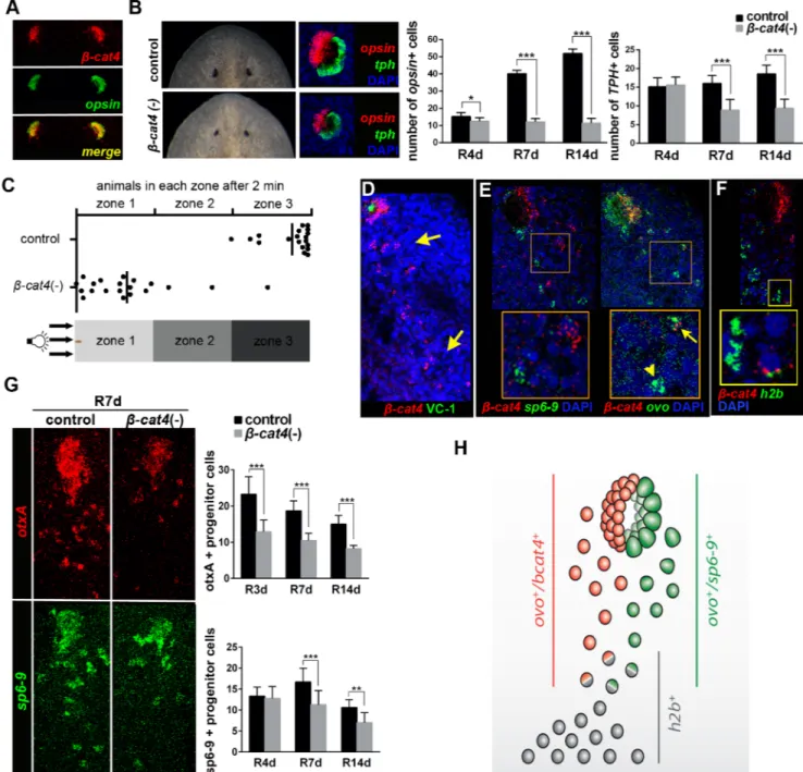
Sinceβ-cat3 and 4 act through inhibition of β-cat1, they could be essential in any of the pro-cesses in whichβ-cat1 is involved, such as posterior identity specification or organogenesis [31–34,36,45,46,48]. Whole-mountin situ hybridization (WISH) in intact and regenerating animals showed thatβ-cat3 and 4 are expressed in the parenchyma and in the central nervous system of intact animals, as well as in the new regenerating brain (S3A and S3B Fig). Remark-ably,β-cat4 is also highly expressed in the eyes (S3A and S3B Fig), specifically in photorecep-tors, since fluorescentin situ hybridization (FISH) analysis demonstrated that it is exclusively expressed inopsin+ cells (Fig 2A) [49]. No apparent defects were observed in regenerating β-cat3 (RNAi) planarians (S3C Fig). In contrast, compared to control animals,β-cat4 (RNAi) animals regenerated smaller eyes, with smaller pigmented spots and missing the periglobular
analyzed as a negative control. (C) Localization ofβ-cat3 and 4 alone or co-expressed with Axin in transfected HeLa cells. In HeLa cells,β-cat3, which localized mainly in the nucleus alone, was recruited to the cytoplasm when co-transfected with Axin.β-cat4, which was more widely dispersed in the cytoplasm, was recruited by Axin and had a punctate distribution. Scale bar = 20μm. (D) TOPflash reporter assay following co-transfection of HEK293T cells withβ-cat1,β-cat2,β-cat3,β-cat4 and reporter plasmids.10 or 30 ng were transfected of eachβ-catenin. Just β-cat1 but notβ-cat3 or 4 activated Wnt/β-catenin signaling.β-cat2 does not show Wnt reporter activity, as reported [40]. (E) TOPflash reporter assay following co-transfection of HEK293T cells withβ-cat1 (20 ng),β-cat2 (10, 20, 30 or 60 ng),β-cat3 (10, 20, 30 or 60 ng),β-cat4 (10, 20, 30 or 60 ng) and reporter plasmids. The co-transfection ofβ-cat3 or 4 inhibitedβ-cat1 reporter activation in a dosage dependent manner. Immunoblot analysis shows the protein expression level ofβ-cat1/2/3/4 in each line. (F) HEK293T cells were transfected with the indicated plasmids, and lysates were immunoprecipitated (IP) with FLAG-M2 beads. Western blot was performed with anti-Myc and anti-Flag antibodies. Upon co-transfection ofβ-cat1 andβ-cat4, the amount of immunoprecipitatedβ-catenins by TCF was less than that during sole transfection of either, supporting that they compete with each other for TCF binding. The relative protein levels of precipitatedβ-catenins by TCF were quantified and normalized against totalβ-catenins in WCL (whole cell lysates). Quantitative results of relative binding ofβ-cat1 orβ-cat4 alone with respect toβ-cat1+β-cat4 derived from four independent experiments.β-cat1, 0.63±0.19;β-cat4, 0.51±0.14 (SD; n = 4).**p<0.05, ***p<0.001 (t test). Relative binding ofβ-cat1 orβ-cat4 alone with respect toβ-cat1+β-cat2 orβ-cat4+β-cat2, respectively, was measured as a negative control.β-cat1, 1.11;β-cat4, 0.91 (n = 1). Blue asterisk indicates non-specific bands.
Fig 2. Smed-β-cat4 is required for planarian photoreceptor specification. (A) Double FISH assay withβ-cat4 (red) and opsin (green), a
marker of photoreceptor cells, in intact animals, showing their co-localization. (B) Left, live and FISH [opsin (red) and tph (green)] images of control andβ-cat4 (RNAi) planarian eyes at 12 days of regeneration. Right, quantification of the number of photoreceptor (opsin+) and pigment (tph+) cells
per eye at 4, 7 and 14 days of regeneration in control andβ-cat4 (RNAi) planarians. opsin+ cells in control R4d, 15.25±2.19 (SD; n = 8 eyes);β -cat4 (RNAi) R4d,12.5±2.07 (SD; n = 8 eyes); control R7d,40.11±2.03 (SD; n = 9 eyes);β-cat4 (RNAi) R7d,12.08±1.98 (SD; n = 12 eyes); control R14d,51.92±2.50 (SD; n = 12 eyes);β-cat4 (RNAi) R14d,11.38±2.83 (SD; n = 8 eyes). tph+ cells in control R4d, 15.13±2.36 (SD, n = 8 eyes);β -cat4 (RNAi) R4d, 15.63±2.07 (SD; n = 8 eyes); control R7d, 16.00±2.12 (SD; n = 9 eyes);β-cat4 (RNAi) R7d, 8.92±2.81 (SD; n = 12 eyes); control R14d, 18.58±2.27 (SD; n = 12 eyes);β-cat4 (RNAi) R14d, 9.38±2.45 (SD; n = 8 eyes).*p<0.05,***p<0.001 (t test). (C) Phototaxis assay ofβ-cat4
(RNAi) animals. Graphical representation of the percentage of control andβ-cat4 (RNAi) planarians found in the different regions in 2 minutes
(n = 20). The schematic of the container indicates the clearest and the darkest zone. (D)β-cat4 (red) FISH followed by immunohistochemistry with
VC-1 (anti-arrestin) antibody (green), to label the visual axons, shows the expression ofβ-cat4 in a trail of isolated cells posterior to the eyes
(yellow arrows) in addition to expression in the photoreceptors in the eyes. Animals were analyzed at 4 days of regeneration. (E) Double FISH ofβ -cat4 (red) and ovo (green), a pan-eye cell marker, or sp6-9 (green), a pigment progenitor cell marker, shows that among all ovo+ cells some of
them co-express withβ-cat4 (ovo+/β-cat4+, yellow arrow) and some of them do not (ovo+/β-cat4-, yellow arrowhead).β-cat4 was never
-unpigmented epidermis, which corresponds to the photoreceptor area (Fig 2B). Importantly, posterior identity specification, which is disrupted inβ-cat1 (RNAi) animals [31–33], was not affected afterβ-cat4 (RNAi) (S3C and S3D Fig). The efficiency and specificity of the RNAi inhibition was assessed by qPCR, showing thatβ-cat4 RNAi animals show highly reduced lev-els ofβ-cat4 but not of β-cat1, 2 and 3 mRNA (S3E Fig). Thus, we focused on the study of β-cat4 function specifically in the eye.
Planarian eyes are simple structures comprising two main, well-characterized cell types: photoreceptors and pigment cells [50,51]. The number of each cell type was quantified during regeneration ofβ-cat4 (RNAi) planarians by analyzing the expression of opsin and tph, specific markers of photoreceptor and pigment cells, respectively [52] (Fig 2BandS4 Fig). Remarkably, RNAi ofβ-cat4 resulted in reduced photoreceptor cells early in regeneration (R4d), whereas pigment cells did not show significant differences at this stage (Fig 2BandS4 Fig). As regener-ation progressed,β-cat4 (RNAi) animals showed a significantly reduced number of pigment cells compared to control (Fig 2BandS4 Fig), possibly due to a non-autonomous effect [53]. Consistent with the effects on photoreceptor cells,β-cat4 (RNAi) animals did not show the proper negative phototaxis behavior (Fig 2C). When exposed to a light gradient, all control animals moved away from the light and remained in the darkest zone (zone 3). Conversely, althoughβ-cat4 (RNAi) organisms seemed to move normally, most of them remained in the clearest zone and did not reach the darkest zone in the same time period (Fig 2CandS1and S2Movies). These data show thatβ-cat4 silencing causes a reduction of photoreceptor cells, followed by a reduction of pigment cells, and influences their normal behavioral responses to light, suggesting its role in photoreceptor specification.
Since planarians continuously remodel their tissues [28], we analyzed whetherβ-cat4 is also required for eye cell maintenance during normal planarian homeostasis. Injection ofβ-cat4 dsRNA over a period of 5 weeks produced a decrease in eye size and in the photoreceptor area (S5A Fig). Quantification of photoreceptor and pigment cells throughopsin and tph FISH over the 5 weeks of the experiment revealed thatβ-cat4 (RNAi) animals always have fewer photore-ceptor cells (S5A Fig). The gradual reduction of photoreceptor cell number observed in control animals (S5A Fig) is due to shrink age of the animals, which remained starved over the 5-week period. According to the phenotype,β-cat4 (RNAi) animals showed a defective negative pho-totaxis response that got worse as the experiment progressed (S5B Fig). Thus, these data dem-onstrate thatβ-cat4 is required for photoreceptor maintenance during homeostasis.
Planarian photoreceptor and pigment cells differentiate from progenitor cells that are located as a trail of cells extending caudally from the eye and express the pan-eye markerovo [54]. A small number of eye progenitor cells co-expressovo with stem-cell specific markers (h2b) and correspond to the specialized eye stem-cells [54]. Eye stem cells acquire the expres-sion of specific determinants that direct their final fate to photoreceptor or pigment cells [52, 55]. In order to understand the mechanism by whichcat4 influences eye regeneration, β-cat4 expression was further studied with FISH. Besides its expression in eye photoreceptor
cat4 (red) and h2b (green) shows thatβ-cat4 is expressed in stem cells. Animals were analyzed at 7 days of regeneration. (G) FISH of otxA (red), a
photoreceptor progenitor cells marker, and sp6-9 (green), in control andβ-cat4 (RNAi) animals, at 7 days of regeneration. The quantification of otxA+ and sp6-9+ cells in the trail posterior to the eyes at different regeneration time points is shown. otxA+ cells in control R3d, 23.25±4.81 (SD; n = 13 eyes;β-cat4 (RNAi) R3d, 12.89±3.26 (SD; n = 9 eyes); control R7d, 18.69±2.72 (SD; n = 13 eyes);β-cat4 (RNAi) R7d, 10.50±1.96 (SD; n = 10 eyes); control R14d, 15.00±2.39 (SD; n = 8 eyes);β-cat4 (RNAi) R14d, 8.20±0.92 (SD; n = 10 eyes). sp6-9+ cells in control R4d, 13.33±2.16 (SD; n = 6 eyes);β-cat4 (RNAi) R4d, 12.75±2.87 (SD; n = 8 eyes); control R7d, 16.71±3.25 (SD; n = 7 eyes);β-cat4 (RNAi) R7d, 11.29±3.35 (SD; n = 7 eyes); control R14d, 10.63±1.85 (SD; n = 8 eyes);β-cat4 (RNAi) R14d, 7.00±2.40 (SD; n = 9 eyes).**p<0.01,***p<0.001 (t test). (H) Schematic ofβ-cat4 expression in the eyes. Stem-cells (h2b+) acquire expression of ovo to become eye progenitors. Eye progenitors became
photoreceptor or pigment cells when acquiring the expression ofβ-cat4 or sp6-9, respectively. In all images anterior is to the top. Scale bars,
50μm (A-F), and 10μm (E and F insets).
cells,β-cat4 was found to be expressed in the trail of eye precursors posterior to the eye both in intact animals and during regeneration (Fig 2DandS5C Fig). Double FISH analysis ofβ-cat4 withovo and sp6-9, a pigment cell determinant, revealed that β-cat4 is exclusively expressed in photoreceptor but not pigment-cell progenitors, since it was always co-expressed with the eye markerovo but never with the pigment-specific marker sp6-9 (Fig 2EandS5D Fig). In addi-tion, a few isolatedβ-cat4+ cells were also found to be h2b+ (Fig 2F), indicating thatβ-cat4 is already expressed in the eye stem cell.
To test whetherβ-cat4 is required for specification of photoreceptor cells from the common eye stem cell precursor, we quantified the number of photoreceptor and pigment-cell progeni-tors in the eye trail ofβ-cat4 (RNAi) animals using the specific markers otxA and sp6-9, which label differentiating photoreceptor and pigment cells, respectively [52,54,55]. As expected, β-cat4 (RNAi) animals had a reduced number of otxA+ cells in the eye from early regeneration stage and a later decrease insp6-9+ cells (S6 Fig). Importantly, the same result was observed when quantifyingotxA+ and sp6-9+ cells in the trail; β-cat4 (RNAi) resulted in failure of pho-toreceptor progenitor-cell specification from the early regeneration stage (R3d), whereas pig-mented cells appeared reduced at a later stage (R7d) (Fig 2GandS6 Fig). Thus,β-cat4 is required for photoreceptor progenitor-cell specification from the common eye stem cell (Fig 2H).
β
-cat4 specifies photoreceptor cells through
β
-cat1 inhibition
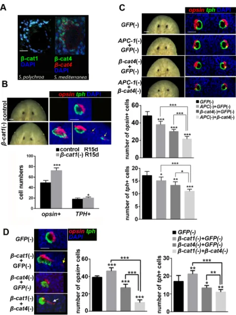
Considering thatβ-cat4 inhibits TCF-mediated β-cat1 activity in cell cultures and that it has lost the C-terminal transactivation domain,β-cat4 could inhibit Wnt signaling in photorecep-tors by competing withβ-cat1 for TCF binding in the nucleus. To test whether this molecular mechanism could be functional in planarians, we first analyzedβ-cat1 and β-cat4 expression in the planarian eye field. FISH forβ-cat1 followed by immunostaining of VC-1, which labels the rhabdomeres of photoreceptor cells [52,56,57], showed thatβ-cat1 was expressed in regener-ating photoreceptor cells (S7A Fig). Moreover, double FISH for bothβ-cat1 and β-cat4 mRNA revealed that both are found in photoreceptor cells (S7A Fig). Using a specific antibody gener-ated in this study (S7B Fig) we could demonstrate thatβ-cat4 protein is localized in the nucleus of photoreceptors (Fig 3A). Immunostaining with aβ-cat1-specific antibody [45], revealed thatβ-cat1 is also localized in the nucleus of photoreceptor cells in S. polychroa, the sister spe-cies ofS. mediterranea (Fig 3A). Thus, our results show that bothβ-cat1 and β-cat4 are local-ized in the nucleus of photoreceptor cells, which is consistent with their nuclear interaction.
Next, we performed RNAi experiments to analyze the functional relationship between the twoβ-catenins. Planarians were decapitated after β-cat1 dsRNA injection and allowed to regenerate. The efficiency of the inhibition was tested by qPCR (S7C Fig). Newly formed heads showed “slanted eyes” with a very thin and elongated periglobular unpigmented epidermis and pigmented cup (Fig 3B). The observed phenotype is very similar to a previously reported β-cat1 (RNAi) [38]. FISH with eye-specific markers confirmed thatβ-cat1 (RNAi) led to a dis-ordered eye structure, in which photoreceptor and pigment cells formed larger eyes and in which ectopic eye cells appeared (Fig 3B,S7D FigandS3andS4Movies). Quantification of opsin+ and tph+ cells present in the eye structure showed an increase with respect to control animals (Fig 3BandS7D Fig). This result is the opposite of that found inβ-cat4 (RNAi) planar-ians, which had a decrease in the number of photoreceptor cells, thus supporting the opposing role of theseβ-catenins.
Since techniques for overexpression are currently unavailable in planarians, we took an indirect approach to up-regulateβ-cat1 through silencing APC-1, the APC homolog in planari-ans [31,34].APC-1 (RNAi) leads to β-cat1 up-regulation and nuclear accumulation [45],
Fig 3.β-cat4 specifies photoreceptor cells throughβ-cat1 inhibition. (A)β-cat1 protein (green) localizes to the nucleus of photoreceptors (image corresponds to intact S. polychroa, syster species of S. mediterranea). β-cat4 protein (green) andβ-cat4 mRNA (red) colocalize in photoreceptor cells, where the protein is found in the
nucleus (images correspond to intact S. mediterranea) (B) Eye phenotype ofβ-cat1 (RNAi) animals at 15 days
of regeneration. Live images and FISH of opsin (red) and tph (green).β-cat1 (RNAi) animals showed elongated
and disorganized eyes, with the appearance of ectopic photoreceptor cells in 50% of the eyes analyzed (yellow arrows in the right image). The respective quantification of opsin+ and tph+ cells per eye is shown. Ectopic cells were not included in the analysis. opsin+ cells in control R15d, 49.25±4.37 (SD; n = 12 eyes);β-cat1 (RNAi)
R15d, 72.50±5.21 (SD; n = 6 eyes). tph+ cells in control R15d, 18.00±2.26 (SD; n = 12 eyes);β-cat1 (RNAi)
R15d, 20.33±1.86 (SD; n = 6 eyes).*p<0.05,***p<0.001 (t test). (C) Double knockdown of APC-1 (RNAi) and β-cat4 (RNAi) in intact animals. Live images and FISH of opsin (red) and tph (green) after the indicated RNAi
treatments with the respective quantification of opsin+ and tph+ cells per eye. opsin+ cells in GFP (RNAi), 48.17 ±4.80 (SD; n = 6 eyes); APC-1;GFP (RNAi), 38.13±3.76 (SD; n = 8 eyes);β-cat4;GFP (RNAi), 30.17±2.40 (SD; n = 6 eyes); APC-1;β-cat4 (RNAi), 21.50±2.35 (SD; n = 6 eyes). tph+ cells in GFP (RNAi), 17.17±1.60 (SD; n = 6 eyes); APC-1;GFP (RNAi), 15.13±1.46 (SD; n = 8 eyes);β-cat4;GFP (RNAi),13.33±1.37 (SD; n = 6 eyes);
APC-1;β-cat4 (RNAi), 10.83±0.75 (SD; n = 6 eyes).*p<0.05,**p<0.01,***p<0.001 (t test). APC-1 (RNAi) and β-cat4 (RNAi) caused smaller eyes than control. Notice that double RNAi of APC-1 andβ-cat4 resulted in a
more severe phenotype than each one alone. (D) Double knockdown ofβ-cat1 (RNAi) andβ-cat4 (RNAi). FISH
of opsin (red) and tph (green) after the indicated RNAi treatments in 9 days regenerating animals. The respective quantification of opsin+ and tph+ cells per eye is shown. opsin+ cells in GFP (RNAi), 39±1.7 (SD; n = 6 eyes);β-cat1;GFP (RNAi), 46.25±3.77 (SD; n = 8 eyes);β-cat4;GFP (RNAi), 27.67±4.03 (SD; n = 6 eyes); β-cat1;β-cat4 (RNAi), 10±3.03 (SD; n = 6 eyes). tph+ cells in GFP (RNAi), 17±3.34 (SD; n = 6 eyes);β-cat1;
resulting in the regeneration of a tail at anterior wounds [31,34,45]. In order to analyze the eye field inAPC-1-knockdown planarians, we performed RNAi experiments in intact animals, since it is known thatAPC-1 silencing during two weeks does not lead to tail-head transforma-tion [45]. We injected dsRNA forAPC-1, β-cat4, and APC-1;β-cat4 for 2 weeks. The efficiency of the inhibition was analyzed by qPCR (S7E Fig). As expected,β-cat4 (RNAi) caused smaller eyes with a decrease in both photoreceptor and pigment cell numbers compared to controls (Fig 3C). Importantly,APC-1 (RNAi) generated the same phenotype (smaller eyes with fewer photoreceptor and pigment cells) (Fig 3C). Co-silencingAPC-1 and β-cat4 caused an even more severe decrease in the number of photoreceptor cells (Fig 3C). This last result is consistent with the hypothesis thatβ-cat4 competes with β-cat1 in the nucleus inhibiting its transcriptional activity. However, it should also be considered that, since bothβ-catenins have the potential to be regulated by the destruction complex, inAPC RNAi animals not only β-cat1 but also β-cat4 could be stabilized.
β-cat1 gain of function through APC-1 (RNAi) results in the same phenotype of diminished eye cell numbers asβ-cat4 (RNAi), whereas β-cat1 loss of function causes the opposite effect. Thus,β-cat1, as a key downstream transcriptional co-activator in Wnt signaling, plays a nega-tive role in planarian photoreceptor development.
To further understand the functional relationship ofβ-cat1 and β-cat4 in photoreceptor cells, the phenotype of the double RNAi was analyzed. The efficiency of the inhibition was ana-lyzed by qPCR (S7F Fig). The result shows that co-silencingβ-cat1 and β-cat4 causes extremely disorganized eyes, with abundant delocalized eye cells, which show a reduction in the number of photoreceptor and pigment cells (Fig 3D). This result does not support the hypothesis of β-cat4 directly acting as a dominant-negative form ofβ-cat1, but suggests alternative competition models (seediscussion). Furthermore, the appearance of ectopic eye cells inβ-cat1 RNAi ani-mals, and the severe disorganization of the eyes ofβ-cat1/β-cat4 RNAi planarians also suggest thatβ-cat1 could exert additional autonomous roles, in pigment or neuronal cells, which influ-ence the localization of photoreceptor cells.
Overall, our results indicate that the activity ofβ-cat1 in the eyes is not only controlled by the elements of theβ-catenin destruction complex, like APC-1, but also by β-cat4, which exerts a negative regulatory effect on a process that isβ-cat1 and APC dependent.
Smed-TCF-2 mediates
β
-cat4 and
β
-cat1 activity during photoreceptor
differentiation
To gain further insight into the mechanism through which planarianβ-catenins specify photo-receptor differentiation, we searched for the TCF transcription factor that acts as a target. Although most invertebrate genomes contain a single TCF/LEF ortholog, we identified three TCF orthologs (Smed-TCF-1 to -3) in theS. mediterranea transcriptome database Planmine [58] (S8A Fig). The corresponding homologs were found in five more planarian species in the same database (S8A Fig). The phylogenetic analysis suggests that the duplications found in pla-narians arise from Platyhelmintes and are independent of the vertebrate TCF expansion (S8A Fig). Protein sequence analysis of the threeS. mediterranea TCFs demonstrated that TCF-2 is the onlyS. mediterranea TCF that conserves all functional domains required to bind to
β-GFP (RNAi), 21.12±1.96 (SD; n = 8 eyes);β-cat4;GFP (RNAi),13.33±1.37 (SD; n = 6 eyes);β-cat1;β-cat4
(RNAi), 11±1.4 (SD; n = 6 eyes).*p<0.05,**p<0.01,***p<0.001 (t test).β-cat1 (RNAi) animals show large
eyes with ectopic eye cells (yellow arrow);β-cat4 (RNAi) animals show small eyes; and doubleβ-cat1;β-cat4
(RNAi) animals show smaller and more disorganized eyes than singleβ-cat4 orβ-cat1 (RNAi) (white arrow
indicates a row of delocalized photoreceptor and eye cells). Anterior is to the top. Scale bar = 20μm (A), 50μm (B, C,D).
catenin, Groucho and DNA (Fig 4AandS9 Fig) [59]. TCF-1 has lost theβ-catenin binding domain and TCF-3 does not conserve the Groucho binding sites (S9 Fig). Analysis of their expression pattern in planarians showed thatTCF-1 was expressed specifically in the planarian brain, as previously reported [60] (S8B Fig).TCF-2 and -3 are also mainly expressed in the CNS and, importantly,TCF-2 is found in photoreceptors (Fig 4B), strongly resembling the β-cat4 expression pattern. Thus, TCF-2 is a candidate to function as a β-cat1 and β-β-cat4 target during photoreceptor development.
The eyes ofTCF-2 RNAi animals were analyzed to understand its possible function. The efficiency of the inhibition was analyzed by qPCR (S8C Fig). Analysis ofTCF-2 RNAi Fig 4. TCF-2 mediatesβ-cat1 andβ-cat4 signaling. (A) TCF-2 conserves the characteristic domains ofβ -catenin and DNA binding (HMG domain). (B) Expression of TCF-2 in the CNS and in the photoreceptors
(yellow arrows in the magnification) (C) Live images and FISH of opsin (red) and tph (green) of TCF-2 (RNAi) and GFP (RNAi) planarians at 7 days of regeneration, with the respective quantification of the opsin+ and tph+ cells per eye. opsin+ cells in control R7d, 35.60±4.17 (SD; n = 10 eyes); TCF-2 (RNAi) R7d, 50.8±4.21 (SD; n = 10 eyes). tph+ cells in control R7d, 15.7±1.06 (SD; n = 10 eyes); TCF-2 (RNAi) R7d, 20.70±2.06 (SD; n = 10 eyes).***p<0.001 (t test). Anterior is to the top. (D) Double knockdown assay ofβ-cat4 (RNAi) and TCF-2 (RNAi). Live images and FISH of opsin (red) and tph (green) to show planarian regenerated eyes after
the indicated RNAi treatment. The respective quantification of opsin+ and tph+ cells is shown. opsin+ cells in
GFP (RNAi), 39.70±4.47 (SD; n = 10 eyes);β-cat4;GFP (RNAi), 21.60±4.48 (SD; n = 10 eyes); TCF-2;GFP (RNAi), 64.64±8.68 (SD; n = 10 eyes); APC-1;β-cat4 (RNAi), 63.50±4.32 (SD; n = 12 eyes). tph+ cells in GFP (RNAi), 17.60±2.01 (SD; n = 10 eyes);β-cat4;GFP (RNAi), 12.89±2.15 (SD; n = 10 eyes); TCF-2;GFP (RNAi), 22.00±3.49 (SD; n = 10 eyes); APC-1;β-cat4 (RNAi), 21.58±2.27 (SD; n = 12 eyes).**p<0.01,***p<0.001 (t test). Scale bars, 250μm (B), 50μm (C, D).
regenerating animals revealed that their eyes were bigger than in controls (Fig 4CandS8D Fig). The brain ofTCF-2 RNAi animals appeared normal (S8E Fig), which suggests that the larger eye phenotype is eye specific. Quantification of the different eye populations demon-strated thatTCF-2 silencing results in an increased number of photoreceptor cells (Fig 4Cand S8D Fig). The number of pigment cells also increased (Fig 4CandS8D Fig), probably due to the cellular relationship between the two compartments, as shown earlier inβ-cat4 RNAi pla-narians. TheTCF-2 (RNAi) phenotype in the eyes phenocopies the β-cat1 (RNAi) phenotype with respect to the number of photoreceptor cells and is consistent with the hypothesis that Wnt/β-cat1 signal inhibition is required for correct planarian photoreceptor specification. To note,TCF-2 RNAi animals showed bigger eyes but not the appearance of ectopic eye cells, sug-gesting that this defect is caused in aTCF-2 independent manner.
To analyze whetherβ-cat4 function in photoreceptor specification also depends on TCF-2, we performed a double RNAi assay to inhibitβ-cat4 and TCF-2 simultaneously during planar-ian regeneration. The efficiency of the inhibition was tested by qPCR (S8F Fig). As expected, β-cat4 (RNAi) planarians had smaller eyes with a reduced number of photoreceptor cells, whereasTCF-2 (RNAi) resulted in larger eyes with an increased number of photoreceptor cells. Remarkably, the size of the eyes in doubleβ-cat4 and TCF-2 RNAi animals, and the number of photoreceptor and pigment cells, resembled the phenotype observed withTCF-2 (RNAi) alone (Fig 4D). This observation is consistent with a role for TCF-2 as the transcription factor downstream ofβ-cat4. Above all, the data suggests that β-cat4, which lacks the C-termi-nal transactivation domain, modulates theβ-cat1/TCF-2-mediated signal for the correct differ-entiation of photoreceptor cells (Fig 5).
Plakoglobin and Neural Arm act as
β-catenin and Arm inhibitors,
respectively, in vitro
To determine whether the existence of inhibitoryβ-catenins could be an evolutionary con-served mechanism for the regulation of Wnt signaling, we investigated the existence of C-Fig 5. Proposed Model for Wnt/β-catenin activity modulation in planarian eyes. During planarian eye
regeneration or maintenance,β-cat4 inhibitsβ-cat1/TCF-2 activity in photoreceptor cells, modulating Wnt signaling to an appropriate level to ensure correct differentiation of photoreceptors.
terminally truncatedβ-catenins in other organisms. The existence ofβ-catenin paralogs is not exclusive to planarians. Theβ-catenin family has undergone a vertebrate-specific subphylum dupli-cation (β-catenin and plakoglobin) [61], two nematode-specific phylum duplications (4β-catenins inC. elegans) [41], and multiple species-specific duplications in the Arthropoda phylum [61] (S10 Fig). Phylogenetic analysis of theβ-catenin family members of different Lophotrocozoa species shows the existence of a unique bi-functionalβ-catenin in all of them except for Platyhelmintes (S10 Fig). The analysis suggests thatβ-catenin underwent two phylum-specific duplications in Pla-tyhelmintes to generateβ-cat1, 2 and 3/4 classes, and that in Triclads a third duplication produced theβ-catenin3 and 4 orthologs (S10 Fig). Furthermore, a genus-specific duplication occurred in theβ-cat3 class (S10 Fig). Thus, aβ-cat3/4 ortholog that retains the signaling domains but not the C-terminal transactivation domain (S11 Fig) exists in all Plathyhelminthes.
Taking into account the number ofβ-catenin duplications found across evolution, we hypothesized that the existence of an inhibitoryβ-catenin to regulate β-catenin-dependent Wnt signaling could be a common mechanism throughout evolution as a result of convergent evolution. A protein sequence analysis of differentβ-catenin orthologs found across the animal phyla shows the shortening of the C-terminal end in several cases, for instance in one of the twoβ-catenins found in the sponge A. quensatlantica or the vertebrate β-catenin duplication Plakoglobin (S11 Fig). Although Plakoglobin shows high protein sequence conservation with β-catenin in the central armadillo repeats, and shares the binding domains for α-catenin, Cadherins and TCF, it has very low amino acid sequence conservation in the C-terminal transcriptional transactivation domain (15%) (Fig 6A) [62]. Accordingly, it has been shown that Plakoglobin has limited transactivation ability compared toβ-catenin [63]. Furthermore, although a uniqueβ-catenin is found in the genome of D. melanogaster (Armadillo, Arm), a C-terminally truncatedArm named Neural Armadillo (NArm) has been reported to occur through alternative splicing [64] (Fig 6BandS11 Fig). It is known thatNArm is expressed in the brain from larval stage [64] but no functional studies have been reported. To analyze whether verte-brate Plakoglobin orDrosophila NArm could have an inhibitory function, we designed in vitro Super-TOPflash experiments. Results showed that whileβ-catenin (S37A), a stabilized β-catenin that cannot be captured by the cytoplasmic destruction complex, could highly activate Wnt sig-naling, Plakoglobin activity was very low (Fig 6A). Interestingly, Plakoglobin co-transfection withβ-catenin (S37A) decreased the reporter signal in a dose-dependent manner (Fig 6A). Sim-ilarly, activation of Wnt signaling by NArm was very weak compared to that induced by Arm. Importantly, co-transfection of both plasmids showed that NArm suppresses the Arm induced TCF-reporter activity in dosage dependent manner (Fig 6B).
Our results demonstrate that Plakoglobin and NArm inhibit the Wnt signal activated by β-catenin/Arm through a TCF transcription factor. This result, together with the presence of β-catenin paralogs in several species, indicates that the existence of an inhibitoryβ-catenin could be a conserved mechanism to fine tuneβ-catenin-dependent Wnt signaling. The phylogenetic relationship between theβ-catenin family members and the existence of splice variants indicates that the inhibitory form ofβ-catenin would not have evolved from a common ancestor but appeared during evolution as a product of unrelated events (species- or phylum-specific genome duplications or alternative splicing), thus representing an example of convergent evolution.
Discussion
Here we show a new role for the Wnt/β-catenin pathway in planarian eyes and demonstrate the existence of a novel mechanism to regulate its activity in a context-specific manner. We have identifiedβ-cat4, a C-terminally truncated β-catenin generated by gene duplication within the planarian group, and we demonstrate that it modulatesβ-cat1 activity during eye
photoreceptor cells specification but not during axial patterning. Wnt/β-catenin activity is modulated through a novel mechanism in whichβ-cat4, a truncated form of β-catenin that lacks the transactivation domain, exerts a negative regulatory effect on aβ-cat1/TCF-2 depen-dent process. The existence of this new Wnt/β-catenin activity regulatory mechanism would ensure the appropriate fine-tuning of nuclearβ-catenin/TCF activity in specific cell types. Genome searches andin vitro assays indicate that use of an inhibitory form of β-catenin could be a conserved mechanism to regulateβ-catenin activity in specific contexts.
The inhibitory role of the C-terminally truncated planarian
β-catenin4
represents a novel mechanism for modulation of nuclear
β-catenin activity.
The multiple roles and complexity of the Wnt/β-catenin signaling necessitate regulation at different levels. Extracellularly, secreted proteins interact with Wnts or their receptors (e.g. DKK1, SFRP, WIF1, notum) to inhibit the ligand-receptor interaction [65,66], and in the Fig 6. Plakoglobin and Neural Arm inhibit Wnt/β-catenin activity ‘in vitro’. (A) Upper panel, schematic
representation of Plakoglobin orβ-catenin proteins, showing the protein interacting domains and the degree of homology of the N-terminal, central Arm repeat and C-terminal region between both proteins. Lower panel, activation of TOPflash reporter signal byβ-catenin (S37A) and Plakoglobin in HEK293T cells. Plakoglobin showed limited capacity to activate reporter signal, and inhibitedβ-catenin (S37A) activity in a dose-dependent manner. (B) Upper panel, schematic representation of the exon composition of Arm and Neural Arm (in which exon 6 is skipped). Lower panel, activation of TOPflash reporter signal by Armadillo and Neural Arm in HEK293T cells. Neural Arm showed limited capacity to activate reporter signal and inhibited Arm activity in a dose dependent manner.
cytoplasm, regulation of theβ-catenin destruction complex determines the amount of β-cate-nin that will escape phosphorylation and degradation and reach the nucleus [5]. However, the modulation ofβ-catenin activity once it reaches the nucleus remains poorly understood. We used planarians to approach this question since, althoughβ-cat1 exerts multiple functions, it is primarily localized to the nucleus [31–33,36,44–46,48], suggesting that mechanisms must be available to modulateβ-cat1 nuclear activity.
Here we found two newβ-catenins cat3 and 4) in planarians, after the two β-catenins (β-cat1 and 2) already described [40], which 1) showed a C-terminal truncated transactivation domain, while conserving the TCF binding amino acid residues, and 2) were mainly expressed in the nervous tissues. Those findings lead to hypothesize thatβ-cat3 and 4 could be acting as inhibitors of the canonicalβ-cat1 in the nucleus and in a tissue-specific manner. Indeed, we could demonstrate thatβ-cat4 is required for normal specification of photoreceptors from the common eye stem cell and that its function relies on the inhibition ofβ-cat1 activity. Impor-tantly,β-cat4 has no role in A-P axial polarity, in contrast to β-cat1.
Our RNAi experiments show that inhibition ofβ-cat4 or APC, which in planarians is demon-strated to increaseβ-cat1 activity [31,34,45], produces a decrease in the number of photoreceptor cells, whereas inhibition ofβ-cat1 produces the opposite phenotype, indicating that β-cat4 exerts a negative regulatory effect on aβ-cat1-dependent transcriptional activity. The finding that inhibi-tion ofTCF-2, the TCF factor identified in the present study, leads to an increase in the number of photoreceptor cells, as observed afterβ-cat1 RNAi, and that RNAi inhibition of β-cat4 together withTCF-2 leads to the same phenotype as TCF-2 RNAi alone, indicates that TCF-2 is the down-stream effector of thecat1 and cat4 action. Although we cannot rule out the possibility that β-cat1 andβ-cat4 could interact in the cytoplasm, since β-cat4 conserves the essential domains to interact with cytosolicβ-catenin destruction elements, our results support the regulative interaction betweenβ-cat1 and β-cat4 at nuclear level. Thus, both β-cat1 and β-cat4 are found in the nucleus of photoreceptor cells (althoughβ-cat1 expression analysis was performed in a S. mediterranea sis-ter species due to technical limitations [45]). Furthermore, although we have not directly demon-strated the binding ofβ-cat1 or β-cat4 to TCF-2, we demonstrate that both planarian β-catenins conserve the TCF binding sites and bind to xTCF-1 in co-immunoprecipitation experiments.
Overall, our results in planaria indicate thatβ-cat1 and β-cat4 regulate in an opposite man-ner the transcription of genes required for photoreceptor specification, and that this action depends on the TCF-2 transcription factor. According to the presented data, three main sce-narios could be considered (Fig 7). In the first one,β-cat4 could act as a dominant negative form ofβ-catenin, which is able to bind to TCF-2 but not to activate transcription, due to the missing C-terminal domain (Fig 7A). The RNAi phenotypes ofβ-cat1, β-cat4, and TCF-2, agree with this possibility. However, the finding that the doubleβ-cat1/ β-cat4 inhibition pro-duces a decrease in the number of photoreceptors cannot be directly explained under this sup-posal. In the second scenario,β-cat4 could be able to directly repress transcriptional targets through binding to TCF-2 (Fig 7B). InDrosophila, it has been shown that Arm and TCF can directly repress transcriptional targets and that this repressive activity of Arm does not require the C-terminal domain [67,68]. Thus, the balance betweenβ-cat1/TCF-2 transcriptional acti-vation andβ-cat4/TCF-2 transcriptional repression would determine the amount of photore-ceptor cells. The finding that double inhibition ofcat1 and cat4 does not phenocopy the β-cat1 RNAi phenotype, favor this hypothesis. However, under this scenario the β-β-cat1/β-cat4 RNAi should produce a similar phenotype to the one produced afterTCF-2 inhibition, which is not the observed. A third possibility would be that the proper transcriptional regulation of photoreceptor targets is achieved by the combinatory action of bothβ-cat1/TCF-2 and β-cat4/ TCF-2 complexes when bind to different TCF responsive elements (Fig 7C). In the future, the analysis of specific downstream targets ofβ-cat1, β-cat4 and TCF-2, and the possibility of
performing tissue specific RNAi and rescue experiments would help to clarify the specific mechanism ofβ-cat1 and β-cat4 activity.
Altogether, our data supports the existence in planarians of a novel level of Wnt/β-catenin signaling modulation in the nucleus through the action ofβ-catenins with different transacti-vation capacity. The regulatory interaction between those differentβ-catenins and TCFs would result in the modulation of the transcriptional rates of downstream Wnt target genes and Wnt-responsive activities.
Regulation ofβ-catenin/TCF transcriptional activity is achieved through nuclear factors that regulateβ-catenin binding properties (e.g. Chibby and ICAT) [20,22–27], or regulation of TCF transcriptional and binding properties [20,22–27]. Accordingly, the existence of TCFs that lack theβ-catenin-binding domain and act as endogenous dominant negative forms [59], and also post-translational modifications of TCFs that modulate their activity have been exten-sively reported [69–74]. The C-terminal truncated inhibitoryβ-cat4 found in this study repre-sents a novel mechanism to modulateβ-catenin/TCF activity that also targets to TCF. As reported for the endogenous dominant negative forms of TCFs, in which alternative splicing and alternative promoters leads to the expansion of the isoforms [59], this level of regulation would allow the refinement of Wnt signaling in time and space.
A C-terminal truncated form of
β-catenin could be an evolutionarily
conserved mechanism to achieve the multiple context-specific roles of
Wnt/β-catenin signaling
Here we have found a new planarianβ-catenin (β-cat4) which represents a novel mechanism to fine tune the activity of nuclearβ-catenin in a context-specific manner. Not surprisingly, Fig 7. Possible Models forβ-cat1 andβ-cat4 transcriptional activity in planarians. (A)β-cat4 could act as a dominant negative form of β-catenin, which is able to bind to TCF-2 but not to activate transcription, due to the missing C-terminal domain. (B)β-cat4 could be able to directly repress transcriptional targets through binding to TCF-2. The balance betweenβ-cat1/TCF-2 transcriptional activation andβ-cat4/ TCF-2 transcriptional repression would determine the amount of photoreceptor cells. (C) Bothβ-cat1/TCF-2 andβ-cat4/TCF-2 complexes could bind to different TCF responsive elements in the same promoter. The proper transcriptional regulation of photoreceptor targets is achieved by the combinatory action of both complexes.
this mechanism acts in neural tissues, whereβ-cat3 and 4 are mainly expressed, the complexity of which requires a more sophisticated regulation. Phylogenetic analysis of theβ-catenin fam-ily in platyhelminthes supports the hypothesis that aβ-cat1, β-cat2 and β-cat3/4 ortholog was present in their common ancestor. Although the expression pattern and functional analysis of theβ-cat3/4 ortholog in other platyhelminth species should be done, our data predicts that the β-cat3/4 ortholog would modulate β-cat1 activity in Platyhelminthes.
Importantly, although the presence of a uniqueβ-catenin with dual adhesion and signaling functions has been proposed to be present in the last common animal ancestor [75], genomic duplications in theβ-catenin family are more common than previously thought. In vertebrates, plakoglobin is a genomic duplication of β-catenin that has suffered a functional specialization, since it is predominantly involved in desmosomes [76,77]. However, Plakoglobin shows lim-ited capacity to activate Wnt signaling [63,78] and, importantly, its nuclear accumulation down-regulatesβ-catenin activity in a dose-dependent manner, as demonstrated by our TOP-flash reporter assays (Fig 6A). Thus, Plakoglobin could be acting as a negative regulator of β-catenin in vertebrates. Since Plakoglobin shows much lower transactivation properties than β-catenin, its presence results in a decrease of the transcriptional activation promoted by β-cate-nin. The real existence and the importance of this regulatory mechanism is further supported by recent studies demonstrating that an increase of Plakoglobin nuclear translocation is associ-ated with diseases such as arrhythmogenic right ventricular cardiomyopathy or head neck can-cer and leads to a suppression of theβ-catenin mediated TCF/LEF transcriptional activity [79, 80]. Taking into account that the C-terminal part of Plakoglobin is the one showing less con-servation with respect toβ-catenin (Fig 6A), the inhibitory function could be associated with the lack of conservation of the transactivation domain, as described in the present study for planarianβ-cat4.
In invertebrates, theβ-catenin family has undergone several species- and phylum-specific duplications [81]. The nematodeC. elegans has four β-catenins with different roles in cell adhe-sion and nuclear signaling [42], and several insects have twoβ-catenins, which have also un-dergone partial subfunctionalization between the cell adhesion and the centrosome separation functions [81]. Importantly, although inDrosophila a unique β-catenin is found (Arm), an alternative splicing which occurs mainly in neural cells has been reported, which is known as Neural Armadillo (NArm) [81]. Interestingly, this alternative splicing deletes exon 6 and results in a C-terminal truncated Arm isoform that, according to our TOPflash reporter assay, exerts an inhibitory role of Arm in a dose dependent manner, as described for planarianβ-cat4 towardsβ-cat1. Although the role of NArm as modulator of Arm should be demonstrated ‘in vivo’, it should be stressed that the presence of the alternative splicing isoform deleting exon 6 is conserved across all insect homologs [81], supporting the evolutionarily pressure to main-tain it and thus its biological implications. Overall, the present data supports the hypothesis that modulation of nuclearβ-catenin transcriptional activity through the action of a C-termi-nally truncatedβ-catenin able to bind to TCF could be a conserved mechanism to regulate the Wnt/β-catenin pathway. Interestingly, the C-terminally truncatedβ-catenin of different species did not arise from any homolog of a common ancestor but originated in different animal groups from different mechanisms of gene diversification, as gene duplication or alternative splicing. Thus, the presence of aβ-catenin with limited transactivation activity, which could compete with the canonicalβ-catenin, in different species represents an example of convergent evolution and supports its importance as a regulatory mechanism.
The existence of inhibitoryβ-catenins and its expression in specific cell types provides a new answer to the important question of how the same transcription factor elicits so distinct responses. It is assumed that the different transcriptomic/proteomic context of each cell could result in differential activation ofβ-catenin target genes. However, few tissue-specific
Wnt/β-catenin regulators have been found, leading to the idea that not only Wnt/β-Wnt/β-catenin regulators but the interplay with the other conserved signaling pathways as BMP would be essential for their complex regulation [82]. Our results provide evidences that the existence of inhibitory β-catenins is not only a new mechanism to regulate Wnt signaling but to modulate it in specific cell types, for instances in specific subsets of neuronal types.
Eye development and Wnt/β-catenin signaling
Our results demonstrate thatβ-cat4 is a new essential element for photoreceptor cell specifica-tion in planarians. Planarian eyes are true cerebral eyes and their simplicity makes them an excellent system to study eye development. They are composed by two cell types: rhabdomeric photoreceptor neurons, which express evolutionarily conserved photoreceptor genes such as otxA or opsin [49,55], and pigment cells, which expressTryptophan hydroxylase (tph), an enzyme involved in the production of melanin, the pigment found in planarian eyes. Several studies demonstrate that both cell types arise from a common eye stem cell that express stem cell markers (h2b) and eye determinants as the transcription factors ovo, eya and six1/2 [54,55, 83]. Among themovo is the only factor exclusively found in the eye cell and specifically essen-tial for eye regeneration and maintenance [54]. The expression ofsp6-9 and dlx in the common eye progenitor determines the pigment fate, whileotxA specifies photoreceptors [52,55]. We demonstrate thatβ-cat4 is a new factor that specifies the photoreceptor fate and that it is expressed in the stem cell precursors (h2b+/ovo+), a fact that has not been yet demonstrated forotxA. Inhibition of β-cat4 results in a decrease in number of photoreceptor progenitor and differentiated cells from the earliest time points analyzed (3–4 days of regeneration), while the number of progenitors and differentiated cells of the pigment lineage only decrease several days after. This observation agrees with the already described mutual dependence of both cell populations in planarians [53]. Thus, althoughβ-cat4 is specifically required for photoreceptor determination, according to its expression in the photoreceptor lineage, the reduced number of photoreceptor cells would lead to the subsequent decrease of the pigment lineage. Whether the origin of this mutual dependence is at the level of the common stem cell progenitors, or at the level of the differentiated cells in the optic cup, remains to be studied. Although not much is known about the mutual dependence between photoreceptor and pigment cells in verte-brates, some studies suggest that it also exists [84].
To date, the main focus on the role of the Wnt/β-catenin signaling pathway in planarians has been in its essential role for axial patterning and posterior identity specification [31–33]. Our results highlight the importance of the regulation of the Wnt/β-catenin pathway in a dif-ferent context, namely during planarian photoreceptors regeneration and maintenance. The finding is not surprising, since appropriate Wnt/β-catenin signaling levels are required for ret-ina progenitor differentiation in several animal models [85–90]. For example, during chicken embryo development,β-catenin overexpression in the central neural retina inhibits the differ-entiation of retinal neurons, while loss ofβ-catenin leads to neural retina enlargement [86]. In zebrafish the ectopic expression of the Wnt antagonists DKK1 or SFRP1 causes expansion of the embryonic retina [91,92], whereas activation of the Wnt/β-catenin pathway leads to lack of expression of eye markers [93]. Here we show that in planariansβ-cat1 inhibition results in larger eyes, whileAPC-1 silencing causes a reduction of photoreceptor cell number. The sim-plicity and approachability of planarian eyes has allowed the identification of a new mecha-nism to fine regulate Wnt/β-catenin pathway activity in the eyes. Thus, the truncated β-cat4 that competes with the canonicalβ-cat1 for binding to TCF-2 allows the required fine-tuning of the Wnt/β-catenin pathway in planarian photoreceptor cells. Since the in vitro experiments showed thatβ-cat4 could bind to E-cadherin and α-catenin, a role of β-cat4 in cell adhesion
should be considered. However,β-cat4 protein was only found in the nucleus of photorecep-tors (Fig 3A), andβ-cat4 RNAi eyes appeared properly patterned with no apparent defects in cell adhesion.
The new described mechanism to fine-tune the Wnt/β-catenin signal in specific cell types implies the existence of Wnt elements specifically expressed in photoreceptor and not in pigment cells. The finding that the phenotype ofβ-cat1 RNAi animals, which show ectopic differentiation of eye cells, differs from theTCF-2 RNAi, which show bigger eyes but properly patterned, indi-cates thatβ-cat1 could exert a TCF-2 independent role during eye regeneration. The cause of this defect could be a non-autonomous role ofβ-cat1, which is broadly expressed in planarians [45], or a role in pigment cells. This finding emphasizes the requirement of a cell-specific regulation of the Wnt/β-catenin signal during development. It will be interesting to study whether in other ani-mal species eye-population specific Wnt elements (or isoforms) are found and to investigate if this novel mechanism ofβ-catenin regulation also takes place during eye development.
Conclusions
Our finding that planarianβ-cat4 functions as a modulator of β-cat1 activity in the nucleus by regulating TCF-2 transcriptional activity has two deep implications: 1) considering its specific role in modulatingβ-cat1 activity during planarian photoreceptor specification, and not in other Wnt/β-catenin dependent processes such as axial patterning, it appears as a tissue-spe-cific manner to fine-tune nuclearβ-catenin activity; and 2) the finding of C-terminal trun-cated/non-conservedβ-catenin forms in other species (NArm or Plakoglobin), which inhibit β-catenin activity in TOPflash experiments, suggests that an inhibitory β-catenin could be an evolutionarily conserved mechanism to regulate the Wnt/β-catenin signaling. The identifica-tion of a novel mechanism of Wnt/β-catenin signaling regulaidentifica-tion has important implicaidentifica-tions in view of the complex and essential roles of this pathway in development and diseases. Thus, the present research represents a starting point to design further studies: 1) to demonstrate whether the C-terminal truncated/non-conservedβ-catenins found in Drosophila (NArm) and vertebrates (Plakoglobin) can compete for Arm orβ-catenin binding, respectively, to the TCF co-factor ‘in vivo’ and assess whether this mechanism provides a tissue-specific manner to modulateβ-catenin/Arm once reaches the nucleus; 2) to study the existence and function of possibleβ-catenin isoforms present in other animal species, as a product of gene duplication or alternative splicing, and understand their real contribution to the regulation of the Wnt/β-catenin signal; and 3) to test whether new drugs mimicking the inhibitory action ofβ-cat4 could be used to fine tune the activity of nuclearβ-catenin in human diseases.
Materials and methods
Planarians
The planarians used in this study belong to an asexual clonal strain ofS. mediterranea BCN-10 biotype. The animals were maintained at 20˚C in PAM (1X) water [94]. Animals were fed with organic veal liver and starved for at least a week before all experiments. A sexual strain ofS. polychroa collected from Sant Celoni (Barcelona, Spain) was used inFig 3A.
Gene identification and phylogenetic analysis
β-Catenin sequences from Planarian species were identified in the Planmine database [58]. β-Catenin sequences from Echinococcus multilocularis and Kronborgia cf. amphipodicola were found in the available databases [95,96]. The rest of the sequences were found in the NCBI. β-Catenin protein sequences from different species were aligned using the MAFFT server
(http://mafft.cbrc.jp/alignment/server/). Neighbor joining distance-based analyses were con-ducted using MEGA version 6 [97], and the support given by bootstrap percentiles of 1000 replicates. BioEdit was used to edit the protein alignments.
Luciferase reporter assay
HEK293T cells were cultured in Dulbecco’s modified Eagle’s medium (DMEM) supplemented with 10% fetal calf serum. To obtain pCS2+-6Myc-Smed-β-catenin3 and 4, the full length Smed-β-catenin3 and 4 was amplified by PCR and inserted into pCS2+-6Myc vector at StuI/ XbaI sites. HEK293T cells were seeded in 96-well plates and transfected in triplicates with pCS2+-6Myc-Smed-β-catenin1/2/3/4 plasmids, together with Super-TOPflash and pRL-TK as the internal control. Firefly and Renilla luciferase activities were measured 36h after transfec-tion using the Dual-Luciferase assay kit (Promega). TOPflash luciferase activity was normal-ized to that of Renilla. 15 ng of Super-TOPflash and 0.5 ng of pRL-TK reporter plasmids were added per well. pCS2+ empty vector was used to adjust the total DNA amount to 150 ng/well. All experiments were repeated at least three times.
Immunofluorescence in cell cultures
HeLa cells were cultured in DMEM with 10% fetal calf serum and grown on glass coverslips. Thirty-six hours post transfection of pCS2+-6Myc-Smed-β-catenin3/4 and pCS2+-HA-mAxin, cells were fixed with4% paraformaldehyde/PBS for 20 min, permeabilized with 0.2% Triton X-100/PBS for 10 min and then blocked with 3% BSA/PBS for 30 min before primary antibodies were applied. A PBS rinse for 5 min between each step was performed. The primary antibodies anti-Myc (mouse, Santa Cruz) and anti-HA (rabbit, Santa Cruz) were incubated for 1h at RT diluted in 1%BSA, 0.1%Tween 20/PBS at 1:100. After washing 5min X 3 times in 3% BSA, 1%TritonX-100/PBS, the fluorophore-conjugated secondary antibodies diluted at 1:400 in 3%BSA/PBS were incubated for 1h. Donkey anti mouse–Alexa Fluor 568 (Molecular Probes) was used to visualizeβ-catenin3/4, and goat anti rabbit–Alexa Fluor 488 (Molecular Probes) to visualize Axin. Cell nuclei were visualized with DAPI staining. Cells were mounted in Prolong Gold antifade reagent (Thermo Fisher Scientific) and stored at 4˚C before imaging. Images were recorded using a Zeiss LSM710 confocal microscope.
Co-immunoprecipitation assay and western blot
HEK293T cells were seeded into 6-well plates and the following plasmids transfected the follow-ing day: pCS2+-6Myc-Smed-β-catenin1/2/3/4, pCS2+-flag-xTCF1, pcDNA3.1-flag-β-Trcp, pcDNA3.1-flag-E-cadherin and pcDNA3.1-flag-α-catenin. Forty-eight hours after transfection, cells were lysed and sonicated in 400μl of lysis buffer/well (50 mM Tris-HCl, pH 7.4, 300 mM NaCl, 1 mM EDTA, pH 8.0, 1% NP-40) containing protease inhibitor mixture (Roche Applied Science) at 4˚C. After centrifugation at 14000 rpm, 4˚C for 15min, 40μl of supernatant was mixed with 10μl 5x SDS-loading buffer and treated at 95˚C for 5min. The remaining supernatant for each well was incubated with 10μl of FLAG-M2 beads (Sigma) at 4˚C for 6h. The beads were then washed three times with lysis buffer at 4˚C for 10min each, and bound proteins were eluted with 40μl of 2x SDS loading buffer at 95˚C for 5min. Immunoprecipitates and total lysates were sepa-rated by SDS-PAGE and analyzed by immunoblot with anti-Myc or anti-Flag specific antibodies.
Xenopus embryo assays
Xenopus embryos were cultured under standard conditions. mRNA was synthesized using a mMESSAGE mMACHINE SP6 kit (Ambion, Austin, TX) according to the manufacturer’s
instructions. Synthetic mRNAs were microinjected into embryos cultured in 2% Ficoll 400 in 0.3× MMR (1× MMR: 100 mM NaCl, 2 mM KCl, 2 mM CaCl2, 1 mM MgCl2) at 8-cell stage, and fixed at stage 20.
In situ hybridization in whole-mount planarians
Colorimetric whole-mountin situ hybridization (WISH) and fluorescent in situ hybridization (FISH) were performed as elsewhere described [98,99]. The following DIG- (Roche), FITC-(Roche), or DNP- (Perkin Elmer) labeled riboprobes were synthesized using anin vitro tran-scription kit (Roche):β-catenin1/3/4, TCF1/2/3; opsin, otxA, Smed-sp6/9 [52];Smed-tph [100];Smed-ovo [54];Smed-h2b [101];Smed-th (tyrosine hydroxylase), Smed-tbh (tryptophan hydroxylase) [102],Smed-pc2 (prohormone convertase 2) [103]. Primers used for their synthesis are indicated (S1 Table). Riboprobes were finally diluted to 250 ng/μL in pre-hybridization solution, stored at -20˚C, and were used at 1:500 in hybridization solu-tion, except forsp6/9 (1:200). Samples were observed through Leica MZ16F (Leica Microsys-tems, Mannhiem, BW, Germany), Zeiss Stemi SV6 stereomicroscopes and a Zeiss Axiophot microscope (Zeiss, Jena, TH, Germany); images were captured with a ProgRes C3 camera from Jenoptik (Jena, TH, Germany), sCMEX 3.0 camera (Euromex, Arnhem, The Nether-lands) and Leica DFC300FX camera (Leica Microsystems, Heerbrugg, CH, Switzerland). Con-focal laser scanning microscopy was performed with a Leica TCS-SP2 (Leica Lasertchnik, Heidelberg, BW, Germany) adapted for an inverted microscope.
Immunohistochemistry in planarians
After FISH, samples were rinsed with TNTx 5min, 50%PBSTx, 50%TNTx (0.1M Tris•HCl pH7.5, 0.15M NaCl, 0.3%TritonX-100) 10min, then PBSTx 10min. 1% BSA or 10% Goat serum were used as blocking reagent for 2 hours, followed by anti-β-cat4 (1:200, diluted in 10% goat serum) or anti-VC-1 (1:15000, diluted in 1% BSA, kindly provided by Hidefumi Orii, Himeji Institute of Technology, Hyogo, Japan) antibody incubation. PBSTx (PBS with 3% TritonX-100) wash for 15min x3. Goat-anti-mouse-488 conjugated antibody (1:400, Molecular Probes) and Goat-anti-rabbit-HRP conjugated antibody (1:500, Pierce) were used as secondary antibody. HRP signal was developed with a tyramide signal amplification kit fol-lowing manufacturer’s recommendations (Perkin Elmer). anti-β-cat1 immunohistochemistry was performed as previously described [45]. Nuclei were counterstained with DAPI (Sigma, 1:5000). The polyclonal anti-β-cat4 antibody was generated against 34 amino acids of the N-terminal part of theβ-cat4 protein (indicated inS1 Fig) (GeneCust, Luxembourg).
RNAi silencing
Double-stranded RNAs (dsRNA) forSmed-β-catenin1/3/4, Smed-TCF2 and Smed-APC-1 were synthesized and delivered as described elsewhere [49]. Primers used for their synthesis are indicated (S1 Table). Control animals were injected with dsRNA for GFP [53]. dsRNA was diluted to 1μg/μl in water. Microinjections were performed as described elsewhere [49] fol-lowing the standard protocol of a 32 nl injection of dsRNA on three consecutive days. On the next day planarians were amputated pre- and post-pharyngeally, and the head, trunk, and tail pieces were allowed to regenerate. When injectingSmed-β-catenin3/4 the same procedure was performed during 2 weeks to improve the penetrance of the phenotype. For experiments in intact planarians, animals were injected 3 consecutive days per week for 5 weeks.