DEPARTMENT OF PHARMACY
Ph. D. THESIS
In Pharmaceutical Sciences
XXXI Cycle
EICOSANOIDS AND CANCER: FOCUS ON
MELANOMA
Ph.D. Ph. D. Tutor: Dott. Giuseppe Ercolano Prof. Angela Ianaro
Programme in Pharmaceutical Sciences Coordinator: Prof. M. Valeria D’Auria
Inflammation plays a key role in tumor promotion and development. Indeed, high levels of cyclooxygenase-2 (COX-2) expression are associated with worse prognosis in several types of cancer including melanoma. The aim of our project, divided in three sections, has been to investigate on the role of COX-2 in melanoma development and progression. In the first phase of the project, we evaluated the expression of both COX-1 and COX-2 in a large panel of human melanoma cells and assessed the effect of COX-2 ablation on cancer cell proliferation and invasiveness by the mean of siRNA technology and by selective inhibition of COX-2 activity. Translation of in vitro data to in
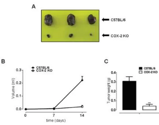
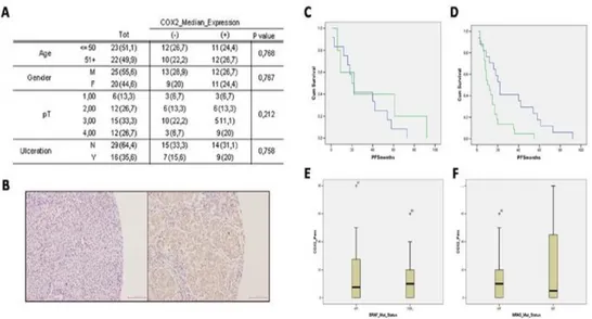
vivo models of cutaneous melanoma showed that in COX-2-/- mice tumor development was almost blunted as compared to littermate control C57Bl/6J. Finally, we performed a retrospective clinical study on 45 human lymph node melanoma metastases and correlated COX-2 expression to progression free survival (PFS). Our results show an inverse correlation between PFS and COX-2 expression suggesting that COX-2 is a negative prognostic factor in metastatic melanoma.
In addition to COX enzymes, also hydrogen sulfide (H2S), an endogenous gasotransmitter, has been recently demonstrated to be involved in human melanoma. Thus, the second phase of the project was focused on the evaluation of the efficacy of a new H2S-releasing nonsteroidal anti-inflammatory drugs (H2S-NSAIDs) named ATB-346, developed by combining naproxen with a chemical moiety that donates hydrogen sulfide. In particular, we used cell culture and a murine melanoma model to evaluate the effect of ATB-346 on: i) proliferation of human melanoma cells; ii) melanoma development in mice. Cell culture studies demonstrated that ATB-346 reduced the proliferation of human melanoma cells and this effect was associated to induction of apoptosis and inhibition of NF-κB activation. Moreover in vivo data showed that
by using this dual approach we propose that COXs and H2S pathway could be innovative therapeutic targets to generate new treatment options based on “combination therapy” for melanoma.
Finally, in the third and last phase of the project, we decided to better define the role of COX-2 in melanoma development. The aim was to establish if this enzyme acts predominantly in the microenvironment rather than in tumor cells. For this purpose we deleted COX-2 in B16/F10 murine melanoma cells by the mean of CRISPR/Cas9 technology. We firstly investigated on the effect of COX-2 knockdown on proliferation, migration, invasion and colonie formation of B16/F10 murine melanoma cells. In vitro studies demonstrated that CRISPR/Cas9-mediated COX-2 knockdown decreased proliferation of B16/F10 cells and inhibited some features of metastatic melanoma such as motility, invasiveness and focus formation. Finally, subcutaneously injection of B16/F10 cells knocked down for COX-2 showed slightly reduced melanoma growth and reduced the CXCL1 chemokine plasma levels. In addition, we also investigated on the expression and role of miR-143-3p in human malignant melanoma that has been shown to be dysregulated in many cancers.Our results showed that the expression of miR-143-3p was lower in human melanoma cells, as well as human tumor biopsy specimens, when compared to normal human melanocytes. Ectopic expression of miR-143-3p in human melanoma cells inhibited proliferation, migration, invasion and promoted apoptosis acting through a molecular mechanism that, at least in part, is dependent on inhibition of the COX-2 gene. Collectively, our findings show that COX-2 has a critical role in modulating melanoma development and progression. Nonetheless, significant challenges still lie ahead for blocking the interactions between the microenvironment and tumors.
When you are forge, stay; when you are hammer bang.
7
TABLE OF CONTENTS
CHAPTER 1: INTRODUCTION
1.1 INFLAMMATION AND CANCER
1.1.1 The inflammatory pathway………..15
1.1.2 Inducers and sensors of inflammation……….18
1.1.3 Mediators of inflammation………..24
1.1.4 Cyclooxygenase………....28
1.1.5 Eicosanoids and cancer………33
1.2 MELANOMA 1.2.1 Generalities and classification...37
1.2.2 Immune response in melanoma………....44
1.2.3 Treatment and immunotherapy in melanoma…………..………..47
1.2.4 The role of inflammation in melanoma and NMSC…….………...51
8
CHAPTER 3: MATERIALS AND METHODS
3.1 IN VITRO EXPERIMENTS
3.1.1 Patients and specimens...57
3.1.2 Tissue micro-array...57
3.1.3 Immunohistochemistry analysis...58
3.1.4 Cell culture and reagents...59
3.1.5 RNA purification and quantitative real-time PCR (qPCR)...60
3.1.6 Proliferation assay...61
3.1.7 COX-2 small interfering RNA transfection of SK-Mel-5...62
3.1.8 Preparation of cell lysates and western blot analysis...63
3.1.9 Cell invasion assay...64
3.1.10 Annexin V-FITC/PI Flow Cytometry...65
3.1.11 Elisa...66
3.1.12 Knockdown of PTGS2 with CRISPR/Cas9...66
3.1.13 Wound healing assay...67
3.1.14 Clonogenic assay...68
3.1.15 miR-143-3p mimics transfection...68
3.1.16 Apoptosis assay……….…………69
3.1.17 Cell migration assay………..…….………69
3.1.18 miRNA target prediction………..………..…………..……….70
3.1.19 Luciferase report activity assay………..………70
9
3.2 IN VIVO EXPERIMENTS
3.2.1 Animals...71 3.2.2 Induction of subcutaneous B16 lesions...71 3.2.3 Statistical analysis...72
CHAPTER 4: RESULTS
4.1 DIFFERENTIAL EXPRESSION OF CYCLOOXYGENASE-2 IN
METASTATIC MELANOMA AFFECTS PROGRESSION FREE
SURVIVAL
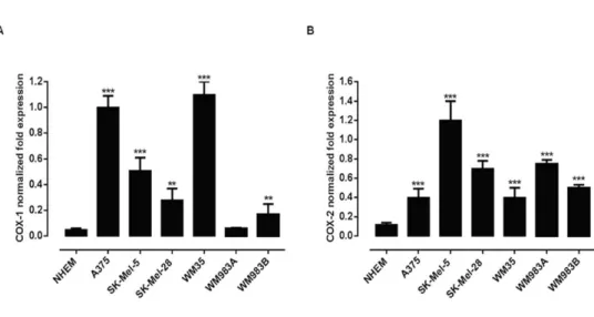
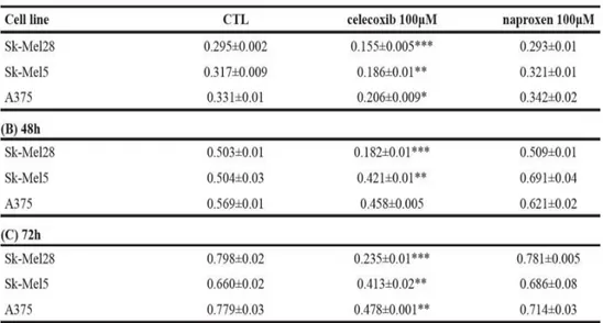
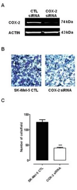
4.1.1 Expression of COX-1 and COX-2 in human melanoma cell lines...73 4.1.2 Selective inhibition of COX-2 activity and expression reduces
human melanoma cell proliferation and invasiveness...75 4.1.3 B16-F10 murine cell induced melanoma is blunted in COX-2-/- mice...78 4.1.4 Human melanoma samples...80 4.1.5 Higher COX-2 expression percentage in lymph node metastases correlates with negative progression free survival (PFS) outcome ...80 4.1.6 BRAF and NRAS mutational status does not correlate with COX-2
10
4.2 ATB-346, A NOVEL HYDROGEN SULFIDE-RELEASING ANTI-INFLAMMATORY DRUG, INDUCES APOPTOSIS OF HUMAN MELANOMA CELLS AND INHIBITS MELANOMA DEVELOPMENT IN VIVO
4.2.1 ATB-346 inhibits human melanoma cell proliferation...84 4.2.2 ATB-346 induces apoptosis of human melanoma cells...87 4.2.3 ATB-346 inhibits NF-𝜅B activation and down-regulates NF-𝜅B dependent anti-apoptotic genes...90 4.2.4 ATB-346 inhibits growth of melanoma tumors in vivo and reduce
plasma levels of melanoma-associated chemokines...93
4.3 PTGS2 KNOCKDOWN BY CRISPR/CAS9 TECHNOLOGY INHIBITS AGGRESSIVENESS AND PROGRESSION OF MELANOMA
4.3.1 Generating a Knockdown PTGS2 Melanoma Cell Line Using the CRISPR/Cas9 System...96 4.3.2 PTGS2 Knockdown Downregulates the Proliferative Potential in
Melanoma Cells...98 4.3.3 PTGS2 Knockdown Thwart Cell Motility and Invasion...99 4.3.4 PTGS2 Knockdown Inhibits Focus Formation of Melanoma Cells...102 4.3.5 PTGS2 Knockdown Inhibits Development of Cutaneous Melanoma In Vivo and Reduces CXCL1 Plasma Levels...104
11
4.4 MicroRNA-143-3p INHIBITS GROWTH AND INVASIVENESS OF MELANOMA CELLS BY TARGETING CYCLOOXYGENASE-2 AND INVERSELY CORRELATES WITH MALIGNANT MELANOMA PROGRESSION
4.4.1 miR-143-3p is downregulated in melanoma cell lines………..……106 4.4.2 miR-143-3p suppresses migration and invasion of human
melanoma cells………...….………109 4.4.3 miR-143-3p targets the 3’-UTR of COX-2………111 4.4.4 miR-143-3p transfection decreases PGE2 levels in A375
cells……….………..….113 4.4.5 Expression of miR-143-3p in human primary melanoma
lesions………114
CHAPTER 5: DISCUSSION...117
13
ABBREVIATIONS
15dPGJ2: 15-deoxy-Δ12,14-PGJ2 AA: Arachidonic acid
ACT: Adoptive cell transfer
AJCC: American Joint Committee on Cancer APL: Acute promyelocytic leukemia
APC: Antigen presenting cells
ATB-346: 2-(6- methoxynapthalen-2-yl)-propionicacid 4 thiocarbamoyl
phenyl ester
BCC: Basal cell carcinoma CDK: Cyclin dependent kinase CLRs: C-type lectin receptors COX: Cyclooxygenase
COX-1: Cyclooxygenase-1 COX-2: Cyclooxygenase-2
CRISPR/Cas9: Clustered regularly interspaced short palindromic repeats
associated nuclease 9
DCs: Dendritic cells
DMEM: Dulbecco’s modified Eagle’s medium EMT: Epithelial-mesenchymal transition FDA: Food and Drug Administration Gr-MDSCs: Granulocytic MDSCs H2S: Hydrogen sulfide
H2S-NSAIDs: H2S-releasing nonsteroidal anti-inflammatory drugs HMGB1: High-mobility group box 1 protein
HPGD: Hydroxyprostaglandin dehydrogenase IAP: Inhibitor of apoptosis
IDO: Indoleamine 2,3-dioxygenase IFNs: Type I interferons
ILCs: Innate lymphoid cells IRF3: IFN regulatory factor 3
LGP-2: Laboratory of genetics and physiology-2 LPS: Lipopolysaccharides
LOX: Lypooxigenase
mAbs: Monoclonal antibodies
MAPK: Mitogen-activated protein kinase MDA5: Melanoma differentiation factor-5 MDSC: Myeloid derived suppressor cells miRNAs: microRNAs
14
MRP4: Multidrug resistance-associated protein 4
MTT: 3-[4,5-dimethyltiazol-2-yl]-2,5-diphenyl tetrazolium bromide MyD88: Myeloid differentiation primary response 88
NALP3: Leucine-rich repeat-and pyrin-domain-containing protein NF-𝜅B: Nuclear factor kappa-light-chain-enhancer of activated B cells NHEM: Normal Human Epidermal Melanocytes
NLRs: NOD-like receptors
NMSC: Non-melanoma skin cancers NO: Nitric oxide
NSAIDs: Non-steroidal anti-inflammatory drugs PAF: Platelet-activating factor
PAMPs: Pathogen-associated molecular patterns
PARP: Poly (adenosine diphosphate-ribose) polymerase PFS: Progression free survival
PGG2: Prostaglandin G2 PGH2: Prostaglandin H2
PI3K/AKT: Phosphatidylinositol-3 kinases PLA2: Phospholipase A2
PPARs: Peroxisomal proliferator activated receptors PRRs: Pattern-recognition receptors
PS: Phosphatidylserine
PTEN: Phosphatase and tensin homolog PTGS: Prostaglandin endoperoxide synthases qPCR: Quantitative real-time PCR
RAF: Rapidly Accelerated Fibrosarcoma RGP: Radial growth phase
RLRs: Retinoic acid-inducible gene I Retinoic acid-inducible gene
(RIG)-I-like receptors
ROS: Reactive oxygen species
SCC: Squamous cell carcinoma T-reg: Regulatory T cells
TBZ: 4-hydroxy-thiobenzamide TGFβ: Transforming growth factor β TILs: Tumor infiltrating lymphocytes TLRs: Toll-like receptors
TMA: Tissue micro-array TME: Tumor microenviroment TNF-α: Tumour-necrosis factor-α
TRIF: TIR-domain-containing adapter-inducing interferon- β VEGF: Vascular endothelial growth factor
VGP: Vertical growth phase
15
CHAPTER 1: INTRODUCTION
1.1 INFLAMMATION AND CANCER
1.1.1 The inflammatory pathway
Inflammation is a protective response involving host cells, blood vessels, proteins and other mediators that is intended to eliminate the initial cause of cell injury, as well as the necrotic cells and tissues resulting from the original insult, and to initiate the process of repair [1]. The first description of inflammation by Cornelius Celsus in the 1st century, defined the four cardinal signs of inflammation: heat (calor), redness
(rubor), swelling (tumor), pain (dolor), and loss of function (functio laesa) then added by Rudolf Virchow in the 19th century [2]. Inflammation is often considered in terms of acute inflammation and chronic inflammation that differ in onset, cellular infiltrate and local and systemic signs [3]. The acute inflammatory response follows within seconds of the tissue injury and lasts for some minutes inducing vasodilation and increase of capillary permeability which allows to augment the blood flow and entry of fluids and diffusible components to the infected area inducing redness and erythema as consequence. This phase usually terminates after several tens minutes and is common in case of minor injuries such as bruising, scratching, cuts, and abrasions. If there has been sufficient damage to the tissues, or if
16 infection has occured, the acute inflammatory response takes place over the next few hours.
In this phase there is the recruitment of leukocytes, particularly neutrophils, in the tissues [4]. The sequence of events in the recruitment and activation of leukocytes consists of different phases: first leukocytes accumulate themselves to the endothelial cells (margination), then begin to tumble on the endothelial surface by the interaction between adhesion molecules expressed on the surface of both leucocytes and endothelial cells (rolling and adhesion) and then migrate through the vessels and move toward sites of infection or injury (diapedesis and chemotaxis). Finally leukocytes bind and ingest most microorganisms and dead cells (phagocytosis) and the subsequent resolution process begins restoringthe normal tissue architecture [5]. If the acute inflammatory response fails to eliminate the pathogen, a chronic inflammatory state ensues where the inflammatory process persists and acquire new characteristics. Here the neutrophil infiltrate is replaced with macrophages, lymphocytes and plasma cells producing inflammatory cytokines, growth factors and enzymes which in turn contribute to the progression of tissue damage and secondary repair including fibrosis and granuloma formation.
There are two types of chronic inflammation:
Non specific proliferative: characterized by the presence of non-specific
17 (lymphocytes, macrophages, plasma cells) and proliferation of fibroblasts, connective tissue, vessels and epithelial cells, for example, an inflammatory polyp-like nasal or cervical polyp and lung abscess.
Granulomatous inflammation: A specific type of chronic inflammation
characterized by the presence of different nodular lesions or granulomas formed with a conglomeration of activated macrophages or its derived cells called epithelioid cells usually enclosed by lymphocytes. The macrophages or epithelioid cells inside the granulomas often combine to form Langhans or giant cells such as foreign body, Aschoff, Reed-Sternberg and Tumor giant cells.
There are two major types of granuloma:
Foreign body granuloma: that are formed to a foreign body or T-cell
mediated immune response such as in sarcoidosis and Crohn’s disease [6, 7].
Infectious granuloma: Granuloma that are formed from chronic
infection such as in tuberculosis and leprosy [8].
Although chronic inflammation progresses silently, it is the cause of most chronic diseases including obesity, type 2 diabetes, atherosclerosis, asthma and neurodegenerative diseases [9].
18
1.1.2 Inducers and sensors of inflammation
A typical inflammatory response consists of four components: inducers, sensors, mediators and the effectors of inflammation. There are different inducers that trigger the inflammatory response. They activate specific sensors, which then stimulate the production of distinct sets of mediators. Inducers of inflammation can be divided in exogenous or endogenous [10].
Exogenus stimuli are both microbial and non-microbial, such as pathogen-associated molecular patterns (PAMPs) and virulence factors. PAMPs are a restricted and specific set of conserved molecular patterns that are carried by all microorganisms and are recognize by the host through the expression of a specific set of receptors known as pattern-recognition receptors (PRRs) [11]. The second class of microbial inducers are virulence factors that are not recognized by specific receptors but by specialized sensors such as NACHT-, leucine-richrepeat-and pyrin-domain-containing protein (NALP3) inflammasome [12]. Non-microbial exogenous stimuli are allergens, irritants, foreign bodies and toxic compounds that simulate the virulence activity of parasites or can act as irritants on the mucosal epithelia [1]. Others exogenous stimuli are: physical (such as extremely low or high temperature or ionizing radiations) and nutritive (e.g. deficiency of oxygen and vitamins). Endogenous stimuli are signals generated by damaged or stressed tissues. For example, the release of ATP and high-mobility group box 1
19 protein (HMGB1) during necrotic death cells, can cooperate with Toll-like receptors (TLRs) to induce an inflammatory response [13]. Or, the formation of urate and calcium crystals that are common in people-eating high purine foods and are responsible of periarticular diseases like gout and pseudogout [14]. These crystals are detected by macrophages activating the NALP3 inflammasome which in turn activates the production of caspase-1 substrates and members of the interleukin 1 (IL-1) family [15].
Pattern-recognition receptors are designed to sense the presence of infectious pathogens and substances released from dead cells. Currently, four different classes of pattern-recognition receptors families have been identified: Toll-like receptors (TLRs), C-type lectin receptors (CLRs), the Retinoic acid-inducible gene (RIG)-I-like receptors (RLRs) and NOD-like receptors (NLRs) [16].
TLRs are one of the best-characterized PRRs and are responsible for sensing invading pathogens outside of the cell and in intracellular endosomes and lysosomes [17]. TLRs are expressed by T cells, B cells neutrophils, mast cells, monocytes and macrophages. There are ten mammalian TLRs, TLR1 –TLR10, which recognize products of bacteria (such as endotoxin and bacterial DNA), viruses (such as double stranded RNA), and other products including lipoproteins and di- and triacyl lipopeptides, fungal zymosan, peptidoglycans, flagellin and different synthetic molecules such as imidazoquinolines and guanosine
20 analogues. These molecules are recognized by distinct TLRs in combination with coreceptors, or by TLR heterodimers [18]. TLR1 is closely related to TLR2 and TLR6 with which it forms heterodimers that recognize bacterial lipoproteins and lipopeptides [19]. TLR4 recognizes lipopolysaccharides (LPS), which are present in the cell wall of Gram-negative bacteria [20]. The TLR5 recognizes flagellin the dominant component of the bacterial flagellum and the structure responsible for motility in different bacterial species [21]. TLR3, TLR7, TLR8, and TLR9 are different from other TLRs since they are not expressed at the cell surface but located in endosomal compartment. TLR3, TLR7, and TLR8 detect double- and single-stranded RNA and TLR9 detects unmethylated CpG DNA [22]. Finally, TLR10 is the only remaining orphan receptor without a known ligand or signaling function. Moreover, it has been recently reported that TLR10 does not activate the immune system and has instead been shown to suppress inflammatory signaling on primary human cells [23].
TLRs signaling is divided into two distinct pathways: the myeloid differentiation primary response 88 (MyD88) and TIR-domain-containing adapter-inducing interferon-β (TRIF) (Figure 1.1). Those signaling pathways lead to activation of nuclear factor kappa-light-chain-enhancer of activated B cells (NF-𝜅B) protein, that induces transcription of a wide range of genes involved in the inflammatory response including cytokines, chemokines, and immunoreceptors [24].
21
22 MyD88 is utilized by all TLRs with the exception of TLR3 and drives NF-𝜅B and mitogen-activated protein kinase (MAPK) activation to control inflammation. TRIF, also known as TICAM1, is utilized by TLR3 and TLR4 and is triggered by dsRNA and LPS, respectively [26].
CLRs are crucial for controlling both innate and adaptive immune response and are expressed by dendritic cells (DCs), monocytes and macrophages [16]. CLRs recognize carbohydrates, such as mannose, fucose, and glucan carbohydrate structures present in bacterial, viral, and fungal components.
RLRs are intracellular receptors for virus recognition. There are three major components of this family: retinoic acid-inducible gene I (RIG-I), melanoma differentiation factor-5 (MDA5), and laboratory of genetics and physiology-2 (LGP-2) that bind to virus double-stranded RNA. Activation of RIG-I and MDA5 increase the secretion of type I interferons (IFNs), such as IFN-β, through the activation of the IFN regulatory factor 3 (IRF3) transcription factor [27]. Conversely, LGP-2 has a negative regulatory role by inhibiting IRF3 and the secretion of IFN-β.
In addition to sensing of microbial RNAs by RLRs, STING signaling is a recent identified pathway that recognize microbial and self-DNA [28]. STING (Stimulator of Interferon Genes, also known as MITA, ERIS, MPYS, or TMEM173) is thought to function as an adaptor protein, which links upstream DNA sensors to downstream IRF-3 and NF-κB pathway
23 activation inducing the expression of IFNs and inflammatory cytokines such as TNF, IL-1β, and IL-6 [29].
NLRs are intracellular sensors localized in the cytoplasm of DCs, macrophages and lymphocytes and are associated with cell stress. There are different members of this family, in particular NOD1, NOD2, and NALP3 [30]. NOD1 and NOD2 recognize intracellular bacterial cell products in both Gram-positive and Gram-negative bacteria driving the activation of NF-𝜅B and MAPK pathways, leading to proinflammatory cytokine secretion [16]. NALP3 responds to a widest array of stimuli through the development, togheter with NLRP1 and NLRC4, of a multiprotein complex named NALP3 inflammasome (Figure 1.1) [30]. Inflammasome induces the cleavage and consequent activation of caspase-1 which in turn cleaves the precursor cytokines pro-IL-1β and pro-IL-18, producing the biologically active cytokines IL-1β and IL-18 and also inducing an inflammatory form of cell death known as pyroptosis [31]. IL-1β is a cytokine that promotes T helper 1 (Th1) and T helper 17 (Th17) differentiation. IL-18 is important for IL-17 expression by Th17 cells and for T cell polarization toward Th1 or Th2 profiles in combination with other cytokines [32].
24
1.1.3 Mediators of inflammation
Inflammatory mediators are soluble, diffusible molecules that can be produced locally by cells at the site of inflammation or derived from circulating inactive precursors to be then activated at the site of inflammation. Some mediators (such as histamine and serotonin) are normally stored in intracellular granules of mast cells, basophils and platelets and are rapidly released upon cellular activation. Others are synthesized de novo or circulate as inactive precursors in the plasma and require a proteolytic cleavage to acquire their biological activity [1]. Inflammatory mediators can be divided into seven groups according to their biochemical properties: vasoactive amines, vasoactive peptides, fragments of complement components, lipid mediators, cytokines, chemokines and proteolytic enzymes.
The vasoactive amines are responsible for the immediate and short-lived responses in inflammation including vasodilation, increase of vascular permeability and smooth muscle contraction. Histamine and serotonin are the most important vasoactive amines and are secreted by mast cells and platelet degranulation [33].
Vasoactive peptides are small proteins, such as substance p that is stored in secretory vesicles and released by sensory neurons transmitting pain signals and regulating vessel tone and vascular permeability. Other vasoactive peptides are bradykinin, fibrinopeptide A, fibrinopeptide B and fibrin degradation products. Bradykinin is the
25 main product of the kallikrein–kinin cascade activated by the Hageman factor together to the clotting, fibrinolytic and the complement system. Like histamine, bradykinin increases vascular permeability, arteriolar dilation, and bronchial smooth muscle contraction [34].
The complement system consists of plasma proteins that play an important role in inflammation. In particular, the complement-derived factors C3a, C4a and C5a (also called anaphylatoxins) increase vascular permeability and cause vasodilation by inducing mast cells to release histamine, activate leukocytes, increasing their adhesion to endothelium, and augment the phagocytosis by neutrophils and macrophages [35].
Lipid mediators are eicosanoids and platelet-activating factor (PAF). Eicosanoids derived from the metabolism of arachidonic acid (AA). AA is a component of cell membrane phospholipids released through the action of phospholipase A2 (PLA2) that is activated by other inflammatory mediators sush as the complement-derived factor C5a [36]. AA is processed by two different enzimes: Cyclooxygenase (COX) and Lypooxigenase (LOX), which in turn produce respectively prostaglandins, thromboxanes and leukotrienes and lipoxins. We will
discuss about eicosanoids and their relationship with cancer later in this thesis. PAF is generated by the acetylation of lysophosphatidic acid and acts directly on target cells through the binding with a specific G protein–coupled receptor. PAF stimulates platelet aggregation, causes
26 bronchoconstriction and induces a potent vasodilatation and increase of vascular permeability.
Another class of inflammatory mediators are cytokines. Inflammatory cytokines are produced by different cell types including in particular macrophages and mast cells. The major cytokines involved in inflammation are tumour-necrosis factor-α (TNF-α), IL-1, IL-6 and many others [37]. TNF-α and IL-1 stimulate the expression of adhesion molecules on endothelial cells, increasing leukocyte binding and recruitment, and augment the production of additional cytokines. Chemokines are produced by different cell types and act as chemoattractants controlling extravasation and chemotaxis of monocytes, lymphocytes, neutrophils, eosinophils, basophils, natural killer cells, dendritic cells, and endothelial cells towards the site of inflammation [38].
Proteolytic enzymes have different roles in inflammation. In particular elastase, collagenase, and cathepsin, secreated by neutrophils and monocytes, destroy phagocytosed substances. Their activity is regulated by antiproteases present in the plasma and tissue fluids such as α1-antitrypsin and α2-macroglobulin.
In addition, other mediators of inflammation are: reactive oxygen species (ROS) and nitric oxide (NO).
27 ROS are released from neutrophils and macrophages and amplify the cascade of inflammatory mediators by inducing adhesion molecule expression, chemokines and cytokines promoting their migration across the endothelial barrier helping also in the clearence of phagocytosed microbes and necrotic cells [39].
NO is a soluble gasotransmitter that possesses cytotoxic properties serving as a killing mechanism against invading microbes. In particular, macrophages produce it as cytotoxic agent for killing microbes and tumor cells [40]. On the other hand, NO is considered as a pro-inflammatory mediator that induces inflammation due to over production in unusual conditions such as in inflammatory disorders of the joint, lungs and gut [41].
28
1.1.4 Cyclooxygenase
COX enzymes, also known as prostaglandin endoperoxide synthases (PTGS), are enzymes involved in the rate-limiting step of prostaglandin production from AA [42]. They exist in two isoforms: COX-1 (also known as PTGS1) and COX-2 (also known as PTGS2). COX-1 is a constitutively-expressed enzyme found in many tissues. This enzyme is often referred as the ‘housekeeping’ form of the COX enzymes and modulates aspects of routine functions such as platelet aggregation and gastric acid secretion by maintaining basal prostanoid levels [43]. By contrast, COX-2 is highly induced by a variety of endogenous and external stimuli that may include growth factors, tumor promoters, viral or bacterial infections as well as a widely range of proinflammatory cytokines [43, 44]. The gene structures of COX-1 and COX-2 are found at distinct genetic loci and differ mainly in size. While COX-1 maps to the long arm of chromosome 9 and measures 22 kb, COX-2 maps to the long arm of chromosome 1 and measures 8.3 kb [45]. By contrast, both COX isoforms share approximately 60% of their protein structure which differs in the substitution of a Valine residue in COX-1 for an Isoleucin at corresponding position 523 within the active site of COX-2. This change, together to the loss of a methyl group, increases the volume of the COX-2 active site by COX-25% allowing the accomodation of larger chemical structures (Figure 1.2) [46].
29
30 These differences have been exploited for the development of COX-2 selective non-steroidal anti-inflammatory drugs (NSAIDs) that are widely used in clinical practice for the treatment of chronic inflammatory diseases such as osteoarthritis, rheumatoid arthritis and gout showing fewer gastrointestinal adverse effects than non-selective NSAIDs [47]. Cyclooxygenase enzymes catalyse the oxidation of arachidonic acid to form the peroxide intermediate prostaglandin G2 (PGG2) which in turn is reduced to prostaglandin H2 (PGH2) (Figure 1.3). The latter acts as a substrate for several prostaglandin synthases including PGE2, PGD2, PGF2α, PGI2, and TXA2 [48].Prostaglandins exert their biological effects in an autocrine or paracrine fashion by binding to cognate membrane bound G-protein-coupled receptors such as DP for PGD2; EP1, EP2, EP3 and EP4 for PGE2; FP for PGF2α; IP for PGI2; and TP for TXA2. Moreover, some prostaglandins and their metabolites bind to intracellular peroxisomal proliferator activated receptors (PPARs) inducing their transcriptional activity in the nucleus [49]. For example, PGI2 and PGE2 can bind to PPARβ while, 15-deoxy-Δ12,14-PGJ2 (15dPGJ2), a PGD2 dehydration product, can bind to PPARγ.
31
32 In addition to their synthesis, the extracellular levels and the inactivation of prostaglandins is regulated by different transporters and enzymes such as the influx prostaglandin transporter PGT; the efflux transporter multidrug resistance-associated protein 4 (MRP4); and the enzyme hydroxyprostaglandin dehydrogenase 15-(NAD) (HPGD; also known as 15-PGDH) that metabolize PGE2 and PGF2α, while other prostaglandins are metabolized in a non-enzymatic manner [51].
Prostaglandins regulate different biological processes in a wide range of tissues under both physiological (mainly ascribed to COX-1 activity) and pathological conditions (mainly ascribed to the induction of COX-2). For example, PGE2 and PGI2 exert cytoprotective effects in the gastrointestinal tract by reducing gastric acid and increasing the release of mucous, while in the kidneys these prostaglandins act as vasodilators that maintain renal homeostasis [48]. Moreover, prostaglandins control the homeostasis of other peripheral tissues such as the cardiovascular system, the lungs, reproductive system and the central nervous system.
33
1.1.5 Eicosanoids and cancer
The concept that inflammation is a critical component of tumour progression is not new. In 1863, Rudolph Virchow was the first to hypothesize that cancer occurred at sites of chronic inflammation, assuming that some classes of irritants, together with the tissue injury and consequent inflammation, release factors stimulating cancer cell proliferation [52]. In 1957, Burnet and Thomas postulated the existence of tumor immunosurveillance: the immunological resistance of the host against the development of cancer. Burnet and Thomas proposed that lymphocyte populations of the immune system continuously recognized and eliminated cancerous and/or precancerous cells arising in the host before they could cause harm. In the last years different data highlighted the concept that inflammation is a critical component of tumour progression and has been reported to be one of the newest “hallmarks of cancer” [53]. Different epidemiological data reported that about 20% of cancer are linked to chronic inflammation [52]. For example, inflammatory bowel diseases are linked to colorectal cancer; bronchitis to lung cancer and prostatitis to prostate cancer [54]. Most precancerous and cancerous lesions show signs of inflammation including innate immune cells, cytokines, and chemokines [55]. For example, macrophages in inflammatory bowel disease are phenotypically different from normal macrophages and produce different cytokines such as IL-1, IL-6, and TNF-α that can promote tumor
34 invasion and metastasis [56]. Among various inflammatory mediators, deregulation of prostaglandin mediated signaling as a result of aberrant COX expression, has been linked to different types of cancer. In 1994 it has been reported that COX-2 is overexpressed in human colorectal adenomas and adenocarcinomas and has a key role in inflammatory bowel disease and colorectal cancer [57]. Moreover COX-2 has been detected in a wide range of cancers including: breast cancer, lung cancer, ovarian cancer, colon cancer, colorectal cancer, gliomas, prostate cancer, esophageal carcinoma, pancreatic cancer, gastric carcinoma, Kaposi’s sarcoma and melanoma [54, 58]. The importance of COX-2 in cancer has been demostrated by treatment with aspirin or other NSAIDs that inhibit prostaglandin synthesis decreasing the incidence of esophageal, colorectal, bladder, lung, and gastric cancer [54]. Moreover, COX-2 selective inhibitors (such as celecoxib) showed inhibition of adenoma growth in patients at high risk for colorectal carcinoma in three different clinical trials. Unfortunately, all three trials were aborted because of cardiovascular and gastrointestinal toxicity highlighting the importance to develop new classes of anti-inflammatory agents for cancer therapy [59-61]. Among prostaglandins, PGE2 is the most common prostaglandin that is found in various human malignancies, including colon, lung, breast, and head and neck cancer, and is often associated with a poor prognosis [50]. PGE2 appears to play essential roles in tumor cell proliferation, invasion, angiogenesis, and
35 epithelial-mesenchymal transition (EMT), all of which are associated with the established or emerging “hallmarks of cancer” [50, 62-64]. Another critical mechanism for the potent pro-tumorigenic activity of PGE2 is the local suppression of the immune responses [65]. PGE2 inhibits macrophages, natural killer cells and T cell activation, resulting in immunosuppressive and pro-tumorigenic activity [66]. Moreover, PGE2, also plays a key role in promoting myeloid derived suppressor cells (MDSC) differentiation from bone marrow stem cells that are responsible for inhibiting the antitumor immune response; this provides a therapeutic approach for anti-cancer therapy [67]. Another strategy to inhibit the pro-tumorigenic activity of PGE2 is the overexpression of 15-PGDH enzyme, which is responsible for the degradation of PGE2. In fact, this enzyme results to be down-regulated in human gastric cancer, breast cancer, lung cancer, and bladder cancer, suggesting its tumor suppressive role [68-71]. Another approach for cancer therapy is the inhibition of PGE2 receptors. Mice deficient for EP1, EP2 or EP4 receptors showed decreased tumorigenesis while administration of PGE2 receptor antagonists (such as ONO-8711 which is selective for EP1 receptor) inhibit tumor growth and can act as chemo-preventive agents [72-75].
In addition to PGE2, there are conflicted data about the role of PGD2 in cancer progression. Disruption or overexpression of hematopoietic prostaglandin D synthase, which synthesizes PGD2, accelerates or
36 suppresses intestinal tumour growth in mice [76]. By contrast, an immunosuppresive effect of PGD2 has been recently reported in human acute promyelocytic leukemia (APL) by involving a new class of innate immune cells named innate lymphoid cells (ILCs) which in turn activate MDSC [77].
While COX-2 has been the most extensively studied enzyme in cancer, few reports have indicated that LOX enzyme has also an important role in tumor progression and survival and is often constitutively expressed in various epithelial cancers [78]. However, due to its association with the inflammatory response under most physiological conditions as well as the overexpression of COX-2 in many tumors, including melanoma, the cyclooxygenase pathway will be given the principle focus throughout this thesis.
37
1.2 MELANOMA
1.2.1 Generalities and classification
Melanocytes are melanin-producing cells located in the basal layer of the skin’s epidermis, the uvea, the inner ear, meninges, bones and heart. The word “melanin” comes from the ancient Greek melanos, meaning 'dark' and is the pigment that provides color and protection against ionizing radiations [79]. Cutaneous melanoma is a type of skin cancer that arises from abnormal melanocytes proliferation and can occur in any anatomic location containing melanocytes. There are four different types of skin cancer: basal cell carcinoma (BCC) and squamous cell carcinoma (SCC) identified as non-melanoma skin cancers (NMSC) or keratinocyte carcinoma; melanoma and other non-epithelial skin cancer. Moreover, there are also other uncommon types of skin cancer, such as Merkel Cell Carcinoma, Cutaneous T-cell lymphoma and Kaposi's sarcoma [80]. BCC and SCC represent approximately 80 % and 16 % of all NMSC, respectively. By contrast, melanoma represents less than 5% of the skin cancer cases, however, it far the most dangerous as it accounts for 80% of all the deaths related to skin cancer [81]. When diagnosed in the early stage, melanoma can be easily cured by surgical resection. However late metastatic stages are often fatal and, currently available or developing treatment strategies, are only able to prolong life for a few months, are expensive and may produce severe adverse effects [82]. In the past few years melanoma has steadily increased
38 worldwide becoming the 5th neoplasia in America [83]. Its incidence varies by geographic location and across different ethnic groups. This variation is partly related to decreased photoprotection to UV radiations that is well known to induce both cell death and malignant transformation of skin cells and are considered the major risk factor for melanoma [84]. In 2017 the American Cancer Society and the National Cancer Institute estimated 87,110 new cases of patients with invasive melanoma and 9,730 deaths only in USA. Melanoma incidence is also dependent on gender, infact in the U.S. is higher in women than in men before age 50, but by age 65, rates in men are double those in women, and by age 80 they are triple. In addition to UV radiations, genetic predisposition and environmental insults contribute to the genesis of melanoma. About 10% of melanoma patients report a family history of melanoma and are associated with germline mutations in cyclin dependent kinase (CDK) gene CDKN2A which is a potent cell cycle inhibitor through a direct negative interaction with CDK4. Moreover, also xeroderma pigmentosum and MC1R genes have been implicated in familial melanomas [85]. These initial observations opened the door to additional efforts in understanding the genetics of melanoma, and several other mutations have been identified since then. For example, the MAPK/ERK and phosphatidylinositol-3 kinase (PI3K/AKT) pathways are one of the first most frequently deregulated signaling pathways in the great majority of melanomas. In particular, the role of the Ras
39 oncogene family in the melanoma development and their effects on downstream signaling were one of the first mutations to be identified, then NRAS mutation which is mutated in 15-30% of tumor samples leading to serial activation of both the downstream components of the RAS effector pathways and the non-MAPK pathways such as PI3K/AKT [86]. In 2002 the mutation called ‘Rapidly Accelerated Fibrosarcoma’ (RAF) were observed and the V600E variant identified as a frequent mutation in cutaneous melanoma [87]. This mutation and the consequent constitutive activation of the MAPK pathway has become the target of multiple pharmaceutical trials of small molecule inhibitors resulting in several new Food and Drug Administration (FDA) approved therapies [88]. The other pathway involved in melanoma development and progression is PI3K/AKT. In particular, in 12% of melanoma patients a lack of the tumour suppressor phosphatase and tensin homolog (PTEN) that antagonizes PI3K activity has been detected. Lack of PTEN antagonism causes increased phosphorylation of AKT promoting cancer cell survival and proliferation [89]. In addition there are also other mutations affecting genes that control growth, such as those of Bcl2 family, production of growth factors and loss of adhesion molecules which favor the alteration of intracellular signaling allowing the melanocytes to escape from the keratinocyte control [90].
Pathological features of melanoma can be divided in 5 steps according to the Clark model: I) Nevi formation of structurally normal
40 melanocytes, II) dysplastic nevi formation, III) radial growth phase (RGP) primary melanoma, IV) vertical growth phase (VGP) involving cells that have invaded the dermis and cells with metastatic potential and V) metastatic melanoma to distant organs (Figure 1.4). The development of this illness can be seen as a disruption of normal homeostatic mechanisms in the skin. Disruption of the homeostatic control also leads to le loss of adhesion molecules such as E-cadherin and the increase of N-cadherin that favors the mobility and invasiveness of melanoma cells thus the development of metastasis [91].
41
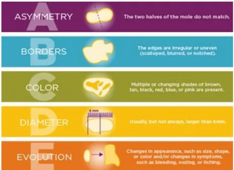
42 In addition, the American Joint Committee on Cancer (AJCC) considers the Breslow index as the most important prognosis factor in primary melanomas. This model measures the melanoma thickness (in millimetres) according to the distance between the upper layer of the epidermis and the deepest point of tumor penetration (Figure 1.4) [93]. According to AJCC the melanoma stages are classified into 4 groups: I and II as primary invasive melanomas and III and IV define local regional and distant metastasis respectively [94]. However, regular self-skin examinations by patients showed to decrease the risk of metastatic melanoma at the time of diagnosis expecially when coupled with regular visits to a dermatologist [95]. The A-B-C-D-E guide, devised by the american cancer society, highlights several clinical features of melanoma, including asymmetry, border, color, diameter and evolution (Figure 1.5).
A = asymmetry, when the two halves of the lesion do not match. B = border, when the edge are irregular or uneven.
C = color, when the lesion doesn’t have an uniform color but rather a combination of black, brown, blue, or even white.
D = diameter, when the lesion is larger than 6 mm.
43
Figure 1.5: The A-B-C-D-E guide
44
1.2.2 Immune response in melanoma
The concept of immune response in tumors was propose for the fist time by Paul Ehrlich in 1907 and later expanded when Coley obtained tumor reduction after injection of bacteria into tumors [96]. This kind of work revealed the importance of studying the relationship of host immune response to tumors and patients’ survival. The work that followed showed that injection of melanoma cell lysates resulted in 2/26 patients with total remission and 5/26 with partial tumor regression. Furthermore, it was observed that patients injected with irradiated melanoma cells increased the index of lymphotoxicity. In all cases histology from regressing tumors showed high lymphocyte infiltration [96]. In fact, the importance of lymphocytes in tumors has been long recognized but their possible role in melanoma prognosis has been debated and only recognized in the past 40 years. Some groups showed increased survival and improved prognosis in patients with mild and high lymphocyte infiltration and finally established 3 categories: Brisk (dense infiltrate), No brisk (mild infiltrate) and absent [97, 98].On the contrary, other groups failed to show a positive correlation between tumor infiltrating lymphocytes (TILs) and prognosis or patients’ survival [99-101]. Discrepancies in those results could be explained by the extreme diversity of the patients included in the studies and the fact that no phenotypic characterization of those TILs was done [102], in fact studies were such characterization was done, showed that CD8+ T cell
45 infiltration improve prognosis [103]. More answers to this might be found in the understanding of the role of inflammatory cytokines in lymphocyte functions, and the immunosuppressive aspects of other cells in the tumor microenvironment [96]. Human tumors bear antigens that can be recognized by autologous T lymphocytes. Some of these antigens appear to be tumor specific (neo-antigens), whereas others are also present on normal tissues such as overexpressed antigens or viral-derived antigens. Specifically in melanoma, several tumor specific antigens have been described making easier the assessment of the anti-tumor immune response [104, 105]. After identification of melanoma antigens several groups reported evidence about the existence of a spontaneous anti-tumoral T cell response in patients. For example it has been reported that tumor-specific CD8+ T lymphocytes from melanoma patients have a memory effector phenotype (TEM) (CCR7+, CD4+45RO+) while healthy donors bear naive tumor-specific T lymphocytes (CCR7+, CD4+45RA+) [106, 107]. In addition TILs of melanoma patients were shown to contain not only CD8+ T cells but also CD4+ T cells recognizing melanocyte differentiation antigens and cancer testis antigens [108]. In conclusion, spontaneous T cell response correlates with a better prognosis, even though the correlation may be obscured in some circumstances by the fact that most metastatic melanoma patients are progressing despite having mounted a quantitatively significant T cell response against their tumor [109]. T cell unresponsiveness does not
46 seem to be due to their failure to reach the tumor sites but the anti-tumoral T cell response must become ineffective at one point, either because the tumor cells have become insensitive to the effector cells or because the effector cells themselves have become unable to be stimulated by the antigen or to exert their function, a state also refered as “exhaustion”. This exhausted state could result from inhibitory processes elicited by tumor cells that were selected by the immune response (immunoediting), the loss of antigen or MHC molecules [110], the expression of immunosuppressive factors such as Indoleamine 2,3-dioxygenase (IDO) and prostaglandins [111, 112], the secretion of immunosuppressive cytokines such as IL-10 and TGFβ [113], arginase expression [114, 115], TREG infiltration [116] or the expression of PDL-1 molecule at the surface of tumor cells among others [117].
47
1.2.3 Treatment and immunotherapy in melanoma
In the last 30 years many drugs and combination of drugs have been introduced in melanoma therapy to improve patient survival for longer time. Cytotoxic chemotherapy was the main approach for metastatic melanoma while radiation therapy is used in 1–6% of patients with melanoma in the US in the setting of inoperable disease [118]. Common antineoplastic agents currently being used include dacarbazine and temozolomide that, in monotherapy, showed low overall response rates (approximately 10%-15%). In the past decades, the increasing knowledge in cellular immunology and tumor led to the development of immunotherapy approaches. One of the first approaches was the discovery of the T cell growth factor IL-2 that was the first agent used to modulate the immune system for the treatment of patients with metastatic melanoma [105]. It was approved by the FDA in 1998 but its response rate was low (approximately 16%) and showed marked secondary effects [119]. In fact, before 2010 none of the systemic therapies approved for metastatic melanoma treatment showed to increase patients’ survival, and only little improvement was observed by using TNF-α as co-adjuvant. Currently, treatment with BRAF inhibitors, which are included in the “target therapy" drugs, have been used to treat melanoma patients bearing this mutation that constitutive activates MAPK pathway. However, BRAF inhibitor Vemurafenib, approved by the FDA in 2011, can only be used in approximately 50% of
48 melanoma patients where the V600E mutation is present [120]. There are also other BRAF inhibitors approved such as Dabrafenib and Trametinib but unfortunately, an important percentage of the patients acquire drug resistance loosing response to the therapy and tumor progression approximately after 6-8 months [121]. After the BRAF inhibitors’ development, the interest about therapy continued to grow and the latest clinical advances in melanoma treatment have targeted immune response by blocking inhibitory checkpoint molecules. In particular, the two best characterized are CTLA-4 and PD-1 for which monoclonal antibodies have been developed and already approved by the FDA. For example, Tremelimumab was the first anti-CTLA-4 agent to be investigated in patients with metastatic melanoma. Although in phase I/II clinical trials, it showed antitumor activity in patients with stage III/IV melanoma, a phase III study failed because tremelimumab didn’t demonstrate any benefit over chemotherapy [122]. More successes has had Ipillimumab (Yervoy®) approved by FDA in 2011. Ipilimumab blocks the CTLA-4 molecule favoring the activation state of T lymphocytes. CTLA-4 or CD152 works as an inhibitory receptor for the co-stimulatory signal delivered by the interaction with B7 expressed on the surface of antigen presenting cells (APC) [123] and has been also reported to be capable of depleting regulatory T cells (T-reg) [124]. In patients Ipillimumab showed an overall response of 11% [125] and, in
49 combination with dacarbazine, it improved patients’ survival for 11,2 months with 24% reduction of patients’ disease relapse [126].
Another inhibitory checkpoint is the PD-1/PDL-1 pathway. Physiologically, PD-1/PDL-1 interaction limits T cell activity in the setting of an inflammatory response. PD-1 is expressed in activated T, B and NK cells, whereas PDL-1 or PDL-2 are expressed on immune cells as well as tumor cells. PD-1 has been shown to be express on TILs while circulating melanoma antigen specific T cells and melanoma tumors express PD-L1 which induce T cell anergy and immunosuppression [105]. Nivolumab (Opdivo®) is directed against PD-1 and showed 28% of clinical response with a dosage of 3 mg/kg bodyweight and with a low rate of toxicity, infact the most common side effects are diarrhea, rush, and pruritus [127]. Pembrolizumab (formerly known as lambrolizumab) is a second anti-PD-1 agent approved by FDA on September 2014. It showed 25% of clinical response at low doses and 52% at high doses and 77% of the patients showed tumor regression [128].
Immunotherapy, based on adoptive cell transfer (ACT) of ex-vivo activated and expanded TILs, has shown promising clinical results in patients with metastatic melanoma. TIL therapy after lymphodepletion showed response rates of around 50% and significant survival benefit in refractory melanoma patients [129]. However TIL-based ACT is an individualized treatment that requires specialized laboratories and is more complex to perform than conventional therapies. Today, properly
50 designed clinical trials are required to formally demonstrate the advantages of this kind of therapies over the classic treatment modalities. In addition, identifying predictors of TIL efficacy and detection of TIL subsets with specific reactivity against tumors is necessary to improve the clinical responses and patients survival [130]. Other types of immunotherapies are cancer vaccines that have been also tested in the past decade and, in the last years, their development has been evolved in a significant manner in antigenic composition, delivery methods, adjuvants, treatment frequency and administration techniques [129]. Nonetheless, this approach has not been very successful in terms of objective response and patients survival. Current immunotherapy approaches for melanoma show encouraging results and the inefficacy of some of these therapies could be explain by different reasons such as the type of adjuvants used during the vaccination process [131], expression of exhaustion markers, loss of functionality, limited access to tumor site and the immunosuppressive tumor microenvironment [132].
51
1.2.4 The role of inflammation in melanoma and NMSC
As previously reported, inflammation is linked with several stages of tumor development, including initiation, promotion, invasion, and metastasis [133]. NF-κB protein is the main player in inflammation and, together with other molecules including cytokines, chemokines, ROS, oncogenes, inflammatory enzymes (COX-2 and LOX), anti-apoptotic proteins, transcription factors (STAT3, AP-1, CREB, NRF2) regulate tumor cell proliferation, transformation, and survival [134]. Cell lines from SCC showed high expression of activated NF-κB while, other cancer cells, including melanoma cells, showed higher expression of STAT-3 which in turn up-regulates anti-apoptotic proteins such as Bcl-2 and controls the expression of cyclins and the proto-oncogene c-Myc [135].
Different cytokines also control the inflammatory milieu promoting tumor progression. For example, IL-6 showed a direct effect on cancer cell growth and survival in melanoma [136]. The expression of inflammatory cytokines is induced by different pathways, including MAPK and in particular RAS, which is mutated in approximately 25 % of all malignancies, and has also been reported to be linked with tumor progression locus 2 (Tpl2) which in turn favors ERK phosphorylation and is associated with resistance to the Raf kinase inhibitor PLX4720 [137]. IL-6 is induced by UV radiation in keratinocytes [138] and promotes angiogenesis in human BCC line by inducing b-fibroblast growth factor (bFGF) via PI3-kinase/Akt pathway. Moreover, silencing of COX-2 by
52 siRNA reduced angiogenic activity of IL-6, suggesting that COX-2 also plays a role in IL-6-induced angiogenesis [139].
COX-2 has also a key role in keratinocyte differentiation and, chronic exposure to UV radiations, induces its over-expression that, together with the accumulation of DNA damage and mutations, causes malignant changes in epidermal keratinocytes and skin cancers [140]. Moreover, COX-2 up-regulation is also induced by different molecules such as TGFβ1 affecting the invasion and the metastatic spread of melanoma cells [141]. In addition, also IL-1α cytokine has been reported to induce the expression of COX-2 [142]. In fact, over-expression of the antagonist of IL-1α in mouse skin carcinoma cell line results in down-regulation of COX-2 expression and slower in vitro and in vivo growth [140]. The important role of COX-2 in tumor promotion has been also demonstrated in vivo by using transgenic mouse model, wherein COX-2 over-expressing transgenic mice are highly susceptible to develop spontaneous skin tumor [143], while COX-2 knockout mice are less inclined to induce tumor development [144].
Increase in COX-2 causes a consequent increase in the level of PGs, which are up-regulated in various premalignant and malignant tissues and are functionally related to mouse skin tumor promotion [145]. PGE2, which is engaged in normal skin homeostasis, has been shown to be a key player mediating the involvement of the COX-2 pathway to cancer development. Elevated levels of PGE2 were observed in
53 melanoma and have been correlated to cancer cell invasion and migration [146]. Several studies have shown increased expression of EP1 receptor in murine skin tumor cells and that this receptor is decisive for the mitogenic effects of PGE2 on these cells in vitro [147]. Moreover, even topical application of PGD2 dehydration product, 15dPGJ2, has been shown to potentiate mouse skin tumorigenesis [148].
Taken together, the relation between inflammation and tumor development and progression has been sustained supported by an extensive number of clinical studies in particular regarding the treatment with both COX-2 selective or not selective NSAIDs. However, these studies do not consent any acumen in the cellular and molecular mechanisms that are at the basis of the relation between tumor progression and inflammation in melanoma and NMSC.
55
CHAPTER 2: AIM
Inflammation has emerged as a major factor promoting cancer development. In the current literature there is an increasing interest for the role played by COX- 1 and COX-2, the key rate-limiting enzymes involved in regulation of PGE2 synthesis. In particular, the COX-2 isoform has been shown to be constitutively expressed in various cancers, predominantly by stromal cells [50]. In melanoma COX-2 expression has been detected in human specimens and murine models [149, 150] and has been proposed to be correlated with the development and progression of disease [151-153]. More recently this concept has been reinforced by the finding that increased levels of PGE2 are associated with enhanced cancer cell survival, growth, invasion, angiogenesis and immunesuppression [50]. In fact it is recognized that the consumption of certain anti-inflammatory drugs, including aspirin, can significantly reduce cancer risk, suggesting that common NSAID and more specific COX-2 inhibitors can be used in cancer prevention [154]. In addition, recent evidences demonstrated that microRNAs (miRNAs), that play a critical role in the post-transcriptional regulation of gene expression and regulate many aspects of tumor progression [155, 156] have a huge impact as important cancer prevention genes. In particular, several miRNAs have been demonstrated to be important direct regulators of COX-2 gene expression in cancer or non cancer cells [157].
56 Starting from this evidence, the aim of my PhD project was to investigate the role of the cyclooxygenase pathway in human melanoma development and progression.
To address this issue we:
1) Evaluated the correlation between COX-2 expression and disease progression in human melanoma.
2) Evaluated the role played by COX inhibitors in preventing melanoma progression.
3) Evaluated the effect of COX-2 deletion in the tumor cells rather than in the tumor microenvironment by using CRISPR/Cas9 technology and investigated the role of miR-143-3p in human malignant melanoma.
57
CHAPTER 3: MATERIALS AND METHODS
3.1 IN VITRO EXPERIMENTS
3.1.1 Patients and specimens
The retrospective study samples consisted of 45 metastatic lymph node samples obtained from melanoma patients who underwent surgical resection from September 2001 to January 2009 in Istituto Nazionale per lo Studio e la Cura dei Tumori “Fondazione G. Pascale”, Naples (Italy) and were enrolled in a specific clinical protocol where all the 45 patients were diagnosed with lymph node metastases after the first surgery (“in progress disease”). PFS was selected as primary outcome. The melanomas were divided according to the AJCC TNM classification for melanoma staging into four groups pT1 (n = 6 melanomas), pT2 (n = 12 melanomas), pT3 (n = 1 5 melanomas) and pT4 (n = 12 melanomas). The number of patients in the different sub-groups were: for levels of COX-2 expression <=9% (n=23); for levels of COX- 2 expression >10% (n=22).
3.1.2 Tissue micro-array
Tissue micro-array (TMA) was built using the two representative areas from each single case. All tumours areas were selected by two experienced pathologists. Finally, two tissue cylinders (diameter 1 mm) were punched from morphologically representative tissue areas of each donor tissue block and brought into one recipient paraffin block using a
58 semi-automated tissue arrayer (Galileo TMA CK3500, Integrated System Engineering srl, Milan, Italy).
3.1.3 Immunohistochemistry analysis
Immunohistochemical staining was carried out on TMA 4-μm section to evaluate the expression of COX-2 marker. Briefly, paraffin slides were deparaffinized in xylene and then rehydrated through alcohol gradient. Antigen retrieval was performed by decloaking chamber™ (Biocr Medical) in 0.01 M citrate buffer for 10 min. After peroxidase and protein block (BSA 5% in 1X PBS), the slides were incubated with primary antibody to human COX-2 (D5H5 XP® Cell Signaling). Antigen expression was evaluated independently and blindly by two experienced pathologists using light microscopy. The percentage of cancer cells with cytoplasmic staining was determined by counting the number of positive cells as a fraction of the total number of cancer cells in tissue cores at ×400 magnification as follow:
The median value of positive expression (9%) was used as the cut-off point for statistical analyses to distinguish tumours with negative or low COX-2 expression (≤9%; COX-2low) from tumours with high COX-2 expression (≥10%; COX-2high).
59
3.1.4 Cell culture and reagents
Normal Human Epidermal Melanocytes (NHEM) were purchased from Lonza (Walkersville, MD, USA) and were grown in Melanocyte growth medium 2 (Lonza). The melanoma cell lines B16/F10, Mel-5 and Sk-Mel-28 were purchased from IRCCS AOU San Martino – IST (Genova, Italy), A375 from Sigma-Aldrich (Milan, Italy) and were cultured in Dulbecco’s modified Eagle’s medium (DMEM) containing 10% fetal bovine serum, 2 mmol/L L-glutamine, 100 μmol/L non essential amino acids, penicillin (100 U/mL), streptomycin (100 μg/mL) and 1 mmol/L sodium pyruvate (all from Sigma-Aldrich, Milan, Italy). WM35, WM983A and WM983B were from Rockland (Limerick, Ireland) and were cultured in Tumor Specialized Media (1:5 Leibovitz’s – MCDB153), containing 2% inactivated FBS and 1,68 mM CaCl2. The cell line PES43 was isolated from a lung metastases of a patient from the National Cancer Institute, G. Pascale Foundation and cultured in Iscove’s modified Dulbecco’s medium (Cambrex Bioscience, Verviers, Belgium) supplemented with heat-inactivated 10% FBS, penicillin, and streptomycin (100 units/mL each). Cells were grown at 37°C in a humidified incubator under 5% CO2. All cell lines used in this study were characterized by the cell bank were they were purchased. Celecoxib (Selleck Chemicals, Munich, Germany) and naproxen (Sigma-Aldrich, USA) were solubilized in H2O. ATB-346 and 4-hydroxy-thiobenzamide (TBZ) were synthesized by Antibe Therapeutics Inc. (Toronto, ON, Canada) and were solubilized in DMSO.
60
3.1.5 RNA purification and quantitative real-time PCR (qPCR)
Total RNA was isolated from cells by use of the TRI-Reagent (Sigma-Aldrich, Milan, Italy), according to the manufacturer’s instructions, followed by spectrophotometric quantization. Final preparation of RNA was considered DNA- and protein-free if the ratio between readings at 260/280 nm was ≥ 1.7. Isolated mRNA was reverse-transcribed by use of iScript Reverse Transcription Supermix for RTqPCR (Bio-Rad, Milan, Italy). The quantitative time PCR was carried out in CFX384 real-time PCR detection system (Bio-Rad, Milan, Italy) with specific primers
(hCOX-1 5′-AAGGTGGCATTGACAAACTCC-3′, 5′-CG
CCAGTGATCCCTGTTGTT-3′; hCOX-2 5′-TAAGTGC GATTGTACCCGGAC-3′,
5′-TTTGTAGCCATAGTCAGCATTGT-3′; mCOX-2
5′-TACCCTCCTCACATCCCTGA -3′, 5′-CCTGCTTGAGTATGTCGCAC-3′) by the use of SYBR Green master mix kit (Bio- Rad, Milan, Italy). Samples were amplified simultaneously in triplicate in one-assay run with a non-template control blank for each primer pair to control for contamination or primer dimer formation, and the ct value for each experimental group was determined. The housekeeping gene (ribosomal protein S16) was used as an internal control to normalize the ct values, using the 2-ΔCt formula.
For the miR-143-3p study, total RNA was isolated from 20-μm sections from formalin-fixed, paraffin-embedded tissue blocks according to the protocol of miRNeasy FFPE Kit (Qiagen, Valencia, CA). Total RNA was
![Figure 1.1: TLR and NLR signaling pathways [25]](https://thumb-eu.123doks.com/thumbv2/123dokorg/2759186.1000/21.892.192.754.201.664/figure-tlr-nlr-signaling-pathways.webp)
![Figure 1.2: Structure of COX-1 and COX-2 isoforms [46]](https://thumb-eu.123doks.com/thumbv2/123dokorg/2759186.1000/29.892.200.739.138.516/figure-structure-cox-cox-isoforms.webp)
![Figure 1.3: Eicosanoid synthesis pathway [50]](https://thumb-eu.123doks.com/thumbv2/123dokorg/2759186.1000/31.892.196.744.172.556/figure-eicosanoid-synthesis-pathway.webp)
![Figure 1.4: Clark and Breslow’s model [92]](https://thumb-eu.123doks.com/thumbv2/123dokorg/2759186.1000/41.892.207.745.208.419/figure-clark-and-breslow-s-model.webp)