Forensic application of isotope ratio mass
spectrometry: development and validation
of GC-C-IRMS based methods
in doping analysis
Loredana Iannella
PhD Program in Pharmaceutical Science XXXIII Cycle
Tutors
Prof. Dr. Claudio Villani
Prof. Dr. Francesco Botrè
VII
VIII
Table of contents
VII
List of Tables
XII
List of Figures
XVI
List of Abbreviations
XXII
General Introduction
1
Chapter 1: Introduction
2
1 General aspects of doping 2 Steroid hormones
3 Ex-vivo degradation of endogenous compounds 4 The GC-C-IRMS analysis
References
IX
Part I: Improved confirmation methods for
compounds ex-vivo produced
55
Chapter 3: Advantages of using the large volume
injection technique in GC-C-IRMS doping control
analyses
56
1 Abstract 2 Introduction
3 Materials and methods 4 Results and discussion 5 Conclusions
References
Chapter 4: Development and validation of a method
to confirm the exogenous origin of prednisone and
prednisolone by GC-C-IRMS
68
1 Abstract 2 Introduction
3 Materials and methods 4 Results and discussion 5 Conclusions
References
Chapter 5: Detecting the abuse of 19-norsteroids
in doping controls: a new GC-C-IRMS method for the
analysis
of
19-norandrosterone
and
19-noretiocholanolone
99
1 Abstract 2 Introduction
3 Materials and methods 4 Results and discussion 5 Conclusions
X References
Part II: The IRMS anti-doping analysis: critical issues
and current challenges
133
Chapter 6: Carbon isotopic characterization of
prednisolone
and
prednisone
pharmaceutical
formulations: implications in antidoping analysis
134
1 Abstract 2 Introduction
3 Materials and methods 4 Results and discussion 5 Conclusions
References
Chapter 7: 5α-reductase inhibitors: evaluation of their
potential confounding effect on GC-C-IRMS doping
analysis
162
1 Abstract 2 Introduction
3 Materials and methods 4 Results and discussion 5 Conclusions
References
General conclusions
201
Annex I
204
XII
XIII
Chapter 4
Development and validation of a method to confirm the exogenous
origin of prednisone and prednisolone by GC-C-IRMS
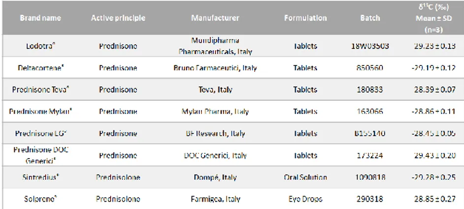
Table 4.1: δ13C values of prednisone and prednisolone pharmaceutical
preparations available in Italy
Table 4.2a: Prednisone: LOQ and recovery. Mean, SD and recovery of three
replicates for each considered spiked positive control urine (USP) at 30, 20 and 10 ng/mL
Table 4.2b: Prednisone: LOQ and recovery. Mean, SD and recovery of three
replicates for each considered spiked positive control urine (USP) at 30, 20 and 10 ng/mL
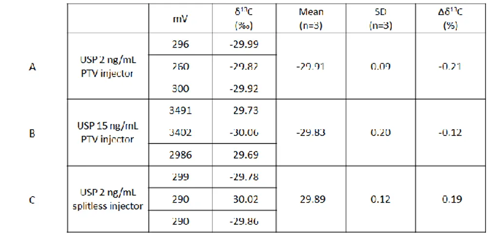
Table 4.3a:Prednisone injected in a splitless (A) or solvent venting mode (B)
Table 4.3b:Prednisolone injected in a splitless (A) or solvent venting mode (B)
Table 4.4:Repeatability. Analysis of samples from four male (M) and four female
(F)
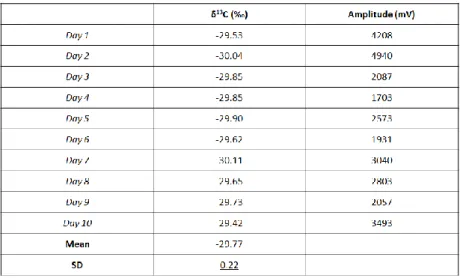
Table 4.5: Repeatability. Analysis of 10 replicated of the same urine spiked at 30
ng/mL
Table 4.6a: Volunteer 1 before and after a Sintredius® 1 mg/mL one vial
administration
Table 4.6b: Volunteer 2 before and after a Deltacortene® 5 mg one tablet
administration
Table 4.7a: Volunteer 1: Δδ13C values (‰) for each ERC-TC pair
Table 4.7b: Volunteer 2: Δδ13C values (‰) for each ERC-TC pair
Table 4.8a: Volunteer 1: endogenous glucocorticoids metabolites δ13C ‰ before
and after a Sintredius® 1 mg/mL one vial administration
Table 4.8b: Volunteer 2: endogenous glucocorticoids metabolites δ13C ‰ before
and after a Deltacortene® 5 mg, one tablet administration
Chapter 5
Detecting the abuse of 19-norsteroids in doping controls: a new
GC-C-IRMS method for the analysis of norandrosterone and
19-noretiocholanolone
XIV
Table 5.1: Collection intervals for the two HPLC purification steps
Table 5.2: LOQ and method linearity in splitless and solvent vent injection mode.
The Δδ13C is against the δ13C value of 19-NE reference standard (-29.70 ± 0.13 ‰).
Table 5.3: δ13C values repeatability
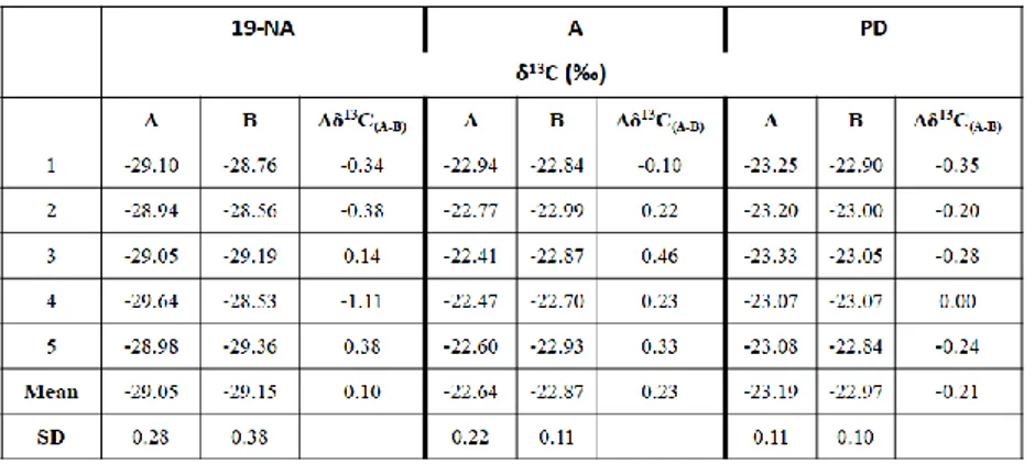
Table 5.4: IRMS analysis of 19-NA, A and PD: comparison between the new
developed procedure (A) and the method previously in use (B)
Table 5.5: Spiked urine sample at 2 ng/mL injected in different GC-C-IRMS
instruments
Chapter 6
Carbon isotopic characterization of prednisolone and prednisone
pharmaceutical formulations: implications in antidoping analysis
Table 6.1: δ13C values of prednisolone and prednisone pharmaceutical
preparations available in Italy, Belgium, Spain, Brazil and India
Table 6.2: δ13C values of PD, PT and THS after the nasal administration of
Sofrasolone®
Table 6.3a: Δδ13C for each ERC-PLONE pair
Table 6.3b: Δδ13C for each ERC-PRED pair
Table 6.4: Volunteer 1: Δδ13C values for each PD-TC pair after the oral
administration of Sintredius® (A) and the combined administration of Sofrasolone® and Sintredius® (B)
Chapter 7
5α-reductase inhibitors: evaluation of their potential confounding
effect on GC-C-IRMS doping analysis
Table 7.1a: Volunteer 1: urinary concentrations ratios and Δδ13C values before the
administration of finasteride of each 5α-, 5β- testosterone metabolites pair
Table 7.1b: Volunteer 1: urinary concentrations ratios and Δδ13C values after the
administration of finasteride of each 5α-, 5β- testosterone metabolites pair
Table 7.2a: Volunteer 2: urinary concentrations ratios and Δδ13C values before the
XV
Table 7.2b: Volunteer 2: urinary concentrations ratios and Δδ13C values after the
administration of finasteride of each 5α-, 5β- testosterone metabolites pair
Table 7.3a: Volunteer 3: urinary concentrations ratios and Δδ13C values before the
administration of finasteride of each 5α-, 5β- testosterone metabolites pair
Table 7.3b: Volunteer 3: urinary concentrations ratios and Δδ13C values after the
administration of finasteride of each 5α-, 5β- testosterone metabolites pair
Table 7.4a: Volunteer 1: 19-NA and 19-NE urinary levels and their corresponding
ratios before the administration of finasteride
Table 7.4b: Volunteer 1: 19-NA and 19-NE urinary levels and their corresponding
ratios after the administration of finasteride
Table 7.5a: Volunteer 2: 19-NA and 19-NE urinary levels and their corresponding
ratios before the administration of finasteride
Table 7.5b: Volunteer 2: 19-NA and 19-NE urinary levels and their corresponding
ratios after the administration of finasteride
Table 7.6a: Volunteer 3: 19-NA and 19-NE urinary levels and their corresponding
ratios before the administration of finasteride
Table 7.6b: Volunteer 3: 19-NA and 19-NE urinary levels and their corresponding
XVI
XVII
Chapter 1
Introduction
Figure 1.1: Gonane nucleus and conventional numbering of the rings and carbons Figure 1.2: Steroidogenesis synthetic pathway
Figure 1.3: Physiological effects (direct and indirect) of testosterone Figure 1.4: The main structural modifications of testosterone
Figure 1.5: 5 Example of the Steroidal Module of the Athlete Biological Passport
Blu lines represent actual test results; red lines indicate individual range116
Figure 1.6: The most common glucocorticoids: chemical structures Figure 1.7: Diagram of cortisol and cortisone metabolism
Figure 1.8: International standards for the most common elements analyzed by
IRMS
Figure 1.9: Carbon isotopic composition of plants and human diets
Figure 1.10: Schematic outline of a GC-C-IRMS system configured for C-isotopic
analysis
Chapter 3
Advantages of using the large volume injection technique in
GC-C-IRMS doping control analyses
Figure 3.1: Schematic representation of the large volume injection phases in a
programmed temperature vaporizer inlet
Figure 3.2a: Instrumental setting in splitless injection mode Figure 3.2b: Instrumental setting in large volume injection mode
Figure 3.3: Chromatographic GC-C-IRMS profile analysis of BM2 (m/z 44 lower and
45/44 upper) after different volumes and injection conditions
Figure 3.4a: β-Bold: comparison of δ13C values, signal and BGD amplitudes using
PTV injection at different volumes (1, 2, 4, 6, 9 µL)
Figure 3.4b: BM2: comparison of δ13C values, signal and BGD amplitudes using PTV
injection at different volumes (1, 2, 4, 6, 9 µL)
Figure 3.5: Chromatographic GC-C-IRMS profile analysis of 19-norandrosterone
XVIII
Chapter 4
Development and validation of a method to confirm the exogenous
origin of prednisone and prednisolone by GC-C-IRMS
Figure 4.1: Instrumental conditions and phases of the PTV injection
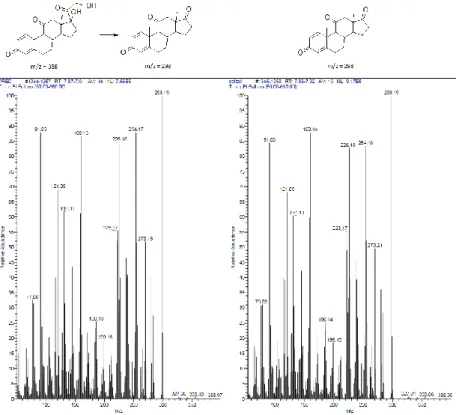
Figure 4.2a: Mass spectra comparison: the prednisone native form and its oxidation
product (on left) and Δ1-adrenosterone (on right)
Figure 4.2b: Mass spectra comparison: the cortisone native form and its oxidation
product (on left) and adrenosterone (on right)
Figure 4.2c: Mass spectra of the prednisolone (on left) and cortisol (on right)
oxidation products
Figure 4.3: HPLC chromatograms of a mixture of standards analysed during the first
purification step. (1) prednisolone; (2) prednisone; (3) cortisol; (4) cortisone; (5) Δ1-adrenosterone; (6) adrenosterone
Figure 4.4: Linearity. (A) The graphs represent the mean measurements of 3
injections of prednisone, the lower and upper acceptance limits (mean ± 0.5 ‰) and the line of best fit; (B) The graphs represent the mean measurements of 3 injections of prednisolone, the lower and upper acceptance limits (mean ± 0.5 ‰) and the line of best fit
Figure 4.5a: Selectivity. Prednisone: the GC-C-IRMS analysis comparison between
the blank and a positive sample
Figure 4.5b: Selectivity. Prednisolone: the GC-C-IRMS analysis comparison between
the blank and a positive sample
Chapter 5
Detecting the abuse of 19-norsteroids in doping controls: a new
GC-C-IRMS method for the analysis of norandrosterone and
19-noretiocholanolone
Figure 5.1a: Linearity of δ13C values: amplitude vs δ13C values. The mean δ13C value
(dotted line), the upper and the lower limit (solid lines; µ ± 0.5 ‰) are represented in the graph
XIX
Figure 5.2a: 19-NA (4 ng/mL): urine sample showing a high background noise
injected after one single HPLC step
Figure 5.2b: 19-NA (4 ng/mL): urine sample showing a high background noise
injected after two sequential HPLC steps in PTV mode
Figure 5.3: 19-NE (4 ng/mL): injection in PTV mode (5 µL)
Figure 5.4: 19-NA (2 ng/mL): injected after two purification steps in PTV mode (9
µL)
Figure 5.5: Volunteer 1: 19-NA and 19-NE urinary excretion profile and their
corresponding ratios. In the box: the last stage of the excretion study; the dotted lines indicate the range within the IRMS procedure should be performed (2.5 – 15 ng/mL)
Figure 5.6: Volunteer 2: 19-NA and 19-NE urinary excretion profile and their
corresponding ratios. In the box: the last stage of the excretion study; the dotted lines indicate the range within the IRMS procedure should be performed (2.5 – 15 ng/mL)
Figure 5.7: Volunteer 1: δ13C values trend of 19-NA and 19-NE
Figure 5.8: Volunteer 2: δ13C values trend of 19-NA and 19-NE
Figure 5.9: Volunteer 3: 19-NA and 19-NE urinary excretion profile and their
corresponding ratios. In the box: the last stage of the excretion study; the dotted lines indicate the range within the IRMS procedure should be performed (2.5 – 15 ng/mL)
Figure 5.10: Volunteer 3: δ13C values trend of 19-NA and 19-NE
Chapter 6
Carbon isotopic characterization of prednisolone and prednisone
pharmaceutical formulations: implications in antidoping analysis
Figure 6.1: Volunteer 1: urinary excretion profile of prednisolone and prednisone
after the nasal administration of Sofrasolone®
Figure 6.2: Volunteer 2: urinary excretion profile of prednisolone and prednisone
after the nasal administration of Sofrasolone®
Figure 6.3: Volunteer 1: urinary excretion profile of prednisolone and prednisone
XX
Figure 6.4: Volunteer 1: δ13C values trend of prednisolone and prednisone after the
nasal administration of Sofrasolone® in comparison with those of PD
Figure 6.5: Volunteer 2: δ13C values trend of prednisolone and prednisone after the
nasal administration of Sofrasolone® in comparison with those of PD
Figure 6.6: Volunteer 1: δ13C values of prednisolone, prednisone and PD after the
oral administration of Sintredius®
Figure 6.7: Volunteer 1: urinary excretion profile of prednisolone and prednisone
after the combined administration of Sofrasolone® nasal spray and Sintredius® oral vial
Figure 6.8a: Volunteer 1: δ13C values of prednisolone after the combined
administration of Sofrasolone® and Sintredius®
Figure 6.8b: Volunteer 1: δ13C values of prednisone after the combined
administration of Sofrasolone® and Sintredius®
Figure 6.9a: Percentage contribution of the two formulations to the resultant δ13C
values of prednisolone after the combined administration
Figure 6.9b: Percentage contribution of the two formulations to the resultant δ13C
values of prednisone after the combined administration
Chapter 7
5α-reductase inhibitors: evaluation of their potential confounding
effect on GC-C-IRMS doping analysis
Figure 7.1a: Volunteer 1: urinary concentrations trend of A and Etio
Figure 7.1b: Volunteer 1: urinary concentrations trend of 5αAdiol and 5βAdiol Figure 7.2a: Volunteer 2: urinary concentrations trend of A and Etio
Figure 7.2b: Volunteer 2: urinary concentrations trend of 5αAdiol and 5βAdiol Figure 7.3a: Volunteer 3: urinary concentrations trend of A and Etio
Figure 7.3b: Volunteer 3: urinary concentrations trend of 5αAdiol and 5βAdiol
Figure 7.4a: Volunteer 1: δ13C values trend of A and Etio
Figure 7.4b: Volunteer 1: δ13C values trend of 5αAdiol and 5βAdiol
Figure 7.5a: Volunteer 2: δ13C values trend of A and Etio
Figure 7.5b: Volunteer 2: δ13C values trend of 5αAdiol and 5βAdiol
Figure 7.6a: Volunteer 3: δ13C values trend of A and Etio
XXI
Figure 7.7: GC-C-IRMS confirmation analysis for detecting the 19-norsteroids
abuse: TC and ERC δ13C values of V1
Figure 7.8: GC-C-IRMS confirmation analysis for detecting the 19-norsteroids
abuse: TC and ERC δ13C values of V2
Figure 7.9: GC-C-IRMS confirmation analysis for detecting the 19-norsteroids
abuse: TC and ERC δ13C values of V3
Annex I
Figure A.1: 1st HPLC chromatogram of TC (PLONE, PRED) and ERC (THS, PT and PD)
in the pre-treatment procedure to detect prednisolone and prednisone abuse; MT is the internal standard
Figure A.2: 2nd HPLC chromatogram of TC (PRED, PLONE) in the pre-treatment
procedure to detect prednisolone and prednisone abuse; DESA is the internal standard
Figure A.3: 1st HPLC chromatogram of TC (19-NE and 19-NA) and ERC (PT, A and PD)
in the new proposed pre-treatment procedure to detect 19-norsteroids abuse; MT is the internal standard
Figure A.4: 2nd HPLC chromatogram of 19-NA in the new proposed pre-treatment
procedure to detect 19-norsteroids abuse; NET is the internal standard
Figure A.5: 2nd HPLC chromatogram of 19-NE, A and PD in the new proposed
pre-treatment procedure to detect 19-norsteroids abuse; NET is the internal standard
Figure A.6: Volunteer 1: carbon isotopic composition of other compounds
(11-β-OH-A; 11-Keto-Etio; T; PT and PD) commonly determined to detect the abuse of pseudo-endogenous steroids. Dotted lines define the individual reference range (µ ± 3SD)
Figure A.7: Volunteer 2: carbon isotopic composition of other compounds
(11-β-OH-A; 11-Keto-Etio; T; PT and PD) commonly determined to detect the abuse of pseudo-endogenous steroids. Dotted lines define the individual reference range (µ ± 3SD)
Figure A.8: Volunteer 3: carbon isotopic composition of other compounds
(11-β-OH-A; 11-Keto-Etio; T; PT and PD) commonly determined to detect the abuse of pseudo-endogenous steroids. Dotted lines define the individual reference range (µ ± 3SD)
XXII
XXIII 11-Keto-Etio 11-keto-etiocholanolone 11β-OH-A 11β-hydroxy-androsterone 11β-HSD 11β-hydroxysteroids dehydrogenase β-Bold boldenone 19-NA 19-norandrosterone 19-NE 19-noretiocholanolone 3β-HSD 3β-hydroxysteroid dehydrogenase 5αAdiol 5α-androstane-3α,17β-diol 5βAdiol 5β-androstane-3α,17β-diol 5α-R 5α-reductase
5-ARI 5α-reductase inhibitor Δ1-SDH Δ1-steroid-dehydrogenation
A Androsterone
AAF Adverse analytical finding AAS Anabolic androgenic steroids ABP Athlete Biological Passport
ACTH Adrenocorticotropic hormone
ADD Boldione
allo-THE allo-tetrahydrocortisone allo-THF allo-tetrahydrocortisol
APMU Athlete Passport Management Unit
AR Androgen receptor
AREs Androgen response elements
ATF Atypical Finding
ATPF Atypical Passport Finding BHP Benign prostatic hyperplasia
BM2 Boldenone metabolite
BUR Blank urine sample
CAM Crassulacean acid metabolism
CIR Carbon isotope ratio
CHR Corticotropin-releasing hormone
COPD Chronic obstructive pulmonary disease
CYP Cytochrome P450
DBS Dried blood spots
DESA Dexamethasone
DHEA Dehydroepiandrosterone
DHT 5α-dihydrotestosterone
DPD DNA-binding domain
XXIV E. Coli Escherichia Coli
EAAS Endogenous anabolic androgenic steroids
ER Smooth endoplasmic reticulum
ERα/β Estrogen receptor alpha/beta
ERC Endogenous reference compound
Etio Etiocholanolone
FSH Follicle-stimulating hormone
GC Gas-chromatography
GCs Glucocorticoids
GC-C-IRMS Gas-chromatography-combustion-isotopic ratio mass spectrometry
GC-MS Gas-chromatography coupled to mass spectrometry GC-MS/MS Gas-chromatography coupled to tandem mass
spectrometry
GnRH Gonadotropin-releasing hormone
GR Glucocorticoid receptor
GRE Glucocorticoids response elements H6PDH Hexose-6-phosphate dehydrogenase HPG-axis Hypothalamic-pituitary-gonadal axis HPLC High performance liquid chromatography
HSD Hydroxysteroid dehydrogenase
Hsp Heat shock protein
IAAF International Association of Athletics Federations IAEA International Atomic Energy Agency
IOC International Olympic Committee
IRMS Isotopic ratio mass spectrometry
ISCCS International Standard for Code Compliance by Signatories
ISL International Standard for Laboratories ISO International Organization for Standardization ISPPPI International Standard for the Protection of Privacy
and Personal Information
ISTDRI Internal standard for IRMS measurements
ISTI International Standard for Testing and Investigations ISTUE International Standard for Therapeutic Use
Exemptions
LBD Ligand-binding domain
XXV
LC-MS Liquid chromatography coupled to mass
spectrometry
LC-MS/MS Liquid chromatography coupled to tandem mass spectrometry
LH Luteinizing hormone
LOD Limit of detection
LOQ Limit of quantification
LVI Large volume injection
MCR2 Melanocortin receptor
MRPL Minimum Required Performance level
MR Mineralocorticoid receptor
MS Mass spectrometry
MT 17α-methyltestosterone
NADPH Nicotinamide adenine dinucleotide phosphate NET 5β-estran-17α-ethynyl-3α,17β-diol
NIST National Institute of Standards and Technology
NTD N-terminal domain
P450scc Cytochrome P-450 side chain cleavage enzyme P450c17 Steroid 17 alpha-hydroxylase/17,20 lyase PAPS 3’–phosphoadenosine-5’-phosphosulfate PBR Peripheral benzodiazepine receptor
PD Pregnanediol POMC Pro-opiomelanocortin PR Progesterone receptor PRED Prednisone PLONE Prednisolone PT Pregnanetriol
PTV Programmed temperature vaporizer
SARM Selective androgen receptor modulator
SG Specific gravity
SHBG Sex hormone binding globulin
SPE Solid phase extraction
SRE Steroid responsive element
SSP Suspicious steroid profile
StAR steroidogenic acute regulatory protein
SULT Sulfotransferases
T Testosterone
TBME tert-butyl methyl ether
XXVI TD2019IRMS Technical Document 2019 Isotopic Ratio Mass
Spectrometry
TD2018EAAS Technical Document 2018 Endogenous Anabolic Androgenic Steroids
TD2019MRPL Technical Document 2019 Minimum required performance levels for the detection and identification of non-threshold substances
TD2019NA Technical Document 2019 Harmonization of analysis and reporting of 19-norsteroids related to nandrolone
THE tetrahydrocortisone
THF tetrahydrocortisol
THS tetrahydro-11-deoxycortisol
TL19 Technical Letter. In situ formation of prednisone and prednisolone
UGT Uridine diphosphate glucuronosyltransferase
USP Positive urine sample
VPA Arginine vasopressin
VPDB Vienna Pee Dee Belemnite
WADA World Anti-Doping Agency
1
2
3
Introduction
1.1 General aspects of doping
1.1.1 Historical perspective
The origin of the word “doping” is still under a controversial etymological investigation but seems to derive from “dope”, a stimulant drink consumed by African tribes in religious ceremonies but also an alcoholic beverage made from grapes skin and used by Zulu warriors during battles. In 1889, doping was first mentioned in the English dictionary to describe a narcotic potion enhancing the racehorse’s performances1. Today, it refers to the occurrence of one or more of the
anti-doping rules violations described from the article 2.1 to the article 2.8 of the Anti-Doping Code (WADC), that are: “(i) the presence of a prohibited substance or its metabolites or markers in an athlete sample; (ii) the use or attempted use by athlete of a prohibited substance or a prohibited method; (iii) refusing or falling without compelling justification to submit to sample collection after notification as authorized in applicable anti-doping rules, or otherwise evading sample collection; (iv) violation of applicable requirements regarding athlete availability of Out-of-Competition Testing, including failure to file required whereabouts information and missed tests which are declared based on rules which comply with the international standard for testing; (v) tampering or attempting to tamper, with any part of doping control; (vi) possession of prohibited substances and prohibited methods; (vii) trafficking or attempted trafficking in any prohibited substance or prohibited method and (viii) administration or attempted administration to any athlete of any prohibited method or prohibited substance”2.
The practice of resorting dietary and medical help to be competitive and enhance performances, even under adverse conditions (injury or illness), is not a modern phenomenon3–9: extracts from the plant Ephedra10, hallucinogenic mushrooms11,
cocoa leaves or various stimulant mixture (strychnine, cocaine, caffeine and alcohol)12,13 were largely used in past during competitions, especially endurance
events. From the 19th century, the misuse of drugs rapidly boosted thanks to the
4 doping-related fatality was reported in 1896, when the Welsh cyclist Arthur Linton died of a combination of caffeine and strychnine overdose during a 600 km race between Bordeaux and Paris12,16. Even if some restrictions were introduced in 1928
by the International Association of Athletics Federations (IAAF) on the use of stimulants and narcotics, appropriate official doping testing procedures had not yet been implemented. The death of the Danish cyclist Knut Jensen during the Rome’s Olympic Game in 1960 and the first televising doping death of the English cyclist Tom Simpson during the Tour de France in 1967, highlighted the urgency of anti-doping policy, leading the International Olympic Committee (IOC) to institute its own Medical Commission and publish the first List of Prohibited Substances (1967)8,17. The List of “Banned Substances Classes and Methods” included five
groups: sympathomimetic amines; stimulants of the central nervous system; analgesic narcotics; anti-depressants and major tranquillizers. The last two categories were removed only one year later; then the list remained practically unchanged until the introduction of anabolic steroids just before the Summer Olympiad in Montreal (1976). Subsequent remarkable changes concerned the prohibition of caffeine (1984)9,18,19, peptide hormones, like human chorionic
gonadotropin20, adrenocorticotropic hormone, human growth hormone12,21,22 (all
in 1989) and erythropoietin (1990)17,23. During the World Conference on Doping in
Sport held in 1999 in Lausanne, Switzerland, the institution of an international anti-doping agency was established in preparation for the imminent Games of the XXVII Olympiad in Sydney in 2000, resulting in the World Anti-Doping Agency (WADA) creation24. WADA has been founded on equal partnership between public
governments and Olympic sport to coordinate the fight against doping and harmonize the Olympic antidoping policies in a single code applicable and acceptable for all the stakeholders4,7,25,26: the World Anti-Doping Code27. It is the
core document of the antidoping community, first adopted in 2003 and subjected to regular reviews and updates over the years28; the last revised version will come
into force on January 2021. It establishes universal anti-doping rules and programs valid for all athletes, regardless of the country in which they compete. The Code acts in conjunction with six International Standards:
5 II. The International Standard for Testing and Investigations (ISTI, a practical directive that preserves the integrity and the identity of the samples from their collection to their analysis)30.
III. The International Standard for Laboratories (ISL, that ensures that all the accredited WADA Laboratory report valid results based on reliable evidentiary tests)31.
IV. The International Standard for Therapeutic Use Exemptions (ISTUE, that describes the conditions in which the possession or the administration of prohibited substances are allowed for therapeutic purposes)32.
V. The International Standard for the Protection of Privacy and Personal Information (ISPPPI, for ensuring appropriate privacy standards to all parties involved in the anti-doping procedures)33.
VI. The International Standard for Code Compliance by Signatories (ISCCS, that describes the rights and the responsibilities for all the Signatories of the Code)34.
1.1.2 The Prohibited List
WADA published its first List of Prohibited Substances and Methods in 2004, following the 2nd World Conference in Doping in Sport (Copenhagen, 2003). Since
then, it is yearly updated according to new doping or new drugs trends showing a potential doping effect35.
In the current version, it is divided in the subsequent sections: Substances and Methods prohibited at all-times:
Prohibited Substances
• S0: Non-Approved Substances • S1: Anabolic Agents
• S2: Peptide Hormones, Growth Factors, Related Substances and Mimetics • S3: Beta-2 Agonist
• S4: Hormone and Metabolic Modulators • S5: Diuretics and Masking Agents
Prohibited Methods
6 • M2: Chemical and Physical Manipulation
• M3: Gene and Cell Doping
Substances and Methods prohibited in competition:
Prohibited Substances
• S6: Stimulants • S7: Narcotics • S8: Cannabinoids • S9: Glucocorticoids
Substances and Methods prohibited in particular sports: • P1: Beta-Blockers
For each class, a list of most representative examples is reported, but compounds with similar chemical or pharmacological activities are also prohibited. Instead, some substances are not yet included in the List, but closely monitored to investigate their pattern of use by athletes (in and/or out of competitions, by systemic or local routes) and verify if they beneficially affect the sportive performance36.
Banned compounds are divided into two groups, depending on whether they may be simply detected and identified (non-threshold substances), or they must reach concentrations above certain cut-off (threshold substances) to provide adverse analytical findings (AAF). This is the case of substances for which is difficult to discriminate the exogenous administration from the physiological production or the pharmacological/social use from a doping violation. Minimum required performance levels have been established for the analysis of the non-threshold substances to harmonize the method sensitivity requirements and the results obtained from all the accredited WADA laboratories37.
Compounds specifically considered in this work are included in the ▪ section S1
19-norandrostenediol 3,17-diol); 19-norandrostenedione (estr-4-ene-3,17-dione); nandrolone (17β-hydroxyestr-4-en-3-one); boldenone (17β-hydroxyandrosta-1,4-dien-3-one); dihydrotestosterone
(17β-hydroxy-5α-7 androstan-3-one); testosterone (17β-hydroxyandrost-4en-3-one) and their metabolites.
▪ section S9
prednisolone (11β,17α, 21-trihydroxypregna-1,4-diene-3,20-dione) and prednisone (17α,21-dihydroxypregna-1,4-diene-3,11,20-trione).
1.1.3 Analytical anti-doping strategies
The current analytical strategies rely on the detection and, in some cases the quantification, of the prohibited substances and methods in biological samples collected in and/or out of competition. The routine anti-doping controls are mainly performed on urinary matrices, because they can be obtained in relatively large volume with non-invasive procedure. Moreover, the concentration of most of the forbidden drugs and their metabolites is higher in urine than in blood. The use of hematological specimens (whole blood, serum and plasma) remains mandatory for some peptide hormones and in case no urinary markers may be detected38. The
anti-doping control is a two-step procedure, in which an initial fast, selective and sensitive screening is followed by a confirmatory process. The first step is applied to all the collected samples to give indication about the presence or the absence of banned substances by limiting the risk of false-negative and false-positive results: the same method is typically applied to a wide number of compounds with similar physical-chemical properties (multitargeted approach). An additional confirmatory test is carried out on suspicious samples to specifically identify the analyte of interest and its metabolites.
Detection of the misuse of banned substances is preferably performed using gas (GC) and liquid (LC) chromatography coupled to mass spectrometry (MS or MS/MS)-based methods39. Some strategies exploit immunoassays and
electrophoretic techniques40. Since many of the banned compounds have their
endogenously produced counterparts, the application of the isotopic ratio mass spectrometry technique has been demanded.
Evidences of the continuous sophistication of the illicit practices and the rise of novel doping agents lead the anti-doping community to opportunely enhance the existing analytical methods and find new strategies41. The selection of alternative
8 DBS), faster and more automated pre-treatment techniques are all approaches recently considered42,43.
1.2 Steroid hormones
Steroid hormones are secreted by adrenal cortex, testes, ovaries, and by placenta during pregnancy. Depending on the receptors to which they bind and consequently on their physiological functions, they can be divided into five groups: glucocorticoids, mineralocorticoids, androgens, estrogens and progestogens. They are all structurally characterized by a tetracyclic hydrocarbon ring, the tetracyclopentanoperhydrophenantrene or gonane core, in which 3 cyclohexane rings (A, B and C) are fused with a cyclopentane ring (D) (Figure 1.1).
Figure 1.1 Gonane nucleus and conventional numbering of the rings and carbons
Steroid hormones are synthesized in the mitochondria and smooth endoplasmic reticulum (ER) from cholesterol, supplied from three different sources. It is provided by acetyl coenzyme A; esterified cholesterol deposits stored within the steroidogenic cells; and low-density lipoproteins derived from dietary cholesterol. The delivery of cholesterol from intracellular stores to the inner mitochondria membrane is the first rate-limiting step of steroidogenesis; the StAR (steroidogenic acute regulatory protein) and PBR (peripheral benzodiazepine receptor) are the two key proteins mainly involved in this process44–47. Cholesterol is then converted
to pregnenolone via three different chemical reactions: two hydroxylations at C-20 and C-22 and a side-chain cleavage event, yielding pregnenolone and isocaproaldehyde. Pregnenolone, once left the mithocondria, may undergo 17α-hydroxylation by P450c17 to obtain 17α-hydroxypregnenolone, or it may be converted to progesterone by 3β-hydroxysteroid dehydrogenase (3β-HSD). A
9 subsequent sequence of hydroxylation and carbon-carbon bond cleavage reactions, mostly catalyzed by CYPs or HSDs, leads to the production of two categories of hormones: 21C steroids
(
progestogens, glucocorticoids and mineralocorticoids, characterized by 21 carbon atoms) and 19C steroids (androgens, that exhibit 19 carbon atoms and from which derive estrogens, 18C steroids). The overall diagram of the human steroidogenesis pathway is represented in Figure 1.2.Figure 1.2 Steroidogenesis synthetic pathway48
The production of steroid hormones is regulated by complex reciprocal interactions among the hypothalamus, anterior pituitary and endocrine glands. Hypothalamic neurons in the supraoptic and paraventricular nuclei secrete hypothalamic releasing hormones that bind membrane receptors on specific subsets of pituitary cells and stimulate the secretion of the related pituitary hormones. The pituitary hormones flow to the target endocrine glands, where they activate the synthesis and the secretion of the target endocrine hormones. Constant negative feedback from the target glands to the anterior pituitary and hypothalamus ensures the body’s homeostasis, turning off the cascade in case of high levels of circulating
10 endocrine hormones. Also the pituitary hormones can act back on the hypothalamus in the so-called short-loop feedback49,50.
1.2.1 Androgens
1.2.1.1 Biosynthesis
Testosterone is the principal secreted androgen, mainly produced in Leydig cells by different pathways (see Figure 1.2). It is primarily derived from pregnenolone in a four steps process: the hydroxylation of the C-17 carbon atom (P450c17); the cleavage of the C-17 and C-20 carbon-carbon (17,20-lyase) bond to form C19 compound DHEA; the conversion of the hydroxyl group at the C-3 carbon into a carbonyl group (3β-HSD) to form androstenedione; the reduction of the 17-oxo
group (17β-HSD)51. Another minor biosynthetic precursor of testosterone is
androstenediol. In women, testosterone is synthetized in corpus luteum and adrenal cortex by similar biosynthetic routes. In detail, it mainly derived from androstenedione conversion in peripheral tissues, while 30 % is produced by adrenal glands and 20 % by ovaries. The serum testosterone concentration ranges from 500 to 7000 ng/dL in men, compared to 30 to 50 ng/dL in women52,53.
Theluteinizing hormone (LH) and the follicle-stimulating hormone (FSH), secreted by the pituitary gonadotropes under the positive regulation of the hypothalamic peptide gonadotropin-releasing hormone (GnRH), stimulate the production of testosterone. The pulsatile secretion of GnRH and pituitary hormones results in a pulsatile daily secretion of testosterone, higher in the morning and lower in the evening. High testosterone and estradiol levels suppress, via negative feedback, the release of LH, FSH and GnRH, inducing the reduction of anabolic androgenic biosynthesis. In women, the secretion of LH is also inhibited by progesterone51,54.
Moreover, in stress instances, glucocorticoids negatively modulate the steroidogenesis cascade at hypothalamic, pituitary and endocrine levels55.
Once secreted, androgens circulate in blood mainly linked to albumin and sex-hormone binding globulin (SHBG), both produced by the liver; only a small portion of them remains unbounded.
1.2.1.2. Mechanism of action
Androgens are the key hormones responsible for the development, maturation and maintenance of male phenotype and sexual and reproductive characteristics and functions. Both in male and in female, they exert anabolic effects on skeletal muscle
11 and bone, by stimulating the linear growth and the protein synthesis56,57. In skeletal
muscle, testosterone directly links the specific receptor and promotes the increase of muscle mass and strength, while, in reproductive tissues, it acts as prohormone of its more potent 5α-reduced derivative, 5α-dihydrotestosterone (DHT). In adipose tissues and parts of brain, testosterone is converted by aromatase to the estrogen, estradiol. In bone, both the direct effect of testosterone and the indirect action mediated by estradiol seem to be relevant (Figure 1.3)54,58,59.
Figure 1.3 Physiological effects (direct and indirect) of testosterone54
At cellular level, the androgen action is mediated by the high affinity binding to the androgen receptor (AR) belonging to the nuclear receptor superfamily of ligand-activated transcription factors, as well as glucocorticoid (GR), mineralocorticoid (MR), progesterone (PR) and estrogen (ERα and ERβ) receptors. It consists of three domains: a N-terminal domain (NTD, containing polyglutamine repeats of different length), a DNA-binding domain (DBD, showing two Zn finger motifs) and a C-terminal ligand-binding domain (LBD). In the absence of ligand, the steroid receptor is located in the cytoplasm as an inactive oligomeric complex with molecular chaperone heat shock proteins Hsp90, Hsp70 and p23, and co-chaperones Hsp40 and Hop, essential to ensure the correct receptor conformation. The hormone binding induces the dissociation of the AR from heat-shock protein complex and its dimerization and translocation to the nucleus. The activated receptor interacts via DBD to androgen response elements (AREs) generally located at the promoter or enhancer regions of AR specific responsive genes, triggering the recruitment of a
12 cluster of gene transcription coregulators (co-activators and co-repressors). This leads to up or down-regulation of the target genes60–64.
Besides their genomic activity, androgens can also influence directly the cells activity in a non-classic pathway, even with no translocation of the AR into the nucleus. The effects tend to be rapid (within seconds to few minutes) and may involve the binding to other membrane-bound androgen receptors, that make androgens no longer able to cross the plasma membrane65–67.
1.2.1.3 Anabolic androgenic steroids
The anabolic androgenic steroids (AAS) are all testosterone derivatives properly synthetized to overcome the pharmacokinetic limitations (rapid first pass metabolism, short half-life) and the androgenic/feminizing side effects occurring after the administration of testosterone (Figure 1.4)68.
Figure 1.4 The main structural modifications of testosterone59
The main structural modifications include 17α-alkylation, 1-methylation, 17β-esterification, addition of a 19-normethyl group, and 7α-methylation69,70. The
substitution of methyl (CH3) or ethyl (C2H5) group for the H on the cyclopentane
ring structure (position 17) hinders the first pass oxidation of the 17β-hydroxy group and confers oral availability. However, after prolonged use at high dosage, these 17α-derivatives (methyltestosterone, methandrostenolone, norethandrolone, fluoxymesterone, danazol, oxandrolone, oxymetholone, stanozol)may cause severe liver disfunction. The attachment of a methyl group on C-1 allows to obtain orally active derivates (methenolone or mesterolone), even if with a weaker pharmacological effect. The esterification of the 17β-hydroxy group
13 with acid moiety of different length leads to more fat-soluble steroids with a much longer half-life (testosterone enanthate, propionate, cypionate, decanoate, isocaproate, phenylpropionate and undecanoate; 19-nortestosterone cyclohexyl propionate, phenylpropionate, decanoate and laurate; methenolone enanthate, boldenone undecylenate, trenbolone acetate, dimeric testosterone). The length of the lateral chain influences the duration of action: more length ensures long-lasting release in the blood stream. Once adsorbed, the 17β-esters are rapidly hydrolyzed by the blood esterases to form the active compounds. They are parentally administered in an oil-based carrier, mainly a mixture of arachis/sesame seed oil and alcohol. Parental preparations show fewer negative effects on hepatic functions. Several other modifications on the ring A, such as junction with pyrazole ring, insertion of methyl group at position C-1, attachment of alkyl substituents or oxygen atom at position C-2, introduction of double bond at position C-1, C-2, and removal of the C-19 methyl group, enhance the anabolic activity relative to androgenic properties71. 19-Nortestosterone (nandrolone) was the first synthetic
testosterone derivative showing an advantageous myotrophic-to-androgenic-ratio in animal experiments. It possesses strong affinity with the androgenic receptor, to which it directly binds in skeletal muscle. Whereas, in androgenic tissues, it is rapidly converted in its 5α-reduced metabolite, that acts on the androgen receptor with weak affinity compared to the parent compound. The insertion of a methyl substituent at the position C-7 of the base structure of 19-nortestosterone (trestolone) has been demonstrated favorable anabolic efficacy68.
1.2.1.4 Metabolism
Anabolic steroids undergo an extensive phase I oxidoreductive reactions principally occurring in the liver72–76. They mainly involve the A and D-rings: C-4, C-5 double
bond reduction (by 5α- and 5β-reductase); C-3 carbonyl group reduction (by 3α- or 3β -hydroxysteroid dehydrogenase); C-1, C-2 double bond reduction (in case of
1,4-diene steroids) and 17β-hydroxy group oxidation (by 17β-hydroxysteroid dehydrogenase) are the most metabolic pathways. The human urine profile of
testosterone includes androsterone, etiocholanolone, dihydrotestosterone, 5α-androstane-3α,17β-diol and 5β-5α-androstane-3α,17β-diol as the more abundant phase I metabolites. The endogenous production of 1,2 dehydrogenated compounds (boldenone and metabolites) by the gut bacteria in humans has been also reported77,78. Metabolism on B-ring preferentially occurs for
6,7-dehydro-14 metabolites (minor pathway). The biotransformation reactions on C-ring are negligible; the hydroxylation at C-12 is the well-studied one. 16-Hydroxylated compounds have been identified for several AAS. Further metabolites can be identified depending on the chemical modifications inserted on the base steroid structure.
Most of endogenous and exogenous androgens are excreted as glucuro- or sulfo-conjugated metabolites72–76,79,80. Both the phase II reactions are enzymatically
controlled. The glucuronidation is catalyzed by uridine diphosphate glucuronosyltransferase (UGTs) membrane bound enzymes through a nucleophilic substitution from the glucuronic acid from uridine diphosphate glucuronic acid to the functional group of the steroid molecule81. Instead, the sulfonation consists in
the transfer of a sulphate moiety by sulfotransferases (SULT), which use the 3’– phosphoadenosine-5’-phosphosulfate (PAPS) as co-substrate, to the acceptor compound82–84. 3α-O-β-Glucuronides, glucuronides and sulfates of the secondary
and tertiary 17β-hydroxy steroids are the major AAS metabolites. Glucuronidation is the preferential metabolic pathway, unless for 3β-hydroxy steroids (such as DHEA, dehydroepiandrosterone85) which are predominantly excreted as sulfate86.
Genetic polymorphism of gene coding metabolizing enzymes may significantly affect urinary androgens levels and cause relevant inter-individual variations. Among the others (CYP17, UGT2B15, UGT2B7, SULT2A1), polymorphism in the UGT2B17 gene has been widely studied, because its deletion drastically reduces the excretion of testosterone and some other related steroids, making more difficult their misuse detection87–89.
1.2.1.5 AAS in clinical practice: therapeutic use and adverse effects
Several evidences about the benefits and the risks associated with the use of AAS in clinical practices are widely reported in literature68,90–95. In the early nineties,
they were usually prescribed by psychiatrists to men suffering depressive disorders, epilepsy and paranoia, also in combination with electroconvulsive therapy. Then, AAS, especially testosterone 17β-esters, 17α-derivatives and nandrolone, have been considered useful in the treatment of cachexia and catabolic conditions in patients with AIDS, chronic obstructive pulmonary disease (COPD), renal and burns failure or malnutrition and growth retardation. AAS (stanozolol) may exert beneficial effects in the treatment of aplastic anemia, even if they have been recently replaced by recombinant human erythropoietin and analogues. The main indication for the administration of androgens remains the male hypogonadism96:
15 testosterone depot preparations (transdermal gel, mucoadhesive buccal tablets, long acting intramuscular injection) show an attractive pharmacokinetic profile to ensure a constant testosterone serum levels in androgens replacement therapies. From the 1990s, however, the selective androgen receptor modulators (SARMs) have become the preferential candidate to treat androgen-deficiency-related diseases: they can be administered orally in low milligram doses and possess high tissue selectivity, consistently reducing the androgenic side effects on prostate, hair and skin97,98.
AAS can cause a wide range of undesirable effects after a prolonged use, an acute overdose or a use for non-medical purposes71,74,90,99–101. They were usually
prescribed in cycles of 6-12 weeks followed by a variable suspension period to avoid remarkable disturbance on the hypothalamic-pituitary-gonadal axis (HPG-axis). The suppression of the release of LH and FSH, that regulate the steroidogenesis and spermatogenesis processes respectively, induces a drastic reduction of fertility, azoospermia and atrophy of testes in men, and amenorrhea in women. As a result of the endogenous aromatization process to estrogens, AAS can lead to gynecomastia and higher voice pitches in men. Androgenic steroids enhance the occurrence of thrombotic events and cardiac damage (left ventricular hypertrophy and heart failure), especially if administered in combination with growth hormone and insulin, also abused for anabolic intent102. The psychological implications are
mostly unpredictable and range from irritability, violent behavior towards himself and the others, to uncontrolled libido. The administration of AAS, particularly the 17α-testosterone derivatives, is also related to a high incidence of hepatotoxicity and increased risk of liver tumor. During puberty, the use of AAS can induce growth stunting, due to a premature closure of the epiphysis. Hirsutism in women and acceleration of baldness in men are adverse effects equally frequent.
1.2.1.6 AAS in doping analysis
The anabolic androgenic steroids represent one of the most group of drugs abused by athletes to improve the sport performances, despite the extent of their adverse effects. In the late 90’s, the administration of androgens for not therapeutic purposes have been significantly increased, especially in the German Democratic Republic and other socialist systems, where many cases of young women athletes submitted to virilization/androgenization programs were reported103. AAS have
been prohibited from 1974; since then, the main analytical challenge has been to differentiate the endogenous from the exogenous steroids, given that they are
16 both excreted in urine as structural identical phase I and phase II metabolites104.
The general instructions for measuring and reporting of endogenous anabolic androgenic steroids (EAAS), also called pseudo-endogenous steroids, have been provided by WADA in the Technical Document TD2018EAAS105. The current strategy
includes an initial testing procedure to estimate the steroid profile of the athlete and a confirmation procedure of any abnormal urinary steroid profiles by GC-C-IRMS analysis. The key parameters of the steroid profile are six steroidal markers (testosterone (T), epitestosterone (E), androsterone (A), etiocholanolone (Etio), 5α-androstan-3α,17β-diol (5αAdiol), 5β-androstan-3α,17β-diol (5βAdiol)) as sum of free and glucuronide fractions) and their relative ratios (T/E, A/T, A/Etio, 5αAdiol/5βAdiol and 5αAdiol/E). More specifically, epitestosterone is the C-17 epimer of testosterone, produced in parallel with T via a not completely known biosynthetic pathway106. Even though the daily production of E is only 3 % of that
of T, the excretion rates of T and E are similar (T/E approximately 1) due to the poor metabolism of E in man. The exogenous administration of testosterone and its precursors in healthy subjects leads to abnormal increase of the urinary concentration of T glucuronide, but does not distinctly affect the E. Therefore, the T/E ratio is considered the most representative first level index of the intake of AAS. The initial cut-off of 6 has been lowered to 4 to avoid false positive results in case of athletes showing a natural elevated T/E ratio107,108. The T/E ratio displays high
population variability, mainly caused by the genetic polymorphism of UGT2B17, and intra-individual fluctuations, especially occurring in female athletes during the different phases of the menstrual cycle109; the 5α- and 5β-metabolites ratio
(5αAdiol/5βAdiol and A/Etio) are more stable steroid profile parameters. They are particularly sensitive to detect application of transdermal formulations of T or DHT administration: DHT is their common precursor, produced from T by the 5α-reductase highly expressed in the skin110. A/T ratio was earlier T/A ratio; the
numerator has been switched with the denominator to reduce the decimals needed. The 5αAdiol/E ratio is the latest parameter added, following the evidence of its usefulness to detect transdermal T-gel111.
These steroid concentrations (adjusted to a urine specific gravity (SG) of 1.020, if SG > 1.018) and ratios are compared to population-based reference range. A sample is suspicious if any of the following parameters are met:
• T/E ratio (calculated from the corrected chromatographic peak areas or height) greater than 4.0;
17 • 5αAdiol/5βAdiol ratio greater than 2.4;
• Concentrations of T or E (adjusted for specific gravity (SG)) greater than 200 ng/mL in males or greater than 50 ng/mL in females;
• Concentrations of A or Etio (adjusted for SG) greater than 10000 ng/mL; • Concentration of 5αAdiol (adjusted for SG) greater than 250 ng/mL in
males or greater than 150 ng/mL in females, combined with a 5αAdiol/E greater than 10 in either sex.
The traditional testing of steroid profile is currently supported by an individual reference range evaluation, the Athlete Biological Passport (ABP), firstly proposed by Donike et al. in 1994 and then developed by Sottas et al.112–114.
The steroidal module of the ABP is a Bayesian screening test for the detection of abnormal values in longitudinal markers. It compares sequential measurements of a steroidal marker with previous readings performed on the same individual: if more measurements are executed, narrower individual reference range can be defined (Figure 1.5)115–117. The responsibility for managing and assessing each
Passport belongs to the Athlete Passport Management Unit (APMU)118.
Figure 1.5 Example of the Steroidal Module of the Athlete Biological Passport
Blu lines represent actual test results; red lines indicate individual range116
The subsequent confirmation procedure is performed when the estimated steroid profile constitutes an Atypical Passport Finding (ATPF) or represents a “suspicious steroid profile” (SSP) finding, or upon direct request from the APMU, the Testing Authority or WADA. It consists of a GC-MS(/MS) full quantitative determination and
18 a GC-C-IRMS analysis of the steroid profile markers to assess their exogenous or endogenous origin105,119.
1.2.2 Glucocorticoids
Glucocorticoids (GCs) are corticosteroids with 21 carbon atoms; cortisol is the main representative one. They have key role in the regulation of carbohydrate, protein, lipid and nucleic acid metabolism53. They promote the gluconeogenesis by liver
from amino acids and glycerol and reduce the utilization of glucose in the peripherical tissues, resulting in the increase of the blood glucose levels. Glucocorticoids stimulate the catabolism of proteins and activate the lipolysis. They induce the mobilization of fatty acids from adipose tissue and their oxidation in cells; excess of cortisol can causes dramatic redistribution of fat in the body, mainly in the back of the neck (“buffalo hump”) and face (“moon facies”). Glucocorticoids are involved in the suppression of inflammatory processes and immune response, since they reduce the release of vasoactive factors, the extravasation of leukocytes and the secretion of lipolytic, proteolytic enzymes and cytokines120. Remarkable
effects are also induced on bone and cardiovascular system, on which high levels of circulating GCs are responsible for osteopenia, osteoporosis and hypertension. Psychiatric manifestations (anxiety, sleep disturbance, mood disorders, psychotic depression and cognitive disfunctions) in patients with GCs excess or deficiency reveal that the central nervous system is another target of this class of hormones. They seem to regulate the neuronal excitability via mechanisms not yet completely understood54.
The molecular pathway by which GCs exert their action at intracellular level involves genomic steps similarly to those previously described for AAS. Briefly, glucocorticoids enter the cell and bind the specific GR receptors inducing its nuclear translocation and the activation or repression of target genes (GRE, glucocorticoids response elements). A non-genomic mechanism including signaling through membrane associated receptors are also reported121–123.
1.2.2.1. Regulation of glucocorticoids secretion
The physiological levels of GCs are regulated by the hypothalamic-pituitary-adrenal (HPA) axis49 in a complex interactions system among the hypothalamic
19 corticotropin-releasing hormone (CRH), arginine vasopressin (VPA) and the pituitary adrenocorticotropic hormone (ACTH), through three different mode:
1. negative feedback mechanism: GCs act at several levels of the HPA axis for decreasing the release of CRH from CRH neurons and ACTH from corticotropes;
2. circadian rhythm:a pulsatile secretion dependent on day-night and sleep-wake patterns. The circulating GCs levels peak at 8 A.M. (140-180 ng/mL, while in the evening 20-40 ng/mL);
3. stress response: stress signals, as injury, hemorrhage, surgery, exercise, pain, anxiety, apprehension, nausea, fever and hypoglycemia overcome the negative feedback regulation and diurnal variation. In stress conditions, cortisol rises consistently.
ACTH (39 amino acids peptide) is released from a larger precursor, the pro-opiomelanocortin (POMC), through sequential proteolytic cleavage steps. The interaction of ACTH with its melanocortin receptor (MCR2, a G protein-coupled receptor) on the adrenal cortex triggers the secretion of GCs, mineralocorticoids and the androgens precursor dehydroepiandrosterone (DHEA, peripherally converted to more potent androgens). More specifically, mineralocorticoids (mostly aldosterone) are synthesized in the glomerulosa outer layer, glucocorticoids (such as cortisone) are produced in the middle fasciculata zona, and the reproductive steroids are secreted by the inner reticularis layer.
See Figure 1.2 for the biosynthetic pathway illustration.
1.2.2.2. Synthetic glucocorticoids: chemical structure and clinical
use
Since cortisone (1948) and cortisol (1951) became the two drugs of choice in the treatment of rheumatoid arthritis, several studies have been implemented to produce derivatives having fewer side-effects, more separated glucocorticoid and mineralocorticoid activities and longer duration of action124. The double bond at
C-4, C-5 and the 3-oxo-group are essential for both the glucocorticoid and mineralocorticoid activities, while the 11β and 17α-hydroxyl groups on ring C and D respectively are responsible for an appreciable glucocorticoid activity and high potency. The introduction of an additional C-1, C-2 double bond (prednisone and prednisolone) and a hydroxyl or methyl group at C-16, selectively increases the
20 glucocorticoid activity, especially if combined with a fluorine atom at C-9 (triamcinolone, dexamethasone, betamethasone). Esterification of the hydroxyl groups at C-17 and C-21 with valerate or propionate enhances the molecular lipophilicity with improved topical/systemic potency ratios125. The most common
GCs are represented in Figure 1.6. Compounds differing in potency, activity and half-life have been synthetized by providing the opportune chemical modifications. Depending on their half-life, GCs can be divided in short- (≤ 12 h; hydrocortisone and cortisone), intermediate- (12-36 h; prednisone, methylprednisolone), and long- (36-72 h; dexamethasone and betamethasone) acting agents.
Figure 1.6 The most common glucocorticoids: chemical structures
Due to their prominent anti-inflammatory and immunosuppressive effects, GCs are mainly used for treatment of several inflammatory (psoriasis, eczema, allergies, asthma) and rheumatic (rheumatoid arthritis) diseases126,127. They may be also
administered in replacement therapy in cases of hypothalamic-pituitary-adrenal axis failure or adrenal insufficiency, and in clinical oncology to reduce the side effects of chemotherapy and treat lymphoproliferative disorders124. The
benefit-to-risk ratio of glucocorticoids, especially after chronic therapies, has been debated for years. Treatment with systemic GCs is associated with a number of adverse reactions and toxicities of different extents: suppression of the HPA axis, bone fragility, growth arrest, behavioral and psychiatric problems, hypertension, hyperglycemia and increased risk of infections are the most significant ones123,126,128–131.
21
1.2.2.3. Metabolism
As already described for AAS, the phase I metabolism of GCs mainly involves the reduction of both the C-4, C-5 double bond and the C-3 carbonyl group. Most of these A-ring reduced compounds are excreted as glucuro- or sulfo- conjugated metabolites76: specifically, all metabolites with a 3α-hydroxyl substituent are
excreted as glucuronide conjugates and those with a 3β-hydroxy-5-ene group are excreted as sulfates. Other cysteinyl conjugates and N-acetylglucosamines types of conjugated metabolites have been recorded132. Metabolism of cortisol and
cortisone is illustrated in Figure 1.7: tetrahydrocortisone (THE), 5α-tetrahydrocortisone (Allo-THE), tetrahydrocortisol (THF), 5α-tetrahydrocortisol (Allo-THF) and 11-desoxy-tetrahydrocortisol (THS) are the main metabolites133.
Corticosteroids with the 11-keto group, as cortisone and prednisone, undergo a pre-receptor metabolism consisting of an enzymatic reduction to the corresponding more active 11β-OH derivatives, cortisol and prednisolone123. The
enzymatic conversion is catalyzed by 11β-HSD type 1 (11β-hydroxysteroids dehydrogenase) in a nicotinamide adenine dinucleotide phosphate
(NADPH)-dependent reaction. 11β-HSD1 is a reductase widely expressed in liver, adipose tissue, muscle, pancreatic islets, adult brain and inflammatory cells. Its activity is supported by the microsomal hexose-6-phosphate dehydrogenase (H6PDH), able to regenerate NADPH and supply the reducing sources. Accordingly, in patients suffering from cortisone reductase deficiency, drugs that do not require the enzymatic activation should be preferentially administered134–136. The conversion
of the oxo precursors in their hydroxy metabolites is inhibited by the 11β-HSD type 2, since it catalyzes the opposite reaction. 11β-11β-HSD type 2 is mainly located in the mineralocorticoids target tissues (kidneys) where the GCs inactivation promotes the effects mediated by the aldosterone-mineralocorticoid receptor (MR) binding.
22
Figure 1.7 Diagram of cortisol and cortisone metabolism133
1.2.2.4. GCs as doping agents
Glucocorticoids exert a beneficial impact on the muscle responsiveness and recovery during sport performances, by increasing the availability of metabolic substrates and reducing the feeling of fatigue and the pain of efforts137. GCs are
widely abused by athletes, despite the not negligible side effects associated with their administration138,139. They are included in the section S9 of the WADA
Prohibited List and banned “in competition” when administered by systemic (oral, intravenous, intramuscular or rectal) routes29. A reporting level of 30 ng/mL has
been established by WADA to discriminate the allowed (topical) from the prohibited (systemic) administration: a concentration higher than 30 ng/mL shall be observed in urine samples to report an adverse analytical finding37. Synthetic
23 based methods is limited by the difficulty in obtaining unique derivatization products143. GCs are also included in multitargeting procedures for the
simultaneous detection of other banned compounds (anabolic agents, β2-agonists, hormone antagonists and modulators, diuretics, stimulants, narcotics and β-blockers)144,145. Moreover, since hydrocortisone and cortisone are naturally
produced in the body, the isotope ratio mass spectrometry analysis can be utilized to determine the abuse of endogenous glucocorticoids by measuring the carbon isotope ratio of their resulting metabolites in human urine samples133,146.
1.3 Ex-vivo degradation of endogenous compounds
The usual non-sterile collection or transportation conditions and the presence of normal or pathogenic microbial flora contamination are all favorable circumstances able to change the composition of the collected urine (increase or depletion of endogenous steroids or even the hydrolysis of conjugated metabolites) and alter specific steroid parameters. The free/glucuronide testosterone and 5α-androstanedione/A and 5β-androstanedione/Etio ratios are all criteria monitoring in doping analyses as markers of the bacterial metabolic activity147–150. Among theex vivo bacterial degradation of endogenous steroids, 19-demethylation and
Δ1-steroid-dehydrogenation have been extensively reported in literature and considered in this PhD thesis. They induce the formation of banned substances (19-norandrosterone, boldenone, prednisolone and prednisone) even if no drugs intake has occurred, affecting the correct interpretation of data.
19-Norsteroids
19-Norsteroids, nandrolone and its precursors, are anabolic androgenic compounds synthetized since the 1950s to enhance the myotrophic action and reduce the androgenic side effects of testosterone, from which they structurally derive151–153. They are primarily metabolized into two glucuronic acid conjugated
products, 19-norandrosterone (19-NA), about 72 %, and 19-noretiocholanolone (19-NE), about 28 %. Unfortunately, the presence of 19-NA and 19-NE in urine could also occur in case of not intentional intake of nandrolone and its precursors154: after the consumption of non-castrated boar edible tissues155–157,
during pregnancy or norethisterone based contraceptive therapy158–162 or as the