original article
Mutations in DSTYK and Dominant Urinary
Tract Malformations
S. Sanna-Cherchi, R.V. Sampogna, N. Papeta, K.E. Burgess, S.N. Nees, B.J. Perry, M. Choi, M. Bodria, Y. Liu, P.L. Weng, V.J. Lozanovski, M. Verbitsky, F. Lugani,
R. Sterken, N. Paragas, G. Caridi, A. Carrea, M. Dagnino, A. Materna-Kiryluk, G. Santamaria, C. Murtas, N. Ristoska-Bojkovska, C. Izzi, N. Kacak, B. Bianco,
S. Giberti, M. Gigante, G. Piaggio, L. Gesualdo, D. Kosuljandic Vukic, K. Vukojevic, M. Saraga-Babic, M. Saraga, Z. Gucev, L. Allegri, A. Latos-Bielenska,
D. Casu, M. State, F. Scolari, R. Ravazzolo, K. Kiryluk, Q. Al-Awqati, V.D. D’Agati, I.A. Drummond, V. Tasic, R.P. Lifton, G.M. Ghiggeri, and A.G. Gharavi
The authors’ full names, degrees, and affiliations are listed in the Appendix. Address reprint requests to Dr. Gharavi at the Division of Nephrology, Columbia University College of Physicians and Surgeons, 1150 St. Nicholas Ave., Russ Berrie Pavilion 413, New York, NY 10032, or at [email protected].
Drs. Sampogna and Papeta and Ms. Bur-gess contributed equally to this article. This article was published on July 17, 2013, at NEJM.org.
N Engl J Med 2013;369:621-9. DOI: 10.1056/NEJMoa1214479
Copyright © 2013 Massachusetts Medical Society. ABS TR ACT
BACKGROUND
Congenital abnormalities of the kidney and the urinary tract are the most common cause of pediatric kidney failure. These disorders are highly heterogeneous, and the etiologic factors are poorly understood.
METHODS
We performed genomewide linkage analysis and whole-exome sequencing in a family with an autosomal dominant form of congenital abnormalities of the kidney or urinary tract (seven affected family members). We also performed a sequence analysis in 311 unrelated patients, as well as histologic and functional studies. RESULTS
Linkage analysis identified five regions of the genome that were shared among all affected family members. Exome sequencing identified a single, rare, deleterious vari-ant within these linkage intervals, a heterozygous splice-site mutation in the dual serine–threonine and tyrosine protein kinase gene (DSTYK). This variant, which resulted in aberrant splicing of messenger RNA, was present in all affected family members. Additional, independent DSTYK mutations, including nonsense and splice-site mutations, were detected in 7 of 311 unrelated patients. DSTYK is highly expressed in the maturing epithelia of all major organs, localizing to cell membranes. Knock-down in zebrafish resulted in developmental defects in multiple organs, which suggested loss of fibroblast growth factor (FGF) signaling. Consistent with this finding is the observation that DSTYK colocalizes with FGF receptors in the ure-teric bud and metanephric mesenchyme. DSTYK knockdown in human embryonic kidney cells inhibited FGF-stimulated phosphorylation of extracellular-signal-regu-lated kinase (ERK), the principal signal downstream of receptor tyrosine kinases. CONCLUSIONS
We detected independent DSTYK mutations in 2.3% of patients with congenital ab-normalities of the kidney or urinary tract, a finding that suggests that DSTYK is a major determinant of human urinary tract development, downstream of FGF sig-naling. (Funded by the National Institutes of Health and others.)
C
ongenital malformations of the kidney and urinary tract contribute to 23% of birth defects1,2 and account for 40 to 50% of pediatric cases and 7% of adult cases of end-stage renal disease (ESRD) worldwide.3,4 These disorders are genetically heterogeneous and encompass a wide range of anatomical de-fects, such as renal agenesis, renal hypodyspla-sia, ureteropelvic junction obstruction, or vesico-ureteral reflux.5 Mutations in genes that cause syndromic disorders, such as HNF1B and PAX2 mutations, are detected in only 5 to 10% of cas-es.6,7 Familial forms of nonsyndromic disease have been reported, further supporting genetic determination8,9; however, owing to locus het-erogeneity and small pedigree size, the genetic cause of most familial or sporadic cases remains unknown.We studied a family with an autosomal dominant form of congenital abnormalities of the kidney and urinary tract (Fig. 1A; and Table S1 in the Supplementary Appendix, available with the full text of this article at NEJM.org).8 Seven family members were classified as affected on the basis of the presence of a solitary kidney, renal hypodysplasia, ureteropelvic junction ob-struction, or vesicoureteral reflux, as document-ed by means of imaging studies. The mean age at diagnosis was 12 years (range, 1 to 37). Three family members (members 7, 8, and 13), all with ureteropelvic junction obstruction, had ESRD at a young age (at 8, 10, and 23 years of age, re-spectively), without other known causes of renal failure, such as diabetes mellitus or uncontrolled hypertension. Epilepsy developed in these three persons during their third decade of life. The other affected family members did not have evi-dence of renal dysfunction at the last follow-up visit. Seven family members were classified as unaffected on the basis of normal ultrasono-graphic studies of the abdomen and normal re-nal function, and six were classified as having an unknown phenotype, owing to unavailable or indeterminate ultrasonographic studies.
A series of 311 patients with congenital ab-normalities of the kidney and urinary tract was studied to identify independent mutations. Ge-netic and clinical data for 7 of these patients (5 of whom had congenital obstructive uropathy) are shown in Table 1 and Figure 1.
METHODS
GENOTYPING AND SEQUENCING
After genomewide genotyping, we conducted a genomewide analysis of linkage, under an auto-somal dominant mode of inheritance, with as-signment of phenotype only to affected persons (affected-only analysis). Rare copy-number vari-ants were excluded with the use of HumanHap 650Y Genotyping BeadChips (Illumina). Whole-exome sequencing and analysis were performed as previously described10,11 in two members of the family (members 13 and 19).
We performed Sanger sequencing of DSTYK to validate exome data and detect independent mu-tations in 311 unrelated patients with congenital abnormalities of the kidney and urinary tract and in 384 healthy European controls matched to patients with DSTYK mutations according to self-reported ethnic group; 96 of these controls were also matched according to recruitment site. Cod-ing exons and flankCod-ing introns of HNF1B, PAX2, and EYA1 were sequenced in the 7 patients car-rying DSTYK mutations. We determined allele frequencies with the use of public databases, scores from Polymorphism Phenotyping, version 2 (PolyPhen-2), and alignment in 22 mammalian species.
Written informed consent was obtained from all the participants. The study was approved by the institutional review board at each participat-ing site. All the authors vouch for the accuracy and completeness of the data and for the fidelity of the study to the protocol.
LYMPHOCYTE IMMORTALIZATION AND DNA ANALYSIS
We generated lymphoblastoid cell lines from 17 family members. Total RNA was isolated, comple-mentary DNA (cDNA) was generated, and Sanger sequencing was performed on the cDNA with the use of primers spanning the exon 2 and 3 bound-aries.
IMMUNOHISTOCHEMICAL AND IMMUNOFLUORESCENCE TESTING
DSTYK expression was examined in adult and embryonic mouse organs (embryonic day 15.5 to 18.5) and in a kidney and ureter from a pediatric donor (3 months of age; International Institute
1 2 3 4 5 6 7 8 9 10 11 12 13
p.W8X c.654+1 G→A c.655−3 C→T p.R29Q p.D200G p.S843L
AGCGGAACTGGAGGTAACGATGCACCATGCTCTCTTACAGGAAGTGGACGTTGTG
AGCGGAACTGGAGGTAACGATGCACCATGCTCTCTTACAGGAAGTGGACGTTGTG
AGCGGAACTGGAGGAAGTGGACGTTGTG Alternative splice site L 1 2 3 4 5 6 7 8 9 10 11 12 13 14 15 16171819202122 DSTYK D LOD 4 0 2 −2 −4 −6 F G H C B A 1 2 3 4 5 6 7 8 9 10 11 12 13 14 15 16 17 18 19 20 E I R L R L R L R L Genomic DNA cDNA c.654+1 G→A c.654+1 G→A Forward c.654+1 G→A Reverse Nonmutant Nonmutant Exon 2 Exon 2 Exon 3
Figure 1. Identification of DSTYK Mutations in a Family with Congenital Abnormalities of the Kidney and Urinary Tract and the Spectrum
of Mutations and Phenotypes in Unrelated Patients.
Panel A shows the pedigree of the study family. Squares represent male family members, circles female family members, black symbols affected persons, white symbols unaffected persons, gray symbols unknown phenotype, red solid circles DSTYK mutation carriers, and red open circles noncarriers of the mutation; slashes indicate deceased family members. Panel B shows the linkage analysis identifying five regions (arrows) of the genome reaching the maximal expected LOD score of 1.5. DSTYK is located in the chromosome 1q25–1q41 locus. Panel C shows a chromatogram of a DSTYK c.654+1 G→A mutation from genomic DNA. Sequence analysis of complementary DNA (cDNA) in mutation carriers shows the use of an alternative splice site in exon 2, leading to a 27-bp deletion (Panel D). Panel E shows the genomic structure of DSTYK and the location of pathogenic mutations identified in the present study. The exons encoding the kinase domain are shown in green. Representative ultrasonographic findings in mutation carriers are shown in Panels F, G, and H, with hypoplasia of the left kidney (Panel F, kidneys outlined by dashed lines) detected at birth in a girl with a p.R29Q mutation, bilateral hydronephrosis (Panel G, arrows) caused by ureteropelvic junction obstruction detected at birth in a girl with a c.655−3 C→T mutation, and hydronephrosis only of the left kidney (Panel H, arrow) caused by ureteropelvic junction obstruction in a 5-year old boy with a p.R29Q mutation. The intravenous pyelogram in Panel I shows blunting of fornices on the right side (white arrow) and calyceal dilatation on the left side (red arrow) in a 2-year-old boy with a p.W8X mutation.
Ta bl e 1. Spectrum of DSTYK Mutations and Associated Phenotypes.* Variant Consequence
PolyPhen-2 Prediction (Score)†
Ancestry‡ Sex Age at Diagnosis Phenotype Familial or Sporadic Disease Chronic Renal Failure or Dialysis Extraurinary Tract Condition Splice-site or truncating mutation c.654+1 G→A (in seven members of the study family)§ p.V210_ Q218del NA Italian M and F 1–37 yr RHD, UPJO, and VUR Familial Ye s (m em be rs 7, 8 , a nd 1 3) Epilepsy (members 7, 8, and 13) c.24G→A p.W8X NA Macedonian M 2 yr UPJO Sporadic No Hemangioma, ataxia (resolved), and hearing loss c.655–3 C→T (in two siblings from another family)¶ — NA Italian F At birth UPJO Familial No Hypercalciuria Missense mutation c.86G→A p.R29Q Probably damaging (0.997) Albanian M 5 yr UPJO Sporadic No — c.86G→A p.R29Q Probably damaging (0.997) Italian M Prenatal RHD Sporadic Yes — c.86G→A p.R29Q Probably damaging (0.997) Albanian F At birth RHD Sporadic No Congenital adrenal hyperplasia c.599A→G p.D200G Probably damaging (0.992) Italian M At birth UPJO Sporadic No Hearing loss c.2528 C→T p.S843L Possibly damaging (0.741) Croatian M 5 mo CHN Sporadic Yes Factor VII deficiency and hypercalciuria * The protein and complementary DNA annotations are based on National Center for Biotechnology Information reference sequence numbers NP_056190.1 and NM_015375.2, respec -tively. All variants in this table were absent in both the Single Nucleotide Polymorphism database for build 137 and the exome variant server and were not detected in 384 European controls. CHN denotes congenital hydronephrosis, NA not applicable, RHD renal hypodysplasia, UPJO ureteropelvic junction obstruction, and VUR vesicoureteral reflux. † Scores from Polymorphism Phenotyping, version 2 (PolyPhen-2), indicate the probability of the mutation causing damage. ‡ Ancestry was self-reported and confirmed by the investigator on the basis of the nationality and region of birth of the index patient and his or her parents and grandparents. § Detailed phenotypes of the seven affected family members are described in Table S1 in the Supplementary Appendix. ¶ T hi s m ut at io n w as fo un d in t w o si bl in gs w ith o bs tr uc tiv e ur op at hy ; o nl y on e si bl in g w as c ou nt ed in t he g ro up o f 3 11 u nr el at ed p at ie nt s. T he c .6 55 –3 C → T va ri an t is p re di ct ed t o de cr ea se the consensus values for the canonical splice site from 96.68 to 89, corresponding to a −7.95% variation (Human Splicing Finder; www.umd.be/HSF/).
for the Advancement of Medicine). Immunohisto-chemical testing was performed with the use of DSTYK antibody (also called RIP5 anti-body; Universal Protein Resource Knowledgebase accession number, Q6XUX3; catalogue number, sc-162109; Santa Cruz Biotechnology) on paraffin-embedded tissues with the use of heat-induced antigen retrieval. Immunofluorescence testing was performed with the use of the following an-tibodies: anti-RIP5 antibody, antibodies to fibro-blast growth factor (FGF) receptors 1 and 2 (anti-FGFR1 and anti-FGFR2 antibodies, respectively), anti–aquaporin-2 antibody, and anti–E-cadherin antibody (see the Supplementary Appendix).
ZEBRAFISH MORPHOLINO KNOCKDOWN
Morpholino oligomers were designed to block the zebrafish dstyk (National Center for Biotech-nology Information reference sequence number, NM_205627.2) exon 9 splice donor and to truncate or delete the ATP-binding kinase domain. Morpho-lino injections were also performed in tumor-suppressor protein (p53)–mutant homozygotes12 to control for nonspecific antisense effects.
SMALL INTERFERING RNA KNOCKDOWN AND WESTERN BLOTTING
Human embryonic kidney 293T cells were grown with the use of standard procedures. The cells were transfected with a mixture of DSTYK small inter-fering RNAs (siRNAs; SMARTpool, Dharmacon); for a negative control, we used a pool of siRNA that was tested by the manufacturer for not inter-fering with any transcript. At 72 to 96 hours after transfection, the cells were starved in serum-free medium and treated with FGF. Blotting was per-formed as above with the use of goat anti-RIP5 and rabbit anti-FGFR2 antibodies; mouse anti– glyceraldehyde-3-phosphate dehydrogenase anti-body; rabbit anti–phosphorylated extracellular-signal-regulated kinase (pERK) 1 and 2 and anti–extracellular-signal-regulated kinase (ERK) 1 and 2 antibodies; and horseradish-peroxidase– labeled sheep antimouse, donkey antigoat, or don-key antirabbit antibody. Detailed methods are in-cluded in the Supplementary Appendix.
R ESULTS
HETEROZYGOUS MUTATIONS IN DSTYK
Genomewide analysis of linkage with the use of an affected-only analysis identified five regions
of the genome that were shared among all seven affected members (Fig. 1). These intervals col-lectively spanned 55.44 Mb (approximately 1.8%) of the genome, containing 645 protein-coding genes (Table S2 in the Supplementary Appendix). Analysis of copy-number variants in all affected persons excluded major genomic imbalances (data not shown).
Whole-exome sequencing, performed in mem-bers 13 and 19 of the study family (Fig. 1A) at an average depth of coverage of approximately 108 times per base, identified 14,943 single-nucleotide variants (SNVs) across the genome, including 709 SNVs that were absent in all public databases. Among these, 24 protein-altering variants were shared by these two patients (missense, nonsense, or splice-site variants) (Table S3 in the Supple-mentary Appendix). Only 2 of these 24 SNVs, both on chromosome 1q25–1q41, mapped to the linkage intervals.
Follow-up testing by means of Sanger sequenc-ing detected both SNVs in all affected persons and the obligate carrier in the family. One of these SNVs (p.A111V in TIMM17A) was common among persons of Sardinian ancestry (minor al-lele frequency, 0.47). The other variant, a ca-nonical splice-donor SNV at the first base after exon 2 (c.654+1 G→A) of DSTYK, encoding a dual serine–threonine and tyrosine kinase, was ab-sent in 48 persons of Sardinian ancestry and 384 European controls (Fig. 1C). The DSTYK mutation was heterozygous in all affected persons, obligate carriers, and two apparently unaffected members (who were 44 and 20 years old at the last follow-up visit).
Sequence analysis of DSTYK cDNA from lym-phoblastoid cell lines in 17 family members showed that all mutation carriers had a hetero-zygous 27-bp deletion resulting from the use of an alternative splice donor within the normal exon 2, which would result in an in-frame deletion of nine amino acids (VTMHHALLQ) in a domain that is highly conserved among mammals (Fig. 1, and Fig. S1 and S2 in the Supplementary Appendix).
We next searched for independent mutations in DSTYK in 311 additional patients with con-genital abnormalities of the kidney and urinary tract. We identified a nonsense mutation (p.W8X) in a patient with ureteropelvic junction obstruc-tion and early-onset ataxia, and a splice-site mu-tation (c.655−3 C→T) in two siblings affected by ureteropelvic junction obstruction. We also
iden-tified, in 5 other patients, three missense variants that occur at completely conserved positions in mammals and that were predicted to be damag-ing accorddamag-ing to PolyPhen-2 (p.R29Q, p.D200G, and p.S843L). These five damaging variants were absent in all public databases and were also not detected in the 384 healthy European controls. Moreover, none of these patients carried delete-rious structural variants13 or point mutations in HNF1B, PAX2, or EYA1. The phenotypic spectrum associated with DSTYK mutations is described in Table 1 and shown in Figures 1F through 1I.
Thus, sequence analysis in an independent cohort of 311 unrelated patients with congenital abnormalities of the kidney and urinary tract identified five DSTYK mutations in 7 patients (2.3% of this independent cohort) (Table 1). As a comparison, the exome variant server (a data-base hosted by the National Heart, Lung, and Blood Institute that contains exome data for 6503 people) does not contain any DSTYK non-sense mutations or variants affecting the three canonical nucleotides flanking splice junctions. Only 0.3% of white persons (14 of 4300) in this database have rare damaging DSTYK missense variants affecting completely conserved positions in mammals (11 variants with a minor allele fre-quency of <0.001), indicating a significant excess burden of rare damaging variants among persons with congenital abnormalities of the kidney and urinary tract (7 of 311 affected persons [2.3%]; odds ratio, 7.1; P = 0.0003 by Fisher’s exact test).
UBIQUITOUS EXPRESSION OF DSTYK ON CELL MEMBRANES
DSTYK has a striking membrane-associated dis-tribution in mesenchymal-derived cells of all major organs (Fig. S3 in the Supplementary Ap-pendix). In the developing mouse kidney, it is expressed at low levels in the nephrogenic zone but is more highly expressed in maturing tubular epithelia, with the most prominent expression in the medulla and the papilla (Fig. S3G, S3H, and S3I in the Supplementary Appendix). In postnatal mouse and human pediatric kidneys, DSTYK is detected in the basolateral and apical membranes of all tubular epithelia (Fig. 2A through 2D, and Fig. S4 in the Supplementary Appendix). It has both a basolateral and a cytoplasmic distribution in the thin ascending limb of the loop of Henle and the distal convoluted tubule, but expression is restricted to apical and basolateral membranes in the collecting duct, including the principal
and intercalated cells (Fig. 2A through 2D, and Fig. S5 in the Supplementary Appendix). DSTYK was also detected in all layers of transitional ure-teric epithelium and in the ureure-teric smooth-mus-cle cells (Fig. 2D).
MORPHOLINO KNOCKDOWN IN ZEBRAFISH
To investigate the role of DSTYK in embryonic development, we performed knockdown of the orthologue in zebrafish. With maximal knock-down, embryos showed growth retardation, as evidenced by small fins, abnormal morphogene-sis of the tail, and loss of heartbeat (Fig. S6 in the Supplementary Appendix); we also observed clo-acal malformations that correspond to lower genitourinary defects in mammals and defects in jaw development, as well as specific loss of the median fin fold. Pericardial effusion was evident in 5-day-old morphant larvae, which was attrib-utable to both heart and kidney failure. These data suggest an essential role of DSTYK in the development of major organs. The observed de-velopmental defects resemble phenotypes produced by global loss of FGF signaling in zebrafish.14-16
COLOCALIZATION OF DSTYK AND FGF RECEPTORS and effect on erk phosphorylation
In the developing mouse nephron (embryonic day 15.5), DSTYK colocalizes with cells that are E-cadherin–positive and those that are E-cad-herin–negative, confirming its expression in both the metanephric mesenchyme and the ureteric bud (Fig. 2G and 2H). DSTYK localization to the cell membrane in the metanephric mesenchyme and ureteric bud highly parallels the known ex-pression pattern of FGF receptors. Joint immu-nostaining confirmed that DSTYK colocalizes with both FGFR1 and FGFR2 in the ureteric bud and comma-shaped bodies (Fig. 2G and 2H, and Fig. S7 in the Supplementary Appendix). Colocal-ization with FGFR2 was also evident in distal tu-bular cells in the adult renal medulla and papilla (Fig. S8 in the Supplementary Appendix). Punc-tate DSTYK staining was seen at apical cell–cell junctions lining the ureteric-bud epithelia (Fig. S9 and S10 in the Supplementary Appendix).
On activation, FGF receptors trigger cytoplas-mic protein kinases, resulting in ERK phosphory-lation, which is the main effector of FGF-induced transcriptional activity.17,18 Because DSTYK en-codes a kinase and colocalizes with FGFR1 and FGFR2, we hypothesized that DSTYK acts as a positive regulator of FGF-mediated signaling in
− − − + + − + + E G H F A Glom MD CD CD Sm Ur FGFR1+DSTYK DSTYK siRNA 105 kDa DSTYK FGF pERK1/2 GAPDH 44/42 kDa 38 kDa 44/42 kDa 96 hr ERK1/2 UB FGFR1+DSTYK DSTYK FGFR1 MM MM FGFR2+DSTYK E-cadherin FGFR2 DSTYK E-cadherin UB DSTYK FGFR2 FGFR2+DSTYK CD AL AL C D I B
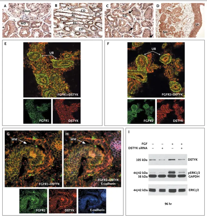
Figure 2. DSTYK Expression in the Human and Mouse Urinary Tract and Small Interfering RNA (siRNA) Knockdown Studies.
Panels A through D show DSTYK expression in the kidney and ureter of a 3-month-old child. Panel A shows the glomerulus (Glom) and macula densa (MD). Panel B shows the renal cortex with ascending limbs of loop of Henle (AL) and collecting ducts (CD). Panel C shows the CD in the renal medulla, with punctate staining at the basolateral side (arrows). Panel D shows the smooth-muscle layer (Sm) and urothelium (Ur) of the ureter. Panels E through H show DSTYK colocalizing with fibroblast growth factor (FGF) receptors. Immunofluorescence analysis in a developing murine kidney (embryonic day 15.5) shows DSTYK (red) colocalization with FGF re-ceptor 1 (FGFR1; green) (Panel E), FGF rere-ceptor 2 (FGFR2; green) (Panels F, G, and H) and E-cadherin (Panel H; blue). In Panel I, DSTYK knockdown is shown to inhibit FGF-mediated phosphorylation of extracellular-signal-regulated kinase 1 and 2 (ERK 1 and 2); siRNA knockdown of DSTYK in 293T cells starved in serum-free medium shows a significant reduction in the DSTYK level at 96 hours after knockdown. FGF stimulation significantly augments phosphorylated ERK (pERK 1 and 2), but DSTYK knockdown abrogates FGF-mediated ERK phosphorylation. Glyceraldehyde-3-phosphate dehydrogenase (GAPDH) was used as a loading control. MM denotes metanephric mesenchyme, and UB ureteric bud.
the kidney. To test this hypothesis, we per-formed siRNA knockdown of DSTYK in the hu-man embryonic kidney-cell line 293T, which re-sulted in a reduction of up to 80% in transcript levels and a pronounced reduction of DSTYK protein levels within the first 96 hours after transfection (Fig. 2I, and Fig. S11 in the Supple-mentary Appendix). FGF stimulation augmented the levels of phosphorylated ERK (pERK) as ex-pected, but siRNA silencing of DSTYK signifi-cantly prevented ERK phosphorylation (Fig. 2I). This effect was not mediated by a direct physical interaction of DSTYK with FGFR2 (Fig. S12 in the Supplementary Appendix). Combined with colocalization of DSTYK with FGFR1 and FGFR2, these data implicate DSTYK downstream of FGF signaling.
DISCUSSION
The development of the kidney and the urinary tract is orchestrated by a series of inductive sig-nals between the metanephric mesenchyme and the ureteric bud, and any disruption of this recipro-cal signaling results in developmental defects.19-22 The diversity of signaling pathways in nephro-genesis explains the significant locus hetero-geneity of congenital abnormalities of the kidney and urinary tract.20 For example, in a recent study involving 522 patients with kidney malformations, we identified 72 distinct copy-number disorders in 87 patients, suggesting that virtually every patient may have a unique genetic diagnosis.13
In the current study, we identified independent mutations in DSTYK in 2.3% of patients with congenital abnormalities of the kidney and uri-nary tract. The identification of a heterozygous nonsense mutation suggests haploinsufficiency as a potential genetic mechanism, underscoring a critical role of DSTYK gene dosage in the devel-opment of the human urinary tract. Mutations were detected in patients with defects in the ure-ter and renal parenchyma, an observation that is consistent with DSTYK expression in the ureteric bud and metanephric mesenchyme. These
find-ings show the effectiveness of exome sequencing for the elucidation of heterogeneous developmen-tal traits with modest-sized pedigrees.
In humans, a total of 22 FGF ligands signal through four FGF receptors, conferring both com-plexity and redundancy to this pathway.17,23 Dif-ferent combinations of FGF ligands are expressed in the ureteric bud, metanephric mesenchyme, and renal stroma,17 and a recessive FGF20 muta-tion was recently reported in a family with renal agenesis.21 FGFR1 and FGFR2 are responsible for most of the FGF signaling during nephro-genesis.17,22,24 Engagement of the FGF receptor results in autophosphorylation and activation of the intracellular mitogen-activated protein kinase cascade, ultimately resulting in the production of pERK, the main effector of the FGF transcriptional program.17,23
Our data indicate that DSTYK is a positive regulator of ERK phosphorylation downstream of FGF-receptor activation. A previous study has suggested a role of DSTYK in the induction of apoptosis, a pathway also regulated by FGF sig-naling.25 Additional studies will therefore be required to delineate the precise role of DSTYK in this signal-transduction cascade. Identification of other components of this pathway may eluci-date additional forms of congenital abnormali-ties of the kidney and urinary tract in humans.
Supported by grants from the National Institutes of Health (1R01DK080099, to Dr. Gharavi; DK071041, to Dr. Drummond), the Italian Telethon Foundation (GGP08050, to Dr. Ghiggeri), the National Human Genome Research Institute Centers for Mendelian Genomics (HG006504, to Dr. Lifton), and the Na-tional Institute of Diabetes and Digestive and Kidney Diseases (K23-DK090207, to Dr. Kiryluk); an American Heart Association Scientist Development Grant (0930151N), an American Heart Association Grant-in-Aid (13GRNT14680075), and an American Society of Nephrology Carl W. Gottschalk Research Scholar Grant (all to Dr. Sanna-Cherchi); and funding from the Fondazi-one Malattie Renali nel Bambino (to Dr. Ghiggeri), the Ameri-can Society of Nephrology and the Doris Duke Charitable Foun-dation (to Dr. Nees), and the Polish Ministry of Health (to Drs. Materna-Kiryluk and Latos-Bielenska).
Disclosure forms provided by the authors are available with the full text of this article at NEJM.org.
We thank the patients and their families for participating in the study; Dr. Jeffrey Newhouse for reviewing ultrasonographic images; and Dr. Virginia Papaioannou for helpful discussions.
APPENDIX
The authors’ full names and academic degrees are as follows: Simone Sanna-Cherchi, M.D., Rosemary V. Sampogna, M.D., Ph.D., Natalia Papeta, Ph.D., Katelyn E. Burgess, B.S., Shannon N. Nees, M.D., Brittany J. Perry, M.A., Murim Choi, Ph.D., Monica Bodria, M.D., Yan Liu, M.S., Patricia L. Weng, M.D., Vladimir J. Lozanovski, M.D., Miguel Verbitsky, Ph.D., Francesca Lugani, M.D., Ph.D., Roel Sterken, Ph.D., Neal Paragas, Ph.D., Gianluca Caridi, B.S., Alba Carrea, B.S., Monica Dagnino, B.S., Anna Materna-Kiryluk, M.D., Ph.D., Giuseppe Santamaria, Ph.D., Corrado Murtas, M.D., Nadica Ristoska-Bojkovska, M.D., Claudia Izzi, M.D., Ph.D., Nilgun Kacak, M.D.,
Beatrice Bianco, M.D., Stefania Giberti, M.D., Maddalena Gigante, Ph.D., Giorgio Piaggio, M.D., Loreto Gesualdo, M.D., Ph.D., Durdica Kosuljandic Vukic, M.D., Katarina Vukojevic, M.D., Ph.D., Mirna Saraga-Babic, M.D., Ph.D., Marijan Saraga, M.D., Ph.D., Zoran Gucev, M.D., Ph.D., Landino Allegri, M.D., Anna Latos-Bielenska, M.D., Ph.D., Domenica Casu, M.D., Matthew State, M.D., Ph.D., Francesco Scolari, M.D., Roberto Ravazzolo, M.D., Ph.D., Krzysztof Kiryluk, M.D., Qais Al-Awqati, M.B., Ch.B., Vivette D. D’Agati, M.D., Iain A. Drummond, Ph.D., Velibor Tasic, M.D., Ph.D., Richard P. Lifton, M.D., Ph.D., Gian Marco Ghiggeri, M.D., Ph.D., and Ali G. Gharavi, M.D.
The authors’ affiliations are as follows: the Divisions of Nephrology (S.S.-C., R.V.S., N.P., K.E.B., S.N.N., B.J.P., P.L.W., M.V., F.L., R.S., N.P., N.K., K.K., Q.A.-A., A.G.G.) and Pediatric Nephrology (P.L.W.) and the Department of Pathology (V.D.D.), Columbia Uni-versity, and the Department of Medicine, St. Luke’s–Roosevelt Hospital Center (S.S.-C.), New York; the Department of Genetics, How-ard Hughes Medical Institute, and Yale Center for Mendelian Genomics, Yale University, New Haven, CT (M.C., M.S., R.P.L.); the De-partment of Biomedical Sciences, Seoul National University College of Medicine, Seoul, South Korea (M.C.); the Division of Nephrol-ogy, Dialysis, and Transplantation (M.B., F.L., G.C., A.C., M.D., C.M., G.P., G.M.G.) and Laboratory of Molecular Genetics (G.S., R.R.), Istituto Giannina Gaslini, the Division of Nephrology, Department of Internal Medicine (M.B.), and Dipartimento di Neuroscienze, Riabilitazione, Oftalmologia, Genetica e Scienze Materno Infantili (R.R.), University of Genoa, and IRCCS San Martino-IST (M.B.), Genoa; Cattedra di Nefrologia, Università di Brescia, Seconda Divisione di Nefrologia Azienda Ospedaliera Spedali Civili di Brescia Presidio di Montichiari, Brescia (C.I., F.S.); the Department of Clinical Medicine, Nephrology, and Health Sciences, Unit of Nephrology, University of Parma, Parma (B.B., S.G., L.A.); the Department of Medical and Surgical Sciences, University of Foggia, Foggia (M.G.); the Department of Emergency and Organ Transplantation, University of Bari, Bari (L.G.); and the Division of Nephrology and Dialysis, Hospital of Alghero, Alghero (D.C.) — all in Italy; the Nephrology Division, Massachusetts General Hospital (Y.L., I.A.D.), and Depart-ment of Genetics, Harvard Medical School (I.A.D.), Charlestown, MA; University Children’s Hospital, Medical School of Skopje, Skopje, Macedonia (V.J.L., N.R.-B., Z.G., V.T.); the Department of Medical Genetics, Poznan University of Medical Sciences, Poznan, Poland (A.M.-K., A.L.-B.); and the Department of Pediatrics, University Hospital of Split (D.K.V., M.S.), and the Department of Anatomy, Histology, and Embryology (K.V., M.S.-B.), School of Medicine (M.S.), University of Split, Split, Croatia.
References
1. Loane M, Dolk H, Kelly A, Teljeur C,
Greenlees R, Densem J. Paper 4: EUROCAT statistical monitoring: identification and investigation of ten year trends of con-genital anomalies in Europe. Birth De-fects Res A Clin Mol Teratol 2011;91: Suppl 1:S31-S43.
2. Birth Defects Monitoring Program
(BDMP)/Commission on Professional and Hospital Activities (CPHA) surveillance data, 1988–1991. Teratology 1993;48:658-75.
3. Ardissino G, Dacco V, Testa S, et al.
Epidemiology of chronic renal failure in children: data from the ItalKid project. Pediatrics 2003;111:e382-e387.
4. Sanna-Cherchi S, Ravani P, Corbani V,
et al. Renal outcome in patients with con-genital anomalies of the kidney and uri-nary tract. Kidney Int 2009;76:528-33.
5. Woolf AS, Winyard PJ. Molecular
mechanisms of human embryogenesis: developmental pathogenesis of renal tract malformations. Pediatr Dev Pathol 2002; 5:108-29.
6. Weber S, Moriniere V, Knuppel T, et
al. Prevalence of mutations in renal devel-opmental genes in children with renal hypodysplasia: results of the ESCAPE study. J Am Soc Nephrol 2006;17:2864-70.
7. Thomas R, Sanna-Cherchi S, Warady
BA, Furth SL, Kaskel FJ, Gharavi AG. HNF1B and PAX2 mutations are a com-mon cause of renal hypodysplasia in the CKiD cohort. Pediatr Nephrol 2011;26: 897-903.
8. Sanna-Cherchi S, Caridi G, Weng PL,
et al. Localization of a gene for nonsyn-dromic renal hypodysplasia to
chromo-some 1p32-33. Am J Hum Genet; 2007; 80:539-49.
9. Weng PL, Sanna-Cherchi S, Hensle T,
et al. A recessive gene for primary vesico-ureteral reflux maps to chromosome 12p11-q13. J Am Soc Nephrol 2009;20: 1633-40.
10. Boyden LM, Choi M, Choate KA, et al.
Mutations in kelch-like 3 and cullin 3 cause hypertension and electrolyte abnor-malities. Nature 2012;482:98-102.
11. Sanna-Cherchi S, Burgess KE, Nees
SN, et al. Exome sequencing identified MYO1E and NEIL1 as candidate genes for human autosomal recessive steroid-resis-tant nephrotic syndrome. Kidney Int 2011;80:389-96.
12. Langenau DM, Feng H, Berghmans S,
Kanki JP, Kutok JL, Look AT. Cre/lox-reg-ulated transgenic zebrafish model with conditional myc-induced T cell acute lym-phoblastic leukemia. Proc Natl Acad Sci U S A 2005;102:6068-73.
13. Sanna-Cherchi S, Kiryluk K, Burgess
KE, et al. Copy-number disorders are a com-mon cause of congenital kidney malforma-tions. Am J Hum Genet 2012;91:987-97.
14. Abe G, Ide H, Tamura K. Function of
FGF signaling in the developmental pro-cess of the median fin fold in zebrafish. Dev Biol 2007;304:355-66.
15. Griffin KJ, Kimelman D. Interplay
be-tween FGF, one-eyed pinhead, and T-box transcription factors during zebrafish posterior development. Dev Biol 2003;264: 456-66.
16. Nissen RM, Yan J, Amsterdam A,
Hopkins N, Burgess SM. Zebrafish foxi one modulates cellular responses to Fgf
signaling required for the integrity of ear and jaw patterning. Development 2003; 130:2543-54.
17. Bates CM. Role of fibroblast growth
factor receptor signaling in kidney devel-opment. Am J Physiol Renal Physiol 2011; 301:F245-F251.
18. Corson LB, Yamanaka Y, Lai KM,
Ros-sant J. Spatial and temporal patterns of ERK signaling during mouse embryogen-esis. Development 2003;130:4527-37.
19. Schedl A. Renal abnormalities and
their developmental origin. Nat Rev Gen-et 2007;8:791-802.
20. Sanna-Cherchi S, Caridi G, Weng PL,
et al. Genetic approaches to human renal agenesis/hypoplasia and dysplasia. Pedi-atr Nephrol 2007;22:1675-84.
21. Barak H, Huh SH, Chen S, et al. FGF9
and FGF20 maintain the stemness of nephron progenitors in mice and man. Dev Cell 2012;22:1191-207.
22. Poladia DP, Kish K, Kutay B, et al.
Role of fibroblast growth factor receptors 1 and 2 in the metanephric mesenchyme. Dev Biol 2006;291:325-39.
23. Guillemot F, Zimmer C. From cradle
to grave: the multiple roles of fibroblast growth factors in neural development. Neuron 2011;71:574-88.
24. Zhao H, Kegg H, Grady S, et al. Role
of fibroblast growth factor receptors 1 and 2 in the ureteric bud. Dev Biol 2004; 276:403-15.
25. Zha J, Zhou Q, Xu LG, et al. RIP5 is a
RIP-homologous inducer of cell death. Biochem Biophys Res Commun 2004;319: 98-303.