Prevalence of Clostridium difficile and ribotype 027
infection in patients with nosocomial diarrhoea
in Southern Italy
Raffaele Del Prete
1,2, Luigi Ronga
2, Grazia Addati
1, Raffaella Magrone
1, Giuseppe Miragliotta
1,2 1Section of Microbiology, Interdisciplinary Department of Medicine (DIM), School of Medicine, University of Bari “Aldo Moro”, Bari, Italy; 2UOC Microbiology and Virology, Azienda Ospedaliera-Universitaria Policlinico of Bari, ItalyINTRODUCTION
Clostridium difficile is a Gram-positive anaerobic bacillus
responsible for a range of clinical conditions from mild diarrhea to psudomembranous colitis, toxic megacolon and bowel perforation, commonly known as C. difficile in-fections (CDI) (Bartlett, 2002; Wilcox, 2003; Lyras et al., 2009; Sunenshine and McDonald, 2006; Bartlett, 2006; Monaghan et al., 2008; Rupnik et al., 2009; Surawicz et
al., 2013). CDI is an intestinal disease mediated by
power-ful cytotoxic enzymes that damage the intestinal mucosa (Rupnik et al., 2009; Chen et al., 2015). These cytotoxic enzymes, named toxin A (TcdA) and toxin B (TcdB) alter cytoskeletal actin, causing a reduction of transepithelial resistance, fluid accumulation, and destruction of the in-testinal epithelium (Rupnik et al., 2009; Carter et al., 2015; Kuehne et al., 2010; Voth and Ballard, 2005).
Worldwide, CDI continues to be an important public health problem (McCollum and Rodriguez, 2012; Cen-ters for Disease Control and Prevention, 2012). European and American studies have reported an increase of CDI in terms of incidence, morbidity and mortality (Loo et al., 2005; Pepin et al., 2004; Kuijper et al., 2007a; McDonald
Corresponding author:
Raffaele Del Prete, MD E-mail: [email protected]
©2017 by EDIMES - Edizioni Internazionali Srl. All rights reserved
et al., 2005; Smith, 2005). In Italy it is difficult obtain an
overview of CDI epidemiology because a national moni-toring program is still lacking (Roncarati et al., 2017; Cioni
et al., 2016, Sansone et al., 2009; Davies et al., 2014). In
fact, the few Italian data on CDI rates come from a single or a small group of hospitals and report a highly variable incidence.
An important consideration in the increasing incidence and severity of CDI is due to the emergence of a hyper-virulent strain, typed as ribotype 027 (Khanna and Pardi, 2012a). This strain is associated with a hyperproduction of toxins A and B, production of a third toxin called binary toxin encoded by tcdC gene and also a delection at nu-cleotide 117 in the same gene (Bartlett, 2006; Kato et al., 2007; Kuijper et al., 2006; Kuijper et al., 2007b; Labbè et
al., 2008; Loo et al., 2005; Kelly and LaMont, 2008; Loo et al., 2005; Pepin et al., 2005; Warny et al., 2005, O’Connor et al., 2009; Warny et al., 2005). In the last ten years, the
inci-dence of diseases associated with 027 strain has increased in Europe, the USA, Canada and Asia (Kuijper et al., 2006; Vonberg et al., 2007; Pituch et al., 2006; McDonald et al., 2006).
Clinical suspicion and timely laboratory diagnosis are cru-cial for the treatment of CDI. Although a wide range of laboratory tests are available, recent advances in CDI di-agnosis include the introduction of molecular tests which have revolutionized the diagnostic approach to CDI (Old-field et al., 2014; Planche et al., 2013).
The aim of this study was to evaluate the prevalence of CDI in clinical samples collected from hospitalized patients in Key words:
C. difficile, Ribotype 027, Nosocomial diarrhoea, Epidemiology.
SUMMARY
Clostridium difficile is an emerging cause of healthcare-associated infections. The increasing frequency
and severity is attributed to highly virulent ribotypes such as 027.
The aim of this study was to retrospectively analyze the prevalence of CDI and ribotype 027 in 481 clinical samples collected from hospitalized patients and sent to the laboratory of molecular biology, UOC Micro-biology and Virology, Azienda Ospedaliera-Universitaria, Policlinico of Bari, Italy. Toxins A+B and DNA
C. difficile detections were performed using immunochromatographic test and a multiplex real-time PCR
assay, respectively.
Overall, 37/366 (10.11%) patients were positive at the immunochromatographic assay. This result was con-firmed in 31 (8.47%) samples from 31 different patients by molecular assay. Logist regression concon-firmed age >50 years (adjusted odds ratio [aOR]: 4.29, 95%CI:1.44-18.50) and hospitalization in the Infectious Diseases (aOR: 3.77, 95%CI: 1.34-9.85) ward were risk factors for CDI. The associated 027 ribotype de-letion D117tcd was detected in seven (22.58%) of 31 positive patients. Exploratory analysis of monthly prevalence of 027 ribotype suggested a slight increase after August 2015.
Our results show that a monitoring program is needed to either better assess the diffusion of CDI and ri-botype 027 or also to establish the risk factors associated with the transmission in our healthcare facilities.
Received May 11, 2017 Accepted August 28, 2017
a small area of Apulia, a region of Southern Italy. More-over, the time series of monthly prevalence values were also evaluated to detect presence of an increasing trend.
MATERIALS AND METHODS
From February 2015 to March 2016, 481 liquid or un-formed stool samples collected from 366 hospitalized pa-tients were analyzed in the Laboratory of molecular biolo-gy, U.O.C. Microbiology and Virolobiolo-gy, Azienda Ospedalie-ro-Universitaria, Policlinico of Bari. In particular, more than one sample (range: 2-6) was collected on 78 patients. Specimens without added transport medium were trans-ported to the laboratory and immediately tested by an immunochromatographic test (Mascia Brunelli, Milano). Following the recent guidelines (Oldfield et al., 2014), sam-ples resulting positive were then confirmed by a molecular method (Xpert C. difficile multiplex real-time PCR assay, Cepheid, Sunnyvale, CA) within 24 hours. A patient was considered infected by C. difficile if at least one sample was positive at both tests.
Sample informations (date of sampling, ward, type of specimen, final testing results) together with the data of patients for whom molecular testing was performed (i.e. age and sex) were recorded in an anonymous database by changing sensitive data into alphanumeric codes. No clin-ical data associated with these specimens was available. As a retrospective study, formal consent is not required.
C. difficile toxin A+B Card
C. difficile toxin A+B Card is a immunochromatographic
rapid test for the qualitative detection of C. difficile toxin A and toxin B in stool samples. Briefly, a small portion of stool sample was transferred to the extraction tube con-tained in the kit, shaken to obtain a homogeneous solution and then 100 µl were dispensed into the card’s well. The reaction mixture flowed through the absorbent device.
Xpert C. difficile multiplex real-time PCR assay
The Xpert C. difficile (Cepheid, Sunnyvale, CA) is a multi-plex real-time PCR assay that detects the toxin B gene (tcd B), the binary toxin gene (cdt) and the tcdC gene deletion at nt 117. DNA extraction and amplification are complete-ly automated and the time is around 45 minutes. Briefcomplete-ly, a swab was dipped into the stool specimen container, then shifted to the sample Xpert C. difficile reagent provided in the kit and capped. The specimen was vortexed for ten seconds and then the solution was pipetted into the re-lated cartridge. Next, the cartridge barcode was scannedand placed in the Gene Xpert Instrument (Cepheid) and the test was performed by the Gene Xpert C. difficile assay program.
Statistical analysis
The independence of categorical and continuous variables was assessed by tailed Fisher’s exact Test and two-tailed Kruskal-Wallis Test, respectively, as appropriate. Strength of associations was assessed by odds ratio and 95% confidence interval. Logistic regression analysis was performed combining statistically significant variables (age >50, Infectious Diseases ward) and gender and Inter-nal Medicine ward as adjusting factors. Calculations of all statistical tests were performed by the open source envi-ronment R (R Core Team, 2016).
Exploratory analysis of time trends of overall and 027 C.
difficile infections was performed by Lowess smoothing
(Locally weighted scatterplot smoothing) on one-month period prevalences. The smoother span value selected for the analysis was 0.8.
RESULTS
From March 2015 to March 2016, 481 stool samples from 366 patients (193 males and 173 females, M/F=1.12) were collected and analyzed.
Median age of men was 60.35 years (Inter Quantile Range: 41.39-43.10) while the median age of women was 63.76 years (42.76-78.79). The age difference was not statistical-ly significant (p value=0.076).
37/366 (10.11%) patients were positive at the immuno-chromatographic assay. However, only 31 (8.47%) samples from 31 different patients were confirmed positive at the molecular assay.
The median age of 31 patients with CDI was 76.43 years (72.20-80.26), whereas the median age of patients without CDI was 60.30 years (41.00-74.21) and such difference was statistically significant (p value<0.001). In particular, age >50 was a risk factor for CDI (7.65% vs 0.82%, Odds Ratio [OR]: 4.74, 95% Confidence Interval [95%CI]: 1.42-24.86,
p value=0.004). Moreover, the prevalence of CDI in male
and female patients was not statistically significant (3.83%
vs 4.65, p value=0.453).
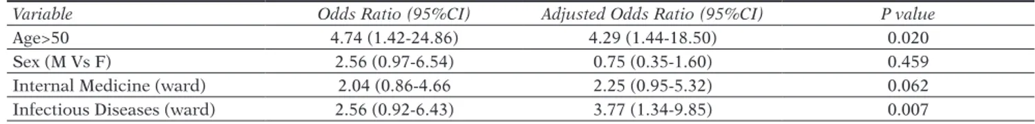
The majority of the 366 patients were hospitalized in the Internal Medicine (91) and Infectious Diseases (48) wards. At univariate analysis, the prevalence of CDI among the 366 patients was not statistically significant for Internal Medicine ward (3.28% vs 5.19%, OR: 2.04, 95%CI: 0.86-4.66, p value=0.080) while for Infectious Diseases it was
Table 1 - Evaluation of the logistic regression model for the presence of Clostridium difficile infection among the 366
ana-lyzed patients.
Variable Odds Ratio (95%CI) Adjusted Odds Ratio (95%CI) P value
Age>50 4.74 (1.42-24.86) 4.29 (1.44-18.50) 0.020
Sex (M Vs F) 2.56 (0.97-6.54) 0.75 (0.35-1.60) 0.459
Internal Medicine (ward) 2.04 (0.86-4.66 2.25 (0.95-5.32) 0.062
Infectious Diseases (ward) 2.56 (0.92-6.43) 3.77 (1.34-9.85) 0.007
Table 2 - Results obtained by GeneXpert C. difficile on 31 samples positive for Clostridium difficile infection.
GeneXpert@ results Number of samples Percentage% (95%CI)
Toxin B DNA (only) 19 61.29 (42.29-77.58)
Toxin B + Binary Toxin DNA 5 16.13 (6.09-34.47)
statistically significant (2.18% vs 6.28%, OR: 2.56, 95%CI: 0.92-6.43, p value=0.045). Logistic regression model con-firmed that age >50 years and the presence of patients in the Infectious Diseases ward were risk factors for CDI
(Ta-ble 1).
Among the 31 patients with CDI, 61.29% had only toxin B DNA and 16.13% both toxin B and binary toxin DNA. Deletion D117tcd which is associated with C. difficile ribo-type 027 was detected in at least 22.58% (Table 2). Exploratory analysis of the monthly prevalence of C.
dif-ficile among the 481 stool samples analyzed revealed a
quite constant trend of C. difficile detection with a min-imum value in December 2015 and a maxmin-imum in Feb-ruary 2016. On the contrary, the trend of 027-associated D117tcd began to increase slightly after August 2015 with a peak value in September 2015 (Figure 1).
DISCUSSION
The present study describes the prevalence of CDI from hospitalized patients and an attempt to evaluate the dis-tribution of ribotype 027 isolated from stool samples from March 2015 to March 2016 in Bari. To our knowledge, this is the first study to evaluate the prevalence of CDI in a important area of South Italy. The prevalence rate of CDI among patients undergoing C. difficile testing was 8.47%. More recent studies showed similar rates with only 5%-10% of samples testing positive (American Society for Mi-crobiology, 2010). This prevalence is also similar to those reported by Roncarati et al. and Tan et al. (Roncarati et
al., 2017; Tan et al., 2014). The prevalence rate of CDI in a
five-year study of Aschbacher et al. in Bolzano was 9.41% with incidence rates ranging from 4.3/10,000 in 2009 to 1.8/10,000 patients/days in 2013 (Aschbacher et al., 2017). On the contrary, a study by Cioni et al. in Internal Med-icine wards reported a prevalence rate of 0.96% with an estimated incidence of 5.3/10,000 patients-days (Cioni et
al., 2016).
An Italian survey revealed that the average percentage of positive tests for C. difficile toxins was 12.2% but only 34% of laboratories used a algorithm for CDI (Spigaglia et al., 2016). Immunochromatographic assay detected toxin A/B
in 37 patients (10.11%) according to the result of Spigaglia
et al. (Spigaglia et al., 2016) but only in 31 was the result
confirmed by the molecular assay.
The increasing incidence and severity of CDI has been at-tributed to the emergence of the hypervirulent strain 027 (McDonald et al., 2005). An update on the spread of C.
dif-ficile ribotype 027 in Europe was published in 2007
(Kui-jper et al., 2007b). We detected seven patients with ribo-type 027-linked deletion D117tcd accounting for 22.58% of C. difficile positive patients. Such result is in line with that reported in a northern Italian study (Baldan et al., 2010). Moreover, C. difficile ribotype 027 and ribotype 078 were firstly detected in Italy in eight and 26 cases of CDI in generally younger patients (Baldan et al., 2010). Ribo-type 027 was also detected in 10/24 stool samples collected from patients with severe diarrhoea and clinical suspicion of hypervirulent strain from seven hospitals in Rome and another multicentre study in six tertiary care hospitals in Rome detected ribotype 027 in 270 of 563 patients with CDI (Falcone et al., 2016, Di Bella et al., 2012). In the sur-vey of Spigaglia et al. ribotype 027 accounted for 8% of the clinical isolates (Spigaglia et al., 2016), whereas ribo-type 027 accounted for 19% of the 1,196 C. difficile clinical isolates from 19 European Countries (Davies et al., 2016). The hypervirulent fluoroquinolone-resistant C. difficile ri-botype 027 is considered a cause of severe outbreaks as well as higher death rates, longer hospital stays, and fre-quent relapses (Bacci et al., 2011). On the contrary, the pathological significance of binary toxin is not yet clear. In fact, Bacci et al. reported a 30-day fatality rate (27.8%) in 72 C. difficile non-027 binary toxin positive patients sim-ilar to 193 C. difficile 027 patients (28.0%). The cumula-tive risk of death after 60 days at Kaplan Meier analysis was 24.5% in 212 C. difficile toxin A and B positive pa-tients, 37.1% in C. difficile 027 patients and 30.5% in C.
difficile non-027 binary toxin positive patients. Barbut et
al. reported an association between binary toxin positive
C. difficile strains (11% of 131 strains) and more severe
diarrhea (RR 3.38) and also an increased fatality rate (RR 2.55) (Barbut et al., 2007). However, other studies failed to detect an increase in mortality or relapse rates. In particu-lar, Walk et al. reported that C. difficile ribotype was not a
Figure 1 - Evaluation of trend
of monthly prevalences of C. difficile and D117tcd detection on 481 stool samples by Low-ess smoothing with smoothing span value 0.8.
predictor of 34 severe CDI cases (Walk et al., 2012). Kim et al. reported that 11 binary toxin positive strains compared to 105 toxin A and B positive strains were associated with a significant increase of leukocytosis and mucoid stool but not with different clinical outcome (Kim et al., 2013). Pilate et al. also failed to detect an increase in mortality of the 33 binary toxin positive patients compared to 66 patients who were carriers of non-toxigenic C. difficile de-spite a higher peripheral leukocytosis (Pilate et al., 2016). Moreover, Reigadas et al. reported no association between 54 non-027 binary toxin-positive patients and poor clin-ical outcome (OR: 0.793, 95%CI: 0.243-2.591) compared to 265 C. difficile-positive binary toxin-negative patients (Reidagas et al., 2016).
Internal Medicine and Infectious Diseases wards were the most affected by episodes of CDI but the logistic model confirmed only Infectious Diseases ward and older age as risk factors for CDI. Several Italian studies showed that the area of medicine (general medicine and long-term care wards) was the most affected by CDI (Roncarati et al., 2017). Moreover, older age, the use of antimicrobials and proton pump inhibitors, hospital stay before CDI, and pre-vious CDI are well known risk factors for CDI (Roncarati
et al., 2017, Cioni et al., 2016). According to some studies,
female gender was associated with CDI but this result was not confirmed in our and other studies (Cioni et al., 2016, Boone et al., 2012, Khanna et al., 2012b).
Exploratory analysis of the monthly prevalence of C.
diffi-cile detection in stools samples revealed a quite constant
trend with a peak number of cases in February 2016. Oth-er Italian studies also showed a constant trend of CDI cases despite major fluctuations (Aschbacher et al., 2017, Mellace et al., 2013). The time series of monthly number of cases of CDI analyzed by Polgreeen et al. revealed a clear pattern of yearly seasonal variation with peaks mostly in March and an increasing trend in accordance with oth-er studies (Polgreeen et al., 2010). On the contrary, the monthly prevalence of ribotype 027 seems to show an in-creasing trend in the last months of the observation period but other studies will be needed to confirm such results. This study shows some weaknesses that should be con-sidered. First, immunochromatographic tests for toxin A/B detections may lack sensitivity (Alcalà et al., 2008). For this reason, the prevalence of CDI was possibly un-derestimated. Secondly, the observation period is quite short and the presence of a seasonal and cyclical pattern may not be revealed. Thirdly, due to the absence of data regarding length of hospitalization and clinical outcomes, it was not possible to estimate the incidence of CDI and mortality rates, especially for hypervirulent ribotype 027. Moreover, we were not able to ribotype the Xpert positive clinical strains due to the retrospective nature of the study. In fact, C. difficile Xpert offers only a very limited discrim-ination of potential ribotypes. It is also possible that C.
difficile Xpert may erroneously classify some ribotypes as
presumptive 027. In fact, several cases of ribotype mis-classification of C. difficile Xpert as presumptive 027 were reported. Moreover, C. difficile Xpert has been reported to be unable to diagnose multiple infections from different C.
difficile strains (McMillen et al., 2015, Mentula et al., 2015,
Tenover et al., 2011). However, some studies showed that ribotypes other than 027 are increasing in Italy (Barbanti
et al., 2016).
Despite this limitations, this is the first study to have esti-mated the prevalence and time trend of CDI and ribotype
027 in South Italy. Moreover, surveillance data are needed to better assess the diffusion of C. difficile 027 and other ribotypes in South Italy and to establish risk factors asso-ciated with their transmission in our healthcare facilities. Such data will be also useful to improve specific antibiotic stewardship guidelines to reduce risk of CDI in most in-terested wards.
Conflict of interest
The authors declare that they have no conflict of interest.
Funding
None
Ethical approval
All procedures performed in studies involving human par-ticipants were in accordance with the ethical standards of the institutional and / or national research committee and with the 1964 Helsinki declaration and its later amend-ments or comparable ethical standards. For this type of study formal consent is not required.
References
Alcalá L., Sánchez-Cambronero L., Catalán M.P., Sánchez-Somolinos M., Peláez M.T., et al. (2008). Comparison of three commercial methods for rapid detection of Clostridium difficile toxins A and B from fecal specimens. J. Clin. Microbiol. 46, 833-835.
American Society for Microbiology. A practical guidance document for the laboratory detection of toxigenic Clostridium difficile, September 21, 2010. Available from: http//www.alere.com/content/dam/alere/docs/ guidelines/2010_ASM_guidelines_dated_9.21.10.pdf.
Aschbacher R, Indra A, Wiedermann CJ, March A, Giacon B, et al. (2017). Predominance of Clostridium difficile 027 during a five-year period in
Bol-zano, Northern Italy. Infez. Med. 25, 13-20.
Bacci S., Mølbak K., Kjeldsen M.K., Olsen K.E. (2011). Binary toxin and death after Clostridium difficile infection. Emerg. Infect. Dis. 17, 976-982.
Baldan R., Cavallerio P., Tuscano A., Parlato C., Fossati L., et al. (2010). First report of hypervirulent strains polymerase chain reaction ribo-types 027 and 078 causing severe Clostridium difficile infection in Ita-ly. Clin. Infect. Dis. 50, 126-127.
Barbanti F., Spigaglia P. (2016). Characterization of Clostridium difficile PCR-ribotype 018: A problematic emerging type. Anaerobe. 42, 123-129.
Barbut F., Gariazzo B., Bonné L., Lalande V., Burghoffer B., et al. (2007). Clinical features of Clostridium difficile-associated infections and molecular characterization of strains: results of a retrospective study, 2000-2004. Infect. Control. Hosp. Epidemiol. 28, 131-139.
Bartlett J.G. (2002). Clostridium difficile-associated enteric disease. Curr.
Infect. Dis. Rep. 4, 477-483.
Bartlett JG. (2006). Narrative review: the new epidemic of Clostridium dif-ficile-associated enteric disease. Ann. Intern. Med. 145, 758-764. Boone J.H., Goodykoontz M., Rhodes S.J., Price K., Smith J., et al. (2012).
Clostridium difficile prevalence rates in a large healthcare system stratified according to patient population, age, gender,
and specimen consistency. Eur. J. Clin. Microbiol. Infect. Dis. 31, 1551-1559. Carter G.P., Chakravorty A. Pham Nguyen T.A., Mileto S., Schreiber F., et al.
(2015). Defining the Roles of TcdA and TcdB in Localized Gastrointes-tinal Disease, Systemic Organ Damage, and the Host Response during
Clostridium difficile Infections. MBio. 6, e00551.
Centers for Disease Control and Prevention (2012). Vital signs: preventing Clostridium difficile infections. MMWR Morb. Mortal. Wkly. Rep. 61, 157-162.
Chen S., Sun C., Wang H., Wang J. (2015). The Role of Rho GTPases in Toxicity of Clostridium difficile Toxins. Toxins (Basel). 7, 5254-5267. Cioni G, Viale P, Frasson S, Cipollini F, Menichetti F, et al. (2016).
Epi-demiology and outcome of Clostridium difficile infections in patients hospitalized in Internal Medicine: findings from the nationwide FA-DOI-PRACTICE study. BMC Infect Dis. 16, 656.
Davies K.A., Longshaw C.M., Davis G.L., Bouza E., Barna Z., et al. (2014). Underdiagnosis of Clostridium difficile across Europe: the European, multicentre, prospective, biannual, point-prevalence study of
Clos-tridium difficile infection in hospitalised patients with diarrhoea
(EU-CLID). Lancet Infect. Dis. 14, 1208-1219.
Davies K.A., Ashwin H., Longshaw C.M., Burns D.A., Davis G.L., et al. (2016). Diversity of Clostridium difficile PCR ribotypes in Europe: results from the European, multicentre, prospective, biannual,
point-prevalence study of Clostridium difficile infection in hospitalised patients with diarrhoea (EUCLID), 2012 and 2013. Euro Surveill. 21. PubMed PMID: 27470194.
Di Bella S., Paglia M.G., Johnson E., Petrosillo N. (2012). Clostridium diffi-cile 027 infection in Central Italy. BMC Infect Dis. 12, 370.
Falcone M., Iraci F., Raponi G., Goldoni P., Belvisi V., et al. (2016). Nursing home residence is associated with spread of Clostridium difficile ribo-type 027 in central Italy. J. Hosp. Infect. 94, 201-203.
Kato H., Ito Y., van den Berg R., Kuijper E.J., Arakawa Y. (2007). First isola-tion of Clostridium difficile 027 in Japan. Euro Surveill. 12: E070111.3. http://www.eurosurveillance.org/ew/2007/070111.asp#3.
Kelly C.P., LaMont J.T. (2008). Clostridium difficile-more difficult than ever. N. Engl. J. Med. 359, 1932-1940.
Khanna S., Pardi D.S. (2012a). Clostridium difficile infection: new insights into management. Mayo Clin. Proc. 87, 1106-1117.
Khanna S., Pardi D.S., Aronson S.L., Kammer P.P., Orenstein R., et al. (2012b). The epidemiology of community-acquired Clostridium difficile infection: a population-based study. Am. J. Gastroenterol. 107:89-95Erratum in: Am J Gastroenterol. 107, 150.
Kim J., Seo MR, Kang J.O., Choi T.Y., Pai H. (2013). Clinical and Microbi-ologic Characteristics of Clostridium difficile Infection Caused by Bi-nary Toxin Producing Strain in Korea. Infect. Chemother. 45, 175-183. Kuehne S.A., Cartman S.T., Heap J.T., Kelly M.L., Cockayne A., Minton
N.P. (2010). The role of toxin A and toxin B in Clostridium difficile infection. Nature. 467, 711-73.
Kuijper E.J., Coignard B., Tull P. (2006). Emergence of Clostridium diffi-cile-associated disease in North America and Europe. Clin. Microbiol.
Infect. 12, 2-18.
Kuijper E.J., van Dissel J.T., Wilcox M.H. (2007a). Clostridium difficile: changing epidemiology
and new treatment options. Curr. Opin. Infect. Dis. 20, 376-383.
Kuijper E.J., Coignard B., Brazier J., Suetens C., Drudy D, et al. (2007b). Update of Clostridium difficile-associated disease due to PCR ribotype 027 in Europe. Euro Surveill. 12, 714.
Labbé A.C., Poirier L., MacCannell D., Louie T., Savoie M., et al. (2008). Clostridium difficile infections (CDI) in a Canadian tertiary care hos-pital before and during a regional epidemic associated with the BI/ NAP1/027 strain. Antimicrob. Agents Chemother. 52, 3180-3187. Loo V.G., Poirier L., Miller M.A., et al. (2005). A predominantly clonal
mul-tiinstitutional outbreak of Clostridium difficile-associated diarrhea with high morbidity and mortality. N. Engl. J. Med. 353, 2442-2449. Lyras D., O’Connor J.R., Howarth P.M., Sambol S.P., Carter G.P., et al.
(2009). Toxin B is essential for virulence of Clostridium difficile.
Na-ture. 458, 1176-1179.
McCollum D.L., Rodriguez J.M. (2012). Detection, treatment, and preven-tion of Clostridium difficile infecpreven-tion. Clin. Gastroenterol. Hepatol. 10, 581-592.
McDonald L.C., Killgore G.E., Thompson A., Owens R.C. Jr, Kazakova S.V., et al. (2005). An epidemic, toxin gene-variant strain of Clostridium dif-ficile. N. Engl. J. Med. 353, 2433-2441.
McDonald L.C., Owings M., Jernigan D.B. (2006). Clostridium difficile infection in patients discharged from US short-stay hospitals, 1996-2003. Emerg. Infect. Dis. 12, 409-415.
McMillen T., Kamboj M., Babady N.E. (2016). Comparison of Multilocus Sequence Typing and the Xpert C. difficile/Epi Assay for Identification of Clostridium difficile 027/NAP1/BI. J. Clin. Microbiol. 54, 775-778. Mellace L., Consonni D., Jacchetti G., Del Medico M., Colombo R., et al.
(2013). Epidemiology of Clostridium difficile-associated disease in in-ternal medicine wards in northern Italy. Intern. Emerg. Med. 8, 717-723.
Mentula S., Laakso S., Lyytikäinen O., Kirveskari J. (2015). Differentiating virulent 027 and non-027 Clostridium difficile strains by molecular methods. Expert. Rev. Mol. Diagn. 15, 1225-1229.
Monaghan T., Boswell T., Mahida Y.R. (2008). Recent advances in Clostrid-ium difficile-associated disease. Gut. 57, 850-860.
O’Connor J.R., Johnson S., Gerding D.N. (2009). Clostridium difficile in-fection caused by the epidemic BI/NAP1/027 strain. Gastroenterology.
136, 1913-1924.
Oldfield E.C. IV, Oldfield E.C. III, Johnson D.A. (2014). Clinical update for the diagnosis and treatment of Clostridium difficile infection. World J.
Gastrointest. Pharmacol. Ther. 5, 1-26.
Pepin J., Valiquette L., Alary M.E., et al. (2004). Clostridium difficile-asso-ciated diarrhea in a region of Quebec from 1991 to 2003: a changing pattern of disease severity. CMAJ. 171, 466-472.
Pepin J., Valiquette L., Cossette B. (2005). Mortality attributable to nos-ocomial Clostridium difficile-associated disease during an epidemic caused by a hypervirulent strain in Quebec. CMAJ. 173, 1037-1042. Pilate T., Verhaegen J., Van Ranst M., Saegeman V. (2016). Binary toxin and
its clinical importance in Clostridium difficile infection, Belgium. Eur.
J. Clin. Microbiol. Infect. Dis. 35, 1741-1747.
Pituch H., Brazier J.S., Obuch-Woszczatynski P., Wultanska D., Meisel-Mi-kolajczyk F. et al. (2006). Prevalence and association of PCR ribotypes of Clostridium difficile isolated from symptomatic patients from War-saw with macrolide-lincosamidestreptogramin B (MLSB) type resis-tance. J. Med. Microbiol. 55, 207-213.
Planche T.D., Davies K.A., Coen P.G., Finney J.M., Monahan I.M., et al. (2013). Differences in outcome according to Clostridium difficile test-ing method: a prospective multicentre diagnostic validation study of C.
difficile infection. Lancet Infect. Dis. 13, 936-945.
Polgreen P.M., Yang M., Bohnett L.C., Cavanaugh J.E. (2010). A time-series analysis of clostridium difficile and its seasonal association with influ-enza. Infect. Control. Hosp. Epidemiol. 31, 382-387.
R Core Team R: A language and environment for statistical computing. R Foundation for Statistical Computing, Vienna, Austria, 2016.URL https://www.R-project.org/.
Reigadas E., Alcalá L., Marín M., Martín A., Iglesias C., Bouza E. (2016). Role of binary toxin in the outcome of Clostridium difficile infection in a non-027 ribotype setting. Epidemiol. Infect. 144, 268-273.
Roncarati G., Dallolio L., Leoni E, Panico M., Zanni A., et al. (2017). Sur-veillance of Clostridium difficile Infections: Results from a Six-Year Retrospective Study in Nine Hospitals of a North Italian Local Health Authority. Int. J. Environ. Res. Public. Health. 14, E61.
Rupnik M., Wilcox M.H., Gerding D.N. (2009). Clostridium difficile infec-tion: new developments in epidemiology and pathogenesis. Nat. Rev.
Microbiol. 7, 526-536.
Sansone S., Aschbacher R., Staffler M., Bombonato M., Girardi L.C., et al. (2009). Nosocomial diarrhea in adult medical patients: the role of
Clostridium difficile in a North Italian acute care teaching hospital. J. Prev. Med. Hyg. 50, 117-120.
Smith A. (2005). Outbreak of Clostridium difficile infection in an English hospital linked to hypertoxin-producing strains in Canada and the US.
Euro Surveill. 10, E050630.2.
Spigaglia P., Barbanti F., Morandi M., Moro M.L., Mastrantonio P. (2016). Diagnostic testing for Clostridium difficile in Italian microbiological laboratories. Anaerobe. 37, 29-33.
Sunenshine R.H., McDonald L.C. (2006). Clostridium difficile-associated disease: new challenges from an established pathogen. Cleve. Clin. J.
Med. 73, 187-197.
Surawicz C.M., Brandt L.J., Binion D.G., Ananthakrishnan A.N., Curry S.R., et al. (2013). Guidelines for diagnosis, treatment, and prevention of Clostridium difficile infections. Am. J. Gastroenterol. 108, 478-498. Tan X.Q., Verrall A.J., Jureen R., Riley T.V., Collins D.A., et al. (2014). The
emergence of community-onset Clostridium difficile infection in a ter-tiary hospital in Singapore: a cause for concern. Int. J. Antimicrob.
Agents. 43, 47-51.
Tenover F.C., Akerlund T., Gerding D.N., Goering R.V., Boström T., et al. (2011). Comparison of strain typing results for Clostridium difficile isolates from North America. J. Clin. Microbiol. 49, 1831-1837. Vonberg R.P., Schwab F., Gastmeier P. (2007). Clostridium difficile in
dis-charged inpatients, Germany. Emerg. Infect. Dis. 13, 179-180. Voth D.E., Ballard J.D. (2005). Clostridium difficile toxins: mechanism of
action and role in disease. Clin. Microbiol. Rev. 18, 247-263.
Walk S.T., Micic D., Jain R., Lo E.S., Trivedi I., et al. (2012). Clostridium difficile ribotype does not predict severe infection. Clin. Infect. Dis. 55, 1661-1668.
Warny M., Pepin J., Fang A., Killgore G., Thompson A., et al. (2005). Toxin production by an emerging strain of Clostridium difficile associated with outbreaks of severe disease in North America and Europe. Lancet.
366, 1079-1084.
Wilcox M.H. (2003). Gastrointestinal disorders and the critically ill. Clos-tridium difficile infection and pseudomembranous colitis. Best. Pract. Res. Clin. Gastroenterol. 17, 475-493.