UNIVERSITÀ DEGLI STUDI DI SASSARI
CORSO DI DOTTORATO DI RICERCA IN SCIENZE BIOMEDICHE
Coordinatore del Corso: Prof. Andrea Fausto Piana CURRICULUM IN FISIOPATOLOGIA MEDICA
Responsabile di Curriculum: Prof. Roberto Manetti
XXIX CICLO
Cardiac and vascular characteristics
and gender-related modifications induced by ageing in the founder population of SardiNIA project (ProgeNIA)
Coordinatore:
Prof. Andrea Fausto Piana
Tutor:
Prof. Antonello Ganau
Tesi di dottorato di: Dott. Giuseppe D. Sanna
INDEX
SUMMARY ... 2
INTRODUCTION ... 5
AIMS OF THE STUDY ... 16
METHODS ... 17 RESULTS ... 22 DISCUSSION ... 30 CONCLUSIONS ... 36 REFERENCES ... 38 TABLES ... 46 LEGENDS ... 57 FIGURES ... 58 APPENDIX ... 77
SUMMARY
Background. Cardiovascular diseases are the leading cause of death and disability in Western countries and this is expected to worsen as the population is aging. Aging induces structural and functional changes in the heart and vessels which increase the likelihood of cardiovascular events. Little data is available about gender-related and population-related differences in cardiovascular adaptation to aging. In 2001 the SardiNIA (ProgeNIA) project began in Sardinia. It’s brief was to identify the genetic determinants of complex traits associated with aging in 6148 volunteers with documented Sardinian parentage, enrolled in four villages in the Ogliastra province. In 2013 echocardiography was introduced to investigate the structural and functional cardiac traits.
Aim. 1) To describe the clinical and echocardiographic characteristics of a large sample of the SardiNIA cohort; 2) To assess the normal values of the echocardiographic parameters and the best method of their indexation for body size (height, weight, body mass index, body surface area), in a sample free of cardiovascular diseases; 3) To investigate the effects of ageing on the heart and aorta and the gender-specific differences.
Methods. Of the original 6148 SardiNIA volunteers, 3131 subjetcs underwent a transthoracic echocardiogram according to a standardized protocol and were enrolled in this study. Complete clinical and laboratory data and good quality 2D and M-mode echocardiographic images were available in 2926 of them (47.6% of the whole SardiNIA cohort). All the echocardiographic images were analyzed by a single expert cardiologist. The following parameters were measured: end-diastolic left ventricular
(LV) internal diameter (EDD) and volume (EDV), interventricular septum, posterior wall thickness, LV mass (LVM), ejection fraction (EF), stroke volume (SV), transmitral early (E) and atrial (A) flow velocity and their ratio (E/A), left atrial diameter (LAD) and volume (LAV), aortic root and ascending aorta dimensions. According to clinical and laboratory data, office blood pressure, ABPM, ECG and echocardiography, 1977 subjects (62% women) were free of diabetes and apparent cardiovascular disease and were considered normal.
Results. The prevalence of some cardiovascular risk factors in the SardiNIA sample group was quite low, probably due to the relatively young mean age of the sample, but genetic and environmental factors may also play a role. Gender-specific normal reference values of the major echocardiographic parameters were obtained, after normalizing for body surface area (BSA). In the Sardinian population ageing is associated with significant left ventricular remodelling, left atrial enlargement and diastolic disfunction, while systolic function is preserved. It is noteworthy that all these changes are quantitatively more pronounced in women.
Discussion. This study provides normal reference values of the echocardiographic indices, specific for the Sardinian population. The best method of their indexation resulted in being the isometric scaling using BSA, whereas the allometric methods of normalizing LVM (LVM/height2.7 and LVM/height1.7) overestimated LVM in the smallest subjects (mostly women) of the Sardinian population, with potential distorsive effect in estimating the true prevalence of LV hypertrophy. This data suggests that the best methods of indexing LVM for body size depend on the anthropometric characteristics of the study population. In the Sardinian population, ageing is associated with significant left ventricular remodelling, left atrial enlargement, aortic dilatation and
diastolic dysfunction, while systolic function is preserved. It is worth noting that all these changes are more pronounced in women.
Keywords: SardiNIA project; echocardiography; left ventricular mass; left ventricular hypertrophy; left ventricular function; isometric and allometric scaling; cardiovascular aging; gender medicine.
INTRODUCTION
Cardiovascular ageing is a major target for preventative cardiology. As age advances, hearts and vessels operate on "the edge of cardiovascular disease."1
The epidemic of cardiovascular (CV) disease has become a global phenomenon, no longer limited to the Western countries, since nowadays they account for 30% of deaths worldwide and by 2020 they are estimated to surpass infectious diseases as the main cause of mortality and disability.1
This situation is expected to worsen as the population is ageing. In the United States, 35 million people are over 65 years old and their number will double by 2030. In Italy, people over 65 years are numbered at 13.4 million (22% of the population), with an average life expectancy at birth equal to 80.1 for men and 84.7 years for women, (ISTAT data 2016).2 The clinical and economic impact of this demographic shift are relevant.
An increasingly widespread opinion is that ageing should be considered per se a disease. Ageing is characterized by the progressive loss and failure of molecular mechanisms that create disorder within the DNA and its environment.1 These pathological/regressive processes do not affect all individuals in the same way, but there are wide differences suggesting the existence of factors favouring or contrasting the damage of senescence ("accelerated" versus "successful ageing ").
The SardiNIA project
In 2001 the National Institute of Health - Division of Research Acquisition (Rokville, Maryland, USA), Istituto di Ricerche sulle Talassemie ed Anemie Mediterranee
(Cagliari, Italy) and Centro Nazionale di Ricerche (Cagliari, Italy) began the research project "Genetics and Epidemiology of Aging Associated Conditions in the Sardinian Population". This study, called "Sardinia Project (ProgeNIA)", was aimed to identify the genetic determinants of complex traits (e.g. cardiovascular) related with ageing. This prospective longitudinal study enrolled 6148 volunteers (about 60% of eligibility), aged 14 to 102 years, who were residents in four small towns in the Ogliastra province (Lanusei, Arzana, Ilbono, Elini)3 and with at least two-generations local Sardinian parental lineage.
The founder populations
Ogliastra is a historical and geographical area located in the central-eastern part of Sardinia, inhabited since the Neolithic period. As well as Iceland, Ogliastra with its unique geographic (insulation and isolation) and demographic characteristics (no immigration phenomena, diffuse endogamy) has attracted the interest of geneticists. In recent decades, scientific interest has focused on the founder populations for the genetic study of complex diseases. Individuals belonging to these populations normally share lifestyles, eating habits and natural environment, thus resulting in reduced environmental variability. Often these populations were founded by a small number of individuals, followed by a long period of isolation, during which there has been a gradual genetic drift. As a consequence, a small number of predisposing genes/alleles cause complex phenotypes, so that modern nonparametric analyses extended to the whole genome (genome-wide) can be applied more easily to locate these genetic variants.4
The need of a founder population for the study of phenotypic traits
The importance of studying a founder population is also clinical, since it allows researchers to better characterize phenotypic traits and to identify ethnicity-specific reference values.
This concept is crucial in the study of cardiovascular diseases and echocardiography and it represents a mandatory methodological step before studying the effects of ageing on the heart and vessels.
The reference values of the main echocardiographic parameters routinely used in clinical practice (left ventricular geometry and function, left atrial size, aortic diameters) are listed in ASE/EACVI recommendations.5 This data comes from population studies
involving thousands of subjects, often belonging to different ethnic groups.
There are potential limitations in applying the same echocardiographic criteria to extremely heterogeneous populations with different body size, as well as genetic and environmental backgrounds. In the NORRE (Normal Reference Ranges for Echocardiography) study the Authors highlighted the importance of developing ethnicity-specific reference values for echocardiografic parameters,6 taking into account the differences between values obtained in specific populations.7
At the moment, there are no specific studies on the reference values of echocardiographic parameters in the Sardinian population, which is unique in having particular anthropometric and genetic traits.
A big issue - normalization of echocardiographic parameters for body size
Accurate quantification of cardiac and vascular size is crucial to distinguish between normal and pathological variants, and normalization for body size is a preliminary and
indispensable step. Although technological advances in echocardiography allow very accurate measurements, there is often a certain degree of ambiguity in their interpretation if the value falls in a gray zone with surface and body composition playing an important role on the "weight" of that measurement.8
It is well known that cardiac dimensions and aortic diameters are influenced by body size and that they increase proportionally with larger size.8 Normalization of echocardiographic parameters is also important for prognostic purposes.9 However, normalization of echocardiographic parameters for body size is still controversial and an unanimous consensus is still lacking among researchers.
To normalize cardiac measurements, various methods have been proposed (isometric and allometric). Since the relationships between body size and dimensions of organs are often non-linear,10 allometric approaches are often employed in which specific parameters (e.g. left ventricular mass) are divided by a body size variable (e.g. height) raised to a scalar exponent (e.g. mass/height 2.7). This approach seems to be accurate in effectively removing the effects of body size on cardiac dimensions.
The use of isometric methods (e.g. mass/body surface area) despite the ASE/ESC5 recommendations, is much debated. The main limitation of normalizing using isometric scaling is linked to an overcorrection in obese patients.11 This limitation has been confirmed in several studies including prognostic data, at least for left ventricular mass9, especially in hypertensive subjects.12
The most controversial topic is the best way to normalize left ventricular mass (LVM). This latest is a powerful predictor of cardiovascular risk.13, 14, 15 In 1992 de Simone et al. showed that indexing for body surface area (BSA) and body weight1.5 underestimates left ventricular mass in overweight subjets, and he proposed a new indexing method
based on height2.7 (LVM2.7).16 Further papers with outcome data confirmed the utility of this approach.9, 17, 18
However, other data published over the recent years have raised questions and concerns about the validity and universal extension of this allometric method. One of the most important limitations when normalizing left ventricular mass for height is related to the fact that indexed mass increases with decreasing body height and therefore it represents a suboptimal method of normalization for body size and to define, for example, left ventricular hypertrophy in children.16, 19
In 2010 Chirinos et al. raised some important doubts about the allometric method proposed by de Simone. They started from the concept that gender is a strong correlate of body size and left ventricular mass and that previous studies did not take into account all these influences. Their study analyzed the echocardiographic data of 523 subjects (265 whites, 133 Chinese, 65 blacks, 60 Hispanics) participants in the MESA study (Multi-Ethnic Study of Atherosclerosis) and other 637 participants from the Asklepios study.
The study is important for several reasons. First of all, it emphasizes the importance of gender as a determinant of left ventricular mass, and it confirms the bias resulting from the use of a normalization method in which the scalar exponent is not gender specific. Chirinos proposed a new allometric exponent for normalization of the left ventricular mass which takes into account sex-related differences (height1.7). An important scientific observation deriving from this study is represented by the fact that normalizing left ventricular mass for body height2.7 showed an artificial negative relationship with the height, not only in the reference sample but also in the entire study population (R = -0.25, p <0.0001). Indexing mass/height 2.7 grossly overestimated the
prevalence of hypertrophy in men and women with shorter body height, while underestimating its prevalence in those with greater body height both in MESA and Asklepios.20
These controversial results and the lack of a defined consensus among experts,21, 22 emphasize the clinical importance of identifying the best method for normalization of left ventricular mass in the Sardinian population, known as having genetic and phenotypic peculiarities. Sardinians, with an average height of 168.5 cm in males, are among the shortest European populations.23
Height is a canonical complex trait, under tight genetic control with heritability of 80-90%.3 In recent research with data deriving from the SardiNIA cohort, two gene variants with major effects on height were identified. The first is a premature stop codon in the GHR gene, relatively frequent in Sardinia (0.87% vs <0.01% elsewhere), which in homozygosity causes the short stature Laron syndrome. In heterozygotes it reduces height by an average of 4.2 cm. The other variant, in the imprinted KCNQ1 gene (7.7% vs <1% elsewhere) reduces height by an average of 1.83 cm when maternally inherited. Furthermore, some well known height-decreasing alleles have a higher frequency in Sardinia than would be expected on the basis of genetic drift. These findings are consistent with selection toward shorter stature in Sardinia and a suggesting example in humans of the so-called "island effect" reducing the size of large mammals.24
Sex-related differences in cardiovascular structure and remodelling.
In recent years, the importance of sex-related differences in cardiovascular biology and pathology has emerged. Gender medicine plays a pivotal role in cardiovascular care as men and women have hearts and arteries which, starting from some baseline
differences, show a different response to external stimuli (pathogenic or not) and ageing.25
Ageing and cardiovascular system.
Ageing induces several morphological and functional changes of the heart and arteries. These changes represent the substrate for the development of many cardiovascular diseases whose prevalence increases with age.
Effects on the heart
Ageing is characterized by a dramatic increase in the prevalence of three pathological conditions: left ventricular hypertrophy, heart failure with preserved ejection fraction and atrial fibrillation.26
Left ventricular hypertrophy is associated with an increased risk of coronary heart disease, sudden death, stroke and overall cardiovascular mortality.27 Heart failure with preserved ejection fraction becomes more frequent with ageing and appears to be more common in women than men.28, 29 Moreover, the prevalence of atrial fibrillation increases with age rising from < 0.5% at 40-50 years to 5-15% at 80 years.30
The development of the above mentioned conditions is linked to structural and functional changes affecting the heart with advancing age.
Cross-sectional studies in normotensive subjects without overt heart disease showed an increase of left ventricular wall thickness with ageing.27 The increased vascular load imposed on the ageing heart seems to play a pivotal role in this phenomenon.26 As already shown by Ganau et al., ageing induces left ventricular concentric remodelling.31 The increase in LV mass observed with ageing does not seem to be related to an
increase of cardiomyocytes whose number reduces with ageing. This phenomenon is more pronounced in men than in women and it is linked to a higher apoptotic index (three times greater) in men.26, 32, 33, 34
These differences at cellular level may justify, at least in part, the sex-related differences in cardiac remodeling. In fact, men show concentric remodelling, while women tend to concentric hypertrophy.
The increase in cardiac mass observed with ageing may be due, at least in part, to an increase of the extracellular matrix (i.e. fibrous component). A pivotal role seems to be played (more than by the entity of this deposition) by the type of collagen in the extracellular matrix. The "cross-linking" phenomenon is characterized by an increase of type I collagen (whose fibers are thicker and insoluble) compared to other forms, a well known phenomenon in heart failure with preserved ejection fraction,35 being able to justify the increased heart stiffness that is the basis of the diastolic abnormalities observed with aging.
The LV diastolic filling rate progressively slows after the age of 20 years, so that by 80 years the rate is reduced, on average, up to 50%.26 With ageing more filling occurs in the late diastole due to a vigorous atrial contraction. However, the increase of atrial contraction is accompanied by atrial hypertrophy and dilatation with a consequently increased arrhythmic risk. Multiple regression analyses suggest that ageing is the major determinant of the E/A ratio on echocardiography.26
LV ejection fraction (EF), the most commonly used measure of LV systolic performance, is preserved during ageing. However, there is an age-associated failure to augment EF during exercise, due to a deficit in the ability to reduce end-systolic volume index (ESVi). The net result of the changes in the regulation of diastolic and
end-systolic volumes with ageing is that stroke volume index is substantially preserved at rest because of a greater use of the Frank-Starling mechanism. However, during exercise this mechanism has proved to be inadequate because of an inability to appropriately reduce the end-systolic volume. Hence, although end-diastolic volume increases to a greater extent during exercise, stroke volume does not.26
Cardiac diameters and volumes tend to be reduced in the elderly, as shown in echocardiographic (M-mode, 2D and 3D)36, 37, 38 and MRI39 studies, although changes induced by ageing are less obvious when considering body size and gender.37 With advancing age there is also an increase of the mass/volume ratio of the LV.38, 39
The ageing heart is characterized by a reduced chronotropic reserve during physical exercise, and by a reduction of heart rate variability, both indexes of autonomic dysfunction and associated with an increased risk of morbidity and fatal outcomes.26 Atrial fibrillation is one of the components of the pathological triad affecting the ageing heart.26 Atrial fibrillation recognizes a left atrial structural substrate.30 With ageing left atrium has a progressive structural (and electric) remodelling. Apart from fibrosis, the most important structural marker (even if hardly detectable also when using advanced imaging techniques such as MRI), the left atrium shows a progressive dilation with ageing.40
Besides these anatomical changes with some sex-related differences (greater dilation in men than women), there are also functional abnormalities. The conduit function, for example, reduces with ageing while the reservoir and pump functions tend to be preserved.41 Recent echocardiographic studies using speckle-tracking confirm these aging-related changes with a overall reduction of strain values, together with sex-related differences.42
Left atrial abnormalities occurring with ageing seem to be related, at least in part, to an abnormal ventricular-vascular coupling, together with the increased mass and stiffness of the left ventricle and diastolic dysfunction. Furthermore, an important role is played by arterial hypertension characterized by an early onset of the above mentioned morphological and functional abnormalities. Hypertensive subjects in the fourth decade have similar atrial volumes compared with normotensive individuals in the eighth decade.43
Effects on arteries
With ageing arteries lose their elastic properties, thicken and dilate.44, 45
Postmortem studies indicate that the aortic wall thickening that occurs with ageing consists mainly of intimal thickening, even in populations with a low incidence of atherosclerosis.46 Intima-media thickness increases by about three times, on average, from 20 to 90 years of age, even in the absence of atherosclerotic plaques.47
Increased arterial stiffness has been attributed to the continous expansion and elastic recoil of the arterial walls during cardiac cycles.48
Some specific gene polymorphisms may also promote arterial stiffness. A genome wide association study involving 4221 subjects from the SardiNIA cohort, showed a highly significant association between the aorto-femoral pulse wave velocity (PWV) and a single nucleotide polymorphism (SNP) of COL4A gene on chromosome 13.49 This gene is responsible for the synthesis of type IV collagen.
Arterial geometry is also important. Smaller aortic diameters are associated with increased pulse pressure and could be the reason why women, who usually have smaller arteries, show an increased pulse pressure with ageing compared with men.50
Ageing and blood pressure
Blood pressure (BP) increases with age both in men and women. Systolic BP shows a steady increase until older age, while diastolic BP increases up to 50 years and then tends to be constant or slightly reduces with ageing. As a consequence, there is an increase in both genders in the prevalence of isolated systolic hypertension, a typical marker of increased arterial stiffness.51 This is due to the fragmentation and loss of elastic fibers, collagen proliferation, calcium deposit, tortuosity and dilatation of the aorta. On the other hand, dilatation increases wall stress and worsens the degeneration of the arterial walls. It triggers a damaging loop which perpetuates and exacerbates the parietal damage. The annual progression of pulse wave velocity is higher in hypertensive patients compared to normotensive subjects demonstrating that hypertension accelerates the progression of vascular stiffness.52 Arterial stiffness can also precede the onset of hypertension and it is a potential risk factor for its development.53
AIMS OF THE STUDY
1. Description of clinical characteristics (including cardiovascular risk factors and prevalence of cardiovascular disease) in a large sample from the SardiNIA cohort. 2. Description of echocardiographic cardiac and vascular characteristics of the SardiNIA cohort, and definition of reference values for the main echocardiographic parameters specific for the Sardinian population.
3. To analyze the relationships between cardiac (e.g. LV mass) and vascular parameters and anthropometric indices (height, weight, body mass index, body surface area), in order to identify the best method of normalization for body size in the Sardinian population.
4. To analyze the cardiovascular effects of ageing in subjects free of overt cardiovascular diseases, together with the study of sex-related differences in cardiac remodelling.
METHODS
The study included 3131 individuals from the entire cohort of 6148 volunteers participating in the SardiNIA study. A complete transthoracic echocardiogram was available in 2926 (47.6% of the entire cohort).
Clinical data included:
• Age, gender, body height and weight, body mass index (BMI) and body surface area (BSA).
• Office blood pressure and 24/h ambulatory blood pressure monitoring (ABPM). This latest was available in 2076 volunteers (70.9% of the whole sample of 2926 subjects included in the echocardiographic study).
• Cardiovascular risk factors: smoking, diabetes, total cholesterol (mg/dl).
The individuals included in the study were divided, according to the presence or absence of arterial hypertension and other cardiovascular diseases, in different subgroups:
1. Subjects free of overt cardiovascular diseases.
2. Subjects with arterial hypertension. This latest has been defined on the basis of elevated office blood pressure in at least three occasions (BP > 140/90 mmHg - average of three consecutive measurements) and on the basis of 24/h ABPM (SBP > 130 mmHg and / or DBP > 80 mmHg), in accordance with the Guidelines of the European Society of Cardiology (ESC).54
3. Subjects with known ischemic heart disease (prior myocardial infarction/angina pectoris).
5. Subjects with hypertrophic cardiomyopathy - HCM (definite or possible based on clinical informations and echocardiographic data). Echocardiographic diagnosis was considered when myocardial wall thickness ≥ 15 mm in one or more segments, according to the Guidelines of the European Society of Cardiology.55
6. Subjects with dilated cardiomyopathy (definite or possible based on clinical information and echocardiographic data). Left ventricular dilatation was defined by left ventricular end-diastolic volumes or diameters > 2DS from normal according to the reference values reported in the ASE/ESC recommendations for chamber quantification,56 and in agreement with the new definition proposed by the working group of the ESC on myocardial and pericardial diseases.57 All subjects were then reclassified in accordante with the reference echocardiographic values found in the sardinian population.
7. Subjects with valvular heart disease (moderate or severe). 8. Subjects with previous stroke history.
9. Subjects with diabetes.
Only group 1 (i.e. 1977 individuals without overt cardiovascular diseases) was included in the analysis of echocardiographic data.
Ambulatory blood pressure monitoring (ABPM - ambulatory blood pressure measurement) .
24-hour ambulatory blood pressure monitoring was performed with an oscillometric device (Spacelabs 90207, Spacelabs Inc., Wokingham, Berkshire, UK), with cuff of appropriate size. Measurements were obtained at 15-minute intervals throughout the day (between 7:00 AM and 10:00 PM), and at 30-minute intervals throughout the night
(from 10:00 PM to 7:00 AM). Total recording of each subject lasted a minimum of 20 h. Subjects were instructed to engage in normal daily activities but to refrain from strenuous exercise, and to keep the arm extended and still at the time of cuff inflation.
Echocardiogram
A complete transthoracic echocardiogram was performed on 2926 individuals. The examinations were performed using an echocardiographer (Siemens Acuson X-300), equipped with cardiac transducer (1.5 MHz and 3.6 MHz) and following a standard protocol.
Cardiac dimensions were measured with both 2D-guided M-mode and 2D techniques. However, for linear dimensions in the statistical analysis of the present study only 2D-guided M-mode measurements were considered.
The geometry and mass of the left ventricle were assessed according to the international recommendations.56 Left ventricular mass was calculated using the Devereux formula.58 The left ventricular systolic function was assessed by measuring the ejection fraction (according to the Teichholz method) and the stroke volume and stroke volume index. Diastolic function was evaluated throughout the study of the mitral inflow profile (E/A ratio) and left atrial volume.
Left atrial size was evaluated by measuring the antero-posterior diameter in the parasternal long-axis view (2D-guided M-mode), and volumes (Simpson’s biplane method).
Valvular flows were evaluated with Doppler method (color, PW and CW), and valvular regurgitation or stenoses have been described in accordance with international recommendations.5960
Echocardiographic examinations were performed by expert cardiologists or by other physicians after proper training as cardiac sonographers.
Echocardiographic measurements were performed online during the examination. The quality of all frames and cine-loops together with the measurements were verified and re-analyzed off-line by a single expert echocardiographer using dedicated software. In all subjects included in the echocardiographic study, blood pressure was measured at the time of examination.
Informed consent
A written informed consent was obtained from all subjects prior to enrollment in the SardiNIA study.
Data analysis and statistics
Statistical analysis was performed using the software STATA 13 (StataCorp College Station, TX, USA). Normal distribution of data was checked using the Shapiro-Francia test. According to the results of this test, values were expressed as mean ± standard deviation (SD). For the echocardiographic parameters values were also expressed as percentiles. We used the Student’s t test to compare sex-related differences.
Pearson’s correlation coefficient (r) was used to assess the relationships between some echocardiographic parameters and their normalization for body size with different scaling methods (i.e LVM, LVMI, LVM2.7, LVM1.7) and various anthropometric parameters (i.e body height and weight, body mass index and body surface area). This test was also employed to study the cardiovascular effects of ageing together with simple regression analysis.
We used the analysis of variance (ANOVA) to study the different behavior of the main methods of normalization of left ventricular mass in both genders stratified by body height and body mass index. We then performed a post-hoc test (Bonferroni).
RESULTS
Baseline clinical characteristics
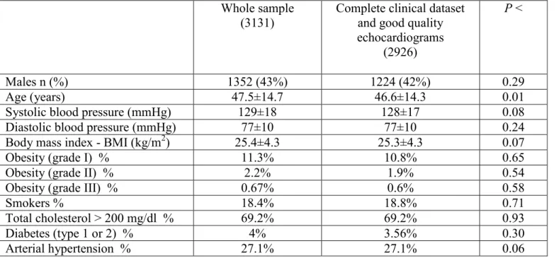
Baseline clinical characteristics of the entire study sample (3131 subjects, 50.9% of the entire SardiNIA cohort) are shown in Table 1. Men were 1352 (43%), women 1779 (57%). Mean age was 47.5±14.7 years. Mean systolic blood pressure (SBP) was 129±18 mmHg, diastolic blood pressure (DBP) was 77±10 mmHg. Body mass index (BMI) was 25.4 kg/m2. Obese patients were 14.17% of the entire sample, smokers 18.4%, diabetic 4%. The prevalence of arterial hypertension was 27.1%. Overall prevalence of cardiovascular diseases was 7.7% (including ischemic heart diseases, heart failure, valvular heart diseases, definite or possible cardiomyopathies, stroke).
Clinical characteristics of men and women with complete and good quality echocardiograms (2926, 47.6% of the entire cohort) are listed in Table 2. Men were 1224 (42%), women 1702 (58%). Mean age was similar in both genders. Overall, men exhibited higher values of body mass index (BMI), blood pressure and had a higher prevalence of cardiovascular risk factors compared with women. The prevalence of cardiovascular diseases in the echocardiographic group was 6.7%. There were no significant clinical differences between the echocardiographic group and all the study sample (see Table 1).
Characteristics of the subjects free of hypertension, cardiovascular diseases or diabetes included in the analysis of echocardiographic data.
Table 3 summarizes the characteristics of the 1977 subjects (67.6% over 2926) included in the analysis of echocardiographic data. Men were 748 (38%), women 1229 (62%).
The mean age was 41.1±12.4 years for men and 42.6±12.4 for women. Women had significantly smaller body surface area, height (157±6 cm vs 169±6.4 cm), and lower BP compared with men.
Reference values for the main echocardiographic parameters in the SardiNIA cohort
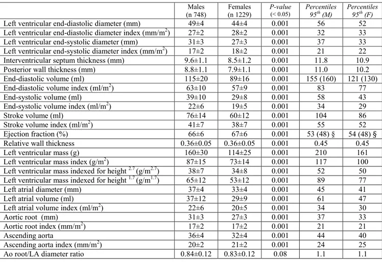
The reference echocardiographic values specific for the Sardinian population are listed in Table 4.
LV wall thickness, mass, diameters and volumes were larger in men compared with women, and gender differences were mantained for most parameters after normalization for the body surface area (BSA). LV ejection fraction (EF) was higher in women. The lower reference values (5th percentile) for LV EF were 53% in men and 54% in women. Left atrial (LA) dimensions and volumes were larger in men than in women, even after normalization for BSA. Aortic diameters resulted larger in men; however, after correction for BSA they resulted equal or slightly larger in women.
Normalization of echocardiographic parameters (i.e. LVM) for body size.
Physiological relation of LVM and its indexations to body height, weight, BMI and BSA.
As shown in Table 5, LVMI, LVM2.7 and LVM1.7 had a significant negative correlation with body height. This correlation was stronger for LVM2.7 (r - 0.34, p < 0.00001 in men; r - 0.36, p <0.00001 in women) compared with LVM1.7 (r - 0.15, p < 0.00001 in men; r - 0.20, p < 0.00001 in women) and LVMI (r - 0.08, p = 0.023 in men; r - 0.13, p < 0.00001 in women). LVM showed a weak positive correlation with body height (r
0.20, p < 0.00001 in men; r 0.09, p-value 0.0013 in women). See Figures 1-4 for details.
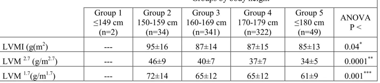
Left ventricular mass (various indexations) in healthy men and women SardiNIA participants stratified by body height.
In order to identify the best method of normalization of LV mass for body size, because of the significant negative correlation between LVMI, LVM2.7, LVM1.7 and body height, we sought to investigate the possible consequences of using allometric methods (e.g. LVM2.7) in our sample whose mean height was only 157 cm for women and 169 cm for men (126 subjects, 6.4% of the entire sample < 150 cm), and thus significantly shorter than other populations. For this purpose, our sample was stratified by body height (≤ 149 cm; 150-159 cm; 160-169 cm; 170-179 cm; ≥ 180 cm). We then analyzed the trends of LV mass (various indexations) among men and women stratified by body height, as shown in Table 6. By applying the analysis of variance (ANOVA), we found significant differences in LV mass among groups (LVMI p 0.04 and 0.0009 respectively in men and women; LVM2.7 p < 0.0001 and 0.0001 in men and women; LVM1.7 p 0.001 and 0.0001 respectively in men and women) with a clear trend toward higher LV mass indexed values in shorter subjects (i.e. < 149 cm). The post hoc analysis (Bonferroni) showed the specific differences among groups with the statistical significance of all single comparisons (see Table 6 for details). These results are also shown in Figures 5-6 clearly showing the relative overestimation of LV mass in shorter subjects with allometric methods compared with normalization for BSA.
Left ventricular mass (various indexations) in healthy men and women SardiNIA participants stratified by body mass index (BMI).
In order to identify the best method of normalization of LV mass for body size, we also stratified our sample by BMI (< 25 kg/m2; 25-30 kg/m2; > 30 kg/m2). As shown in Table 7, subjects with higher BMI showed higher values of LV mass independently from the indexation method. By applying the analysis of variance (ANOVA), we found significant differences in LV mass among BMI groups (LVMI p 0.002 and < 0.0001 in men and women respectively; LVM2.7 and LVM1.7 p < 0.0001 in both genders). The post hoc analysis (Bonferroni) showed the specific differences among groups with the statistical significance of all single comparisons (see Table 7 for details). Despite a common trend characterized by a direct relationship between LV mass and BMI (i.e. higher the BMI and higher the LV mass), values were proportionally higher when using allometric scaling for normalization. This should be related to the potential overcorrection in obese patients when using isometric methods. Figures 7-8 summarize the above mentioned trends.
The optimal scaling exponent in the SardiNIA cohort by gender
We found that body surface area (BSA) provides the best correction for body structure in our population, representing the best way for normalization of cardiac structures (i.e. LV mass) in the SardiNIA cohort. However, indexation for BSA showed residual relationship with body structure in both genders (Figure 1). We identified a scalar exponent able to null these residual effects specific for men (BSA1.1) and for women (BSA1.4). Figures 9-10 show the relationships between these exponents and BSA, body height and weight (i.e for BSA1.1 in men r 0.01 and p = 0.77 with BSA, r – 0.11 and p =
0.0017 with body height, r 0.06 and p = 0.08 with body weight; for BSA1.4 in women r 0.0056 and p = 0.84 with BSA, r - 0.23 and p < 0.00001 with body height, r 0.10 and p < 0.0004 with body weight). We also identified a scalar exponent (BSA1.6) able to almost null the residual effects of BSA with body structure in all subjects (men and women together). This scalar exponent almost nulls the residual relationship with BSA (r 0.077, p = 0.0006 - Figure 9) and the negative relationship with body height (r – 0.02, p = 0.30 - Figure 10), while showing a positive relationship with BMI (r 0.15, p < 0.00001 - Figure 11).
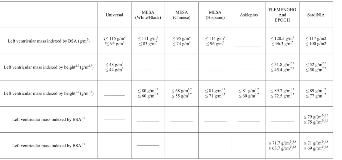
Left ventricular mass in the SardiNIA cohort by using different normalization methods proposed in the literature (BSA, height2.7, height1.7, BSA1.8) plus BSA1.6 (optimal scaling exponent in the Sardinian population). Comparison between different population studies.
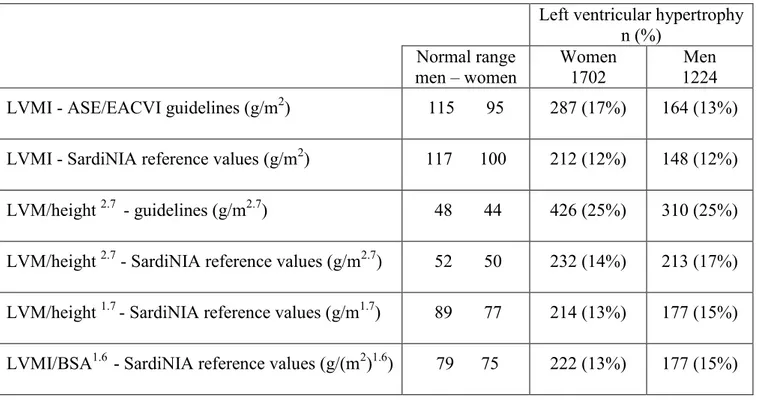
Table 8 shows the 95th percentiles of LV mass with different normalizations for body size (isometric and allometric methods) in various populations. Our study provides reference values for all the main normalization methods, while in other studies only normalization by BSA, height2.7 or by the ethnicity specific scalar exponent found (i.e. height1.8) were reported. Table 8 also shows the reference values for LV mass by BSA1.6 in the Sardinian population (≤ 79 g/(m2)1.6 in men and ≤ 75 g/(m2)1.6 in women). This table emphasizes the importance of reference values ethnicity-specific, as demonstrated by the different cut-point for LV hypertrophy.
(The NORRE study was not included because only reference values for LV mass were reported).
Frequency of left ventricular hypertrophy in the entire population (2926 subjects) according to various indexation methods for left ventricular hypertrophy.
Table 9 emphasizes the potential clinical impact of normalizing LV mass with different methods. When applying various indexation methods to the entire sample study (2926 subjects including those with cardiovascular risk factors and diseases), we found different frequencies of LV hypertrophy (LVH). We found the highest frequency of LVH when indexing by height2.7.
Cardiovascular effects of ageing
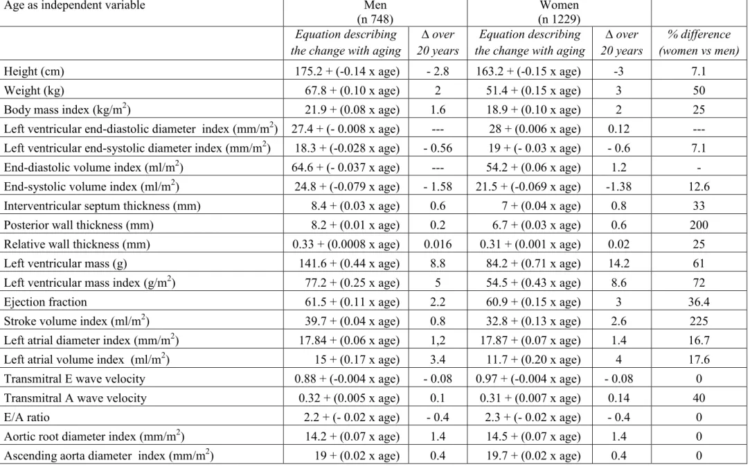
The relationships of ageing with the main anthropometric cardiac and vascular echocardiographic parameters in men and women are shown in Table 10 and Figures 12-19, while Table 11 emphasizes the sex-related differences. We noticed some changes of body structure with advancing age (reduced body height, increased weight and BMI) in both genders.
We observed several changes in cardiac structure and function with advancing age. There was an increase in wall thickness in both genders (interventricular septum r 0.31 and 0.37, p < 0.00001 in both genders; posterior wall r 0.18 in men and 0.33 in women, p < 0.00001). Furthermore, there was a trend toward a reduction of left ventricular end-systolic diameters (r - 0.16 in men and - 0.19 in women, p < 0.00001) and volumes (r - 0.17, p < 0.00001) in both genders, while end-diastolic diameters and volumes showed a similar (but not statistically significant) trend in males and a slight increase in females (diameter r 0.03, p = 0.31; volume r 0.08, p = 0.004). Taken together, all this data showed a trend toward reduction in LV cavity size with advancing age. Accordingly, we found that ageing induces concentric remodelling of the LV, as demonstrated by the
increase in relative wall thickness similar in both genders (r 0.18 in men and 0.22 in women respectively, p < 0.00001). LV mass increased with advancing age in both genders, despite a significant difference in the amount of this growth which was greater in females (LV mass r 0.18 in men vs 0.35 in women, p < 0.00001). By using simple regression analysis we defined a gender-specific equation (LV mass = 141.6 + (0.44 x age) for men; LV mass = 84.2 + (0.71 x age) for women) in order to estimate the amount of increase in LV mass every 10 years. According to these equations, women’s heart increases its mass of 7.1 gr every 10 years, while in men LV mass increases of 4.4 g (that is 61.97 % of 7.1) in the same time. LVMI increases of 2.5 g/m2 in men every ten years (77.26 + (0.25 x age)) and of 4.3 g/m2 in women (54.5 + (0.43 x age)) – Figure 20. Due to the potential clinical relevance of these results and in accordance with them, we propose normal reference values of LV mass index (LVMI) corrected by age in men and women. Table 12 shows the reference values of LVMI by decades based on the data of our population, together with the values of estimated LVMI obtained by applying the above-mentioned equation to estimate the 10-year increase of LVMI in both genders. It is noteworthy that women in the highest decade show values of LV mass similar or even highest to young men.
Our data was consistent with significant gender-related differences in cardiac remdelling with advancing age (i.e concentric remodeling in men vs concentric hypertrophy in women). LV ejection fraction (EF) increased with age in both genders (r 0.23 in men and 0.31 in women, p-value 0.001). However, its increase together with that of stroke volume index (SVI) were higher in women. Left atrial (LA) size and volumes increased with age in both genders but these changes were greater in women (diameter r 0.32 vs 0.36; volume r 0.33 vs 0.47 in men and women, all p < 0.00001).
Diastolic function was characterized by a reduction in E wave velocity (r - 0.36 in men and - 0.37 in women, p < 0.00001), an increase of A wave velocity (r 0.55 vs 0.59, p < 0.00001), with a clear reduction of the Doppler transmitral E/A ratio (r - 0.64 vs - 0.63, p < 0.00001) in both genders. Finally, aortic diameters increased in both genders with advancing age (aortic root r 0.45 vs 0.41, p < 0.00001; ascending aorta r 0.10 vs 0.12, p = 0.008 in men and 0.0006 in women).
Figures 21 and 22 emphasize the gender-related differences in cardiac structure and function with advancing age.
DISCUSSION
This study investigates the cardiovascular characteristics in a large sample representative of the SardiNIA cohort. Our data includes some considerations. The prevalence of arterial hypertension was 27.1% in the sample included in the echocardiographic study, lower than (> 30%) reported by Scuteri et al. in the entire cohort61,62 and lower than the overall prevalence of arterial hypertension in Italy (33.8% in the South according to different macro-areas).63 Diabetes had a frequency of 4% in the entire study sample, lower than the overall prevalence reported in Italy (> 5%).64 Notably, Sardinia represents the major exception to the general north-south gradient of diabetes incidence in Europe, showing a much higher frequency of the disease compared with surrounding Mediterranean regions.65 Despite this well known and established data, the prevalence of diabetes in our sample was quite low.
The overall prevalence of some cardiovascular risk factors (i.e. arterial hypertension, diabetes) and the burden of cardiovascular diseases were lower in our sample compared with national (italian) data or other populations. For example in Italy data collected by the Osservatorio Epidemiologico Cardiovascolare/Health Examination Survey (OEC/HES) in the general population aged 35-74 years showed a much higher prevalence of arterial hypertension (51% in men and 37.2% in women in the last survey 2008-12).66 Similarly the prevalence of diabetes was very high among people aged 75-79 years (27.7% in men and 18.9% in women).66 The relatively low mean age of our sample should explain, at least in part, these results and differences. It is well known that the prevalence of cardiovascular risk factors increases with age. In effect, the Asklepios study (2524 volunteers aged 35-55 years) with a mean age of participants of
45.9 years showed results to some extent more similar to ours in terms of prevalence of cardiovascular risk factors.67 However, genetic and environmental factors might also play a role. The central-eastern part of Sardinia has been identified in recent years as a “Blue Zone”, being characterized for an extreme longevity with a high number of centenarians. The estimated life expectancy in the “Blue Zone” is longer than in the remaining territory of the island especially for men, and the male to female ratio among centenarians born in this area is 1.35 compared to 2.43 in the rest of Sardinia.68
The present study provides reference values for the main echocardiographic parameters (i.e. LV chamber quantification, LA size, aortic dimensions) in a large sample representative of the Sardinian population. In our study, both genders were well represented despite a predominance of females. Overall upper reference limits were higher in men compared with women; these differences were mantained for most parameters after normalization for the body surface area. Our results emphasize the importance of developing ethnicity-specific reference values, as highlighted in previous studies.6 Our data shows some relevant differences with the reference values recommended by the European Association of Cardiovascular Imaging (EACVI) and the American Society of Echocardiography (ASE).22, 56 Notably, when using the 95th percentiles to define the upper reference limits we found significant differences with the commonly used cut-off for relative wall thickness (RWT), LV mass and its various indexations. This data, together with the fact that the sex-related differences in LA size and aortic diameters showed an inversion after normalization for body surface area should be, at least in part (because geometry - e.g. RWT - tends to be constant independently from body size), the result of specific anthropometric characteristics of our population. These characteristics seem to play a pivotal role in the normalization of
the echocardiographic parameters. Normalization for body size is very important but whether to use isometric or allometric methods is a matter of controversy, especially regarding LV mass.16, 20, 69, 70 Despite the potential limitations when using isometric methods (e.g. overcorrection in obese patients), recently published data has raised concerns about the validity and universal extension of some allometric methods (e.g. LV mass/height2.7). In 2010 Chirinos et al. described a negative relationship between LV mass and body height, and they found that indexing mass/height2.7 grossly overestimated LV mass and the prevalence of hypertrophy in men and women with shorter body height.20 Our results in a large sample from the SardiNIA cohort whose mean height was only 157 cm for women and 169 cm for men (6.4% of the entire sample < 150 cm), and thus significantly shorter than other populations16, seem to confirm these findings. When we stratified our sample by body height (≤ 149 cm; 150-159 cm; 160-169 cm; 170-179 cm; ≥ 180 cm) and we analyzed the trends of left ventricular mass (various indexations) among women and men, we found a clear trend toward higher LV mass in shorter subjects (i.e. < 149 cm). Our results clearly show a relative overestimation of LV mass in shorter subjects when using allometric methods compared with normalization for BSA. When we stratified our sample by BMI (< 25 kg/m2; 25-30 kg/m2; > 30 kg/m2), we found that subjects with higher BMI show higher values of LV mass independently from the indexation method. Despite a common trend characterized by a direct relationship between LV mass and BMI (i.e. higher the BMI and higher the LV mass), values were proportionally higher when using allometric methods. This should be related to the well known overcorrection in obese patients when using isometric methods. However, taken together our results suggest that normalizing echocardiographic measurements (i.e. LV mass) for body surface area
(BSA) is superior to the use of allometric methods in removing (or attenuating) the effects of body size in our sample and thus it should be considered as the method of choice in the Sardinian population.
In our study, indexation for BSA showed residual relationship with body structure in both genders. We identified a scalar exponent in men (BSA1.1) and women (BSA1.4) able to almost null these residual effects, providing the best way to normalize LV mass in the SardiNIA cohort. These results emphasize the concept that gender is a strong correlate of boy-size and left ventricular mass and the need of a gender specific scaling exponent. However, due to the need of simplicity when transferring these concepts into clinical practice and in order to make them really useful in the echocardiographic laboratories, we found a scalar exponent (BSA1.6) not gender-specific and more easily applicable. Our results slightly differ from those recently published by Kuznetsova T et al.70 in a sample of 656 healthy individuals. With regard to left ventricular mass they found that the allometric exponents for BSA ranged from 1.7 to 1.8. These differences probably reflect the characteristics (e.g. anthropometric) of the populations, making it difficult to find a scalar exponent universally acceptable.
Our study provides useful data regarding the relationships between ageing and cardiovascular system in both genders. We observed several changes in cardiac structure and function with advancing age. Overall, we noticed a cardiac remodelling (reduced cavity size and increased relative and absolute wall thickness) in both genders. Our data confirms that ageing induces left ventricular cocentric remodelling in normotensive subjects as previously described by Ganau et al. in 430 clinically healthy subjects aged 16-85 years.31 Furthermore, we found some significant gender-related differences in cardiac structural changes with advancing age (tendency to concentric
remodelling in men vs concentric hypertrophy in women). LV mass increased with ageing in both genders, but this growth was greater in females than males. We also quantified the amount of increase in LV mass every 10 years. The increase in LV mass observed with ageing does not seem to be related to an increase of cardiomyocytes whose number reduces with ageing. This phenomenon is more pronounced in men than in women and it is linked to a higher apoptoctic index (three times greater) in men, as previously shown by Olivetti et al.32 and Mallat et al.33
We also found other gender-related differences with advancing age. LV ejection fraction and stroke volume index increases more in women than men suggesting a greater functional systolic reserve in women. Notably, we found gender differences in left atrial size and diastolic function. With advancing age the left atrium enlarges in both genders; however the amount of increase in LA size is greater in women. This result, together with a greater increase in the velocity of transmitral. A wave velocity suggests a higher “left atrial stretch” in women compared with men, together with a greater impairment of diastolic function in females. Our results are consistent with the higher prevalence of heart failure with preserved ejection fraction (HFpEF) in women.
All together, our data highlights that normalization for body size should be performed along with age-gender specific assessment for most echocardiographic parameters.
Study limitations and future perspectives
Our study has some limitations. First, despite the large size of the study sample, it does not include all the SardiNIA cphort. Furthermore, the mean age of our sample was relatively young with a relatively small percentage of people aged 65 years or more. This should have potential limitations when studying the cardiovascular effects of
ageing and when trying to define age-specific reference values for some echocardiographic parameters, due to the relatively low number of old subjects.
Finally left ventricular volumes and ejection fraction were assessed with 2D guided M-mode using the Teichholz method, making a strict comparison with current guidelines hard where 2D measurements are reported.
The extension of the study to the entire SardiNIA cohort will overcome most of the above mentioned limitations. The development of age-gender specific reference values for most echocardiographic parameters will be useful for clinical and prognostic purposes.
In the future, prognostic data will help us to clarify the clinical significance of all the cardiac and vascular changes observed with aging in the SardiNIA cohort.
The genome-wide association study (GWAS) of the entire SardiNIA cohort will help us to identify the genetic determinants of the main cardiovascular traits and echocardiographic parameters.
CONCLUSIONS
This study described the clinical cardiological characteristics in a large sample representative of the SardiNIA cohort. Furthermore, it provided the reference values by sex, together with their normalization for body size, for the main echocardiographic parameters, both cardiac (e.g. wall thickness, cardiac diameters and volumes, left ventricular mass, left atrial volumes, diastolic profile) and vascular (aortic root and ascending aorta diameters). We also identified the best method of normalization for body size in the Sardinian population. Our study demonstrated that some allometric methods of normalization for body size of the echocardiographic parameters (e.g. LV mass) proposed in the literature (e.g. LVM2.7) are not universally applicable. In particular, in the Sardinian population they may overestimate left ventricular mass and, by consequence, the true prevalence of left ventricular hypertrophy. The best method for normalization in our population is the isometric scaling using body surface area (BSA). We identified a scalar exponent of BSA (i.e. BSA1.6) specific for our cohort. It seems to be able to provide a better correction compared to BSA itself.
Finally, we defined the effects of ageing on cardiovascular structure and function (left ventricular remodeling; abnormal diastolic relaxation; left atrial enlargement; increase in aortic diameters), showing significant gender-related differences in cardiac structural changes associated to advancing age. In particular, while relative wall thickness increases with age similarly in men and women, women exhibit an almost double increase in LV mass (4.3 g/m2 every 10 years) compared to men (2.5 g/m2 every 10 years), confirming our previously report of aging induced concentric remodelling in men (increase in relative wall thickness with no or mild increase in LV mass) and
concentric hypertrophy in women (increase in both relative wall thickness and LV mass).31
The strong and quantitatively relevant dependence of left ventricular mass and relative wall thickness with age highlights the need for age-gender reference values for most echocardiographic parameters.
REFERENCES
[1] Lakatta EG. So! What’s aging? Is cardiovascular aging a disease? J Mol Cell Cardiol 2015;83:1-13.
[2] https://www.istat.it
[3] Pilia G, Chen WM, Scuteri A, Orrú M, Albai G, Dei M, Lai S, Usala G, Lai M, Loi P, Mameli C, Vacca L, Deiana M, Olla N, Masala M, Cao A, Najjar SS, Terracciano A, Nedorezov T, Sharov A, Zonderman AB, Abecasis GR, Costa P, Lakatta E, Schlessinger D. Heritability of cardiovascular and personality traits in 6,148 Sardinians. PloS Genet 2006;2(8):e132.
[4] Heutink P, Oostra BA. Gene finding in genetically isolated populations. Hum Mol Genet 2002;11(20):2507-15.
[5] Lang RM, Badano LP, Mor-Avi V, Afilalo J, Armstrong A, Ernande L, Flachskampf FA, Foster E, Goldstein SA, Kuznetsova T, Lancellotti P, Muraru D, Picard MH, Rietzschel ER, Rudski L, Spencer KT, Tsang W, Voigt JU. Recommendations for cardiac chamber quantification by echocardiography in adults: an update from the American Society of Echocardiography and the European Association of Cardiovascular Imaging. J Am Soc Echocardiogr 2015;28(1):1-39.
[6] Kou S, Caballero L, Dulgheru R, Voilliot D, De Sousa C, Kacharava G, Athanassopoulos GD, Barone D, Baroni M, Cardim N, Gomez De Diego JJ, Hagendorff A, Henri C, Hristova K, Lopez T, Magne J, De La Morena G, Popescu BA, Penicka M, Ozyigit T, Rodrigo Carbonero JD, Salustri A, Van De Veire N, Von Bardeleben RS, Vinereanu D, Voigt JU, Zamorano JL, Donal E, Lang RM, Badano LP, Lancellotti P. Echocardiographic reference ranges for normal cardiac chamber size: results from the NORRE study. Eur Heart J Cardiovasc Imaging 2014;15(6):680-90.
[7] Daimon M, Watanabe H, Abe Y, Hirata K, Hozumi T, Ishii K, Ito H, Iwakura K, Izumi C, Matsuzaki M, Minagoe S, Abe H, Murata K, Nakatani S, Negishi K, Yoshida K, Tanabe K, Tanaka N, Tokai K, Yoshikawa J; JAMP Study Investigators. Normal values of echocardiographic parameters in relation to age in a healthy Japanese population: the JAMP study. Circ J 2008;72(11):1859-66.
[8] Dewey FE, Rosenthal D, Murphy DJ Jr, Froelicher VF, Ashley EA. Does size matter? Clinical applications of scaling cardiac size and function for body size. Circulation 2008;117(17):2279-87.
[9] de Simone G, Kizer JR, Chinali M, Roman MJ, Bella JN, Best LG, Lee ET, Devereux RB; Strong Heart Study Investigators. Normalization for body size and population-attributable risk of left ventricular hypertrophy: the Strong Heart Study. Am J Hypertens 2005;18(2 Pt 1):191-6.
[10] Gutgesell HP, Rembold CM. Growth of the human heart relative to body surface area. Am J Cardiol 1990;65:662-8.
[11] Zong P1, Zhang L2, Shaban NM3, Peña J3, Jiang L4, Taub CC5. Left heart chamber quantification in obese patients: how does larger body size affect echocardiographic measurements?
J Am Soc Echocardiogr 2014;27(12):1267-74.
[12] Cuspidi C1, Meani S, Negri F, Giudici V, Valerio C, Sala C, Zanchetti A, Mancia G. Indexation of left ventricular mass to body surface area and height to allometric power of 2.7: is the difference limited to obese hypertensives? J Hum Hypertens 2009;23(11):728-34.
[13] Gardin JM, Arnold A, Gottdiener JS, Wong ND, Fried LP, Klopfenstein HS, O’Leary DH, Tracy R, Kronmal R. Left ventricular mass in the elderly: the Cardiovascular Health Study. Hypertension 1997;29:1095-1103.
[14] Koren MJ, Devereux RB, Casale PN, Savage DD, Laragh JH. Relation of left ventricular mass and geometry to morbidity and mortality in uncomplicated essential hypertension. Ann Intern Med 1991;114:345-352.
[15] Bluemke DA, Kronmal RA, Lima JA, Liu K, Olson J, Burke GL, Folson AR. The relationship of left ventricular mass and geometry to incident cardiovascular events: the MESA (Multi-Ethnic Study of Atherosclerosis) Study. J Am Coll Cardiol 2008;52:2148-2155.
[16] de Simone G, Daniels SR, Devereux RB, Meyer RA, Roman MJ, de Divitiis O, Alderman MH. Left ventricular mass and body size in normotensive children and adults: assessment of allometric relations and impact of overweight. J Am Coll Cardiol 1992;20(5):1251-60.
[17] de Simone G, Devereux RB, Maggioni AP, Gorini M, de Divitiis O, Verdecchia P. Different normalizations for body size and population attributable risk of left ventricular hypertrophy: the MAVI Study. Am J Hypertens 2005;18:1288-1293.
[18] de Simone G, Devereux RB, Daniels SR, Koren MJ, Meyer RA, Laragh JH. Effect of growth on variability of left ventricular mass: assessment of allometria signals in adults and children and their capacity to predict cardiovascular risk. J Am Coll Cardiol 1995;25:1056-1062.
[19] Foster BJ1, Mackie AS, Mitsnefes M, Ali H, Mamber S, Colan SD. A novel method of expressing left ventricular mass relative to body size in children. Circulation 2008;117(21):2769-75.
[20] Chirinos JA, Segers P, De Buyzere ML, Kronmal RA, Raja MW, De Bacquer D, Claessens T, Gillebert TC, St John-Sutton M, Rietzschel ER. Left ventricular mass:
allometric scaling, normative values, effect of obesity, and prognostic performance. Hypertension 2010;56(1):91-8.
[21] Lang RM, Badano LP, Mor-Avi V, Afilalo J, Armstrong A, Ernande L, Flachskampf FA, Foster E, Goldstein SA, Kuznetsova T, Lancellotti P, Muraru D, Picard MH, Rietzschel ER, Rudski L, Spencer KT, Tsang W, Voigt JU. Recommendations for cardiac chamber quantification by echocardiography in adults: an update from the American Society of Echocardiography and the European Association of Cardiovascular Imaging. J Am Soc Echocardiogr 2015;28(1):1-39.
[22] Marwick TH, Gillebert TC, Aurigemma G, Chirinos J, Derumeaux G, Galderisi M, Gottdiener J, Haluska B, Ofili E, Segers P, Senior R, Tapp RJ, Zamorano JL. Recommendations on the Use of Echocardiography in Adult Hypertension: A Report from the European Association of Cardiovascular Imaging (EACVI) and the American Society of Echocardiography (ASE). J Am Soc Echocardiogr 2015;28(7):727-54.
[23] Arcaleni E. Secular trend and regional differences in the stature of Italians. J Anthropol Sci Riv Antropol JASS Ist Ital Antropol 2012;90:233-237.
[24] Zoledziewska M, Sidore C, Chiang CW, Sanna S, Mulas A, Steri M, Busonero F, Marcus JH, Marongiu M, Maschio A, Del Vecchyo DO, Floris M, Meloni A, Delitala A, Concas MP, Murgia F, Biino G, Vaccargiu S, Nagaraja R, Lohmueller KE; UK10K Consortium., Timpson NJ, Soranzo N, Tachmazidou I, Dedoussis G, Zeggini E; Understanding Society Scientific Group., Uzzau S, Jones C, Lyons R, Angius A, Abecasis GR, Novembre J, Schlessinger D, Cucca F. Height-reducing variants and selection for short stature in Sardinia. Nat Genet 2015;47(11):1352-6.
[25] Keller KM, Howlett SE. Sex Differences in the Biology and Pathology of the Aging Heart. Can J Cardiol 2016;32(9):1065-73.
[26] Lakatta EG, Levy D. Arterial and cardiac aging: major shareholders in cardiovascular disease enterprises: Part II: the aging heart in health: links to heart disease. Circulation 2003;107(2):346-54.
[27] Levy D, Garrison RJ, Savage DD, Kannel WB, Castelli WP. Prognostic implications of echocardiographically determined left ventricular mass in the Framingham Heart Study. New Engl J Med 1990;322(22):1561-6.
[28] Kitzman DW, Gardin JM, Gottdiener JS, Arnold A, Boineau R, Aurigemma G, Marino EK, Lyles M, Cushman M, Enright PL; Cardiovascular Health Study Research Group. Importance of heart failure with preserved systolic function in patients > or = 65 years of age. CHS Research Group. Cardiovascular Health Study. Am J Cardiol 2001;87(4):413-9.
[29] Ponikowski P, Voors AA, Anker SD, Bueno H, Cleland JG, Coats AJ, Falk V, González-Juanatey JR, Harjola VP, Jankowska EA, Jessup M, Linde C, Nihoyannopoulos P, Parissis JT, Pieske B, Riley JP, Rosano GM, Ruilope LM, Ruschitzka F, Rutten FH, van der Meer P; Authors/Task Force Members. 2016 ESC
Guidelines for the diagnosis and treatment of acute and chronic heart failure: The Task Force for the diagnosis and treatment of acute and chronic heart failure of the European Society of Cardiology (ESC)Developed with the special contribution of the Heart Failure Association (HFA) of the ESC. Eur Heart J 2016;37(27):2129-200.
[30] European Heart Rhythm Association.; European Association for Cardio-Thoracic Surgery., Camm AJ, Kirchhof P, Lip GY, Schotten U, Savelieva I, Ernst S, Van Gelder IC, Al-Attar N, Hindricks G, Prendergast B, Heidbuchel H, Alfieri O, Angelini A, Atar D, Colonna P, De Caterina R, De Sutter J, Goette A, Gorenek B, Heldal M, Hohloser SH, Kolh P, Le Heuzey JY, Ponikowski P, Rutten FH. Guidelines for the management of atrial fibrillation: the Task Force for the Management of Atrial Fibrillation of the European Society of Cardiology (ESC). Eur Heart J 2010;31(19):2369-429.
[31] Ganau A1, Saba PS, Roman MJ, de Simone G, Realdi G, Devereux RB. Ageing induces left ventricular concentric remodelling in normotensive subjects. J Hypertens 1995;13(12 Pt 2):1818-22.
[32] Olivetti G, Giordano G, Corradi D, Melissari M, Lagrasta C, Gambert SR, Anversa P. Gender differences and aging: effects on the human heart. J Am Coll Cardiol 1995;26(4):1068-79.
[33] Mallat Z, Fornes P, Costagliola R, Esposito B, Belmin J, Lecomte D, Tedgui A.
Age and gender effects on cardiomyocyte apoptosis in the normal human heart. J
Gerontol A Biol Sci Med Sci 2001;56(11):M719 –23.
[34] Piro M1, Della Bona R, Abbate A, Biasucci LM, Crea F. Sex-related differences in myocardial remodeling. J Am Coll Cardiol 2010;55(11):1057-65.
[35] López B, Querejeta R, González A, Larman M, Díez J. Collagen cross-linking but not collagen amount associates with elevated filling pressures in hypertensive patients with stage C heart failure: potential role of lysyl oxidase. Hypertension 2012;60(3):677-83.
[36] Slotwiner DJ, Devereux RB, Schwartz JE, Pickering TG, de Simone G, Ganau A, Saba PS, Roman MJ. Relation of age to left ventricular function in clinically normal adults. Am J Cardiol 1998;82(5):621-6.
[37] Khouri MG, Maurer MS, El-Khoury Rumbarger L. Assessment of age-related changes in left ventricular structure and function by freehand three-dimensional echocardiography. Am J Geriatr Cardiol 2005;14(3):118-25.
[38] Kaku K, Takeuchi M, Otani K, Sugeng L, Nakai H, Haruki N, Yoshitani H, Watanabe N, Yoshida K, Otsuji Y, Mor-Avi V, Lang RM. Age- and gender-dependency of left ventricular geometry assessed with real-time three-dimensional transthoracic echocardiography. J Am Soc Echocardiogr 2011;24(5):541-7.
[39] Nikitin NP, Loh PH, de Silva R, Witte KK, Lukaschuk EI, Parker A, Farnsworth TA, Alamgir FM, Clark AL, Cleland JG. Left ventricular morphology, global and