UNIVERSITY OF CATANIA
INTERNATIONAL PhD IN CHEMICAL SCIENCES
XXX CYCLE
CURRICULUM: ORGANIC CHEMISTRY
Luana Pulvirenti
NATURAL OR BIO-INSPIRED POLYPHENOLS IN THE
SYNTHESIS OF POTENTIAL CHEMOTHERAPEUTIC
AGENTS
Final report of research activity
Tutor: Prof. CORRADO TRINGALI Coordinator: Prof. SALVATORE SORTINO
This thesis is dedicated to the memory of Prof. Carmela Spatafora, a special and talented scientist who
ABBREVIATIONS 9
ABSTRACT 1
1. INTRODUCTION 3
1.1 Polyphenols 5
1.1.1 Natural polyphenols as antioxidative agents 10
1.1.2 Lignans and neolignans 13
1.1.3 Benzoxanthenes lignans 20
1.1.4 Bisphenol neolignans 25
1.1.4.1 Magnolol analogues as -glucosidase inhibitors 27 1.1.5 Ellagitannins: bioactive plant polyphenols 30
1.1.5.1 Stereochemical consideration on ellagitannins 32
1.1.7 Aims of the PhD research activity 34
2. RESULTS AND DISCUSSION 37
2.1 Synthesis and biological evaluation of bioinspired benzo[k,l]xanthene lignans and
related phenazines 37
2.1.1 Biomimetic synthesis of benzo[k,l]xanthene lignans 37 2.1.1.1 Synthesis of dimethyl 6,9,10-trihydroxybenzo[k,l]xanthene-1,2-dicarboxylate
(36) 38
2.1.1.2 Synthesis of diphenethyl
6,9,10-trihydroxy-benzo[k,l]xanthene-1,2-dicarboxylate (38) and its methylated derivative (43) 39 2.1.1.3 Synthesis of dibutyl 6,9,10-trihydroxybenzo[k,l]xanthene-1,2-dicarboxylate
(39) 40
2.1.1.4 Synthesis of diethyl 6,9,10-trihydroxybenzo[k,l]xanthene-1,2-dicarboxylate
(61) 40
2.1.1.5 bis(4-methoxybenzyl)6,9,10-trihydroxybenzo[kl]xanthene-1,2-dicarboxylate
(63) 41
2.1.1.6 bis(benzyl)6,9,10-trihydroxybenzo[kl]xanthene-1,2-dicarboxylate (65) 42 2.1.1.7 Synthesis of bis(4-hydroxybutyl)
6,9,10-trihydroxybenzo[kl]xanthene-1,2-dicarboxylate (67) 43
2.1.2 Synthesis of phenazine derivatives of benzoxanthene lignans 44 2.1.2.1 Preliminary screenings: benzo[k,l]xanthene oxidation 44
2.1.2.2 Enzymatic methods 45
2.1.2.3 Chemical methods 46
2.1.2.4 Synthesis of dibutyl
6-hydroxybenzo[4,5]chromeno[2,3-b]phenazine-1,2-dicarboxylate (69) 48
2.1.2.5 Synthesis of diethyl
6-hydroxybenzo[4,5]chromeno[2,3-b]phenazine-1,2-dicarboxylate (71) 51
2.1.2.6 Synthesis of diphenethyl 6-hydroxybenzo[4,5]chromeno-
[2,3-b]phenazine-1,2-dicarboxylate (73) 54
2.1.2.7 Synthesis of dibutyl
12-(3,4-diaminophenyl)-6-hydroxybenzo[4,5]chromeno[2,3-b]phenazine-1,2-dicarboxylate (75 and 77) 57 2.1.3 Study of the interaction of benzoxanthenes with DNA G-quadruplex 60 2.1.4 Study of the interaction of benzoxanthenes with Bile X receptors 67 2.1.5 Study of the antimicrobial properties of benzoxanthenes 70
2.2 Biomimetic synthesis of dimeric neolignans inspired by magnolol and their
biological activities 74
2.2.1 Chemoenzymatic synthesis and selective hydroxylation of dimeric neolignans
inspired by magnolol 75
2.2.1.1 IBX in the simple synthesis of catechol compounds 77 2.2.1.2 Selective hydroxylation of magnolol (44) mediated by IBX 79
2.2.1.2.1 Synthesis of compounds 81 and 82 82
2.2.1.3 Preliminary study of the synthetic route to magnolol-inspired compounds 84
2.2.1.3.1 Preliminary study on synthetic route a 85
2.2.1.3.2 Preliminary study on synthetic route b and synthesis of the neolignans 84
and 85 86
2.2.1.4 Chemoenzymatic synthesis of bis-phenol neolignans 86 - 93 89 2.2.1.4.1 Biomimetic synthesis of bis-phenol neolignans 86 - 89 90 2.2.1.4.2 Enzymatic acetylation of 77 and 78 91 2.2.1.4.3 Biomimetic synthesis of bis-phenols 88 and 89 92 2.2.1.4.3.1 selective hydroxylation of 88 and 89 93 2.2.1.4.4 Enzymatic alcoholysis of 90 and 91 95
2.2.1.5 Synthesis of compounds 94 - 96 95
2.2.2 Biochemical evaluation of magnolol analogues as α-glucosidase inhibitors 97 2.2.3 Chain-breaking antioxidant activity of hydroxylated and methoxylated magnolol
derivatives 101
2.2.3.1 Kinetics and stoichiometry of the reaction with peroxyl radicals 103
2.2.3.2 FT-IR measures 105
2.2.3.3 Theoretical Calculations 106
2.2.4 Inverse Virtual Screening of magnolol analogues and their biological activities on
bromodomain, tankirase and caseinase 111
2.2.4.1 Inverse Virtual Screening 112
2.2.4.1.1 Assay on Tankyrase 2 114
2.2.5 Magnolol derivatives as inhibitors of the human breast cancer resistance protein
(BCRP/ABCG2) 117
2.3 IBX-mediated synthesis of a new hydroxylated dihydrobenzofuran neolignan 120 2.4 Contribution on total synthesis of ellagitannins 126
2.4.1 Vescalin total synthesis 127
2.4.1.1 Synthesis of the glucose precursor 108 128 2.4.1.2 Synthesis of the galloyl precursor 111 130
2.4.1.3 Multi-step synthesis of vescalin (54) 131
2.4.2 Synthesis of a vescalagin-fucose conjugate 135 2.4.2.1 Synthesis of the fucose precursor 122 137 2.4.2.2 Synthesis of the aromatic precursor 127 138
2.4.2.3 Synthesis of the linker precursor 139
2.4.2.4 New synthetic approach to obtain a vescalagin-fucose conjugate 141
3. CONCLUSIONS AND PERSPECTIVES 145
3.1 Benzo[k,l]xanthene lignans 145
3.3 Contribution to the total synthesis of vescalin 150
3.4 Final comment 151
4. EXPERIMENTAL SECTION 154
4.1 General Experimental Procedures 154
4.2 Biomimetic synthesis of benzo[k,l]xanthene lignans 36, 38, 39, 41, 60, 63, 65 and 67 156 4.3 Synthesis of benzo[k,l]xanthene phenazine derivatives 69, 71, 73, 74 and 75 161 4.4 Chemoenzymatic synthesis of bis-phenol neolignans 81, 82 and 84 - 96 167
4.5 α-Glucosidase inhibition assay 181
4.6 Vescalin total synthesis 182
4.7 Vescalagin fucose synthesis 188
5. SUPPORTING MATERIAL 193
5.1 Appendix A 193
5.1.1 Compound 36 193
Figure 1S: 1H-NMR spectrum (500 MHz, acetone-d6) of compound 36. 193
Figure 2S: 13C-NMR spectrum (125 MHz, acetone-d6) of compound 36. 193
5.1.2 Compound 38 194
Figure 3S: 1H-NMR spectrum (500 MHz, acetone-d
6) of compound 38. 194
Figure 4S: 13C-NMR spectrum (125 MHz, acetone-d
6) of compound 38. 194
5.1.3 Compound 39 195
Figure 5S: 1H-NMR spectrum (500 MHz, acetone-d
6) of compound 39. 195
Figure 6S: 13C-NMR spectrum (125 MHz, acetone-d
6) of compound 39. 195
5.1.4 Compound 61 196
Figure 7S: 1H-NMR spectrum (500 MHz, acetone-d
6) of compound 61. 196
Figure 8S: 13C-NMR spectrum (125 MHz, acetone-d
6) of compound 61. 196
5.1.5 Compound 63 197
Figure 9S: 1H-NMR spectrum (500 MHz, acetone-d
6) of compound 63. 197
Figure 10S: 13C-NMR spectrum (125 MHz, acetone-d
6) of compound 63. 197
5.1.6 Compound 65 198
Figure 11S: 1H-NMR spectrum (500 MHz, acetone-d
6) of compound 65. 198
Figure 12S: 13C-NMR spectrum (125 MHz, acetone-d6) of compound 65. 198
5.1.7 Compound 67 199
Figure 13S: 1H-NMR spectrum (500 MHz, acetone-d
6) of compound 67. 199
Figure 14S: 13C-NMR spectrum (125 MHz, acetone-d
6) of compound 67. 199
5.2 Appendix B 200
5.2.1 Compound 69 200
Figure 15S: ESI-Mass spectrum of 69. 200
Figure 16S: 1H-NMR spectrum (500 MHz,CDCl
Figure 17S: 13C-NMR spectrum (125 MHz,CDCl
3) of 69. 201
Figure 18S: gCOSY spectrum of compound of 69. 202 Figure 19S: gHSQCAD spectrum of compound of 69. 203 Figure 20S: gHMBCAD spectrum of compound of 69. 204
5.2.2 Compound 71 205
Figure 21S: ESI-MS spectrum of 71. 205
Figure 22S: 1H-NMR spectrum (500 MHz,CDCl3 and 1% of CD3OD) of compound
71. 205
Figure 23S: 13C-NMR spectrum (125 MHz, CDCl
3 and 1% of MeOD) of 71. 206
Figure 24S: gCOSY spectrum of compound of 71. 207 Figure 25S: gHSQCAD spectrum of compound of 71. 208 Figure 26S: gHMBCAD spectrum of compound of 71. 209
5.2.3 Compound 71 210
Figure 27S: ESI-MS spectrum of 73. 210
Figure 28S: 1H-NMR spectrum (500 MHz,CDCl
3) of compound 73. 210
Figure 29S: 13C-NMR spectrum (125 MHz,CDCl
3) of 73. 211
Figure 30S: gCOSY spectrum of compound of 73 212 Figure 31S: gHSQCAD spectrum of compound of 73. 213 Figure 32S: gHMBCAD spectrum of compound of 73. 214
5.2.4 Compound 74 and 75 215
Figure 33S: ESI-MS spectrum of 74 or 75. 215
Figure 34S: 1H-NMR spectrum (500 MHz, CD3COD + CDCl3) of 74 or 75. 215
Figure 35S: 13C-NMR spectrum (125 MHz, CD
3COD + CDCl3) of 74 or 75. 216
Figure 36S: gCOSY spectrum of compound of 74 or 75. 217 Figure 37S: gHSQCAD spectrum of compound of 74 or 75. 218 Figure 38S: gHMBCAD spectrum of compound of 74 or 75. 219
5.3 Appendix C 220
5.3.1 Compound 81 220
Figure 39S: ESIMS spectrum of compound 81. 220
Figure 40S: 1H NMR spectrum (500 MHz, CDCl
3) of compound 81. 220
Figure 41S: 13C NMR spectrum (125 MHz, CDCl
3) of compound 81. 221
Figure 42S: gCOSY spectrum of compound 81. 222
Figure 43S: gHSQCAD spectrum of compound 81. 223 Figure 44S: gHMBCAD spectrum of compound 81. 224
5.3.2 Compound 82 225 Figure 45S: 1H NMR spectrum (500 MHz, CDCl 3) of 82. 225 Figure 46S: 13C NMR spectrum (125 MHz, CDCl 3) of 82. 225 5.3.3 Compound 84 226 Figure 47S: 1H NMR spectrum (500 MHz, CDCl 3) of 84. 226 Figure 48S: 13C NMR spectrum (125 MHz, CDCl 3) of 84. 226 5.3.4 Compound 85 227 Figure 49S: 1H NMR spectrum (500 MHz, CDCl3) of 85. 227 Figure 50S: 13C NMR spectrum (125 MHz, CDCl 3) of 85. 227 5.3.5 Compound 86 228
Figure 51S: HRMS spectrum of compound 86. 228
Figure 52S: 1H NMR spectrum (500 MHz, acetone-d6) of compound 86. 228
Figure 53S: 13C NMR spectrum (125 MHz, acetone-d
6) of compound 86. 229
Figure 54S: gCOSY spectrum of compound 86. 230
Figure 55S: gHSQCAD spectrum of compound 86 231
Figure 56S: HRMS spectrum of compound 87. 232 Figure 57S: 1H NMR spectrum (500 MHz, acetone-d
6) of compound 87. 232
Figure 58S: 13C NMR spectrum (125 MHz, acetone-d
6) of compound 87. 233
Figure 59S: gCOSY spectrum of compound 87. 234
Figure 60S: gHSQCAD spectrum of compound 87. 235
5.3.7 Compound 88 236
Figure 61S: ESIMS spectrum of compound 88. 236
Figure 62S: 1H NMR spectrum (500 MHz, acetone-d
6) of compound 88. 236
Figure 63S: 13C NMR spectrum (125 MHz, acetone-d
6) of compound 88. 237
Figure 64S: gCOSY spectrum of compound 88. 238
Figure 65S: gHSQCAD spectrum of compound 88. 239
5.3.8 Compound 89 240
Figure 66S: ESIMS spectrum of compound 89. 240
Figure 67S: 1H NMR spectrum (500 MHz, acetone-d
6) of compound 89. 240
Figure 68S: 13C NMR spectrum (125 MHz, acetone-d
6) of compound 89. 241
Figure 69S: gCOSY spectrum of 89. 242
Figure 70S: gHSQCAD spectrum of 89. 243
5.3.9 Compound 90 244
Figure 71S: ESIMS spectrum of compound 90. 244
Figure 72S: 1H NMR spectrum (500 MHz, acetone-d
6) of compound 90. 244
Figure 73S: 13C NMR spectrum (125 MHz, acetone-d
6) of compound 90. 245
Figure 74S: gCOSY spectrum of 90. 246
Figure 75S: gHSQCAD spectrum of 90. 247
5.3.10 Compound 91 248
Figure 76S: ESIMS spectrum of compound 91. 248
Figure 77S: 1H NMR spectrum (500 MHz, acetone-d6) of compound 91. 248
Figure 78S: 13C NMR spectrum (125 MHz, acetone-d
6) of compound 91. 249
Figure 79S: gCOSY spectrum of 91. 250
Figure 80S: gHSQCAD spectrum of 91. 251
Figure 81S: gHMBCAD spectrum of 91. 252
5.3.11 Compound 92 253
Figure 82S: ESIMS spectrum of compound 92. 253
Figure 83S: 1H NMR spectrum (500 MHz, acetone-d
6) of compound 92. 253
Figure 84S: 13C NMR spectrum (125 MHz, acetone-d6) of compound 92. 254
5.3.12 Compound 93 255
Figure 85S: ESIMS spectrum of compound 93. 255
Figure 86S: 1H NMR spectrum (500 MHz, acetone-d
6) of compound 93. 255
Figure 87S: 13C NMR spectrum (125 MHz, acetone-d6) of compound 93. 256
5.3.12 Compound 96 257
Figure 88S: 1HNMR spectrum (500 MHz, in acetone-d
6) of 96. 257
Figure 89S: 13C NMR spectrum (125 MHz, in acetone-d
6) of 96. 257
5.3.13 Compound 99 258
Figure 90S: ESIMS spectrum of 99. 258
Figure 91S: 1HNMR spectrum (500 MHz, in acetone-d
6) of 99. 258
Figure 92S: 13C NMR spectrum (125 MHz, in acetone-d
6) of 99. 259
Figure 93S: gCosy spectrum of 99 and key correlations 260
Figure 94S: gHSQCAD spectrum of 99. 261
Figure S95: gHMBCAD spectrum of 99 and key correlations 262
5.3 Appendix D 263
Figure 96S: 1HNMR spectrum (300 MHz, in CDCl 3) of 108. 263 Figure 97S: 13C NMR spectrum (75 MHz, in CDCl 3) of 108. 264 5.3.2 Compound 111 264 Figure 98S: 1HNMR spectrum (300 MHz, in CDCl 3) of 111. 264 Figure 99S: 13C NMR spectrum (75 MHz, in CDCl 3) of 111. 265 5.3.2 Compound 114 265
Figure 100S: 1HNMR spectrum (300 MHz, in acetone-d6) of 114. 265
Figure 101S: 13C NMR spectrum (75 MHz, in acetone-d
6) of 114. 266 5.3.2 Compound 127 266 Figure 102S: 1HNMR spectrum (300 MHz, in CDCl3) of 127. 266 Figure 103S: 13C NMR (75 MHz, in CDCl 3) of 127. 267 6. ACKNOWLEDGMENTS 271 7. REFERENCES 271
ABBREVIATIONS
AbL Agaricus bisporus Laccase
BXL Benzoxanthene
CAL Candida antarctica Lipase
C NMR Carbon Nuclear Magnetic Resonance
gCOSY Gradient Correlation Spectroscop
CD DMC Circular Dichroism Dimethylcarbonate DMAP 4-Dimetilamminopiridina; DCC Dicyclohexylcarbodiimide DMSO Dimethylsulfoxide EDCCI:HCl N-(3-Dimethylaminopropyl)-N′-ethylcarbodiimide hydrochloride
EtOAc Ethyl acetate
EtOH Ethanol
EP Petroleum ether
gHSQCAD Gradient Heteronuclear Single Quantum Coherence
Adiabatic
gHMBCAD Gradient Heteronuclear Multiple-Bond Correlation
Adiabatic
G-Q G-quadruplex
HHDP hexahydroxydiphenoyl
H NMR Proton Nuclear Magnetic Resonance
HPLC High Performance Liquid Chromatography
HRP Horseadish Peroxidase
IDA iodobenzene diacetate
NHTP nonahydroxyterphenoyl
ODN Oligodeoxyribonucleotide
PE Petroleum ether
PoL Pleurotus ostreatus Laccase
PcL Pseudomonas cepacea lipase
rt Room Temperature
SIBX Stabilized
1-hydroxy-1-oxo-3H-1λ⁵,2-benziodaoxol-3-one
SPR Surface Plasmon Resonance
TBAF tetra-n-butylammonium fluoride
TBSCl tert-Butyldimethylsilyl chloride
TLC Thin Layer Chromatography
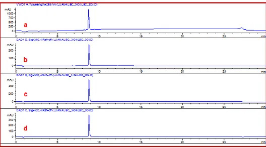
tR Time of retention
1
ABSTRACT
My doctoral research activity was focused mainly on natural or bioinspired polyphenols, aimed to the synthesis of new bioinspired compounds and to their evaluation as potential chemotherapeutic agents. More specifically, my research activity was devoted to lignans and neolignans, synthetized by chemical and/or enzimatic methodology. Hence, in particular, two polyphenol groups were studied, namely benzoxanthene lignans, their related phenazines and bisphenol neolignans; furthermore during the synthesis of magnolol-inspired neolignans, an unexpected dihydrobenzofuran neolignan was also obtained and characterized. The products have been studied in collaboration with others research team for the interaction with G-quadruplex DNA, as α-glucosidase inhibitors, as ABCG2 inhibitors, as agonist/antagonist of the Bile X receptors FXR and LXRα/β, antioxidant and antimicrobial agents. Further goal of this work was an inverse virtual screening focused on magnolol analogues, carried out during my short internship in the lab of prof. G. Bifulco (University of Salerno). In addition, the last part of my work was carried out at the University of Bordeaux, under the guidance of Prof. Stéphane Quideau, and was devoted to synthetic work on ellagitannins, an important subgroup of tannin family. In this context I gave a contribution to the total synthesis of vescalin and a vescalagin conjugate.
2
3
1. INTRODUCTION
The use of extracts from plants or other organisms for the treatment of diseases is as old as human history, but only in relatively recent times the active ingredients present in these natural remedies have been isolated and identified. After decades of research on the isolation and structural characterization of biologically active natural compounds, now we know the structure of more than 200,000 metabolites, and in the future this number could raise to more than 500,000.[1] In recent years there has been a renewed attention to bioactive natural products,[2] and one of the reasons is the observation that the structural diversity (also called ‘chemodiversity’) of natural products is far superior to that of the compounds that can be obtained by synthetic methods, including combinatorial synthesis.[3] One further reason is the awareness that many natural products may be considered as ‘lead compounds’ selected through millions of years of evolution; in fact, natural products have given an 'evolutionary advantage' to plants and other living organisms that produce them. These organisms have evolved in parallel with other species incorporating 'biological targets' for the products of their metabolism,[4] e.g. some alkaloids produced by plants are very toxic to herbivorous predators because the biosynthesis of these substances has evolved in parallel to that of the proteins 'target' of predators.[5] On the other hand, many antibiotics produced by fungi or other microorganisms are probably chemical defense systems against other competing microorganisms. The development of these ‘natural leads’ was exploited in drug discovery: in fact, modifying and optimizing key features of selected natural compounds it is possible to improve their biological activity,
4
bioavailability, the mode of administration, etc. A variety of examples can be cited in this regard. Probably the most popular semi-synthetic derivative of a naturally occurring compound is acetylsalicylic acid (1) or aspirin®, the first of the non-steroidal anti-inflammatory drugs (NSAIDs), synthesized by chemists at Bayer more than a century ago with a simple modification of salicylic acid, itself a product of oxidation of salicin (2), the natural metabolite found in willow (Salix alba var. tristis) extracts.[6]
Although the efficacy of willow extract as antipyretic is reported in the Ebers Papyrus (1550 BC), and aspirin has now been used by more than 100 years as an antipyretic and anti-inflammatory agent, only recently its mechanism of action has been defined. This molecule acts as an inhibitor of cyclooxygenase (COX-1 and COX-2), the key enzymes in the biosynthesis of pro-inflammatory prostaglandins but also of thromboxanes, which facilitate the coagulation of the blood. As a matter of fact, today aspirin is also used to prevent cardiovascular diseases, such as myocardial infarction. Many compounds with antitumor properties have been derived from bioactive natural products: eg. flavopiridol (3), an inhibitor of cyclin-dependent kinases (CDKs) is a semisynthetic derivative marketed under the name of alvocibid® and undergoing clinical evaluation for some forms of leukemia,[7] which has been developed from the natural alkaloid rohitukin (4), isolated from the bark of Amoora
5
The anticancer drug etoposide phosphate (5) (etopophos®) has been selected through the synthesis of hundreds of compounds derived from podophyllotoxin (6), a lignan isolated from the rhizomes of
Podophyllum peltatum. Interestingly, molecular modeling studies and
experimental evidence has established that podophyllotoxin acts as an inhibitor of tubulin polymerization,[8] while etoposide is a topoisomerase II inhibitor.
1.1 Polyphenols
Among the various families of natural products, phenolic compounds (and in particular plant polyphenols) have recently received increasing attention from researchers working in different fields: in fact, many polyphenols found in edible fruits, herbs, vegetables, as well as in foods and beverages derived from them, have been the subject of studies
6
indicating their role in the chemoprevention of degenerative diseases, such as cardiovascular diseases, cancer or Alzheimer's disease. Some of these polyphenols are called 'nutraceuticals', because they are constituents of foods and beverages able to play a 'functional' role in the body. In many cases, these natural polyphenols are very effective antioxidants (radical scavengers) and many studies support the hypothesis that they are also able to counteract pathologies such as inflammation, carcinogenesis or neurodegenerative disorders. Phenolic compounds are widespread mainly in the Plant Kingdom and include more than 8000 known compounds. Their role in the plant is presumably defensive but they may also have other biological activities in interspecies relationships. This group of compounds is one of the most studied worldwide and many publications report beneficial effects of polyphenols on various aspects of human health and well-being.[9]
The growing interest in (poly)phenolic compounds and their exploitation in the fields of agro-food, cosmetic and drug industry has led to a broader (and sometimes inappropriate) use of the term ‘polyphenols’ with respect to the original definition of ‘plant polyphenols’, later expanded by E. Haslam,[10] and recently by S. Quideau.[11] Originally, the ‘plant polyphenols’ were substantially equivalent to ‘vegetable tannins’, with reference to the tanning action of some plant extracts that had been employed for centuries in the leather-making process. However, this definition has subsequently been broadened in the common use to include low-molecular weight phenolic molecules as well, not necessarily water-soluble or exerting a ‘tanning’ action. Consequently, the common feature of polyphenols has been reconfigured with regard to their biosynthetic origin, thus including phenolic metabolites biosynthetically derived through the shikimate and/or the acetate/malonate pathways. The
7
Scheme 1 summarizes very shortly the biosynthesis of phenolic compounds, mainly through the shikimate pathway (Scheme 1).[12]
Scheme 1: shikimic acid secondary metabolism
Some examples of bioactive polyphenols are reported below. Probably the best know polyphenol is resveratrol (7), present in grapes and red wine, considered cardioprotective and anticarcinogenic, which has become very popular due to the so-called ‘French paradox’ (ie, the lower risk of the French people towards cardiovascular diseases, attributed to their consumption of red wine). A recent review by J. Pezzuto cites 512 references on its cancer chemopreventive properties.[13] A further well-known phenolic compound is curcumin (8), the golden yellow pigment of turmeric, which is cited in hundreds of scientific
8
papers for a variety of biological properties including neuroprotective and anticarcinogenic activity. Genistein (9) is an isoflavone present in soybean (Glycine soja) and considered a phytoestrogen able to relieve menopause symptoms and prevent some estrogen-dependent cancers, such as breast cancer.[14] Ellagic acid (10), found in many fruits and pomegranate juice is a powerful antioxidant and is considered a preventive agent for prostate cancer.[15]
Many polyphenols are esters or amides of phenolic acids: for instance, cynarin (1,3-O-dicaffeoyil quinic acid, 11), present in artichoke (Cynara scolymus), was recently reported as inhibitor of P-glycoprotein (P-gp), a membrane transporter involved in ʻmultidrug resistanceʼ.[16] CAPE (caffeic acid phenethyl ester, 12), found in propolis, a substance produced by bees, is known as a potent antioxidant and antitumor compound;[17] many scientific reports are dedicated to CAPE. A very recent work reports that 12 built on nanoparticles could be a promising candidate in the chemotherapy of cancer with anti-metastatic activity of tumor cell lines of colorectal CT26.[18]
9
An epidemiological study showed a lower incidence of estrogen-dependent tumors, monitoring the different eating habits of people who adopt a diet rich in polyphenols, especially rich plant lignin, compared with people who adopt a diet low in polyphenols. This study was corroborated by the observation that protection may result from the presence in biological fluids, of the so-called ‘mammalian lignans’, namely enterolactone (13) and enterodiol (14). These are actually products of metabolic transformations in charge of intestinal microflora, of plant lignans taken with food; these compounds show a considerable anti-estrogenic activity,[19] which prompted some research groups to their evaluation for treatment of breast cancer.
The above reported examples give an idea of the promising properties of many natural phenolic compounds; nevertheless, poor bioavailability and fast metabolic conversion are frequently observed for natural polyphenols. Thus many research groups have carried out studies aimed at obtaining bioactive compounds derived or inspired from natural
10
polyphenols, although showing a higher metabolic stability and possibly enhanced biological activity. A number of semisynthetic analogues of natural polyphenols have been prepared, in particular with antitumor properties.[20] The preparation of libraries of analogues may also support structure-activity relationship (SAR) studies and allow a better understanding of the molecular mechanisms of action of the natural polyphenols. In addition, optimized analogues may possess improved activity even through a different, more effective, mechanism of action; consequently, the phenolic compounds can be used as building blocks in the synthesis of more complex molecules with promising biological activity. Some of these researches were devoted to the synthesis of bioinspired polyphenol analogues, obtained starting from simple phenolic compounds; many of these products belong to the family of lignans and neolignans (see Section 1.1.2) and were obtained through biomimetic methodologies.
1.1.1 Natural polyphenols as antioxidative agents
It is well known that many phenolic compounds, some of which mentioned above for their chemopreventive properties, are powerful antioxidants. Actually there is a precise relationship between antioxidant activity and chemoprevention of degenerative diseases. Antioxidants are good ‘radical scavengers’ and consequently can 'capture' some highly reactive radical species which may be dangerous for the cells and living organisms, namely reactive oxygen species (ROS, and in particular OH• hydroxyl, alkoxy RO•, ROO• peroxyl and superoxide •O2-) but also reactive nitrogen species (RNS). These reactive radicals can damage cells by reacting with the DNA, proteins or cell membrane constituents.[21] In particular, damage towards DNA or enzymes able to repair damaged
11
DNA may cause the process of 'initiation' of a cell that subsequently, through the 'promotion', may convert into a neoplastic cell capable of proliferating in an uncontrolled manner.[22] Recently, antioxidative phytochemicals have been seriously considered also as supplements for animal nutrition. Phytochemicals have been shown to exert their positive antioxidant benefits towards animals in terms of favored performance, production quality, and enhanced endogenous antioxidant system.[23] Polyphenols are an important group of natural antioxidants because they are good donors of a hydrogen atom, and are able to replace ROS or RNS with phenoxy radicals that are much less reactive and consequently less dangerous for the cell (Scheme 2, A). The main reason of the low reactivity of phenoxy radicals is the stabilizing effect of resonance. In particular, catecholic polyphenols bearing two hydroxyl groups in ortho, are very effective antioxidants because they benefit of a further effect of stabilization of the phenoxy radical, due to intramolecular hydrogen bonding, as illustrated in Scheme 2, B. The better antioxidative properties of ortho-diphenols have also been corroborated through theoretical studies.[24]
Scheme 2
Just to name a few of the best known catecholic antioxidants we have to mention chlorogenic acid (15), piceatannol (16), quercetin (17)
12
and hydroxytyrosol (18). Chlorogenic acid (15), (an ester of caffeic acid with quinic acid), found in many plants including the coffee (Coffea
arabica)[25] and the artichoke (Cynara scolymus), has widely been studied as antioxidative agents and there are evidences that it may decrease the risk of prostate cancer (up to 60%) for the habitual coffee drinkers compared to non-drinkers.[26] Piceatannol (16) a naturally occurring hydroxylated analogue of resveratrol, is less studied than resveratrol but displays a wide spectrum of biological activities. It has been found in various plants, including grapes, passion fruit, white tea, and Japanese knotweed. In addition to antioxidant activity, piceatannol (16) blocks proliferation of a wide variety of tumor cells, including leukemia, lymphoma, cancers of the breast, prostate, colon and melanoma.[27] Also the well-known antioxidant quercetin (17) was reported as antitumor agent. It is reported that 17 triggers apoptosis in various tumor cells.[28] Data in literature indicate the potent ‘in vitro’ antioxidant activity of hydroxytyrosol (18);[29] in addition, 18 prevents oxidative damage in human erythrocytes[30] and is also considered an important cancer chemopreventive component of extra-virgin olive oil.[31] Antioxidant polyphenols are also reputed able to prevent neurodegenerative diseases[32] or diabetes.[33]
13
1.1.2 Lignans and neolignans
Lignans and the related neolignans represent an important group of polyphenols frequently found in vascular plants and in particular in some toxic or not edible plants. These compounds are probably produced by the plant as chemical defense agents and are known for interesting biological activities, such as cytotoxic,[34] antiangiogenic,[35] antioxidant, hepatoprotective and antiviral activity.[36] These dimeric or oligomeric compounds present a large structural variety, although their molecular backbone consists normally of simple phenylpropane (C6C3) building blocks: structural differences are mainly due to their biosynthetic mechanism, based on radical oxidative coupling reactions mediated in nature by peroxidases or laccases; these enzymes have different action mechanisms, and bear different metal cations in their active site, Fe3+ in the former and Cu2+ in the latter. According to the 2000 IUPAC recommendations, lignans are the dimers in which the new primary C-C bond is formed between the C-8 (or C-β) of one and the C-8' (or C-β’) of the other monomer.[37] Lignans originated by monomer connect through a bond other than the 8-8' (or β-β’) bond are called neolignans. Neolignans
14
in which the two monomers are connected through a primary C-O bond are specifically named ‘oxyneolignans’. Therefore there are different combinations of these radicals which lead to different regioisomeric dimers. The most frequently found structures in lignans are based on 8-8', 8-5' and 8-Ο-4' coupling. Coupling at position 5 is only possible when this position is unoccupied. On the other hand, coupling between O atoms or between C atoms both in position 1 (1-1' coupling) have not been observed in lignans because in the former case it would create a highly unstable peroxy dimer, whereas in the latter case it could not be effected due to steric hindrance because both monomers bear a propanoid side chain in position 1.[38]
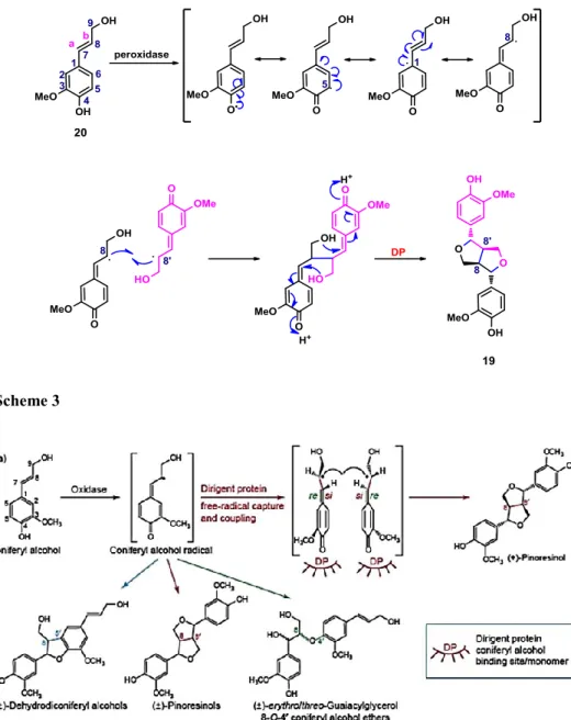
The biosynthetic pathway leading to lignans by coupling of phenylpropanoid units has been largely studied and is exemplified by the biosynthesis of (+)-pinoresinol (19) by 8-8’ (or β-β’) coupling of two conyferil alcohol (20) units in the presence of a peroxidase enzyme, generating the radical species (Scheme 3). A reactive quinone–methide intermediate undergoes intramolecular cyclization, thus affording (+)-pinoresinol (19)¸ interestingly, it has been showed that a protein called ‘Dirigent Protein’ (DP, Figure 1), and not the enzyme, controls the stereochemistry of the reaction; in the absence of this protein, racemic mixtures are obtained.[39] The DP hosts two coniferyl radicals in binding site ‘si-face to si-face’ in order to determine the stereochemical asset of the quinone-methide intermediate and consequently, the stereogenic centres of final product, as in (+)-pinoresinol (19) biosynthesis.
15 Scheme 3
Figure 1: stereochemical control through Dirigent protein - Adapted from: Science, 1997, 275, 362-366
One of the most studied bioactive lignans is podophyllotoxin (6); the optimization of this natural lead, obtained through the synthesis of hundreds of analogues, has afforded anticancer drugs such as etoposide (21), teniposide (22), and etopophos (5).[40]
16
Among the neolignans, those with a dihydrobenzofuran core are worthy of particular attention for the wide range of their biological activities, including antioxidant,[41] antibacterial,[42] anti-inflammatory,[43] cardiovascular,[44] and cytotoxic effects.[45] The 8-5' coupling originates neolignans with this structural core, as reported below for the biosynthesis of (+)-dehydroconiferyl alcohol (23), (Scheme 4).
Scheme 4
Frequently cited examples of bioactive dihydrobenzofuran neolignans are bohemenan H (24) and bohemenan K (25), strong cytotoxic agents against HeLa, Hep-2 and A-549 cell lines.[46]
17
The promising biological activities of dihydrobenzofuran neolignans have prompted many research groups to develop efficient methodologies for their synthesis to obtain not only natural products, but also their synthetic analogues and hybrids with other small molecules. With regard to the preparation of synthetic analogues of natural dimeric polyphenol, worth noting are the ‘biomimetic’ syntheses, mediated by metals or enzymes, and mimicking the biosynthetic coupling pathway. These synthetic methodologies are often carried out on natural precursors with the aim to obtain their correspondents analogues, which yet retain a natural basic skeleton, and possibly have a profile of bioactivity similar to, or better than the natural precursor. It is worth highlighting that this dimerization, even in enzyme-mediated reactions, occurs with regio- and diastereoselectivity, but not enantioselectivity, and consequently affords
trans-substituted racemic mixtures. When eco-friendly syntheses are
planned, these kind of reactions should be mediated by oxidase enzymes, instead metal-based oxidative reagents.[47] The most common enzymes employed in reactions of oxidative dimerization are laccases, which are able to convert atmospheric oxygen (as an oxidizing agent) into water,[48] or peroxidases, which oxidize aromatic substrates in the presence of hydrogen peroxide, converting it, also in this case, in water.[49]
The structure of the enzymatic site and the mechanism of reaction today is well known and reported below in Figure 2 and in Figure 3.
18
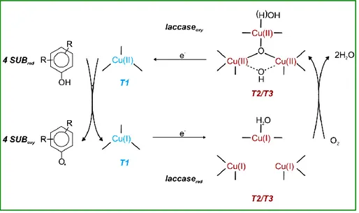
Precisely, laccase catalyzes the one-electron oxidation of four reducing-substrate molecules with the concomitant four-electron reduction of molecular oxygen to water (Figure 2), resulting in a green cycle.[50]
Figure 2: Schematic representation of a fungal laccase catalytic cycle. Two molecules of water result from the reduction of molecular oxygen (at T2/T3) and the concomitant oxidation (at the T1 copper site) of four substrate molecules to the corresponding radicals.
Peroxydases are eme-protein working through the formation of a phenoxy radical and concomitant reduction of hydrogen peroxide (H2O2) to water (Figure 3).[51] The application of enzyme-mediated reactions is common to various fields, including synthetic and analytical purposes,[52] environmental purpose for the wastewater treatment,[53] and biotechnology purpose for must and wine stabilization.[54] Also these
enzymes have been used for biomimetic synthesis of
19 Figure 3: Schematic representation of Horseradish Peroxidase (HRP) catalytic cycle. Abbreviations: SH = substrate, S• = radical substrate.
A representative example is the synthetic dimer 26, that it was tested as anticancer agent following the protocol of the National Cancer Institute (NCI) and showed a GI50 average of 3 M on 60 cancer cell lines and nanomolar scale values towards three breast cancer cell lines and some lines of leukemia. Further studies indicated that the antiproliferative activity of 26 was due to the inhibition of tubulin polymerization. In a study of a series of lipophilic analogues of 26, the synthetic neolignan 27 showed a strong activity towards Plasmodium
falciparum (antimalaric) and Leishmania donovani
20
In particular, a simple preparation of the neolignan 28 (8-5’ coupled diethyl diferulate), with HRP/H2O2, gave a better yield (50%) than the previously used Ag2O-promoted reaction (30%).[56] Another work, on the use of laccase in a biphasic system, showed a rapid formation of the racemic bis-lactone on lignan 29 (from ferulic acid) and 30 (from synapic acid).[57] These reports are only a few examples of the many papers devoted to dihydrobenzofuran neolignans which thus appear as an attractive target for chemical synthesis or modification.
1.1.3 Benzoxanthenes lignans
A part of my PhD research activity has been focused on benzo[k,l]xanthene lignans (in the following, simply ‘BXL’), a subgroup of lignans scarcely reported in the literature, because of their rarity in nature. Only five natural BXLs have been reported in the literature until recent times, namely: mongolicumin A (31), rufescidride (32), yunnaneic acid (33), chiliantin D (34), and dodegranoside (35) respectively isolated from the following species: Taraxacum mongolicum,[58] Cordia
rufescens,[59] Salvia yunnanensis,[60] Rhoiptelea chilianta,[61] e
Dodecadenia grandifora.[62] A simple derivative of mongolicumin A, 36, previously synthesized by the research team guided by Prof. C. Tringali,
21
has been recently identified as a new natural product isolated from plant belonging to the family of Orobanchaceae.[63]
These compounds have been isolated with low yields and long extraction procedures; due to their rare diffusion in nature and the poor availability, they have not been subjected to any evaluation of biological activity. Only for the compound 32 a patent referring to its antimicrobial activity is reported.[64] In a recent study, the above cited research group[47] developed a method of biomimetic synthesis (dimerization by oxidative coupling) to obtain BXLs from esters of caffeic acid. A mechanism for the formation of these dimers has been proposed and corroborated by experimental data and calculations. It is worth noting here that an “orientation” effect towards 8–8’ coupling was observed in the presence of Mn(OAc)3. This mechanism favours the oxidative coupling 8-8' between the caffeate radicals, followed by intramolecular cyclization processes and oxidative additional steps (Scheme 5).
22 Scheme 5
The reaction affords essentially two products: the benzoxanthene lignan as main product and a lignan with an aryldihydronaphthalene core as minor product. It is worth noting that the formation of the aryldihydronaphthalene derivative is a strong indication of the biomimetic nature of the synthesis; in fact, the natural BXL 33 was isolated along with the related aryldihydronaphthalene lignan rabdosiin (37) from the same plant Salvia yunnanensis and this supports the hypothesis that these natural products are formed in nature through a mechanism that is very similar to that proposed here. So the “unnatural” BXLs are mimetic of known natural products and, in principle, may be biogenetically “natural” products and not yet discovered in nature, as confirmed by the recent isolation of compound 36
23
.
In the above cited work the BXLs 36, 38 and 39 and subsequently series of their analogs (40 - 43) were obtained with this biomimetic procedure.
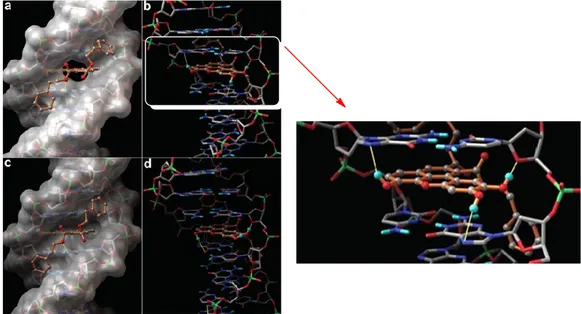
In the frame of a collaboration of Prof. Tringali’s team with that of Prof. G. Bifulco at the University of Salerno these synthetic benzoxanthenes were evaluated in a study on the interaction with DNA, based on STD-NMR experiments and molecular docking calculations (Figura 4);[65] a parallel study of their antiproliferative activity was carried out in collaboration with Prof. Latruffe at the University of Burgundi (Dijon, France). The compounds were evaluated in vitro on two different cell lines of human cancer namely SW 480 (human colon carcinoma) and HepG2 (human hepatoblastoma). Compound 38 resulted
24
the most potent, with a GI50 = 2.57 μM on SW 480 e GI50 = 4.76 μM on HepG2. A three-dimensional model of the ligand-DNA complex was built which made it possible to shed light on the structural elements important for the interaction with the biological target. The results obtained from the STD-NMR measures are compatible with the calculations of molecular docking. In fact, both studies confirmed that the parallel planar structure of benzoxantenes intercalates between the bases of DNA, by establishing a series of π-π interactions, but also the pendants ester contribute to the binding, establishing interactions through Van der Waals forces and hydrogen bonds with the minor groove. This also highlighted the important role of phenolic OH, in the interaction with DNA. In fact, the methylated derivatives showed a lower affinity for the biological target and turned out to be inactive against tumor cells.
Figura 4: Molecular modeling (docking) of complex 38 - DNA
Further work on BXLs as potential anticancer agents was carried out by collaboration with Prof. D. F. Condorelli (University of Catania); a
25
small library of BXLs with different pendant chains were synthetized and evaluated towards a panel of cancer cell lines, namely HT29 (Caucasian
colon adenocarcinoma grade II), Caco-2 (Caucasian colon
adenocarcinoma), HCT-116 (human colon tumor), H226 (lung squamous carcinoma) and A549 (lung carcinoma). The antiproliferative activity data and lipophilicity measurements showed that the most active compounds are also those more lipophilic.[66] The CAPE-derived benzoxanthene 38 again resulted the most potent, and was also the most lipophilic. This benzoxanthene was also the subject of studies on its anticancer properties[67, 68] in collaboration with Dr. G. Srinivas (Cancer Research Program, Rajiv Gandhi Centre for Biotechnology, Thiruvananthapuram, India). BXLs were also studied, in collaboration with Prof. R. Amorati (University of Bologna) as a new class of antioxidant polyphenols, able to effectively react with peroxyl radicals.[69] These studies revealed benzoxanthene lignans as a new class of bioactive natural products, and for this reason we planned, as part of this PhD project, to expand the biological studies on BXLs, as will be detailed below.
1.1.4 Bisphenol neolignans
Diaryl motifs are featured in numerous natural and synthetic compounds used, inter alia, as drugs, agrochemical agents, polymeric materials, and various material additives.[70] In particular, natural C-C or C-O bisphenol neolignans have normally monomeric phenolic precursors, and their biosynthesis is thus viewed to rely on oxidative coupling processes (Scheme 6).
26 Scheme 6: bisphenol biosynthesis through oxidative coupling
Among the biarylic neolignans, in recent years two simple dimeric compounds, magnolol (44) and honokiol (45) gained growing attention by researchers, and a literature search on these compounds affords today more than 2000 results. Both are natural products originally isolated from the bark of Magnolia officinalis,[71] a plant used in Japanese and Chinese traditional medicine for various diseases such as gastrointestinal disorders, anxiety and allergic diseases; M. officinalis bark is reported for a number of biological activities including cancer, anti-inflammatory, anti-depressant and anti-platelet activity.[72]
Magnolol (44) is probably the most cited among M. officinalis constituents, and a non-exhaustive list of its properties includes antitumor,[73] anti-inflammatory,[74] anti-angiogenic,[75] antimicrobial,[76] antiviral[77] and antioxidant[78] activity, as well as prevention of
27
inflammation-induced tumorigenesis,[79] inhibition of osteoclast differentiation,[80] reduction of multidrug resistance through P-glycoprotein modulation[81] and protection against cerebral ischaemic injury.[82] A comparable variety of biological properties has also been reported for honokiol. These properties prompted a number of researchers to synthetize magnolol and honokiol analogs and evaluate their biological properties: this afforded new bisphenol neolignans and derivatives with antimicrobial/antiproliferative,[83, 84] neuroprotective,[85] anti-inflammatory[86] and antioxidant activity, cytotoxicity against cancer cell lines,[87, 88] modulation of GABA receptors[89] and insecticidal activity.[90] Interestingly, only one report deals with inhibition of -glucosidase by honokiol derivatives (namely, dimers and trimers),[91] notwithstanding that a potent inhibitory activity has been reported for honokiol and especially magnolol isolated from Trichilia cannaroides.[92] In view of the diverse pharmacological activities of magnolol scaffold we felt the need for further structural manipulations towards development of improved structures derived from 44, and in particular given the activity of magnolol (44) on -glucosidase, we were interested to study the potential activity as -glucosidase inhibitors, of its synthetic analogues.
1.1.4.1 Magnolol analogues as
-glucosidase inhibitorsThe search for new and effective -glucosidase inhibitors is rapidly growing in the last decade, in view of the epidemic diffusion of diabetes and consequently of the efforts devoted to the discovery of potent glucosidase inhibitors able to retard glucose absorption and reduce blood glucose levels.[93] Diabetes mellitus is a chronic metabolic disease associated with disorders of carbohydrate metabolism and characterized by hyperglycemia. The control of postprandial blood glucose excursions
28
has come to the fore of the treatment of diabetes. One of the therapeutic approaches to reduce postprandial hyperglycemia is to retard digestion and absorption of dietary carbohydrates by inhibiting digesting enzymes, such as α-glucosidase and α-amylase, in the digestive organs.[94] Infact, inhibition of intestinal α-glucosidases delays the digestion of starch and sucrose, flattens the postprandial blood glucose excursions, and thus mimics the effects of dieting on hyperglycaemia, hyperinsulinaemia and hypertriglyceridaemia. Therefore, the mechanism of α-glucosidase inhibition represents the pharmacological optimization of the dietary principle of delayed carbohydrate absorption. Furthermore, the treatment with α-glucosidase inhibitors does not only could improve the metabolic state but it has also the potential to delay, or possibly prevent, the development of diabetic complications (Figure 5).[95]
Figure 5: Schematic diagram of enzymatic hydrolysis of oligosaccharides and competitive inhibition of intestinal brush-border α-glucosidases. Adapted from H. Bischoff, Act Endokr Stoffw 1991;12:25-32.
29
Since the 1960s, considerable efforts have been devoted to the studies of glucosidase inhibition, aiming at the discovery of potent glucosidase inhibitors for the treatment of diabetes through the retardation of glucose absorption and lowering of blood glucose levels. Various types of glucosidase inhibitors, have been extensively studied and reviewed in the past few decades, and among these acarbose (Glicobasey,46), miglitol (Glyset, 47) or voglibose (Prandial, 48) have been successfully commercialized as anti-glucosidase drugs against type-2 diabetes. However, the effectiveness of these drugs is compromised by their deleterious side effects.[96]
As a new class of α-glucosidase inhibitors, polyphenols are attracting great interest for understanding the mechanisms of action of glucosidase inhibition, and for developing alternative drugs to prevent and treat diabetes and obesity.[97] Recently some efforts have been devoted from the research group guided by Prof. C. Tringali to investigate resveratrol-related synthetic glycosides[98] and natural polyphenols[99] as inhibitors of yeast -glucosidase, the enzyme most frequently employed in the preliminary steps of the search for new antidiabetic drugs. On the
30
basis of the above, as further goal of my research activity and as a continuation of the studies to develop new -glucosidase inhibitors, we planned here the chemo-enzymatic synthesis of a series of bisphenols (or related compounds) inspired by magnolol (44) and their evaluation as yeast -glucosidase inhibitors.
1.1.5 Ellagitannins: bioactive plant polyphenols
Ellagitannins belong to the hydrolyzable tannins family, a subclass of the group of tannin molecules. Research interest in these plant polyphenols initially emerged from the discovery of their occurrence in numerous herbal remedies used in oriental traditional medicine and the remarkable biological activities related to their antioxidant,[100] antiviral,[101] and host-mediated anti-tumor properties.[102] In particular, several reports of Quideau’s group showed an interesting targeting ability of some wine ellagitannins toward the human topoisomerase II, a nuclear enzyme involved in DNA processes such as replication, transcription, chromosome condensation and segregation, suggesting a potential proliferative activity and potential use of these molecules as new anti-cancer drugs.[103] Furthermore, the same group discovered that vescalagin (49) is capable to inhibit the activity of certain cells (endothelial and smooth muscle cells) by dismantling their actin cytoskeleton, opening the way towards a potential novel therapy against osteoporosis.[104] To date, after more than 50 years of investigations, more than 1000 members of this subclass of hydrolyzable tannins have been isolated from various plant sources and fully characterized, thus constituting by far the largest group of known tannin molecules.[11, 105, 106] The ellagitannin chemical structures are basically composed of a central sugar core, typically D-glucopyranose, to which are esterified gallic acid units that are further
31
connected together through C–C biaryl and C–O diaryl bonds as a result of intra- and intermolecular oxidative coupling processes. The biosynthetic pathway (Scheme 7) starts from a common penta-O-galloyl-α-D-glucopyranosidic (β-PGG) precursor, which generates the so-called hexahydroxydiphenoyl (HHDP) moiety through an intramolecolar oxidative C-C coupling of appropriately juxtaposed galloyl groups, as proposed by Schimdt and Mayers.[107] The HHDP biaryl unit is a structural determinant to define ellagitannins ‘hydrolyzable tannins’: in fact, hydrolytic release of HHDP units from ellagitannins causes their simple conversion into the bis-lactone ellagic acid (10). Representative compounds of the ellagitannin’s family are peduncalagin (50), stachyurin (51), castalagin (52), vescalagin (49), castalin (53), vescalin (54), grandinin (55) and roburin E (56) in which D-glucose could be in open or closed form. The HHDP and NHTP (nonahydroxyterphenoyl) units possess an axial chirality (atropisomerism) giving the possible production of different stereoisomeric forms.
32
1.1.5.1 Stereochemical consideration on ellagitannins
When a molecule has a biaryl bond, it is possible to observe an axial chirality or atropisomerism, and for each biaryl bond, there are two possible atropisomers, distinguished as S and R.[108] Vescalagin (49), its C-1 epimer, castalagin (52), and their hydrolysis derivatives, castalin (53) and vescalin (54), were first isolated in 1967 from the same oak specimen
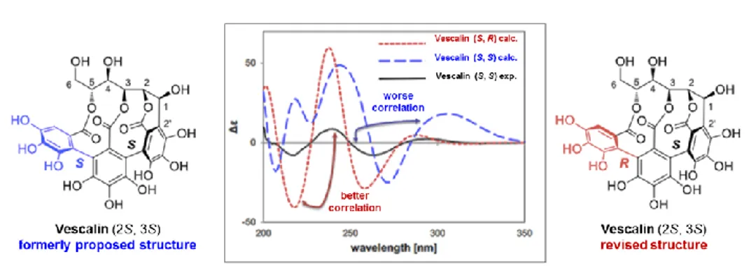
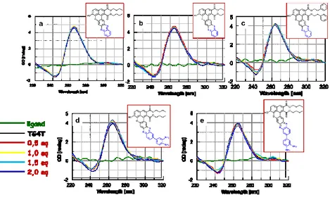
Quercus Sesselijlora from Mayer et al.[109] Molecular structure of these compounds was elucidated using hydrolytic studies combined with spectroscopic analytical methods.[110] In most of the books and papers, reported in literature, the NHTP-atropisomer of vescalagin (49) was defined as (2S,3S)-isomer, together to others ellagitannins correlated with its structure.[11, 105, 111] However, in 1995, a revised study concerning a conformational analysis was published by N. Vivas[112] team; the results of this study, based on molecular mechanics calculations (MM2 force field), showed that the most stable atropoisomer of vescalagin (49) and castalagin (52) is (2S, 3R)-isomer. This axial stereochemistry concerning only one of the two biaryl bonds of the terphenoyl group has been reinvestigated also by the group of T. Tanaka[113] using computational methods (Figure 6). This structure revision is based on the comparative study between a simulation of the circular dichroism spectra of each possible atropoisomer (by TDDFT) and the experimental results (by
33
ECD) obtained from the natural vescalin (54) isolated by extraction process (Figure 7). A comparison between calculated and experimental data shows a much better agreement of the experimental data with those calculated for the revised atropoisomer (2S,3R) than with those calculated for the original (2S,3S) structure.
.
Figure 6: 3D structures of vescalagin (49) proposed by N. Vivas (on the left) and T. Tanaka (on the right)
Figure 7: Experimental and calculated ECD spectra of vescalin (54); adapted from Org. Lett. 2015, 17, 46−49.
The ambiguity about the atropisomerism of triphenoyl moiety of all the ellagitannins isolated until now, prompted a number of researchers
34
in the challenge of the total synthesis of these compounds, and in the synthesis of ellagitannin-bearing devices in proteomics with the aim to study them by crystallography. Part of my Doctorate research activity during the third year was focused on that goal; this part of my work was carried out during my stay at the University of Bordeaux, France (January – July, 2017), under the supervision of Prof. Stéphane Quideau.
1.1.7 Aims of the PhD research activity
On the basis of the above, the main goal of the present research project was to obtain new potential chemotherapeutic agents starting from natural or bio-inspired polyphenols. This objective has been pursued following two parallel, although distinct, strategies:
a) modification of natural polyphenols to obtain optimized analogues with promising bioactivity, for possible use as chemotherapeutic agents.
b) synthesis of new potential chemotherapeutic agents through a chemo-enzymatic approach.
In both cases, chemical and/or enzymatic methodologies have been employed. The compounds obtained have been characterised by spectral analysis and, through collaboration with other laboratories, they have been evaluated for properties of biomedical interest, namely antioxidant, α-glucosidase inhibitor, antiproliferative, antiviral and antibacterial activity. Molecular modelling and structure-activity relationship (SAR) studies have been carried out in selected cases. Further goals of this work were an inverse virtual screening focused on magnolol analogues, and a contribution to the total synthesis of vescalin (54) and a vescalagine probe carried out at the laboratory of Prof.
35
Stéphane Quideau (University of Bordeaux, France). In the following, the results of my work are discussed in detail.
36
37
2. RESULTS AND DISCUSSION
2.1 Synthesis and biological evaluation of
bioinspired benzo[k,l]xanthene lignans and related
phenazines
On the basis of what we have reported in the Introduction on benzo[k,l]xanthene lignans (BXLs) (Section 1.1.3), part of my research activity has been devoted to different studies aimed to highlight new properties of BXLs of potential pharmacological interest. To this purpose we planned the biomimetic synthesis of selected BXLs and related phenazines. The compounds obtained were purified and spectroscopically characterized. Subsequently, by collaboration with other laboratories, BXLs and their analogues were evaluated as:
potential G-quadruplex DNA ligands;
agonist/antagonist of FXR and LXR receptors;
antimicrobial agents.
In some cases, the biochemical or biological assays were limited to the compounds available at the moment of the experiments, so not all compounds were subjected to the same assays.
2.1.1 Biomimetic synthesis of benzo[k,l]xanthene
lignans
In order to evaluate new biological properties of BXLs, the synthesis of new and previously reported benzoxanthene has been planned. The synthesis of BXLs (36, 38, 39, 60, 63, 65 and 67) was carried out employing the Mn-mediated biomimetic methodology, summarized in Scheme 8. In the general procedure, the synthesis of a
38
suitable caffeic ester was carried out as first step, using a Fisher esterification or in selected cases the Steglich esterification. Then we employed the methodology previously reported by the group where I carried out my PhD project and based on biomimetic oxidative coupling of caffeic esters, mediated by Mn3+.[47] In the following, the details are discussed.
Scheme 8: (a) ROH, H2SO4, reflux temperature, 24 h; (b) ROH, DCC, dry THF , 24 h;
(c) Mn(OAc)3, CH3Cl.
2.1.1.1 Synthesis of dimethyl 6,9,10-trihydroxybenzo-[k,l-]xanthene-1,2-dicarboxylate (36)
As reported in Scheme 8, the caffeic methyl ester 58 was obtained through Fischer esterification using caffeic acid (57) and methanol in the presence of concentrated H2SO4 as catalyst. The purified product showed NMR data in perfect agreement with those reported in literature.[114] Compound 58 was submitted to dimerization in presence of an excess of
39
Mn(OAc)3 in CHCl3. The yellow-brown residue obtained, was chromatographically purified to recover the compound 36. The 1H and 13C NMR spectra are respectively reported in Figures 1S and 2S ( Appendix A); the spectroscopic data, compared with the data previously obtained and reported in the literature,[47] confirmed the structure of the benzo[k,l]xanthene lignan 36.
2.1.1.2 Synthesis of diphenethyl 6,9,10-trihydroxy-benzo-[k,l-]xanthene-1,2-dicarboxylate (38) and its methylated derivative (43)
The procedure for the preparation of compound 38 (Scheme 8) is similar to the previous, with the only difference that CAPE (12, caffeic acid phenethyl ester), the substrate of this reaction, was not synthesized because it is a commercially available. Thus, compound 12, in presence of Mn(OAc)3 afforded the expected benzoxanthene 38. After purification, this latter was subjected to 1H and 13C NMR spectral analysis; NMR spectra are reported in Figures 3S and S4 (see Appendix A) and are in agreement with those previously reported in the literature,[47] confirming the structure of the compound 38. This compound was treated with treated with dimethyl sulphate, as previously reported in literature,[65] to prepare its permethylated 43, in order to check the possible role of a free/blocked catechol group for antimicrobial activity (Scheme 9, see below for biological evaluation).
40 Scheme 9
2.1.1.3 Synthesis of dibutyl 6,9,10-trihydroxybenzo-[k,l]-xanthene-1,2-dicarboxylate (39)
Caffeic acid (57) was treated with butanol in presence of concentrated H2SO4 to obtain the caffeic acid butyl ester 59 (Scheme 8). The NMR data are in perfect agreement with those reported in the literature.[115] The ester 59 was employed for the dimerization reaction with Mn(OAc)3. After purification, afforded the pure compound 39. The1H and 13C NMR spectra of 39 was respectively reported in Figures 5S and 6S (see Appendix A). The NMR data are in agreement with those reported in literature and confirmed the structure of compound 39.[66]
2.1.1.4 Synthesis of diethyl 6,9,10-trihydroxybenzo-[k,l]-xanthene-1,2-dicarboxylate (61)
The synthesis of benzoxanthene 61 is reported in Scheme 8. Briefly, caffeic ethyl ester (60) was obtained through Fischer esterification reaction starting from caffeic acid (57). The NMR data of the product are in perfect agreement with those reported in literature.[115] The monomer 60 was treated with Mn(OAc)3 in CHCl3 and after reaction, the purification afforded the pure compound 61. The 1H-NMR and 13 C-NMR spectra are respectively reported in Figures 7S and 8S (see
41
Appendix A); these data confirmed the structure of the benzo[k,l]xanthene lignan 61, and are in perfect agreement with those reported in the literature.[66]
2.1.1.5 bis(4-methoxybenzyl)6,9,10-trihydroxybenzo-[kl]-xanthene-1,2-dicarboxylate (63)
To expand the structural variety of BXLs we planned to synthetize two new analogues of the above cited benzoxanthenes. These syntheses are reported here.
To a solution of caffeic acid (57) in THF, was added N,N’-dicyclohexylcarbodiimide (DCC) in presence of the commercial 4-metoxybenzyl alcoholto obtain the 4-metoxybenzyl caffeate 62 (Scheme 8). The NMR data are in perfect agreement with those reported in the literature.[116] The ester 62 was employed for the dimerization reaction with Mn(OAc)3. The new benzoxanthene 63, recovered after purification, was fully characterized by 1D and 2D NMR spectroscopy; in Figures 9S and 10S (see Appendix A) the 1H and 13C NMR spectra of 63 are reported.
The 1H NMR and 13C spectra of 63 showed the typical signals for the benzoxanthene core: three singlets at 8.17, 7.34 8 and 6.70 ppm at for H-3, H-8 and H-11 respectively in the 1H NMR spectrum and, the resonances at 129.8, 112.4 and 104.9 ppm for C-3, C-8 and C-11 in the 13C NMR spectrum; two doublets at 7.49 and 7.30 ppm mutually coupled (J = 8.5 Hz; see COSY spectrum) for H-4 and H-5 respectively, and the corresponding 13C resonaces at 122.3 and 120.7 ppm. Also the resonances for aromatic chains were detected: two doublets at 7.44 and 6.96 ppm mutually coupled (J = 8.7 Hz) for H-2IV/6IV and H-3IV/5IV respectively and the corresponding 13C resonances at 131.27, 114.8 ppm; two doublets at 7.32 and 6.88 ppm mutually coupled (J = 8.7 Hz) for H-2VI/6VI and
H-42
3VI/5VI and the corresponding 13C resonances at 131.22 and 114.7 ppm. These resonances are unambiguously assigned studying HMBC correlations. In the upper fields region (5.27 – 3.77 ppm) of 1H NMR spectrum the resonances for alkyl chains were detected: two differently shielded singlets for the α-methylene protons (respect to ester functions) at 5.26 (67.5 ppm, C-1’’), and 5.27 ppm (68.3 ppm, C-1’’’) for H-1’’ and H-1’’’ respectively; finally, two singlets integrating for three protons, namely for the two –OMe in 4IV/4V-CH3 3.81 (55.6 ppm, -OMeIV-4), and 3.77 ppm (55.5 ppm, -OMeV-4) ppm.
All resonances were assigned through the study of HSQC correlations . All the NMR data are in agreement confirmed the structure of compound 63.
2.1.1.6 bis(benzyl)6,9,10-trihydroxybenzo-[kl]-xanthene-1,2-dicarboxylate (65)
To a solution of caffeic acid (57) in THF, was added N,N’-dicyclohexylcarbodiimide (DCC) in the presence of the commercial benzyl alcohol to obtain the benzyl caffeate 64 (Scheme 8). The NMR data are in perfect agreement with those reported in the literature.[117]The ester 64 was employed for the dimerization reaction with Mn(OAc)3. After purification, the pure compound 65 was obtained. The new benzoxanthene was fully characterized by 1D NMR spectroscopy (see 11S – 12S in Appendix A). All the NMR data confirmed the structure of compound 65. The structure of 65 differs than 63 only for the aromatic pendants which they do not present the para-methoxy groups.