Daily Rhythms of the Expression of Key
Genes Involved in Steroidogenesis and
Gonadal Function in Zebrafish
Viviana Di Rosa1, Jose Fernando López-Olmeda1, Ana Burguillo1, Elena Frigato2,
Cristiano Bertolucci2, Francesc Piferrer3, Francisco Javier Sánchez-Vázquez1* 1 Department of Physiology, Faculty of Biology, Regional Campus of International Excellence“Campus Mare Nostrum”, University of Murcia, Murcia, Spain, 2 Department of Life Sciences and Biotechnology, University of Ferrara, Ferrara, Italy, 3 Institut de Ciències del Mar, Consejo Superior de Investigaciones Científicas (CSIC), Barcelona, Spain
Abstract
Fish present daily and seasonal rhythms in spawning and plasmatic levels of steroids that control reproduction. However, the existence of the rhythms of expression of the genes that underlie the endocrine mechanisms responsible for processes such as steroidogenesis and reproduction in fish have still been poorly explored to date. Here we investigated the daily pattern of the expression of key genes involved in sex steroid production that ulti-mately set the sex ratio in fish. Adult zebrafish were maintained under a 12:12 h light-dark cycle at a constant temperature of 27°C and were sampled every 4 h during a 24-hour cycle. The expression of key genes in the gonads and brains of female and male individuals were analyzed. In gonads, the expression of aromatase (cyp19a1a, ovarian aromatase) and the antimüllerian hormone (amh, testis) was rhythmic, with almost opposite acro-phases: ZT 5:13 h (in the light phase) and ZT 15:39 h (at night), respectively. The expres-sion of foxl2 (forkhead box L2) was also rhythmic in the ovary (acrophase located at ZT 5:02 h) and the expression of dmrt1 (doublesex and mab-3-related transcription factor 1) was rhythmic in testes (acrophase at ZT 18:36 h). In the brain, cyp19a1b (brain aromatase) and cyp11b(11beta-hydroxylase) presented daily differences, especially in males, where the expression peaked at night. These results provide the first evidence for marked time-of-the-day-dependent differences in the expression of the genes involved in sex ratio control, which should be considered when investigating processes such as reproduction, sex differ-entiation and steroidogenesis in fish.
Introduction
Animals that live according to environmental cycles have developed rhythmic physiological and behavioral processes that are driven by an internal time-keeping system. Biological clocks allow animals to anticipate these cyclic events (e.g., day/night or season) and to cope with them
a11111
OPEN ACCESS
Citation: Di Rosa V, López-Olmeda JF, Burguillo A, Frigato E, Bertolucci C, Piferrer F, et al. (2016) Daily Rhythms of the Expression of Key Genes Involved in Steroidogenesis and Gonadal Function in Zebrafish. PLoS ONE 11(6): e0157716. doi:10.1371/journal. pone.0157716
Editor: Nicholas S Foulkes, Karlsruhe Institute of Technology, GERMANY
Received: February 18, 2016 Accepted: June 5, 2016 Published: June 20, 2016
Copyright: © 2016 Di Rosa et al. This is an open access article distributed under the terms of the Creative Commons Attribution License, which permits unrestricted use, distribution, and reproduction in any medium, provided the original author and source are credited.
Data Availability Statement: All relevant data are within the paper.
Funding: This research was funded by Spanish Ministry of Economic Affairs and Competitiveness (MINECO) by project“Cronosolea” AGL2010-22139-C03-01 and“Solembryo” AGL2013-49027-C3-1-R, cofunded with FEDER fund; and by Project 18963/ JLI/13 granted by the Agencia de Ciencia y Tecnología de la Región de Murcia (Fundacion Seneca) and a research fellowship granted by MINECO (Juan de la Cierva Program) to JFLO. The funders had no role in study design, data collection
better. Alternation of light and darkness (the 24-hour LD cycle) represents the main time cues capable of entraining biological clocks, and allows the phase of endogenous rhythms to be set with the phase of cyclic events in the environment [1]. In ectothermic animals, such as fish, temperature cycles play a relevant role as the water dynamics creates an ecosystem in which animals have to adapt to live [2–3].
Fish reproduction exhibits both daily and seasonal rhythms in most species. A number of investigations have reported daily variations in sex steroids, such as estradiol (E2), testosterone, 11-ketotestosterone (11-KT) and progesterone, in different fish species: Japanese charr (Salveli-nus leucomaenis) [4], wrasse (Pseudolabrus sieboldii) [5], catfish (Heteropneustes fossilis) [6] and Senegalese sole (Solea senegalensis) [7]. Daily changes in oocyte maturation and secretion of sex steroids have also been reported in bamboo leaf wrasse (Pseudolabrus japonicas) [8], snapper (Pagrus auratus) [9], kisu (Sillago japonica) [10] and gilthead sea bream (Sparus aur-ata) [11]. In accordance with fish seasonal reproduction, annual changes in sex steroids have been reported in European sea bass (Dicentrarchus labrax) [12–13], rainbow trout (Oncor-hynchus mykiss) [14] and Senegalese sole [7]. Previous research has also highlighted the effect of photoperiod and temperature as having a potential influence on fish sex ratios [15–23]. Daily cycles of environmental factors have been reported to act differently during sex differen-tiation; e.g. thermo-cycles induce a high proportion of females in zebrafish, whereas constant temperature leads to more males [24].
Zebrafish is mostly a diurnal species [25], but is capable of displaying either diurnal or noc-turnal behavioral rhythms (i.e., nocnoc-turnal self-feeding) [26]. Daily thermocycles can also drive behavioral rhythms in zebrafish [27–28]. When this species is submitted to a long photoperiod (LD 14:10 h), fish spawn at the beginning of the light phase. The diurnal spawning rhythm is maintained in the light phase, even when zebrafish are fed at night, and despite their locomotor activity becoming nocturnal. Zebrafish’s ability to sustain a diurnal spawning rhythm confirms the strong influence of the LD-cycle on the entrainment of spawning rhythms [29]. Daily varia-tions in fsh (follicle-stimulating hormone) and lh (luteinizing hormone) production in the zeb-rafish pituitary have been reported, which could be related with the daily rhythms of zebzeb-rafish spawning [30]. However, nothing is known about rhythmicity in other factors involved in sex steroid production in this species, especially at the gonadal level.
Timing of reproduction and production of sex steroids is controlled by the hypothalamic-pituitary-gonad axis (BPGa), which is necessary for multiple processes like sex differentiation, gonad maturation and spawning [11,31]. Aromatase plays a key role during sex differentiation and gonad maturation, and in most fish it presents two different genes: cyp19a1a and cyp19a1b. The former is also called“ovarian aromatase” as it is expressed mainly in differentiating and adult gonads of teleost fish. It is an enzymatic complex that facilitates estrogen synthesis from testosterone or androstenedione [32]. Cyp19a1b is also called“brain aromatase” because this gene is highly expressed in the teleost brain of both females and males [33]. In fish, cyp19a1a is essentially expressed only in gonads, while cyp19a1b is expressed mostly in the brain, but can also be found in other tissues [34]. In testes, the antimüllerian hormone (amh) initiates the regression of Müllerian ducts and inhibits the expression of aromatase (cyp19a1a) to avoid the transformation of androgens into estrogens. Foxl2 (forkhead box L2) is a transcription factor that is known as a potent transcriptional activator of cyp19a1a [35]. Thus foxl2 plays an impor-tant role in those species in which temperature affects the sex ratio [36]. High temperature dur-ing the thermosensitive period suppresses cyp19a1a gene expression to result in low aromatase activity and E2 levels [37]. Dmrt1 (doublesex and mab-3 related transcription factor 1) has been found expressed in Japanese medaka (Oryzias latipes), is an inhibitor of germ cell proliferation, and is expressed only during testicular differentiation. In Nile tilapia (Oreochromis niloticus), this factor down-regulates cyp19a1a during testicular differentiation [38]. In zebrafish, dmrt1 is
and analysis, decision to publish, or preparation of the manuscript.
Competing Interests: The authors have declared that no competing interests exist.
not only associated with testis development, but may be important in ovary differentiation [39]. The cyp11b (11beta-hydroxylase) gene contributes to the synthesis of 11-KT from testosterone, which is the most potent androgen in teleost fish, with a higher expression in male gonads than in females [40]. Although rhythms at plasmatic levels of sex steroids and fish reproduction have been described in many species, the rhythmic nature of these key enzymes involved in sex ste-roidogenesis remains unknown to date.
The aim of this research was to investigate, for the first time, the existence of daily expres-sion patterns of six specific genes that play a key role in fish reproduction and steroidogenesis by using zebrafish as the most appropriate experimental model. These genes were analyzed in two different tissues, gonads and brain, from both sexes. In the ovary we looked at the expres-sion of cyp19a1a and foxl2, whereas in testes we examined the expresexpres-sion of amh, dmrt1 and cyp11b. In the brain of both males and females we analyzed the expression of cyp19a1b and cyp11b.
Materials and Methods
Ethics Statement
The present research was carried out in the Chronobiology laboratories at the University of Murcia (Spain). All husbandry and experimental procedures complied with European Legisla-tion for the ProtecLegisla-tion of Animals used for Scientific Purposes (Directive 2010/63/EU). The experimental protocol was previously authorized by the Spanish National Committee on Ani-mal Welfare (RD 1201/2005 and law 32/2007) and the Bioethical Committee of the University of Murcia (Spain).
Animal rearing
Adult wild-type zebrafish (Danio rerio) of mixed sexes were obtained from a local provider (Jumipez S.A., Murcia, Spain). Approximate fish body weight was 0.75 g, with a total length of 4 cm. Fish were housed for 6 months in our laboratories. Four weeks before sample collection, fish were divided (N = 160) into two 60-liter aquaria (60x30x32 cm) according to sex, with males placed in one aquarium and females in the other. Each aquarium had a closed water circulation system provided with aeration, and also with mechanical and biological filters. Aquaria were kept in a chronolab, a completely isolated room, where light and temperature were strictly controlled. Temperature was recorded by an underwater data logger (HOBO PENDANT1Onset Computer Corporation, Massachusetts, USA) and was maintained at 27 ±0.5°C using a water heater (100 W, Askoll, Italy). Lighting conditions were set according to a 12:12 h LD cycle, with light onset at 8 am (Zeitgeber Time 0 h, ZT 0 h). Fish were fed by an automatic feeder (Eheim, Germany) located in the upper part of the aquaria, which released food (Tropical fish flakes, Prodac, Italy) at a quantity daily rate of 1% of total biomass in each aquarium once a day at ZT 4 h. Each aquarium was equipped with an infrared photocell con-nected to a computer to record locomotor activity. The photocell was located in the middle of the large side of each aquarium to detect the movement of fish when they interrupted the infra-red beam. Interruptions were recorded as signals and were stoinfra-red in the computer every 10 minutes.
Sampling and analysis
Samples were collected throughout a 24-hour cycle at six different time points (ZT 2, 6, 10, 14, 18 and 22 h). At each time point, 10 fish (five of each sex) were anesthetized with eugenol (clove oil essence, Guinama, Valencia, Spain) at a concentration of 50μL/L and sacrificed by
decapitation. Fish manipulation and tissue collection in the dark phase were performed under a dim red light. Brains and gonads (ovaries from females, testes from males) were extracted from each fish. Total RNA was isolated from each sample using Trizol reagent (Invitrogen, Carlsbad, CA, USA) following the manufacturer’s instructions. The amount, quality and com-position of isolated RNA were analyzed by Nanodrop ND-1000 (Thermo Fisher Scientific Inc., Wilmington, USA). Total RNA (1μg) was incubated with DNase I (Invitrogen) at room tem-perature for 30 min and then at 85°C for 15 min to inactivate the enzyme. DNase-treated RNA was used to perform cDNA synthesis in a final volume of 20μl using the QuantiTect Reverse Transcription Kit (Qiagen, USA). The reaction was performed at 42°C for 30 min, followed by a 5-min inactivation step at 85°C. Then cDNA was PCR-amplified with the StepOnePlus Real-Time PCR System (Applied Biosystems, Foster City, CA, USA) using the SYBR-green primer master mix according to the manufacturer’s recommendations (Applied Biosystems, Foster City, CA, USA). The thermal cycling conditions were as follows: 15 min of denaturation at 95°C, followed by 40 cycles of a 15-s denaturation step at 95°C, and then by an annealing-elon-gation step for 30 s at 60°C. After amplification, a melting curve analysis was performed to con-firm amplicon specificity. All the samples were run in triplicate. The gene-specific primers for cyp19a1a, cyp19a1b, cyp11b, dmrt1 and foxl2 were designed with the primer Express software (Applied Biosystems) (Table 1). The primer sequences for amh were retrieved from the litera-ture [41]. Efficiency of primers was verified by constructing standard curves for all the investi-gated genes. The dissociation curve was used to confirm amplicon specificity. The relative expression levels of each sample were calculated by the 2–ΔΔCTmethod [42]. As housekeeping genes,βactin was used in the gonad samples and loopern4 was used in the brain samples [43]. Housekeeping genes were selected after checking that the coefficient of variation (C.V.) for each gene within each tissue was lower than 5%. The second normalization in the 2–ΔΔCT calcu-lations was performed using the sample with the lowest value within each gene and tissue as the reference.
Statistical analysis
All the results were expressed as mean±SEM. The significance threshold (α) was set at 0.05 in all the statistical tests. The gene expression data were first subjected to the D'Agostino-Pearson normality test and Bartlett's test of homocedasticity. All the data fulfilled the assumptions of normality of distributions (D'Agostino-Pearson test, p>0.05) and homogeneity of variances (Bartlett's test, p>0.05). Then the data from each gene were subjected to a one-way analysis of variance (ANOVA) to determine of the existence of statistically significant differences between time points. The values from each gene and tissue were subjected separately to a one-way ANOVA. After checking with the one-way ANOVA that the data showed significant differ-ences (p<0.05), a Tukey´s HSD post hoc test was used for multiple comparisons between groups (time points). D'Agostino-Pearson tests, Bartlett's test and one-way ANOVAs were per-formed with SPSS 15.0 (SPSS Inc., Chicago, IL, USA).
The Cosinor analysis was run with the El Temps software (v. 275, Prof. A. Díez-Noguera, University of Barcelona, Spain) to determine whether the daily expression of the genes fitted a cosine function: Y = M+A x[cos (Ot+F)], where M is mesor, A is amplitude, O is angular fre-quency (360°/24 h for the circadian rhythms) andF is acrophase. A cosine function was selected because it is the simplest mathematical model that explains the rhythmic process and that which avoids prejudging the correlation/equation to a higher extent. The Cosinor analysis provides a fit value (%V) that indicates the percentage of the variance of the experimental data explained by the cosine equation. This %V is the equivalent to the degree of fitness to the calculated cosine function of the experimental data. The Cosinor analysis also provided the
statistical significance of the rhythm through an F-test of the variance accounted for by the waveform versus a straight line of zero-amplitude (null hypothesis). Therefore, if under a sta-tistical significance of p<0.05 this null hypothesis was rejected, amplitude could be considered as differing from 0, thereby constituting evidence for the existence of a statistically significant rhythm of the given period under study. The locomotor activity data were analyzed, and acto-grams and meanwaves were plotted with El Temps.
Results
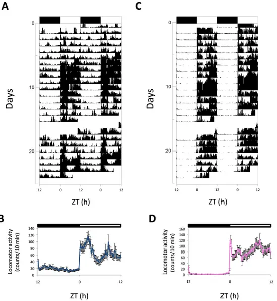
All the male and female zebrafish displayed a diurnal rhythm of locomotor activity and exhib-ited most of their activity in the light phase, as revealed in the actograms (Fig 1A and 1Cfor male and female, respectively). Activity increased sharply after lights were switched on, with a high level maintained in the light phase (Fig 1B and 1D).
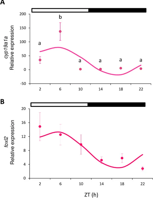
All the analyzed genes showed either daily rhythmicity (Cosinor, p<0.05) or significant dif-ferences among the time points (one-way ANOVA, p<0.05), or both. In females, cyp19a1a was expressed in the ovary and peaked in the middle of the light phase (ML, ZT 6 h) (one-way ANOVA p<0.001;Fig 2A). The analysis of this gene also revealed a sinusoidal rhythmic pat-tern with the acrophase at ZT 5:13 h (Cosinor, p<0.003;Table 2,Fig 3). No differences were observed in foxl2 expression (as assessed by ANOVA), but sinusoidal rhythmicity was shown with the acrophase at ZT 5:02 h (Cosinor, p<0.021,Fig 2B,Table 2), which approached the achrophase of cyp19a1a.
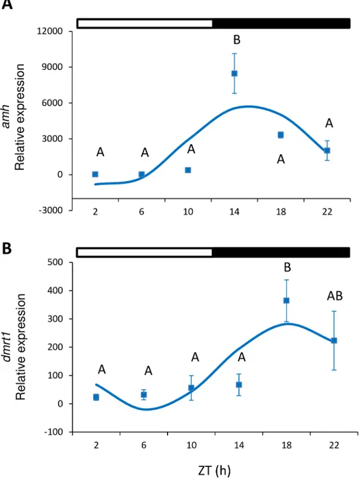
In males, amh expression showed a significant peak of expression in testes at the beginning of the dark phase (ZT 14 h) (one-way ANOVA, p<0.001;Fig 4A). In addition, amh presented a significant sinusoidal rhythm, with the acrophase located at ZT 15:39 h (Cosinor, p<0.05; Table 2,Fig 3). Dmrt1 displayed significant differences during the ZT (one-way ANOVA, p<0.005,Fig 4B), peaked at ZT 18 h and also showed a sinusoidal rhythmic pattern with acro-phase at ZT 18:36 h (Cosinor, p<0.002,Table 2). Cyp11b expression was investigated in testes but displayed no significant differences, and either depended on the time of day (one-way ANOVA, p<0.05, Tukey’s post hoc, p = 0.053) or sinusoidal rhythmicity (Table 2).
Regarding cyp19a1b expression, in the brain both females and males showed statistically sig-nificant differences depending on the time of day. In males, the peak occurred in the dark phase (ZT 22 h), whereas values remained constant and the only decrease in expression was detected at ZT 10 h in females (one-way ANOVA, p<0.005;Fig 5A). Cyp11b expression
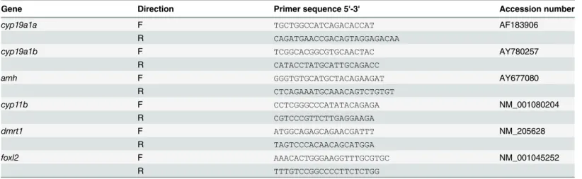
Table 1. Primer sequences used for the quantitative PCR analyses.
Gene Direction Primer sequence 5'-3' Accession number
cyp19a1a F TGCTGGCCATCAGACACCAT AF183906
R CAGATGAACCGACAGTAGGAGACAA
cyp19a1b F TCGGCACGGCGTGCAACTAC AY780257
R CATACCTATGCATTGCAGACC
amh F GGGTGTGCATGCTACAGAAGAT AY677080
R CTCAGAAATGCAAACAGTCTGTGT cyp11b F CCTCGGGCCCATATACAGAGA NM_001080204 R CGTCCCGTTCTTGAGGAAGA dmrt1 F ATGGCAGAGCAGAACGATTT NM_205628 R TAGTCCCACAACAGCATGGA foxl2 F AAACACTGGGAAGGTTTGCGTGC NM_001045252 R TTTGTCCGGCCCCTTCTCTGG doi:10.1371/journal.pone.0157716.t001
presented time-dependent differences in both males and females. The highest expression in females occurred at ZT 10 h, immediately before light offset (one-way ANOVA, p<0.0001;Fig 5B), but occurred at ZT 18 h (in the middle of the night; MD) in males (one-way ANOVA, p<0.006;Fig 5B). For cyp19a1b and cyp11b, no significant daily rhythms were detected (Cosi-nor, p>0.05;Table 2).
Discussion
Although the characterization, localization and expression of the key genes involved in ste-roidogenesis and reproduction have been extensively studied in many fish species, their daily
Fig 1. Representative actograms and mean waveforms of locomotor activity of zebrafish males (A, B) and females (C, D). Zebrafish were subjected to 12:12 LD and fed daily at ZT 4 h. Actograms were double-plotted (time scale 48 h). The height of each point represents the number of infrared light-beam interruptions/ 10 min. Each horizontal line shows one experimental day on the vertical axis, and the hours of the day are represented on the X-axis. The black bar at the top of each actogram represents the dark phase, and the white one represents the light phase of the LD conditions. In the meanwaves, each point was calculated as the mean±S.D from the 10-minute binned data across all the experimental days. Each waveform is represented as single-plotted.
rhythms have been poorly explored to date. The present findings revealed that the expression profiles of these genes are not flat, but change during a 24 h cycle. In the ovary, analyzed genes cyp19a1a and foxl2 oscillated with a similar phase. The same occurred in the testis for amh and dmrt1 expressions. However, the phases of these genes were opposite between sexes, as ovarian cyp19a1a and foxl2 presented diurnal peaks, whereas amh and dmrt1 in testes displayed noc-turnal peaks.
Fig 2. Relative gene expression of cyp19a1a (A) and foxl2 (B) in ovary. Circles indicate the relative gene expression (mean±SEM) at each sampled time point. Different letters indicate the statistically significant differences between time points (one-way ANOVA, p<0.05). The sinusoidal curve calculated by Cosinor (p<0.05) is indicated by the continuous line. The white and black bars above the graphs represent the light and dark phase, respectively.
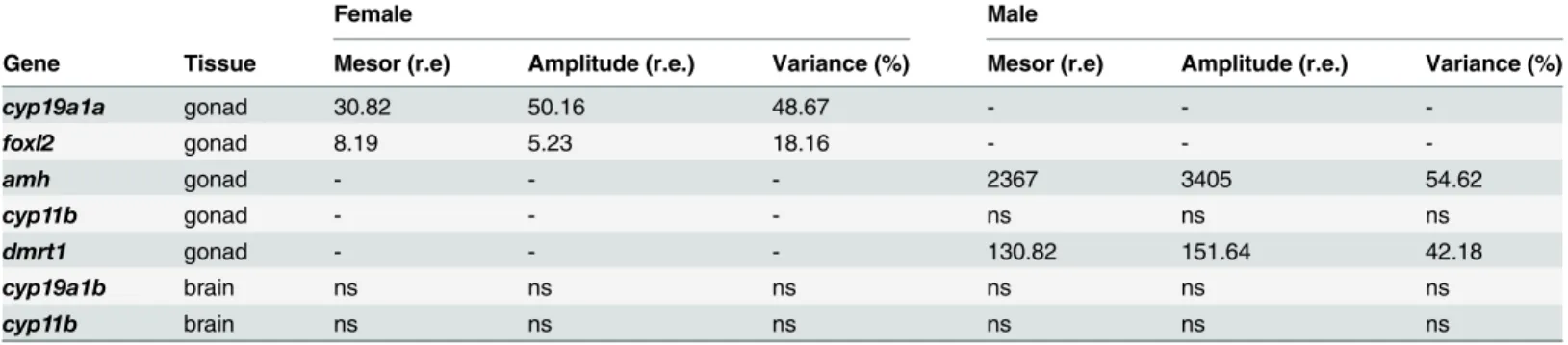
Table 2. Mesor and amplitude values, given as relative expressions (r.e.), calculated by the Cosinor analysis. The degree of adjustment of the experi-mental data to the cosine function calculated by Cosinor is given by the percentage of variance. Values are indicated only for the parameters that showed a significant daily rhythm (Cosinor, p<0.05). NS indicates the genes that show no statistically significant rhythmicity (Cosinor, p>0.05), and a dash denotes the genes that were not analyzed.
Female Male
Gene Tissue Mesor (r.e) Amplitude (r.e.) Variance (%) Mesor (r.e) Amplitude (r.e.) Variance (%)
cyp19a1a gonad 30.82 50.16 48.67 - - -foxl2 gonad 8.19 5.23 18.16 - - -amh gonad - - - 2367 3405 54.62 cyp11b gonad - - - ns ns ns dmrt1 gonad - - - 130.82 151.64 42.18 cyp19a1b brain ns ns ns ns ns ns cyp11b brain ns ns ns ns ns ns doi:10.1371/journal.pone.0157716.t002
Fig 3. Map of acrophases. Only the genes that displayed a statistically significant daily rhythm (Cosinor, p<0.05) are included in the graph. The acrophase is indicated by symbols and the fiducial limits (set at 95%) are indicated by lateral bars. The pink circles and blue squares indicate the genes analyzed in the ovary and testes, respectively. The white and black bars above the graph represent the light and dark phase, respectively.
Estrogens are essential hormones for fish reproduction, and aromatase is the key enzyme involved in their conversion from androgens [37,44]. Cyp19a1a is present only in the ovaries of females and its expression follows a rhythmic pattern. Gonadal aromatase presented its max-imum expression level in the light phase at ML and showed a diurnal pattern in phase with the other genes investigated in female gonad foxl2. However, cyp19a1a expression in the ovary did not occur in phase with amh expression in testes, which peaked at night. In contrast, brain
Fig 4. Relative gene expression of amh (A) and dmrt1 (B) in testes. Squares indicate the relative gene expression (mean±SEM) at each sampled time point. Different capital letters indicate the statistically significant differences between time points (one-way ANOVA, p<0.05). The sinusoidal curve calculated by Cosinor (p<0.05) is indicated by the continuous line. The white and black bars above the graphs represent the light and dark phase, respectively.
cyp19a1b, the“neural aromatase”, presented similar profiles in both sexes. It should be noted that in previous studies cyp19a1b displayed the same level range of expression in both females and males [45–46]. While the role of aromatase in gonads has been largely studied, many ques-tions about its role in the brain remain to be answered. There is evidence that aromatase plays a role in the control of reproduction by the brain [47], and displays different activity depending on the sex and life stage. In 1-year-old European sea bass (Dicentrachus labrax), brain aroma-tase activity was higher in premature males than in females and imnmature fish [48]. Seasonal variations in brain aromatase have also been detected, with maximum values observed during the spawning season [48].
Previous studies on steroid rhythmic levels in other fish species (e.g. S. senegalensis) have revealed that testosterone (T) and estradiol (E2) are both secreted mostly in a rhythmic manner Fig 5. Relative gene expression of cyp19a1b (A) and cyp11b (B) in the brain. Brain samples were analyzed separately for females (pink circles) and males (blue squares). Data are expressed as mean±SEM. Different letters indicate the statistically significant differences between time points in the female (lower case letters) or male (capital letters) brains (one-way ANOVA, p<0.05) within each gene. The white and black bars above the graphs represent the light and dark phase, respectively.
[7]. According to Bayarri et al. [49] in European sea bass, plasma T in males, plasma and pitui-tary LH, and pituipitui-tary GnRH display daily rhythms with different acrophases. In snapper (Pagrus auratus), plasma E2and T change during the day [9]. Japanese whiting (Sillago
japon-ica) also presents a diurnal pattern of plasma E2[50]. Hormonal rhythmicity is connected
directly to rhythms of physiological and behavioral processes, such as reproduction and spawn-ing, which usually coincide with the phase of the LD cycle that displays the greatest locomotor activity. In gilthead sea bream, spawning starts in the afternoon and continues for a few hours after night falls [51]. In Senegalese sole, spawning takes place when night falls and correlates with its nocturnal locomotor activity [7]. In sole, the daily peaks observed in T and E2precede
the time of spawning by a few hours [7,52]. The European sea bass spawns at nighttime [53], which is preceded by a peak in plasma LH observed at dusk [49]. However, T and GnRH do not correlate with spawning, and peak when night ends and during the day, respectively [49]. In zebrafish, spawning takes place at dawn [29]. Ovulation in zebrafish shows a daily rhythm with the acrophase and coincides with spawning: end of darkness and beginning of the light phase [54]. Pituitary fsh and lh [30], and amh and dmrt1 expression (Fig 4), peak at nighttime and precede spawning by several hours, while ovarian cyp19a1a and foxl2 peak several hours after spawning (Fig 2). Although all these results illustrate a complex mechanism of regulation, it is important to emphasize that they highlight that the HPG axis of fish presents daily rhythms at all its levels. These rhythms on the HPG axis seem to be coordinated to lead to the timing of the spawning event, which occurs at the time of day at which success is higher, espe-cially for spawn survival.
In fish, water temperature strongly influences the processes related with reproduction, such as sex differentiation [21,55–57]. In zebrafish, water temperature has been reported to affect the sex ratio in early life stages [58]. It is most interestingly that daily thermocycles also affect sex differentiation compared with constant water temperatures [24]. Thus the sex ratio is affected not only by temperature itself, but also by the time of day at which temperature rises. These findings suggest the existence of a window of sensitivity at specific times of the day. The present results supported this hypothesis since both cyp19a1a and amh displayed daily rhythms with acrophases at different times of day. These findings indicate the complex mecha-nisms by which temperature may induce these sex ratio changes.
In summary, our results revealed, for the first time, the importance of time of day on the daily expression of the genes involved in reproduction, which should be considered when investigating sex steroid production and fluctuations of gene expression over a 24-hour cycle.
Acknowledgments
The authors wish to thank Dr. Luisa Vera Andujar for her help with sampling and Jose Anto-nio Oliver for his help with the molecular analyses.
Author Contributions
Conceived and designed the experiments: VDR AB FP FJSV. Performed the experiments: VDR JFLO AB EF. Analyzed the data: VDR JFLO AB EF CB. Contributed reagents/materials/analy-sis tools: FJSV CB. Wrote the paper: VDR JFLO FP FJSV.
References
1. Dunlap JC, Loros JJ, De Coursey PJ, editors. Chronobiology. Biological timekeeping. Sunderland, MA: Sinauer Associates; 2004.
2. López-Olmeda JF, Sánchez-Vázquez FJ. Thermal biology of zebrafish (Danio rerio). J Therm Biol. 2011; 36:91–104.
3. Schaefer J, Ryan A. Developmental plasticity in the thermal tolerance of zebrafish Danio rerio. J Fish Biol. 2006; 69:722–34.
4. Yamada H, Satoh R, Ogoh M, Takaji K, Fujimoto Y, Hakuba T, et al. Circadian changes in serum con-centrations of steroids in Japanese char Salvelinus leucomaenis at the stage of final maturation. Zool Sci. 2002; 19:891–8. PMID:12193805
5. Ohta K, Mine T, Yamaguchi A, Matsuyama M. Sexually dimorphic expression of pituitary glycoprotein hormones in a sex-changing fish (Pseudolabrus sieboldi). J Exp Zool A. 2008; 309:534–41.
6. Lamba VJ, Goswami SV, Sundararaj BI. Circannual and circadian variations in plasma levels of ste-roids (cortisol, estradiol-17 beta, estrone, and testosterone) correlated with the annual gonadal cycle in the catfish, Heteropneustes fossilis (Bloch). Gen Comp Endocrinol. 1983; 50:205–25. PMID:6862170 7. Oliveira C, Vera LM, López-Olmeda JF, Guzmán JM, Mañanós E, Ramos J, et al. Monthly day/night
changes and seasonal daily rhythms of sexual steroids in Senegal sole (Solea senegalensis) under nat-ural fluctuating or controlled environmental conditions Comp Biochem Physiol A. 2009; 150:168–75. 8. Matsuyama M, Morita S, Nasu T, Kashiwagi M. Daily spawning and development of sensitivity to
gonadotropin and maturation-inducing steroid in the oocytes of the bamboo leaf wrasse, Pseudolabrus japonicus. Env Biol Fish. 1998; 52:281–90.
9. Carragher JF, Pankhurst NW. Plasma-levels of sex steroids during sexual-maturation of snapper, Pagrus auratus(Sparidae), caught from the wild. Aquaculture. 1993; 109:375–88.
10. Kobayashi M, Aida K, Furukawa K, Law YK, Moriwaki T, Hanyu I. Development of sensitivity to matura-tion-inducing steroids in the oocytes of the daily spawning teleost, the Kisu Sillago Japonica. Gen Comp Endocrinol. 1988; 72:264–71. PMID:3197947
11. Gothilf Y, Meiri I, Elizur A, Zohar Y. Preovulatory changes in the levels of three gonadotropin-releasing hormone-encoding messenger ribonucleic acids (mRNAs), gonadotropinβ-subunit mRNAs, plasma gonadotropin, and steroids in the female gilthead seabream, Sparus aurata. Biol Reprod. 1997; 57:1145–54. PMID:9369182
12. Mañanós EL, Zanuy S, Carrillo M. Photoperiodic manipulations of the reproductive cycle of sea bass (Dicentrarchus labrax) and their effects on gonadal development, and plasma 17beta-estradiol and vitellogenin levels. Fish Physiol Biochem. 1997; 16:211–22.
13. Prat F, Zanuy S, Carrillo M, De Mones A, Fostier A. Seasonal changes in plasma levels of gonadal ste-roids of sea bass, Dicentrarchus labrax L. Gen Comp Endocrinol. 1990; 78:361–73. PMID:2347486 14. Baynes SM, Scott AP. Seasonal variations in parameters of milt production and in plasma
concentra-tion of sex steroids of male rainbow trout (Salmo gairdneri). Gen Comp Endocrinol. 1985; 57:150–60. PMID:3972242
15. Pavlidis M, Komoundouros G, Sterioti A, Somarakis S, Divanach P, Kentouri M. Evidence of tempera-ture dependent sex determination in the European sea bass Dicentrarchus labrax. L. J Exp Zool. 2000; 287:225–32. PMID:10900442
16. Aida K, Amano M. Salmon GnRH gene expression following photoperiod manipulation in precocious male Masu salmon. In: Goetz FW, Thomas P, editors. Proceedings of the fifth International Symposium on the reproductive Physiology of fish. Austin: University of Texas; 1995. p. 161–3.
17. Blázquez M, Zanuy S, Carrillo M, Piferrer F. Effects of rearing temperature on sex differentiation in the European sea bass Dicentrarchus labrax L. J Exp Zool. 1998; 282:207–16.
18. Blázquez M, Navarro-Martín L, Piferrer F. Expression profiles of sex differentiation- related genes dur-ing ontogenesis in the European sea bass acclimated to two different temperatures. J Exp Zool. 2009; 312:686–700.
19. Bromage NR. The advancement of puberty or time of first-spawning in female rainbow trout Salmo gairdnerimaintained on altered seasonal-light cycles. In: Idler DL, Crim LW, Walsh JM, editors. Pro-ceedings of the Third International Symposium on the Reproductive Physiology of Fish. St. John’s: Memorial University of Newfoundland; 1987. p. 303.
20. Colombo L, Barbaro A, Francescon A, Libertini A, Bortolusi M, Argenton F, et al. Towards an integration between chromosome set manipulation, intergeneric hybridization and gene transfer in marine fish cul-ture. In: Bartley D, Basurco B, editors. Genetics and breeding of Mediterranean aquaculture species, Cahiers Options Méditerranéennes Vol. 34. Zaragoza: CIHEAM; 1998. p. 77–122.
21. Ospina-Álvarez N, Piferrer F. Temperature-dependent sex determination in fish revisited: Prevalence, a single sex ratio response pattern, and possible effects of climate change. PLoS ONE. 2008; 3(7): e2837. doi:10.1371/journal.pone.0002837PMID:18665231
22. Tranger GL, Daae H, Jørgensen KO, Hansen T. Effects of continuous light on growth and sexual matu-ration in sea water reared atlantic salmon In: Goetz FW, Thomas P, editors. Procedings of the fifth Inter-national Symposium on the reproductive Physiology of fish. Austin: University of Texas; 1995. p. 200.
23. Tranger GL, Haux C, Stefansson SO, Bjorsoon BT, Walther BT, Hansen T. Abrupt changes in photope-riod affect age at maturity, timing of ovulation and plasma testosterone and estradiol-17β profiles in Atlantic salmon, Salmo salar. Aquaculture. 1998; 162:85–98.
24. Villamizar N, Ribas L, Piferrer F, Vera LM, Sánchez-Vázquez FJ. Impact of daily thermocycles on hatching rhythms, larval performance and sex differentiation of zebrafish. PLoS ONE 2012; 7(12): e52153. doi:10.1371/journal.pone.0052153PMID:23284912
25. Hurd MW, DeBruyne J, Straume M, Cahill GM. Circadian rhythms of locomotor activity in zebrafish. Physiol Behav. 1998; 65:465–72. PMID:9877412
26. Del Pozo A, Sánchez-Férez JA, Sánchez-Vázquez FJ. Circadian rhythms of self-feeding and locomotor activity in zebrafish (Danio rerio). Chronobiol Int. 2011; 28:39–47. doi:10.3109/07420528.2010.530728 PMID:21182403
27. López-Olmeda JF, Sánchez-Vázquez FJ. Zebrafish temperature selection and synchronization of loco-motor activity circadian rhythm to ahemeral cycles of light and temperature. Chronobiol Int. 2009; 26:200–18. doi:10.1080/07420520902765928PMID:19212837
28. López-Olmeda JF, Madrid JA, Sánchez-Vázquez FJ. Light and temperature cycles as zeitgebers of zebrafish (Danio rerio) circadian activity rhythms. Chronobiol Int. 2006; 23:537–50. PMID:16753940 29. Blanco-Vives B, Sánchez-Vázquez FJ. Synchronisation to light and feeding time of circadian rhythms
of spawning and locomotor activity in zebrafish. Physiol Behav. 2009; 98:268–75. doi:10.1016/j. physbeh.2009.05.015PMID:19486906
30. So WK, Kwok HF, Ge W. Zebrafish gonadotropins and their receptors: II. Cloning and characterization of zebrafish follicle-stimulating hormone and luteinizing hormone subunits—their spatial-temporal expression patterns and receptor specificity. Biol Reprod. 2005; 72:1382–96. PMID:15728794 31. Falcón J, Besseau L, Sauzet S, Bouef G. Melatonin effects on the hypotalamo-pituitary axis in fish.
Trends Endocrinol Metabol. 2007; 18:81–8.
32. Conley A, Hinshelwood M. Mammalian aromatase. Reproduction 2001; 121:685–95. PMID:11427156 33. Patil JG, Gunasekera RM. Tissue and sexually dimorphic expression of ovarian and brain aromatase
mRNA in the Japanese medaka (Oryzias latipes): Implications for their preferential roles in ovarian and neural differentiation and development. Gen Comp Endocrinol. 2008; 158:131–7. doi:10.1016/j.ygcen. 2008.05.016PMID:18599055
34. Blázquez M, Piferrer F. Sea bass (Dicentrarchus labrax) androgen receptor: cDNA cloning, tissue-spe-cific expression, and mRNA levels during early development and sex differentiation. Mol Cell Endocri-nol. 2005; 237:37–48. PMID:15878229
35. Wang DS, Kobayashi T, Zhou LY, Paul-Prasanth B, Ijiri S, Sakai F, et al. Foxl2 up-regulates aromatase gene transcription in a female-specific manner by binding to the promoter as well as interacting with Ad4 binding protein/steroidogenic factor 1. Mol Endocrinol. 2007; 21:712–25. PMID:17192407 36. Yamaguchi T, Yamaguchi S, Hirai T, Kitano T. Follicle-stimulating hormone signaling and Foxl2 are
involved in transcriptional regulation of aromatase gene during gonadal sex differentiation in Japanese flounder, Paralichthys olivaceus. Biochem Biophys Res Commun. 2007; 359:935–40. PMID:17574208 37. Guiguen Y, Fostier A, Piferrer F, Chang CF. Ovarian aromatase and estrogens: A pivotal role for
gonadal sex differentiation and sex change in fish. Gen Comp Endocrinol. 2010; 165:352–66. doi:10. 1016/j.ygcen.2009.03.002PMID:19289125
38. Wang DS, Zhou LY, Kobayashi T, Matsuda M, Shibata Y, Sakai F, et al. Doublesex- and Mab-3-related transcription factor-1 repression of aromatase transcription, a possible mechanism favoring the male pathway in tilapia. Endocrinol. 2010; 151:1331–40.
39. Guo Y, Cheng H, Huang X, Gao S, Yu H, Zhou R. Gene structure, multiple alternative splicing, and expression in gonads of zebrafish Dmrt1. Biochem Biophys Res Commun. 2005; 330:950–7. PMID: 15809088
40. Ijiri S, Kaneko H, Kobayashi T, Wang DS, Sakai F, Paul-Prasanth B, et al. Sexual dimorphic expression of genes in gonads during early differentiation of a teleost fish, the Nile tilapia Oreochromis niloticus. Biol Reprod. 2008; 78:333–41. PMID:17942796
41. Wang XG, Orban L. Anti-Müllerian hormone and 11beta-hydroxylase show reciprocal expression to that of aromatase in the transforming gonad of zebrafish males. Dev Dyn. 2007; 236:1329–38. PMID: 17393497
42. Livak KJ, Schmittgen TD. Analysis of relative gene expression data using real-time quantitative PCR and the 2(-delta delta C(T)) method. Methods. 2001; 25:402–8. PMID:11846609
43. Vanhauwaert S, Van Peer G, Rihani A, Janssens E, Rondou P, Lefever S, et al. Expressed repeat ele-ments improve RT-qPCR normalization across a wide range of zebrafish gene expression studies. PLoS ONE. 2014; 9(10):e109091. doi:10.1371/journal.pone.0109091PMID:25310091
44. Piferrer F. Endocrine sex control strategies for the feminization of teleost fish. Aquaculture. 2001; 197:229–81.
45. Jørgensen A, Morthorst JE, Andersen O, Rasmussen LJ, Bjerregaard P. Expression profiles for six zebrafish genes during gonadal sex differentiation. Reprod Biol Endocrinol. 2008; 6:25. doi:10.1186/ 1477-7827-6-25PMID:18590525
46. Sawyer SJ, Gerstner KA, Callard GV. Real-time PCR analysis of cytochrome P450 aromatase expres-sion in zebrafish: Gene specific tissue distribution, sex differences, developmental programming, and estrogen regulation. Gen Comp Endocrinol. 2006; 147:108–17. PMID:16458310
47. Tomy S, Wu GC, Huang HR, Dufour S, Chang CF. Developmental expression of key steroidogenic enzymes in the brain of protandrous black porgy fish, Acanthopagrus schlegeli. J Neuroendocrinol. 2007; 19:643–55. PMID:17620106
48. González A, Piferrer F. Aromatase activity in the European sea bass (Dicentrarchus labrax L.) brain. Distribution and changes in relation to age, sex, and the annual reproductive cycle. Gen Comp Endocri-nol. 2003; 132:223–30. PMID:12812769
49. Bayarri MJ, Rodríguez L, Zanuy S, Madrid JA, Sánchez-Vázquez FJ, Kagawa H, et al. Effect of photo-period manipulation on the daily rhythms of melatonin and reproductive hormones in caged European sea bass (Dicentrarchus labrax). Gen Comp Endocrinol. 2004; 136:72–81. PMID:14980798 50. Matsuyama M, Adachi S, Nagahama Y, Maruyama K, Matsura S. Diurnal rhythm of serum steroid
hor-mone levels in the Japanese whiting, Sillago japonica, a daily-spawning teleost. Fish Physiol Biochem. 1990; 4:329–38.
51. Meseguer C, Ramos J, Bayarri MJ, Oliveira C, Sanchez-Vázquez FJ. Light synchronization of the daily spawning rhythms of gilthead sea bream (Sparus aurata) kept under different photoperiod and after shifting the LD cycle. Chronobiol Int. 2008; 25:666–79. doi:10.1080/07420520802380018PMID: 18780197
52. Oliveira C, Dinis MT, Soares F, Cabrita E, Pousão-Ferreira P, Sánchez-Vázquez FJ. Lunar and daily spawning rhythms of Senegal sole Solea senegalensis. J Fish Biol. 2009; 75:61–74. doi:10.1111/j. 1095-8649.2009.02263.xPMID:20738482
53. Villamizar N, Herlin M, López MD, Sánchez-Vázquez FJ. Daily spawning and locomotor activity rhythms of European sea bass broodstock (Dicentrarchus labrax). Aquaculture 2012; 354–355:117– 20.
54. Wang Y, Ge W. Developmental profiles of activinβA, βB, and follistatin expression in the zebrafish ovary: Evidence for their differential roles during sexual maturation and ovulatory cycle. Biol Reprod. 2004; 71:2056–64. PMID:15329331
55. Baroiller JF, D’Cotta H. Environment and sex determination in farmed fish. Comp Biochem Physiol C. 2001; 130:399–409.
56. Baroiller JF, Guiguen Y, Fostier A. Endocrine and environmental aspects of sex differentiation in fish. Cell Mol Life Sci. 1999; 55:910–31.
57. Devlin RH, Nagahama Y. Sex determination and sex differentiation in fish: an overview of genetic, physiological, and environmental influences. Aquaculture. 2002; 208:191–364.
58. Sfakianakis DG, Leris I, Mylonas CC, Kentouri M. Temperature during early life determines sex ratio in zebrafish, Danio rerio (Hamilton, 1822). J Biol Res-Thessalon. 2012; 17:68–73.